spargel dpgc-1 is a new downstream effector in the insulin tor signaling pathway … ·...
TRANSCRIPT
INVESTIGATION
Spargel/dPGC-1 Is a New Downstream Effector inthe Insulin–TOR Signaling Pathway in Drosophila
Subhas Mukherjee and Atanu Duttaroy1
Department of Biology, Howard University, Washington, DC 20059
ABSTRACT Insulin and target of rapamycin (TOR) signaling pathways converge to maintain growth so a proportionate body form isattained. Insufficiency in either insulin or TOR results in developmental growth defects due to low ATP level. Spargel is the Drosophilahomolog of PGC-1, which is an omnipotent transcriptional coactivator in mammals. Like its mammalian counterpart, Spargel/dPGC-1 isrecognized for its role in energy metabolism through mitochondrial biogenesis. An earlier study demonstrated that Spargel/dPGC-1 isinvolved in the insulin–TOR signaling, but a comprehensive analysis is needed to understand exactly which step of this pathwaySpargel/PGC-1 is essential. Using genetic epistasis analysis, we demonstrated that a Spargel gain of function can overcome the TORand S6K mediated cell size and cell growth defects in a cell autonomous manner. Moreover, the tissue-restricted phenotypes of TORand S6k mutants are rescued by Spargel overexpression. We have further elucidated that Spargel gain of function sets back themitochondrial numbers in growth-limited TOR mutant cell clones, which suggests a possible mechanism for Spargel action on cells andtissue to attain normal size. Finally, excess Spargel can ameliorate the negative effect of FoxO overexpression only to a limited extent,which suggests that Spargel does not share all of the FoxO functions and consequently cannot significantly rescue the FoxO pheno-types. Together, our observation established that Spargel/dPGC-1 is indeed a terminal effector in the insulin–TOR pathway operatingbelow TOR, S6K, Tsc, and FoxO. This led us to conclude that Spargel should be incorporated as a new member of this growth-signalingpathway.
TO attain proper cellular growth, availability of nu-trients is imperative, because the nutrient supply fuels
energy metabolism. Thus lack of nourishment cause limitedgrowth of the organism due to reduced energy metabolism(Hietakangas and Cohen 2009; DePalma et al. 2012). Sens-ing and transport of growth signals happen in two distinctpathways: at the cellular level, the TOR pathway governsgrowth (Saltiel and Kahn 2001; Grewal 2009), whereas in-sulin signaling is responsible for subsequent adjustment ofthe cellular metabolism causing growth at a more systemiclevel. Ultimate convergence of these two signaling pathwaysleads to balanced growth. Therefore each member of theinsulin–TOR pathway has recognized influence on cell sizeand cell growth as demonstrated either in cell clones or atthe whole organism level. Severe growth defect during pre-natal development such as intrauterine growth restriction
and low birth weight have been linked to paucity of insulin–Tor signaling (Murakami et al. 2004; Gannage-Yared et al.2012) and ATP production (Selak et al. 2003). Like in mostanimals, the insulin–TOR signaling pathway in Drosophila isdedicated to the control of growth and metabolism (Figure1A). With its enriched genetic and genomic resources, flieshave contributed significantly toward the understanding ofnutritional physiology and cell growth control (Hietakangasand Cohen 2009).
Peroxysome proliferator-activating receptor gamma coac-tivator 1 (PGC-1) is a key transcriptional coactivator inmammals, which is involved in energy homeostasis (Linet al. 2005), gluconeogenesis, fatty acid oxidation, regula-tion of thermal tolerance, and a more recent report suggeststhat PGC-1 is capable of responding to environmental cues,such as nutrients (Bhalla et al. 2011). The single DrosophilaPGC-1 homolog is designated as Spargel (Tiefenbock et al.2010), which shares significant homology with PGC-1 at theRNA recognition motif (RRM) and serine–arginine repeat(RS) (Gershman et al. 2007). Previous observation claimedthat Spargel functions in the insulin signaling pathway be-cause reduced Spargel expression abrogates the cellular
Copyright © 2013 by the Genetics Society of Americadoi: 10.1534/genetics.113.154583Manuscript received June 19, 2013; accepted for publication July 29, 2013Supporting information is available online at http://www.genetics.org/lookup/suppl/doi:10.1534/genetics.113.154583/-/DC1.1Corresponding author: Department of Biology, Howard University, Washington, DC20059. E-mail: [email protected]
Genetics, Vol. 195, 433–441 October 2013 433
overgrowth resulting from overexpression (OE) of insulinreceptor (InR) (Tiefenbock et al. 2010) because Spargel actsdownstream to InR. In mammalian hepatocytes, PGC-1 isknown to act in the TOR pathway (Cunningham et al.2007; Lustig et al. 2011), although how PGC-1 is relatedto growth remains undefined. An important first questionremains: Where in the insulin–TOR signaling pathway isSpargel action required? We pursue this question with thehelp of genetic epistasis analysis.
Insulin–TOR signaling is central to cell growth and cellsize determination, hence the loss of function of some of itsmembers and gain of function of the others, all influencingthe cell size as demonstrated either at the whole organismlevel or in cell clones (Hietakangas and Cohen 2009).Taking advantage of their effect on cell size, we tested theaction of spargel hypomorph and/or Spargel gain of functionon TOR, S6K, Tsc, and FoxO-induced cell growth defects. IfSpargel functions downstream of any of these then Spargeloverexpression should rescue or at least amend TOR, S6K,Tsc, and FoxO mutants’ effect on cell size, whereas, a non-rescue will mean Spargel action is required upstream.
Materials and Methods
Stocks
UAS-srl+-, Tsc2RNAi-, and FoxO-null flies were obtainedfrom the Christian Frei, Morris Birnbaum, and Linda Partridgelaboratories respectively. Act-GAL4, ap-GAL4,MS1096-GAL4,EP-srl+,UAS-TORTED,UAS-S6kSTDETE,UAS-S6kKQ, Act-CD2.GAL4,hsp-FLP: UAS-GFP, UAS-FoxO, GMR-GAL4, and UAS-srlRNAi fly stocks were obtained from the BloomingtonDrosophila Stock Center. Fly cultures were maintained instandard media at 23 6 1� temperature. Transgenic ex-pression of GFP–Spargel fusion protein was achieved bycloning the spargel cDNA into UASP–Gateway vector withN-terminal GFP.
Somatic clones and immunocytochemistry
Clones were generated in fat body cells by adopting the FLP-out technique (Zhang et al. 2006). Individual UAS lineswere crossed with hsp-FLP UAS-GFP; Act . CD2 . GAL4flies, so clones were formed ubiquitously in the F1 progenies.Fat body tissue obtained from the F1 larvae were fixed in 4%paraformaldehyde/13 PBS/0.2% Triton-X 100. For phalloi-din staining, fat bodies were permeabilized with 0.3%TX-100 in 13 PBS for 10 min and incubated for 2 hr in1 mM rhodamine-tagged phalloidin at room temperature.Fat bodies were washed, mounted in Vectashield with DAPI
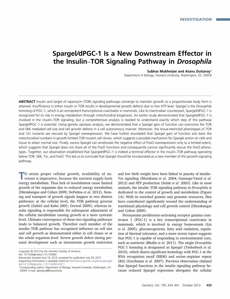
Figure 1 Cellular localization of Spargel follows PGC-1. (A) Schematicrepresentation of the insulin–TOR signaling pathway: insulin and TORsignaling pathway acts together to control growth in Drosophila andmammals. The insulin-like growth factors/insulin turns on this pathway,depending on the availability of glucose in the system. Details of insulinsignaling have been reviewed elsewhere (Bier 2005; Grewal 2009). TORplays a central role in regulating cell growth and control of mitochondrialenergy. Each member of this pathway can control cell growth in a cellautonomous manner (Hennig et al. 2006). (B) GFP–Spargel fusion proteinis localized exclusively inside the nucleus where it is not homogenouslydistributed; instead Spargel appears in the form of punctate structures.(C and D) Pre-mRNA splicing factor SC-35 (red), which is localized exclu-sively inside the nucleus in the form of nuclear speckles, colocalized withSpargel (merge) in many of these puntae. Bar, 40 mm. (E) At highermagnification (Bar, 20 mm) Spargel (green) and SC-35 (red) mergeappears as yellow signals. As shown here inside a polytene nucleus,Spargel and SC35 are quite intimately associated, indicating a conservedrole of PGC-1 and Spargel in the splicing complex. (F) Spargel carriesa 16-amino-acid nuclear localization signal (NLS) as predicted by Predict-
Protein and NLS Mapper software. (G) Cells expressing spargel RNAi (twoGFP positive cells) appeared much smaller in size, suggesting that like allother insulin family members Spargel functions in cell growth in a cellautonomous manner. (H) Quantification of cell size confirmed that boththe cytoplasmic area (CA) and nuclear area (NA) were reduced in spargelRNAi cell clones as compared to the control (non-GFP cells). Bar, 40 mm.N, number of cell clones counted .25. ***P , 0.001.
434 S. Mukherjee and A. Duttaroy
(Vector Lab), and visualized under confocal microscope(Nikon).
Antibody
Phospho-4EBP (1:200) and Phospho-S6K (1:200) antibodieswere obtained from Cell Signaling Technology (Miron et al.2003). Antiactin (1:5000) antibody was obtained fromAbcam.
Direct mitochondria visualization
Fat bodies were fixed with 4% paraformaldehyde and 20 mMformic acid. ATP5A (1: 250) (MitoSciences) antibody wasused to detect mitochondria (Cox and Spradling 2009).
Microscopy
Visualization and analysis of the clones were done usinga Nikon (EZ-C1) confocal microscope. Measurement ofclone size and mitochondrial fluorescence intensity quanti-tation was done with Nikon NIS-Element software.
RT–PCR
RNA extraction was done using Qiagen RNeasy spin kit(catalog no.74104). Thirty-five flies were homogenized in250 ml of buffer RLT with b-marcaptoethanol. Homogenatewas centrifuged at full speed for 10 min at 4�. The superna-tant was added to the g-DNA spin column and centrifugedfor 1 min at 11,000 rpm. The flow through was mixed withequal volume of 70% ethanol and added to the RNAeasyspin column. After centrifugation, 700 ml of buffer RW1was added to the column and centrifuged for 1 min at11,000 rpm. 500 ml of buffer RPE was added, centrifuged,and the step was repeated. The columns were added ina new tube and centrifuged for 1 min at full speed. Thecolumns were transferred to a new tube, 35 ml of RNase-free water was added, and centrifuged at 11,000 rpm for1 min. Collected RNA was quantified and 2 mg of RNA wasused to make the cDNA.
Western blot
Total protein was isolated by homogenization of �30 flies ina prechilled extraction buffer (10 mM DTT, 4% glycerol, and0.15 M Tris-Cl pH 7.5). Homogenates were spun down at14,000 rpm for 10 min in 4�. Supernatant was collected andprotein concentration was adjusted using the Bio-Rad quickstart Bradford assay. Protein samples were denatured byboiling and were then electrophoresed (2 mg/ml) on anSDS–PAGE (14% denaturing and 4% stacking gel) at 120V for 1 hr at room temperature. Samples were run simulta-neously alongside a protein standard (BioRad Precision Plus250, 10 kDa). Following electrophoresis, proteins weretransferred to Millipore’s Immobilion-PSQ (0.2 m pore size)PVDF membrane for 60 min in cold (4�) at 120 V. The PVDFmembrane was then blocked with 5% fat-free milk dissolvedin 13 TTBS (100 mM Tris-Cl pH 7.5, 0.9% NaCl and 0.1%v/v Tween 20) overnight in 4�. The membrane was washedtwice (5 min each) with 13 TTBS and probed with primary
antibody (1 ml Ab in 5 ml TTBS containing 1% fat-free milk)for 1 hr. Excess primary antibody was washed off by rinsingthree times (5 min each) with 13 TTBS. The membrane wasthen incubated in secondary antibody (1 ml Ab in 5 ml TTBScontaining 1% fat-free skimmed milk) for 1 hr, and thenrinsed three times with 13 TTBS. Lastly for detection,�4 ml of ECL was added (Amersham Biosciences) per blot.To test the phosphorylation status of S6k and 4EBP, anti-S6kantibody (1:500) (Cell Signaling) and anti-4EBP (1:500)(Cell Signaling) antibodies were used.
Statistics and software
Measured cellular and nuclear areas were compared usingStudent’s t-test. Significance of pupal and adult rescues wasdetermined using the chi-square test. Nuclear localizationsignal (NLS) in Spargel was predicted using two independentsoftware programs, PredictProtein (http://www.predictprotein.org/) and NLS Mapper (http://nls-mapper.iab.keio.ac.jp/cgi-bin/NLS_Mapper_form.cgi) under high stringency. The possibi-lity of Spargel localization in the nucleus was checked with twodifferent software programs, NucPred (http://www.sbc.su.se/~maccallr/nucpred/) and ESLPred (http://www.imtech.res.in/raghava/eslpred/).
Results and Discussion
Subcellular localization of Spargel and PGC-1is comparable
Significant structural homologies exist between Spargel andPGC-1 (Gershman et al. 2007). We noticed with interest thatthese two molecules share a comparable subcellular locali-zation, which may have some greater functional signifi-cance. Ubiquitous expression of a GFP-tagged Spargelprotein with Act-GAL4 driver displayed that Spargel is local-ized exclusively within the nucleus (Figure 1B), the same asPGC-1, which is largely located in the nucleus (Monsalveet al. 2000). Interestingly, Spargel localization in the nucleusis predictable with Nucpred and ESLpred software (Bhasinand Raghava 2004; Brameier et al. 2007) with almost 93–94% accuracy. Nuclear localization of Spargel was furtherconfirmed with a specific nuclear protein SC35, a pre-mRNAsplicing factor (Fu and Maniatis 1992) (Figure 1C). In mam-malian cells, PGC-1 has been shown to be associated withthe splicing complexes because PGC-1 and SC35 are colo-calized to the same punctate structures (Monsalve et al.2000). To test whether Spargel maintains a similar charac-teristic, we found that Spargel and SC35 are also colocalizedto the same speckles almost perfectly (Figure 1D). Underhigher magnification, salivary gland polytene nuclei dis-played more intimate association between SC35 and Spragel(Figure 1E, yellow signals), which help us to predict thatboth Spargel and PGC-1 are conserved constituents of thesplicing complex. We believe that such discrete localizationof Spargel into the nucleus is possibly resulting from anembedded 16-amino-acid-long Nuclear Localization signal
Spargel/dPGC-1 in Growth Signaling 435
(NLS) (amino acids 734–750) as predicted by PredictProteinand NLS Mapper software (Rost and Liu 2003; Kosugi et al.2009) (Figure 1F). Contrary to our observation, an earlierstudy claimed that a HA-tagged Spargel protein is localized
in the cytoplasmic compartment, which gets transported in-side the nucleus following activation by insulin receptor(Tiefenbock et al. 2010). Based on colocalization data andother characteristics, nuclear localization of Spargel appears
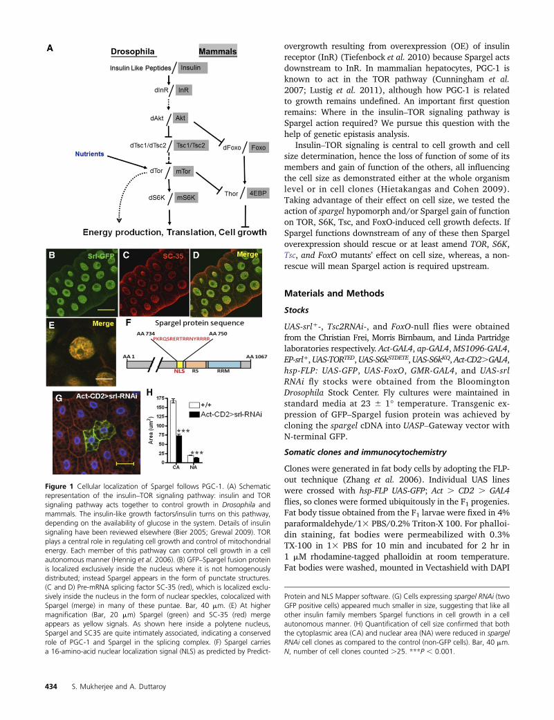
Figure 2 Spargel is downstream of Tor. (A) Spragel overexpression (OE) was achieved by activating an EP(gy2) insertion in the 59-UTR of spargel(CG9809) with a Act-GAL4 driver. (B and C) RT–PCR analysis of spargelOE with EP(gy2)srl+ and UAS-srl+ insertions with Act-GAL4 driver confirmed thatthe EP-srl+ element overexpresses Spargel at the same level as the UAS-srl+. About twofold more spargel mRNA expression was achieved from both EPand UAS lines. Since EP-srl+ is located on the third chromosome, and UAS-srl+ is located on the second, the former insertion was chosen since it makesthe genetic manipulations easier. **P , 0.01; ***P , 0.001. (D) Cells expressing TOR(DN) (two GFP-positive cells) mutation in the fat body tissueappeared much smaller in size with tiny nuclei (blue). (E) Spargel OE helps to recover the reduced cell size of TOR(DN) cell clones (GFP positive), whichnow appears normal in size. (F) Comparison of the nuclear area (NA) and cytoplasmic area (CA) of TOR(DN) cell clones before and after Spargel OEconfirmed that a significant increase in cell size occurs following Spargel OE (N . 25). Bar, 40 mm. CA, cytoplasmic area normalized with control; NA,nuclear area normalized with control. *P , 0.05, **P , 0.01, ***P , 0.001. (G) Expression of TOR(DN) in the wings with MS-1094-GAL4 causesshortened and misshaped wings due to cell growth defect. (H) Following Spargel OE, the wings of TOR(DN) attained their normal shape and size(N . 25).
436 S. Mukherjee and A. Duttaroy

to be more logical; however, future studies should be able toresolve this issue further.
Spargel acts downstream of TOR and S6kinase: TOR is atthe core of cell and tissue growth as TOR can influence cellgrowth in a cell autonomous manner (Wullschleger et al.2006; Zhang et al. 2006). Absence of the TOR nutrient sen-sor causes smaller cell size and fully penetrant larval lethal-ity (Zhang et al. 2000; Hennig et al. 2006). We wanted totest the interaction between TOR and Spargel on cell growthbecause many of TOR’s diverse effects on cellular physiologyoverlap with Spargel (Zhang et al. 2000; Schieke et al.2006). For example, clones of cells with reduced Spargelexpression appear smaller in size (Figure 1, G and H) andspargel hypomorphic adults have smaller body size (SupportingInformation, Figure S1E). This led us to conclude that Spargelcan control cell growth in a cell autonomous manner similar toTOR.
Cell clones expressing TOR-dominant negative [TOR(DN)] mutation (Hennig and Neufeld 2002) (also knownas TOR toxic effector domain, TORTED) appeared muchsmaller in size due to reduced cell growth (Figure 2D).TORTED expresses the 754-amino-acid central domain ofTOR that acts in a dominant negative fashion because it isthought to sequester signaling factors. Overexpression ofSpargel protein (Figure 2, A–C) in the TOR(DN) cells helps�80% of the small-sized TOR(DN) cell to attain normal size(Figure 2, E and F). This led us to wonder whether Spargelis a downstream mediator of TOR-induced cell growth. So,we attempted to restore the tissue-restricted phenotypes ofTOR(DN) with Spargel. When expressed in the wings withwing-specific MS-1096-GAL4 driver, TOR(DN) causes a wingdefect (Figure 2G) due to severe restriction of cell growthduring wing development (Hennig and Neufeld 2002). Co-
overexpression of Spargel and TOR(DN) with the sameGAL4 driver suppresses the wing phenotype and results ina completely normal wing shape (Figure 2H). As a finalattempt, we wanted to rescue the lethal effect of TOR(DN)with excess Spargel. TOR(DN) is early lethal (Hennig andNeufeld 2002) so no pupae are recovered in this mutant(Table S1) (Hennig and Neufeld 2002). Ubiquitous overex-pression of Spargel with the help of Act-GAL4 driver in theTOR(DN) embryos resulted in 94% pupal formation ofwhich 10–12% actually eclosed as adults, which is unprec-edented (Table S1). Spargel-rescued TOR(DN) adults stillappeared smaller in body size (Figure S1D) and they sur-vived for a brief length of time. These data imply that Spar-gel can only take over certain functions of TOR, such as cellgrowth control, as documented. Yet TOR also controlsa large array of cellular processes (DePalma et al. 2012;Laplante and Sabatini 2012) and given that the rescuewas not complete, we interpret the result to indicate thatSpargel does not function in all the effects mediated by TOR.This interpretation is further supported by the fact that Spar-gel overexpression does not influence the phosphorylationstatus of 4EBP (Figure S2), meaning TOR regulates 4EBPphosphorylation in a Spargel-independent manner. Wetherefore conclude that TOR’s action on cell size and cellgrowth is mediated through Spargel and thus Spargel isan important downstream effector of TOR for cell growthsignaling.
The above conclusion will be best supported if spargel-RNAi can block the TOR overexpression phenotype. SinceTOR overexpression is lethal, we used the tuberous sclerosiscomplex (Tsc), which works upstream of TOR and nega-tively regulates TOR function (DePalma et al. 2012). Thusinactivation of Tsc2 through RNAi (also known as gigas-RNAi)in the eye tissue causes an eye enlargement effect (Figure 3,
Figure 3 Reduced Spargel ex-pression suppresses the Tsc2RNAi-mediated overgrowth. (A)Normal eye. (B) Activation ofTsc2-RNAi in the eye with GMR-GAL4 causes cellular overgrowthso the eyes appear much largerin size. (C) The overgrowth effectof Tsc-2 reduction is mendedwhen spargel expression is re-duced with spargel RNAi so theeyes turned normal (N . 25). (D)Tsc-2RNAi effect and cosuppres-sion of Tsc-2; spargel was nor-malized with control eye. Barfor eye, 100 px. (E) Tsc2 RNAiexpression in cell clones (GFPpositive) causes over growth ofthose cells (compare with non-GFP cells). (F and G) Activationof spargel RNAi in Tsc-2RNAi cellclones ameliorates the overgrowthphenotype. Bar for cell clones,40 mm. *P, 0.05, ***P, 0.001.
Spargel/dPGC-1 in Growth Signaling 437
A and B), the same as Tsc2 mutants (Potter et al. 2001).When Spargel levels are reduced (with spargel RNAi) inconjunction with Tsc-2 RNAi, this double mutant has thesame phenotype as that of spargel-RNAi because the excesscell growth defect of Tsc-2 was abrogated, causing the eyesto attain their normal shape (Figure 3, C and D). Similarly,the overgrowth phenotype of the Tsc2 RNAi cell clones(Figure 3E) in the fat body tissue are suppressed by spargelRNAi (Figure 3, F and G) with �80% efficiency. These datasuggest that Spargel action is necessary for Tsc-2 to imposeits effect on cell growth and most importantly provide fur-ther support that loss of Spargel can counteract the highTOR activity that is already known to be induced by theloss of Tsc.
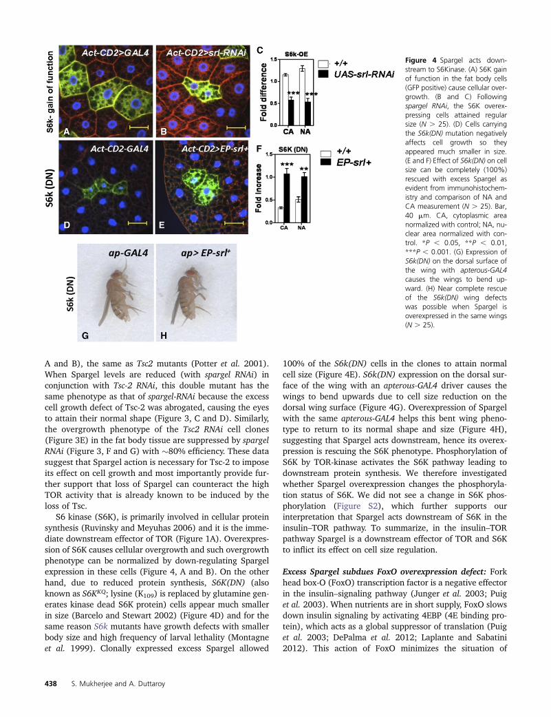
S6 kinase (S6K), is primarily involved in cellular proteinsynthesis (Ruvinsky and Meyuhas 2006) and it is the imme-diate downstream effector of TOR (Figure 1A). Overexpres-sion of S6K causes cellular overgrowth and such overgrowthphenotype can be normalized by down-regulating Spargelexpression in these cells (Figure 4, A and B). On the otherhand, due to reduced protein synthesis, S6K(DN) (alsoknown as S6KKQ; lysine (K109) is replaced by glutamine gen-erates kinase dead S6K protein) cells appear much smallerin size (Barcelo and Stewart 2002) (Figure 4D) and for thesame reason S6k mutants have growth defects with smallerbody size and high frequency of larval lethality (Montagneet al. 1999). Clonally expressed excess Spargel allowed
100% of the S6k(DN) cells in the clones to attain normalcell size (Figure 4E). S6k(DN) expression on the dorsal sur-face of the wing with an apterous-GAL4 driver causes thewings to bend upwards due to cell size reduction on thedorsal wing surface (Figure 4G). Overexpression of Spargelwith the same apterous-GAL4 helps this bent wing pheno-type to return to its normal shape and size (Figure 4H),suggesting that Spargel acts downstream, hence its overex-pression is rescuing the S6K phenotype. Phosphorylation ofS6K by TOR-kinase activates the S6K pathway leading todownstream protein synthesis. We therefore investigatedwhether Spargel overexpression changes the phosphoryla-tion status of S6K. We did not see a change in S6K phos-phorylation (Figure S2), which further supports ourinterpretation that Spargel acts downstream of S6K in theinsulin–TOR pathway. To summarize, in the insulin–TORpathway Spargel is a downstream effector of TOR and S6Kto inflict its effect on cell size regulation.
Excess Spargel subdues FoxO overexpression defect: Forkhead box-O (FoxO) transcription factor is a negative effectorin the insulin–signaling pathway (Junger et al. 2003; Puiget al. 2003). When nutrients are in short supply, FoxO slowsdown insulin signaling by activating 4EBP (4E binding pro-tein), which acts as a global suppressor of translation (Puiget al. 2003; DePalma et al. 2012; Laplante and Sabatini2012). This action of FoxO minimizes the situation of
Figure 4 Spargel acts down-stream to S6Kinase. (A) S6K gainof function in the fat body cells(GFP positive) cause cellular over-growth. (B and C) Followingspargel RNAi, the S6K overex-pressing cells attained regularsize (N . 25). (D) Cells carryingthe S6k(DN) mutation negativelyaffects cell growth so theyappeared much smaller in size.(E and F) Effect of S6k(DN) on cellsize can be completely (100%)rescued with excess Spargel asevident from immunohistochem-istry and comparison of NA andCA measurement (N . 25). Bar,40 mm. CA, cytoplasmic areanormalized with control; NA, nu-clear area normalized with con-trol. *P , 0.05, **P , 0.01,***P , 0.001. (G) Expression ofS6k(DN) on the dorsal surface ofthe wing with apterous-GAL4causes the wings to bend up-ward. (H) Near complete rescueof the S6k(DN) wing defectswas possible when Spargel isoverexpressed in the same wings(N . 25).
438 S. Mukherjee and A. Duttaroy

cellular energy expenditure under stress. Although FoxO hasbeen claimed to be a negative regulator of Spargel transcrip-tion activity in Drosophila S2 cells (Gershman et al. 2007),we noticed no changes in Spargel transcription activity inthe FoxO-null situation (Figure 5, I and J). To obtain furtherinsight on FoxO–Spargel interaction, we co-overexpressedthem in the same cell. FoxO overexpression in larval fatbody causes the cells to be unhealthy with very tiny nucleiand little to no cytoplasmic area, when compared to theneighboring control cells outside the clone (Figure 5A).When Spargel is produced in excess, we found �11% ofFoxO-OE cell clones attain their normal size (Figure 5, A–C). Similarly, excess Spargel is capable of slightly improvingthe FoxO-mediated phenotypic severities in the wing andthe eye tissue (Figure 5, D–H). While excess Spargel ameli-orates the severity of FoxO overexpression in a tissue-limitedfashion, the lethal effect of ubiquitous FoxO overexpressioncannot be rescued with Spargel (Table S2). As a transcrip-tion factor, FoxO regulates multiple targets, which in turninfluences various cellular processes in vital tissues (Acciliand Arden 2004; Greer and Brunet 2005). Our experimentsshow that levels of Spargel overexpression that were able torescue the TOR and S6K effects in the fat body are notcapable of producing a similar high percentage of rescueof the FoxO overexpression phenotype. Therefore we sug-gest that Spargel does not share all of the FoxO functionsand consequently cannot significantly rescue the FoxO phe-notypes, or it is possible that Spargel function is not enough
to take over any of these functions, hence only a limitedrescue happened.
Spargel is involved in growth signaling through mito-chondrial biogenesis: Based on the above observations, wepropose an extension of the insulin–TOR signaling pathwayin which Spargel is incorporated as a new terminal effectorin the cell growth signaling pathway (Figure 6E). Althoughreduced expressions of many insulin–TOR signaling partnersnegatively influence cell growth and Spargel does the same,Spargel gain of function is not lethal, nor does it cause anycellular or organismal overgrowth (Figure S1, A–C) (Reraet al. 2011). We now have reasons to believe that Spargelcontrols the energy flow in this system as mitochondrialenergy production is upheld during Spargel overexpressionwith increased ATP generation (Figure 6A). This observationfollows some earlier claims where Spargel overexpressionwas related to higher activity of electron transport com-plexes (Rera et al. 2011). Conversely, reduced Spargel levelnegatively impacted the electron transport chain enzymesand ATP synthesis (Tiefenbock et al. 2010). How is this excessATP utilized during Spargel overexpression? One clue camefrom the muscle-specific overexpression of Spargel, whichprolongs the vertical climbing behavior in flies (Tinkerhesset al. 2012), possibly due to excess ATP production.
When cells need to grow, TOR is activated throughphosphorylation either by activated insulin signals or byamino acids. An activated TOR maintains nutrient and
Figure 5 Spargel and FoxO in-teraction. (A) Overexpression ofFoxO severely affects cell growth(arrows). It is possible that theFoxO OE cells will die beforeeven reaching the adult stage.(B) Following Spargel OE, someof these tiny cells (�11%)attained increased cellular andnuclear areas as quantitated inC. (D) Normal eye. (E) FoxO OEin the eye with GMR-GAL4 drivershows a significant reduction inommatidial numbers. (F) FoxOOE effect on ommatidial num-bers can be ameliorated by Spar-gel OE (N . 50). (G) Severe wingphenotype resulted when FoxOis overexpressed with apterous-GAL4 driver. (H) Excess Spargelpartially fixes the wing defect inH. (I and J) RT–PCR analysis ofspargel mRNA expression inFoxO null background revealedthat Spargel is not regulated byFoxO. Bar for cell clones, 25 mm;bar for eye, 100 px. CA, cyto-plasmic area normalized withcontrol; NA, nuclear area nor-malized with control. ***P ,0.001; NS, not significant.
Spargel/dPGC-1 in Growth Signaling 439
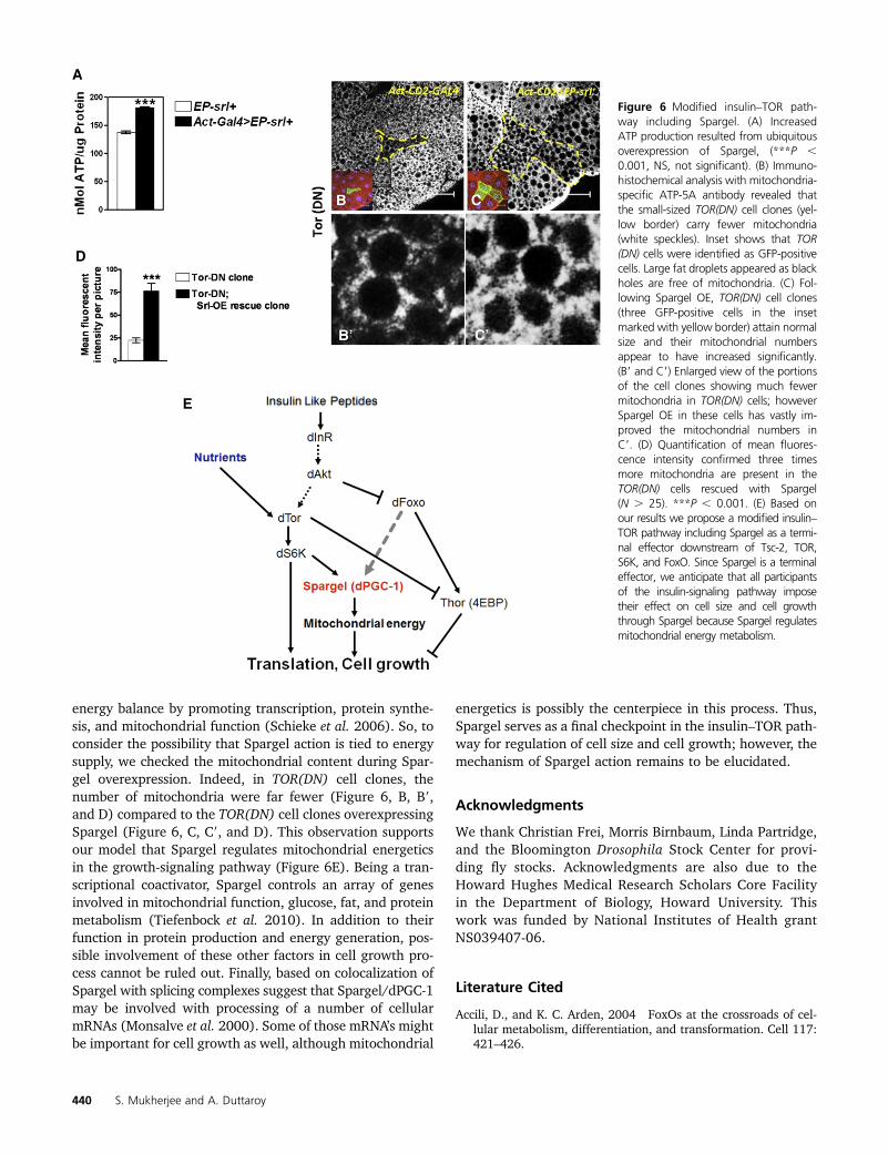
energy balance by promoting transcription, protein synthe-sis, and mitochondrial function (Schieke et al. 2006). So, toconsider the possibility that Spargel action is tied to energysupply, we checked the mitochondrial content during Spar-gel overexpression. Indeed, in TOR(DN) cell clones, thenumber of mitochondria were far fewer (Figure 6, B, B9,and D) compared to the TOR(DN) cell clones overexpressingSpargel (Figure 6, C, C9, and D). This observation supportsour model that Spargel regulates mitochondrial energeticsin the growth-signaling pathway (Figure 6E). Being a tran-scriptional coactivator, Spargel controls an array of genesinvolved in mitochondrial function, glucose, fat, and proteinmetabolism (Tiefenbock et al. 2010). In addition to theirfunction in protein production and energy generation, pos-sible involvement of these other factors in cell growth pro-cess cannot be ruled out. Finally, based on colocalization ofSpargel with splicing complexes suggest that Spargel/dPGC-1may be involved with processing of a number of cellularmRNAs (Monsalve et al. 2000). Some of those mRNA’s mightbe important for cell growth as well, although mitochondrial
energetics is possibly the centerpiece in this process. Thus,Spargel serves as a final checkpoint in the insulin–TOR path-way for regulation of cell size and cell growth; however, themechanism of Spargel action remains to be elucidated.
Acknowledgments
We thank Christian Frei, Morris Birnbaum, Linda Partridge,and the Bloomington Drosophila Stock Center for provi-ding fly stocks. Acknowledgments are also due to theHoward Hughes Medical Research Scholars Core Facilityin the Department of Biology, Howard University. Thiswork was funded by National Institutes of Health grantNS039407-06.
Literature Cited
Accili, D., and K. C. Arden, 2004 FoxOs at the crossroads of cel-lular metabolism, differentiation, and transformation. Cell 117:421–426.
Figure 6 Modified insulin–TOR path-way including Spargel. (A) IncreasedATP production resulted from ubiquitousoverexpression of Spargel, (***P ,0.001, NS, not significant). (B) Immuno-histochemical analysis with mitochondria-specific ATP-5A antibody revealed thatthe small-sized TOR(DN) cell clones (yel-low border) carry fewer mitochondria(white speckles). Inset shows that TOR(DN) cells were identified as GFP-positivecells. Large fat droplets appeared as blackholes are free of mitochondria. (C) Fol-lowing Spargel OE, TOR(DN) cell clones(three GFP-positive cells in the insetmarked with yellow border) attain normalsize and their mitochondrial numbersappear to have increased significantly.(B9 and C9) Enlarged view of the portionsof the cell clones showing much fewermitochondria in TOR(DN) cells; howeverSpargel OE in these cells has vastly im-proved the mitochondrial numbers inC9. (D) Quantification of mean fluores-cence intensity confirmed three timesmore mitochondria are present in theTOR(DN) cells rescued with Spargel(N . 25). ***P , 0.001. (E) Based onour results we propose a modified insulin–TOR pathway including Spargel as a termi-nal effector downstream of Tsc-2, TOR,S6K, and FoxO. Since Spargel is a terminaleffector, we anticipate that all participantsof the insulin-signaling pathway imposetheir effect on cell size and cell growththrough Spargel because Spargel regulatesmitochondrial energy metabolism.
440 S. Mukherjee and A. Duttaroy
Barcelo, H., and M. J. Stewart, 2002 Altering Drosophila S6 ki-nase activity is consistent with a role for S6 kinase in growth.Genesis 34: 83–85.
Bhalla, K., B. J. Hwang, R. E. Dewi, L. Ou, W. Twaddel et al.,2011 PGC1alpha promotes tumor growth by inducing geneexpression programs supporting lipogenesis. Cancer Res. 71:6888–6898.
Bhasin, M., and G. P. Raghava, 2004 ESLpred: SVM-basedmethod for subcellular localization of eukaryotic proteins usingdipeptide composition and PSI-BLAST. Nucleic Acids Res. 32:W414–W419.
Bier, E., 2005 Drosophila, the golden bug, emerges as a tool forhuman genetics. Nat. Rev. Genet. 6: 9–23.
Brameier, M., A. Krings, and R. M. Maccallum, 2007 NucPred–predicting nuclear localization of proteins. Bioinformatics 23:1159–1160.
Cox, R. T., and A. C. Spradling, 2009 Clueless, a conserved Dro-sophila gene required for mitochondrial subcellular localization,interacts genetically with parkin. Dis. Model. Mech. 2: 490–499.
Cunningham, J. T., J. T. Rodgers, D. H. Arlow, F. Vazquez, V. K.Mootha et al., 2007 mTOR controls mitochondrial oxidativefunction through a YY1-PGC-1alpha transcriptional complex.Nature 450: 736–740.
DePalma, M. J., J. M. Ketchum, T. R. Saullo, and B. L. Laplante,2012 Is the history of a surgical discectomy related to thesource of chronic low back pain? Pain Physician 15: E53–E58.
Fu, X. D., and T. Maniatis, 1992 The 35-kDa mammalian splicingfactor SC35 mediates specific interactions between U1 and U2small nuclear ribonucleoprotein particles at the 39 splice site.Proc. Natl. Acad. Sci. USA 89: 1725–1729.
Gannage-Yared, M. H., J. Klammt, E. Chouery, S. Corbani, H. Megarbaneet al., 2012 Homozygous mutation of the Insulin-like GrowthFactor-I Receptor gene (IGF1R) in a patient with severe pre- andpostnatal growth failure, and congenital malformations. Eur. J.Endocrinol 168: K1–K7.
Gershman, B., O. Puig, L. Hang, R. M. Peitzsch, M. Tatar et al.,2007 High-resolution dynamics of the transcriptional responseto nutrition in Drosophila: a key role for dFOXO. Physiol. Ge-nomics 29: 24–34.
Greer, E. L., and A. Brunet, 2005 FOXO transcription factors at theinterface between longevity and tumor suppression. Oncogene24: 7410–7425.
Grewal, S. S., 2009 Insulin/TOR signaling in growth and homeo-stasis: a view from the fly world. Int. J. Biochem. Cell Biol. 41:1006–1010.
Hennig, K. M., and T. P. Neufeld, 2002 Inhibition of cellulargrowth and proliferation by dTOR overexpression in Drosophila.Genesis 34: 107–110.
Hennig, K. M., J. Colombani, and T. P. Neufeld, 2006 TOR coordi-nates bulk and targeted endocytosis in the Drosophila melanogasterfat body to regulate cell growth. J. Cell Biol. 173: 963–974.
Hietakangas, V., and S. M. Cohen, 2009 Regulation of tissuegrowth through nutrient sensing. Annu. Rev. Genet. 43: 389–410.
Junger, M. A., F. Rintelen, H. Stocker, J. D. Wasserman, M. Veghet al., 2003 The Drosophila forkhead transcription factorFOXO mediates the reduction in cell number associated withreduced insulin signaling. J. Biol. 2: 20.
Kosugi, S., M. Hasebe, N. Matsumura, H. Takashima, E. Miyamoto-Sato et al., 2009 Six classes of nuclear localization signals spe-cific to different binding grooves of importin alpha. J. Biol.Chem. 284: 478–485.
Laplante, M., and D. M. Sabatini, 2012 mTOR signaling in growthcontrol and disease. Cell 149: 274–293.
Lin, J., C. Handschin, and B. M. Spiegelman, 2005 Metabolic con-trol through the PGC-1 family of transcription coactivators. CellMetab. 1: 361–370.
Lustig, Y., J. L. Ruas, J. L. Estall, J. C. Lo, S. Devarakonda et al.,2011 Separation of the gluconeogenic and mitochondrialfunctions of PGC-1{alpha} through S6 kinase. Genes Dev. 25:1232–1244.
Miron, M., P. Lasko, and N. Sonenberg, 2003 Signaling from Aktto FRAP/TOR targets both 4E-BP and S6K in Drosophila mela-nogaster. Mol. Cell. Biol. 23: 9117–9126.
Monsalve, M., Z. Wu, G. Adelmant, P. Puigserver, M. Fan et al.,2000 Direct coupling of transcription and mRNA processingthrough the thermogenic coactivator PGC-1. Mol. Cell 6: 307–316.
Montagne, J., M. J. Stewart, H. Stocker, E. Hafen, S. C. Kozma et al.,1999 Drosophila S6 kinase: a regulator of cell size. Science285: 2126–2129.
Murakami, M., T. Ichisaka, M. Maeda, N. Oshiro, K. Hara et al.,2004 mTOR is essential for growth and proliferation in earlymouse embryos and embryonic stem cells. Mol. Cell. Biol. 24:6710–6718.
Potter, C. J., H. Huang, and T. Xu, 2001 Drosophila Tsc1 functionswith Tsc2 to antagonize insulin signaling in regulating cellgrowth, cell proliferation, and organ size. Cell 105: 357–368.
Puig, O., M. T. Marr, M. L. Ruhf, and R. Tjian, 2003 Control of cellnumber by Drosophila FOXO: downstream and feedback regu-lation of the insulin receptor pathway. Genes Dev. 17: 2006–2020.
Rera, M., S. Bahadorani, J. Cho, C. L. Koehler, M. Ulgherait et al.,2011 Modulation of longevity and tissue homeostasis by theDrosophila PGC-1 homolog. Cell Metab. 14: 623–634.
Rost, B., and J. Liu, 2003 The PredictProtein server. Nucleic AcidsRes. 31: 3300–3304.
Ruvinsky, I., and O. Meyuhas, 2006 Ribosomal protein S6 phos-phorylation: from protein synthesis to cell size. Trends Biochem.Sci. 31: 342–348.
Saltiel, A. R., and C. R. Kahn, 2001 Insulin signalling and theregulation of glucose and lipid metabolism. Nature 414: 799–806.
Schieke, S. M., D. Phillips, J. P. Mccoy, Jr., A. M. Aponte, R. F. Shenet al., 2006 The mammalian target of rapamycin (mTOR)pathway regulates mitochondrial oxygen consumption and oxi-dative capacity. J. Biol. Chem. 281: 27643–27652.
Selak, M. A., B. T. Storey, I. Peterside, and R. A. Simmons,2003 Impaired oxidative phosphorylation in skeletal muscleof intrauterine growth-retarded rats. Am. J. Physiol. Endocrinol.Metab. 285: E130–E137.
Tiefenbock, S. K., C. Baltzer, N. A. Egli, and C. Frei, 2010 TheDrosophila PGC-1 homologue Spargel coordinates mitochon-drial activity to insulin signalling. EMBO J. 29: 171–183.
Tinkerhess, M. J., L. Healy, M. Morgan, A. Sujkowski, E. Matthyset al., 2012 The Drosophila PGC-1alpha homolog spargel mod-ulates the physiological effects of endurance exercise. PLoS ONE7: e31633.
Wullschleger, S., R. Loewith, and M. N. Hall, 2006 TOR signalingin growth and metabolism. Cell 124: 471–484.
Zhang, H., J. P. Stallock, J. C. Ng, C. Reinhard, and T. P. Neufeld,2000 Regulation of cellular growth by the Drosophila target ofrapamycin dTOR. Genes Dev. 14: 2712–2724.
Zhang, Y., C. J. Billington, Jr., D. Pan, and T. P. Neufeld,2006 Drosophila target of rapamycin kinase functions as a mul-timer. Genetics 172: 355–362.
Communicating editor: T. Schüpbach
Spargel/dPGC-1 in Growth Signaling 441
GENETICSSupporting Information
http://www.genetics.org/lookup/suppl/doi:10.1534/genetics.113.154583/-/DC1
Spargel/dPGC-1 Is a New Downstream Effector inthe Insulin–TOR Signaling Pathway in Drosophila
Subhas Mukherjee and Atanu Duttaroy
Copyright © 2013 by the Genetics Society of AmericaDOI: 10.1534/genetics.113.154583
S. Mukherjee and A. Duttaroy 2 SI
E
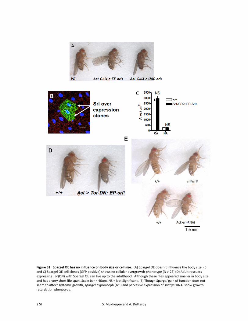
Figure S1 Spargel OE has no influence on body size or cell size. (A) Spargel OE doesn’t influence the body size. (B and C) Spargel OE cell clones (GFP positive) shows no cellular overgrowth phenotype (N > 25) (D) Adult rescuers expressing Tor(DN) with Spargel OE can live up to the adulthood. Although these flies appeared smaller in body size and has a very short life span. Scale bar = 40um. NS = Not Significant. (E) Though Spargel gain of function does not seem to affect systemic growth, spargel hypomorph (srl1) and pervasive expression of spargel RNAi show growth retardation phenotype.
C

S. Mukherjee and A. Duttaroy 3 SI
Figure S2 Spargel over expression does not influence phosphorylation of S6k and 4EBP. S6k and 4EBP are phosphorylated by TOR kinase. Spargel OE causes no change in the phosphorylation status of either S6K or 4EBP. This observation agrees well with the genetic analysis since Spargel is downstream to TOR, so it doesn’t influence the Tor kinase activity.
S. Mukherjee and A. Duttaroy 4 SI
File S1
Supporting Methods
GFP tagged Spargel: spargel cDNA was cloned in UASpN terminal GFP tag gateway vector (DGRC) and injected into fly
lines (Best Gene). Flies were crossed with Lsp-‐GAL4 and 3rd inster larvae were dissected, fixed with 4%
paraformaldehyde in 1X PBS; washed with 0.3% PBX and mounted for visualization under confocal microscope.

S. Mukherjee and A. Duttaroy 5 SI
Table S1 Spargel OE improve the effects of TOR (DN) during development
Genotypes # of pupae formed
# of adults eclosed
Tor and Spargel interaction:
Act-‐Gal4 > UAS-‐Tor (DN) 0 0
Act-‐Gal4 > UAS-‐Tor (DN);EP-‐srl+ 71*** (94.6%) 9** (12%)
UAS-‐Tor (DN); EP-‐srl+/ TM6, Tb (Control)
75 75 (100%)
Chi-‐square test determined the significance of the spargel-‐mediated rescue. ** P < 0.01, *** P < 0.001. DN =
Dominant Negative, OE = Overexpression. Parentheses show percentage of pupae and adults.
S. Mukherjee and A. Duttaroy 6 SI
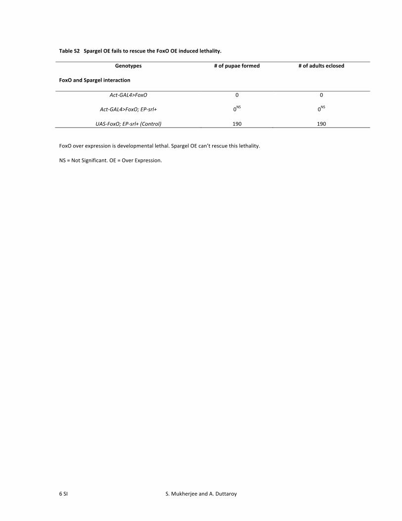
Table S2 Spargel OE fails to rescue the FoxO OE induced lethality.
Genotypes
# of pupae formed # of adults eclosed
FoxO and Spargel interaction
Act-‐GAL4>FoxO
0 0
Act-‐GAL4>FoxO; EP-‐srl+
0NS 0NS
UAS-‐FoxO; EP-‐srl+ (Control) 190 190 FoxO over expression is developmental lethal. Spargel OE can’t rescue this lethality.
NS = Not Significant. OE = Over Expression.