spatial overlap of peptide hotspots and canonical … overlap of peptide hotspots and canonical drug...
TRANSCRIPT

Spatial overlap of peptide hotspots and
canonical drug pockets in a model enzyme
Walraj S. Gosal June 2015
1
In collaboration with:
With funding from:
From molecular display peptides to small molecule inhibitors.
| partnerships in biologics discovery

fold into precise 3D structures
chemical reactions of life through the precise arrangement & dynamics of atoms
Primary seq. 100 amino acid protein = 1x10130 sequences
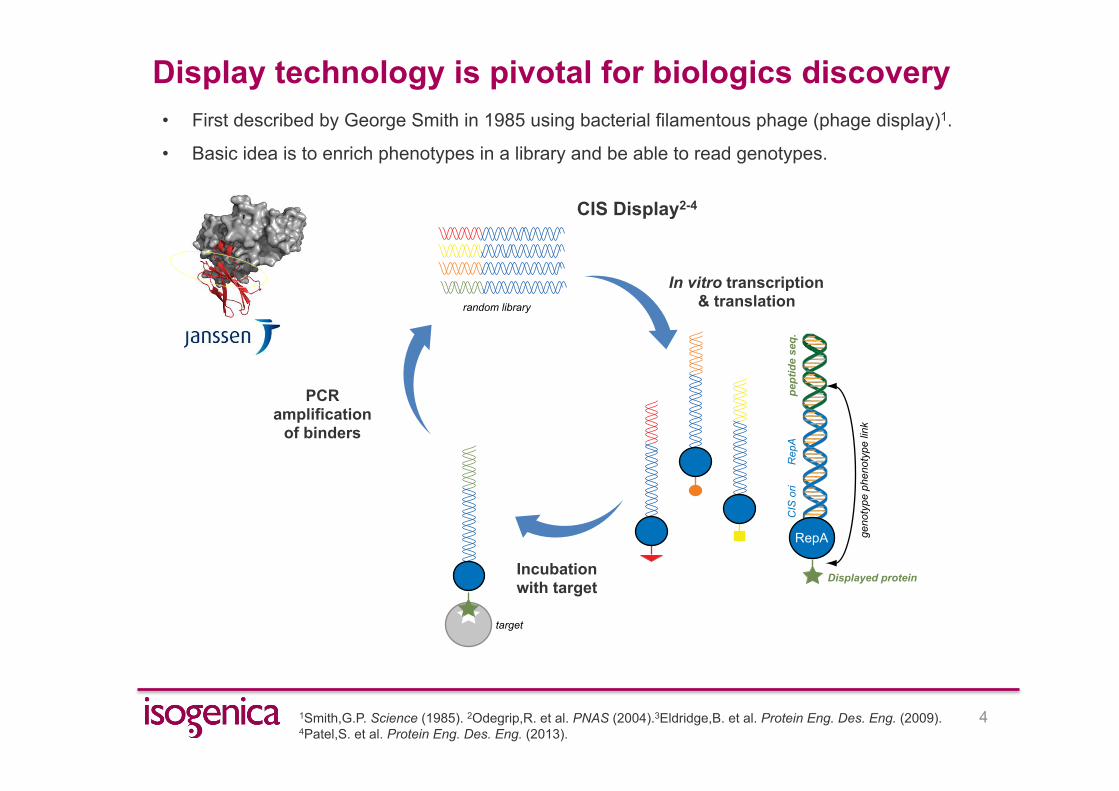
Display technology is pivotal for biologics discovery
2
Humira FAB
TNFα"
Approved: RA, UC, CD
• Humira (monoclonal antibody) is the biggest selling drug worldwide ($13bn in sales).
• Computational approaches are showing evermore promise (e.g. Rosetta)1,2.
Design
Crystal structure
1Kuhlman,B. et al. Science (2003). 2Fleishman,S.J. et al. Science (2011).

Display technology is pivotal for the emergence of biologics • Recent emergence of over a dozen scaffolds ranging in size (~ 20 to 170 amino acids)1,2.
1Lofblom,J. et al. Current Opinion in Biotechnology (2011). 2Gronwall, C. et al. Journal of Biotechnology (2009).
Humira FAB
TNFα"
Approved: RA, UC, CD
Knottin
e.g. Ziconotide (Approved: pain relief)
DARPin e.g. Abicipar pegol (Phase III:
macular degeneration)
e.g. SOBI002 (Phase I: inflammation)
Affibody
Anticalin e.g. PRS-050 (Phase I:
angiogenesis)
Adnectin / Centyrin e.g. Angiocept (phase II
glioblastoma)
Display technology is pivotal for biologics discovery
4
• First described by George Smith in 1985 using bacterial filamentous phage (phage display)1.
• Basic idea is to enrich phenotypes in a library and be able to read genotypes. RepA
CISori
RepA
genotypephenotypelink
peptideseq.
Displayed protein
target
random library
random library
RepA
CISori
RepA
genotypephenotypelink
peptideseq.
Displayed proteintarget
In vitro transcription & translation
RepA
CISori
RepA
genotypephenotypelink
peptideseq.
Displayed protein
target
random library
random library
RepA
CISori
RepA
genotypephenotypelink
peptideseq.
Displayed proteintarget
Incubation with target
1Smith,G.P. Science (1985). 2Odegrip,R. et al. PNAS (2004).3Eldridge,B. et al. Protein Eng. Des. Eng. (2009). 4Patel,S. et al. Protein Eng. Des. Eng. (2013).
CIS Display2-4
RepA
CISori
RepA
genotypephenotypelink
peptideseq.
Displayed protein
target
random library
random library
RepA
CISori
RepA
genotypephenotypelink
peptideseq.
Displayed proteintargetPCR
amplification of binders
RepA
CISori
RepA
genotypephenotypelink
peptideseq.
Displayed protein
target
random library
random library
RepA
CISori
RepA
genotypephenotypelink
peptideseq.
Displayed proteintarget

Can molecular display be used to inform small molecule discovery?
Protein target
3. Structure determination(NMR or X-ray)
1. CIS display& NGS
3D structure:Protein-Peptide complex
2. Peptide hit validation(ELISA, truncation,binding, activity,epitope mapping)
Allostericsitebinders
Activesitebinders
RepA
CISori
genotypephenotypelink
peptideseq.
peptide
target
Target-binding peptide candidates
selection, amplifiction & NGS
random peptidelibrary
VLSEGEWQLVLHVWAKVEADV
AQGAMNKALELFRKDIAAKYK105
1
Small target-binding peptides
site II
site I
CIS display & NGS
3D structure:Protein-Peptide complex
Allostericsitebinders
Activesitebinders
X-ray / NMR
3D structure:Protein-Peptide complex
Allostericsitebinders
Activesitebinders
4. Residue hotspots & field calculation(mutational analysis, Rosetta, & XED force field)
5. Virtual smallmolecule screen
(>107 small molecules)
6. Focused experimental screen(~100 small molecules)
7. Validation & ‘hit to lead’ optimisation (inhibitionmodality, Kd, Ki, X-ray Crystallography) Lead compound
Screening hits
5
1
h1
h2
h3
h4
Peptide field map
hydrophobic
+ve/HBA
VdW surface
-ve/HBD
Screening candidates
100
1O
NH
O
OH
NH
HN NH2
N
N
N
HN
O
NH2
N
N
S O
O
O
NH
NH2
Cl
NH
O
O
NH
NH2
NH
O
O
NH
Computation & screening

Target of choice: thrombin - a serine protease
6
• 40 year race in the drug industry to replace anticoagulants - Warfarin (1953) & Heparin (1937)1,2.
• Large body of public information that covers all major discovery platforms (fragment screening, structure-
based design & HTS).
1Gustafsson,D. Nature Reviews Drug Discovery 3 649-659 (2004). 2Nar, H. Trends in Pharm. Sci. 33 279-268 (2012). 3Huntington, J. A. Thromb. Haemost., 111 583-9 (2014).
Substrate: FPA
S1
S2
S4
Dabigatran Melagatran
S1
S2 S4
S1
S2
S4
s-variegin3 (32 aa) Tropical bont tick
Hirudin3 (65aa) Medicinal leech
Anophelin3 (31 aa) Mosquito
• Unique structure based solutions from Nature3.

Summary & Acknowledgements
Contact Walraj S. Gosal [email protected] www.isogenica.com
7
1. In a retrospective study, CIS display peptide ‘hot-spots’ spatially overlap with known
drug pockets in thrombin, although novel pockets are induced.
2. Peptides cover the “chemical solution space” for the primary S1 pocket.
3. Field patterns based on peptides can be used to find new inhibitors.
Tom Blundell Jim Huntington Ty Adams
Robert Scoffin Andy Vinter Mark Mackey
Steve Gardner Dirk Gewert Peter Campbell
Chris Ullman Neil Cooley Kevin Matthews Gabriela Ivanova Amanda Hallott Shabana Vohra
With funding from:
Gordon Woodrow Stephan Krapp