spebrutinib (cc-292) affects markers of b cell activation ...and fc receptor signaling pathways....
TRANSCRIPT

ORIGINAL RESEARCH
Spebrutinib (CC-292) Affects Markers of B CellActivation, Chemotaxis, and Osteoclasts in Patientswith Rheumatoid Arthritis: Results from aMechanisticStudy
Peter H. Schafer . Alan J. Kivitz . Jianglin Ma . Shimon Korish .
Donna Sutherland . Li Li . Ada Azaryan . Jolanta Kosek . Mary Adams .
Lori Capone . Eun Mi Hur . Douglas R. Hough . Garth E. Ringheim
Received: July 16, 2019� The Author(s) 2019
ABSTRACT
Introduction: Spebrutinib (CC-292) is an orallyadministered, covalent, small-molecule inhibitorof Bruton’s tyrosine kinase (BTK), part of the B-celland Fc receptor signaling pathways. This studyevaluated spebrutinib pharmacology and mecha-nism of action over a 4-week treatment period inpatients with active rheumatoid arthritis (RA).Methods: Primary human B cells, T cells, naturalkiller cells, macrophages, dendritic cells, baso-phils, and osteoclasts were treated with spebruti-nib in vitro. Clinical pharmacodynamics werestudied in 47 patients with active RA on
background methotrexate therapy randomized tooral spebrutinib 375 mg/day or placebo.Results: In vitro, spebrutinib inhibited B-cellproliferation more potently than T-cell prolifera-tion and reduced both lymphoid and myeloidcytokine production and degranulation, as well asosteoclastogenesis.Clinicalefficacytrendedhigherin spebrutinib-treated RA patients, with 41.7% (10/24) achieving C 20% improvement in ACRresponse criteria (ACR20) versus 21.7% (5/23) ofplacebo patients at week 4 (P = 0.25). Treatment-emergent adverse events were comparablebetween treatment groups. In spebrutinib-treatedpatients, median BTK occupancy in peripheralblood was 83%, and significant increases in totalCD19? and mature-naive CD27-CD38-IgD? Bcells and decreases in transitional CD27-CD38? Bcells were observed. Spebrutinib significantlyreduced serum chemokines chemokine ligand 13(CXCL13), macrophage inflammatory protein-1b(MIP-1b), and the bone resorption biomarker car-boxy-terminal collagen cross-linking telopeptide(CTX-I) (P\0.05). Clinical response to spebruti-nib was associated with lower increases in CD19?Bcells and greater decreases in CXCL13 and MIP-1bfrom baseline to week 4. High CD19? B cells andlow CTX-I at baseline were associated with betterspebrutinib clinical response.Conclusions: Spebrutinib inhibited variousleukocyte responses in vitro, including those ofB cells and osteoclasts. In this small study in RApatients, spebrutinib was well tolerated, showeda downward trend for symptoms, significantly
Enhanced Digital Features To view enhanced digitalfeatures for this article go to: https://doi.org/10.6084/m9.figshare.10049777.
Electronic Supplementary Material The onlineversion of this article (https://doi.org/10.1007/s40744-019-00182-7) contains supplementary material, which isavailable to authorized users.
P. H. Schafer (&)Celgene Corporation, 86 Morris Avenue, L Building,Summit, NJ, USAe-mail: [email protected]
A. J. KivitzAltoona Center for Clinical Research, Duncansville,PA, USA
J. Ma � S. Korish � D. Sutherland � L. Li � A. Azaryan� J. Kosek � M. Adams � L. Capone � E. M. Hur �D. R. Hough � G. E. RingheimCelgene Corporation, Summit, NJ, USA
Rheumatol Ther
https://doi.org/10.1007/s40744-019-00182-7

modulated B-cell populations, and reducedmarkers of chemotaxis and osteoclast activity.Trial Registration: NCT01975610.
Keywords: Background methotrexate therapy;Bruton’s tyrosine kinase inhibitor; CC-292;Rheumatoid arthritis; Spebrutinib
Key Summary Points
Why carry out this study?
Bruton’s tyrosine kinase (BTK) has beenproposed as a therapeutic target for thetreatment of autoimmune andinflammatory diseases such as rheumatoidarthritis (RA), based on its role in B-cellreceptor and Fc receptor signaling.
Spebrutinib (CC-292) is an oral smallmolecule that inhibits BTK activity byirreversible covalent binding with highaffinity to the BTK adenosinetriphosphate binding site in B andmyeloid cells.
This phase 2a clinical study was conductedto evaluate spebrutinib efficacy and safetyin female patients with active RA whowere receiving a stable methotrexate doseas background therapy. The study alsoevaluated spebrutinib pharmacodynamiceffects on circulating levels of BTK, B-cellsubsets, and signaling factors essential toBTK activity.
What was learned from the study?
Spebrutinib treatment led to statisticallysignificant changes versus placebo acrossseveral biomarkers of anti-inflammatoryactivity, confirming its mechanism ofaction and potential therapeuticbiological effects in RA
The pharmacodynamics of BTK inhibitionin RA patients demonstrated a significantimpact on B-cell differentiation,chemokine expression, and osteoclastactivity that potentially provides a set ofbiomarkers useful in developing BTKinhibitors for the treatment of RA.
INTRODUCTION
Rheumatoid arthritis (RA) is a chronic, systemicautoimmune disorder initiated by dysregulationof T and B lymphocytes. Its pathology includesexpansion of B cells, which leads to formationof autoantibodies [1]. Bruton’s tyrosine kinase(BTK) is found in B cells but not in T cells ornatural killer (NK) cells within the lymphoidlineage. BTK is required for B-cell activation byengagement of the B-cell antigen receptor (BCR)and is involved in B-cell differentiation,chemotaxis, and trafficking [2, 3]. Inhibition orlack of BTK is marked by increased numbers of Bcells in circulating blood and reduced B-cellchemotaxis and accumulation in secondarylymphoid organs [1, 4]. Inhibition of BTKactivity is associated with reduced lym-phadenopathy in patients with chronic lym-phocytic leukemia [5]. BTK is also expressed inmyeloid cells, where it is involved in activatingFc-gamma and Fc-epsilon receptors (FccR andFceR) pathways in macrophages, neutrophils,and monocytes [1]. Together, these propertiessuggest BTK is an attractive target for B-cellinhibition in RA [1].
Spebrutinib (CC-292) is an oral small mole-cule which inhibits BTK activity by irreversiblecovalent binding with high affinity to the BTKadenosine triphosphate binding site in B andmyeloid cells. By providing rapid, complete,and prolonged inhibition of BTK activity, spe-brutinib may be therapeutically active in RAand other B-cell-mediated autoimmune disor-ders. Preclinical studies have shown thatadministration of spebrutinib led to blockade ofBCR-dependent B-cell activation, inhibition ofFccR-induced inflammatory cytokine produc-tion in myeloid cells, and a reduction in osteo-clastogenesis [6–8]. Spebrutinib hasdemonstrated BCR selectivity, potency, andefficacy in a collagen-induced arthritis model inmice [7]. In a small, first-in-human safety andpharmacokinetic/pharmacodynamics analysisof healthy volunteers, spebrutinib led to near-complete BTK occupancy 8–24 h after adminis-tration [7].
This phase 2a multicenter, US-based clinicalstudy was conducted to evaluate spebrutinib
Rheumatol Ther

efficacy and safety in female patients withactive RA who were receiving astable methotrexate dose as background ther-apy. The study also evaluated spebrutinibpharmacodynamic effects on circulating levelsof BTK, B-cell subsets, and signaling factorsessential to BTK activity.
METHODS
Preclinical Pharmacology
Human primary B cells (CD19?) were isolatedfrom leukopacks from healthy donors obtainedfrom Biological Specialty (Colmar, PA, USA).Peripheral blood mononuclear cells (PBMC)were first isolated by density gradient centrifu-gation using Ficoll (GE Healthcare, Chicago, IL,USA) followed by the EasySep Human Naive BCell Enrichment Kit (Stemcell Technologies,Vancouver, BC, Canada) according to manu-facturing protocol. CD4? and CD8? T cells wereisolated from PBMC using EasySep HumanCD4? and CD8? T Cell Enrichment Kits. NKcells were purified from PBMC using the Easy-Sep Human NK Cell Enrichment Kit.
Immunoblot Analysis of Phospho-phospholipase CcQuantitation of phospho-phospholipase Cc wasby Western-blot analysis of a-IgM-stimulated Bcells, as described previously [7].
B- and T-Cell ProliferationB cells (0.2 9 106 cells in 200 ll) or T cells(0.2 9 106 cells in 100 ll) were incubated for 1 hwith spebrutinib (0.0001–100 lM) followed bystimulation of B cells with a-IgM (10 lg/ml) andCpG (10 lg/ml), or T cells with a-CD3 (1 lg/ml),and a-CD28 (3 lg/ml) for 3 days. 3H-thymidine(1 lCi/well) was added the last 24 h, harvested,and measured for incorporation.
T-Cell Interferon-c SecretionHuman primary T cells (CD3?) were isolatedfrom leukopacks from healthy donors using theEasySep Human T Cell Enrichment Kit (Stemcell
Technologies, Vancouver, BC, Canada) accord-ing to manufacturing protocol. T cells wereincubated for 1 h with spebrutinib followed bystimulation with soluble a-CD3 (3 lg/ml) and a-CD28 (1 lg/ml) for 48 h. Supernatants wereanalyzed in duplicate for interferon-c cytokineproduction in a magnetic multiplex bead for-mat using the MagPix instrument (Millipore,Billerica, MA, USA). Data analysis was per-formed using Milliplex Analyst Software(Millipore).
NK- and CD81 T-Cell DegranulationNK and CD8? cells were incubated for 1 h withspebrutinib followed by addition of anti-CD107a antibody for 1 h, then monensin, Bre-feldin A, and stimulation with 5 lg/ml of anti-CD3 and 1 lg/ml of anti-CD28 (for CD8? Tcells) and 0.1 9 106 K562 cells (for NK cells) for5 h to initiate degranulation. Cell surface mar-ker CD107a was measured by flow cytometry.
Plasmablast Differentiation and IgG and IL-6SecretionPlasmablast generation and IgG production weremeasured by preincubation of B cells for 1 h withspebrutinib followed by addition of anti-IgM, CD-40, interleukin (IL)-21 and IL-2 for 5 days, asdescribed previously [9]. On day 5, IgG wasmeasured by enzyme-linked immunosorbentassay (ELISA) and plasmablast levels by flowcytometry of CD20–CD38? gated cells. Super-natants were measured on day 2 for IL-6 by ELISA.
B-Cell ActivationCD19? B cells were incubated 1 h with spebru-tinib followed by stimulation of B cells with a-IgM (10 lg/ml) and CpG (10 lg/ml) for 24 h andmeasurement of CD86, CD40, CD54, and CD69by flow cytometry.
FccR-Stimulated Macrophage TNF-a SecretionHuman primary monocytes were isolated fromPBMC using the EasySep Negative SelectionHuman Monocyte Isolation Kit (Stemcell Tech-nologies) and cultured in Petri dishes in thepresence of 5-lg/ml granulocyte–macrophagecolony-stimulating factor (GM-CSF) in medium
Rheumatol Ther

(RPMI1640, 10% fetal bovine serum, 2-mM L-glutamine, 25-mM HEPES, 100 U/ml each ofpenicillin/streptomycin). Cells were given 5-mladditional medium containing 5-lg/ml GM-CSFtwice per week for & 2 weeks for differentiationto macrophages.
Human primary monocytes or macrophagesdifferentiated from monocytes in vitro were re-suspended in complete culture medium coun-ted and diluted to 1 3 106/ml; 50 ll of cells inmedium was seeded into 96-well polypropyleneculture plates and 25 ll of culture medium wasadded to the wells, then spebrutinib 25 ll (fourtimes the final concentration) was added andincubated for 30 min. For IgG-coated platespreparation, 96-well polystyrene plates werecoated with human IgG (200 ll of 100 lg/ml inPBS) for 2 h before washing three times withPBS. Cell plates were then transferred to IgG-coated plates and incubated for 24 h beforeremoving culture medium for measuring tumornecrosis factor (TNF)-a by ELISA (QuantikineELISA Kit; R&D Systems, Minneapolis, MN,USA).
Cell Viability
Cell viability was measured using Promega’sCellTiter 96 Aqueous One Solution Cell Prolif-eration Assay (MTS). In all, 75 ll of medium wasremoved for ELISAs and 75 ll of fresh, warmedmedium placed back into the wells followed by20 ll of the kit solution. Cells were placed backinto the tissue culture incubator for & 1 h oruntil an OD490[1.0 was reached whereon thewhole plate was quantified by reading theOD490 on a spectrophotometer plate reader.
CpG Stimulation of Toll-Like Receptor9-Activated Myeloid-Derived Dendritic CellsMonocytes were purified by the Human CD14Selection Kit and differentiated into myeloid-derived immature dendritic cells by incubationwith IL-4 (35 ng/ml) and GM-CSF (50 ng/ml) for5 days. Cells were harvested and plated at1 3 104/400 ll in deep-well plates. Cells wereincubated for 1 h with spebrutinib followed byaddition of CpG (5 lM) or lipopolysaccharide(1 lg/ml) and incubated for 24 h. Cells were
then harvested and CD86 measured by flowcytometry.
Basophil DegranulationHeparinized whole blood from healthydonors was pre-incubated with spebrutinibfor 1 h at 37 �C followed by stimulation withanti-IgE 0.312 lg/ml for 20 min. Cells werethen put on ice to stop degranulation andstained with a-IgE-PE and a-CD63 antibody-FITC for 20 min. Whole blood was lysed,fixed, and washed. Cells were re-suspended inbuffer and analyzed by flow cytometry within2 h. Data were analyzed and calculated usingFlowJo software. Raw data were normalized topositive control (dimethyl sulfoxide only)and half maximal inhibitory concentration(IC50) was calculated with GraphPad Prism.All treatment groups were compared withdimethyl sulfoxide by one-way analysis ofvariance followed by Dunnett’s multiplecomparison post-test.
Osteoclast DifferentiationBone marrow mononuclear cells were obtainedfrom Lonza (Walkersville, MD, USA) and grown4 days in the presence of the receptor activator ofnuclear factor kappa-B ligand (50 ng/ml) and GM-CSF (50 ng/ml) followed by addition of spebruti-nib or alendronate at indicated concentrationsand incubated another 3 days. Plates were stainedwith tartrate-resistant acid phosphatase andosteoclasts were photographed and counted usingNikon NIS software.
Clinical Study in RA Patients
This study enrolled adult female patients only(C 18 to 80 years of age); men were excludedfrom participating based on a preclinical toxi-cology finding of maturing spermatid degener-ation in a 28-day study in mice. All patients hadan RA diagnosis (based on the 2010 AmericanCollege of Rheumatology [ACR]/European Lea-gue Against Rheumatism [EULAR] ClassificationCriteria for Rheumatoid Arthritis)for C 6 months. Patients had to have active RAat the time of randomization, defined as at leastsix swollen joints and at least six tender joints,
Rheumatol Ther
despite C 3 months of methotrexate therapy(7.5–25 mg/week, oral or parenteral) at astable dose for C 4 weeks before randomization.In addition to background methotrexate ther-apy, treatment with non-steroidal anti-inflam-matory drugs and pain medications,sulfasalazine and hydroxychloroquine orchloroquine, ondansetron and loperamide, low-dose corticosteroids (prednisone B 10 mg/dayor equivalent), and antacids as needed ([2 hbefore or after dosing with study drug) waspermitted.
Study Design
This phase 2a, multicenter, double-blind, pla-cebo-controlled, proof-of-concept study wasconducted at 12 research sites in the UnitedStates from October 23, 2013, to February 11,2016 (NCT01975610). The study was done inaccordance with the International Conferenceon Harmonisation E6 requirements for GoodClinical Practice and in accordance with theethical principles outlined in the Declaration ofHelsinki. The study protocol, amendments, andinformed consent form were approved by theinstitutional review board at each investiga-tional site or by a central review board, and allenrolled patients provided written informedconsent before starting the study. SchulmanAssociates IRB in Cincinnati, OH (IRB #1 regis-tration number 00000971) was the main ethicscommittee. The names of the independentethics committee and/or institutional reviewboard at each investigational site are listed inthe Supplementary Material.
Patients were screened and then randomized(1:1) via an interactive voice response system tospebrutinib 375 mg/day (administered in divi-ded doses of 250 mg in the morning and125 mg in the evening) or matching placebocapsules for 4 weeks in addition to theirstable background methotrexate therapy(7.5–25 mg/week, oral or parenteral).
Exclusion Criteria
Patients were not eligible for enrollment if theyhad any other autoimmune disease or prior
inflammatory joint disease other than RA; hadused any biologic agent within 8 weeks or fivehalf-lives of randomization; were receivingtreatment with a disease-modifying anti-rheu-matic drug (other than sulfasalazine, hydroxy-chloroquine or chloroquine, or methotrexate)or any other contraindicated medications ormedical conditions; or had laboratory abnor-malities or psychiatric illness that may affectparticipation in the study. Enrollment could bestopped after 48 patients were randomized foroperational or administrative reasons. Patientsexperiencing nausea, diarrhea, vomiting, ele-vated liver function tests, or abnormal renalfunction during active treatment could havetheir dose of spebrutinib reduced to 125 mg, tobe taken in the morning. One spebrutinib-treated patient had a dose reduction because ofabnormal renal function parameters (i.e., Mod-ification of Diet in Renal Disease estimatedglomerular filtration rate C 70 ml/min/1.73 m2
and albumin/creatinine level between 61 and300 mg/g). At the end of the double-blind, pla-cebo-controlled treatment period, patientscontinued into a 4-week, post-dose, observa-tional follow-up period.
Efficacy Assessments
The primary endpoint was the proportion ofpatients who achieved C 20% improvement inACR response criteria (ACR20) at week 4 withspebrutinib versus placebo. Secondary efficacyendpoints included the proportions of patientswho achieved a C 50% and C 70% improve-ment in ACR response criteria (ACR50 andACR70) at week 4. A post hoc analysis comparedACR20 response rates at week 4 in patients withand without prior biologics.
Safety Assessments
At the time of study initiation, available toxi-cology study data for spebrutinib supported a4-week proof-of-concept study and not a longerperiod. Secondary endpoints assessing thesafety and tolerability of spebrutinib comparedwith placebo included collection of treatment-emergent adverse events (TEAEs), physical and
Rheumatol Ther

ophthalmologic examinations, 12-lead electro-cardiograms, and laboratory assessments.
Subgroup Analyses
Subgroup analyses assessed patient demo-graphics and disease characteristics at baselinein ACR20 responders and non-responders.Additional post hoc analyses compared thebiomarker changes from baseline to week 4 inresponders versus non-responders and com-pared baseline biomarker levels betweenresponders and non-responders.
Pharmacodynamic Assessments
Samples for pharmacodynamic and biomarkerassessments were collected before the morningdose. The primary pharmacodynamic assess-ment was the percent BTK target site occupancyby spebrutinib, as previously described [7].Additional assessments included the ratio offree:total BTK and changes in the concentrationof serum carboxy-terminal collagen cross-link-ing telopeptide (CTX-I), a collagen type I frag-ment released by bone-resorbing osteoclasts.Circulating B-cell subsets examined by flowcytometry included total B cells, mature naive Bcells, transitional B cells, and circulating class-switched and activated memory B cells. Circu-lating factors assessed by cytometric bead arrayincluded von Willebrand factor, eosinophilchemotactic protein-1, macrophage inflamma-tory protein-1b (MIP-1b), haptoglobin, IL-18,matrix metalloproteinase-9, stem cell factor,brain-derived neurotrophic factor, IL-6, regu-lated on activation normal T cell (expressed andsecreted), tissue inhibitor of metalloproteinase-1, factor VII, hyaluronic acid, and ferritin.Chemokine ligand 13 (CXCL13) was measuredby high-sensitivity Simoa assay (Myriad Rules-Based Medicine, Austin, TX, USA).
Statistical Analysis
The target sample size of 80 patients (i.e., 40 pergroup) was based on the assumption thatACR20 response rates would be 60% for thespebrutinib group and 30% for the placebo
group and would allow for approximately 80%statistical power to detect a treatment differencein the ACR20 response rate (based on a Chi-square test with a two-sided significance level ofa = 0.1). Study enrollment was prematurelyhalted due to slow recruitment.
Efficacy analyses were performed on the fullanalysis set, which included all randomizedpatients. The primary efficacy endpoint wasevaluated using a Chi-square test with conti-nuity correction and using non-responderimputation, where P\ 0.1 was considered sta-tistically significant, which is equivalent to aone-sided test at 0.05. A post hoc analysis wasalso performed for ACR20 at week 4 comparingprior biologics use (yes vs. no). The analyses ofsecondary efficacy endpoints of ACR50 andACR70 response rates at week 4 were performedin a manner similar to the analysis of the pri-mary endpoint. Post hoc biomarker analyses ofpatients achieving an ACR20, ACR50, or ACR70response at week 4 were evaluated usingdescriptive statistics. Secondary safety end-points were evaluated with descriptive statisticsin the safety population (i.e., all patients whowere randomized and received at least one doseof study medication).
Considering the low recruitment in thisstudy, we were interested in exploring howmany patients would have been required topower the study to detect a statistically signifi-cant between-group difference based onobserved ACR20 response rates for each group.A post hoc analysis showed 80 patients wouldhave achieved a P value of 0.051 based on a two-sided Chi-square test, assuming the observed20% between-group effect size for the ACR20response rate was maintained.
For pharmacodynamic analyses, the phar-macodynamic population included all patientswith baseline data and at least one post-baselinesample collected for any biomarker. Descriptivestatistics were performed using GraphPad Prismversion 7.03 (GraphPad Software, Inc., La Jolla,CA, USA). P values (two-tailed) were calculatedbased on comparisons of the active treatmentversus placebo groups using a Wilcoxon signed-rank test on the median change from baselinevalues.
Rheumatol Ther
RESULTS
Preclinical Pharmacology
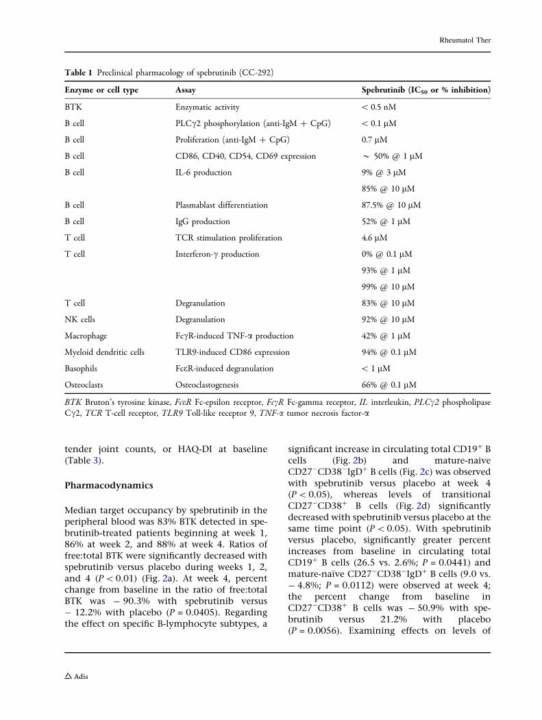
To explore spebrutinib pharmacology onimmune responses, various primary humancellular models were tested using lymphoid andmyeloid cells in vitro. Spebrutinib inhibitedB-cell proliferation with an IC50 of 0.7 lM;reduced expression of activation markers CD86,CD40, CD54, and CD69; and inhibited IL-6production (Table 1). B-cell differentiation toplasmablasts was inhibited, as was their abilityto secrete IgG. T-cell proliferation was inhibitedwith an IC50 of 4.6 lM, which was 6.5-fold lesspotent than inhibition of B-cell proliferation.T-cell interferon-c production, as well asdegranulation of T cells and NK cells, wasinhibited by spebrutinib in the 1- to 10-lMrange. In myeloid cells, spebrutinib reducedFccR-stimulated macrophage TNF-a productionand Toll-like receptor 9-stimulated dendriticcell CD86 expression. Potent inhibition ofFCeR-induced basophil degranulation(IC50\ 1 lM) and osteoclastogenesis wereobserved (66% at 0.1 lM) (Table 1). This phar-macologic effects pattern, including suppres-sion of adaptive and innate immune responses,combined with inhibition of osteoclastogenesisand good in vivo efficacy in the collagen-in-duced arthritis model [7], led to testing spe-brutinib in a phase 2 proof-of-concept clinicaltrial of patients with RA.
Clinical Study in RA Patients
Forty-seven patients were randomized (placebo:N = 23; spebrutinib: N = 24) and 44 (22 in eachgroup) completed treatment during the studyperiod. Three patients were discontinued for aprotocol violation (placebo: N = 1) and with-drawal of consent (spebrutinib: N = 2).
Baseline patient demographics and diseasecharacteristics were well balanced betweentreatment groups (Table 2). Mean age forpatients was 54.8 years; most (66.0%) patientswere 40–65 years of age and the majority(78.7%) were white. Mean body mass index ofparticipants was 31.1 kg/m2.
ACR Responses
The primary endpoint, ACR20 response at week4, was achieved by 41.7% (10/24) of spebruti-nib-treated patients versus 21.7% (5/23) of pla-cebo patients. Although the trend favoredtreatment with spebrutinib, the differencebetween spebrutinib and placebo (20%) was notstatistically significant (P = 0.2493) (Fig. 1).Some spebrutinib-treated patients achieved anACR20 response as early as week 1, andresponses continued through week 2 and week4. For secondary efficacy endpoints, week 4ACR50 response rates were 16.7% (4/24) withspebrutinib and 8.7% (2/23) with placebo, whileweek 4 ACR70 response rates were 8.3% (2/24)and 4.3% (1/23), respectively. Percent differ-ences between spebrutinib and placebo regard-ing ACR50 response (8.0%) and ACR70 response(4.0%) were not statistically significant(P = 0.7029 and P = 1.0000, respectively)(Fig. 1).
In post hoc analyses comparing week 4ACR20 response rates in patients with andwithout prior biologic therapy, the effectobserved in spebrutinib-treated patients versusplacebo patients was consistent with the pri-mary efficacy results. Specifically, 60% (3/5) ofspebrutinib-treated patients and 20% (1/5) ofplacebo patients previously treated with bio-logics achieved an ACR20 response at week 4.
Subgroup Analyses Among ACR20Responders
Subgroup analyses of responder versus non-re-sponder patients demonstrated that respondersin both the placebo and spebrutinib treatmentarms had a shorter duration of disease com-pared with non-responders. The mean durationof RA disease was 3.4 years among responders inboth the placebo and spebrutinib treatmentarms, compared with 7.6 years for non-respon-ders in the placebo arm and 9.7 years for non-responders in the spebrutinib treatment arm(Table 3). There was no difference in diseaseseverity between responders and non-respon-ders, as evidenced by the baseline 28-jointcount Disease Activity Score, swollen and
Rheumatol Ther
tender joint counts, or HAQ-DI at baseline(Table 3).
Pharmacodynamics
Median target occupancy by spebrutinib in theperipheral blood was 83% BTK detected in spe-brutinib-treated patients beginning at week 1,86% at week 2, and 88% at week 4. Ratios offree:total BTK were significantly decreased withspebrutinib versus placebo during weeks 1, 2,and 4 (P\0.01) (Fig. 2a). At week 4, percentchange from baseline in the ratio of free:totalBTK was - 90.3% with spebrutinib versus- 12.2% with placebo (P = 0.0405). Regardingthe effect on specific B-lymphocyte subtypes, a
significant increase in circulating total CD19? Bcells (Fig. 2b) and mature-naiveCD27-CD38-IgD? B cells (Fig. 2c) was observedwith spebrutinib versus placebo at week 4(P\0.05), whereas levels of transitionalCD27-CD38? B cells (Fig. 2d) significantlydecreased with spebrutinib versus placebo at thesame time point (P\0.05). With spebrutinibversus placebo, significantly greater percentincreases from baseline in circulating totalCD19? B cells (26.5 vs. 2.6%; P = 0.0441) andmature-naıve CD27-CD38-IgD? B cells (9.0 vs.- 4.8%; P = 0.0112) were observed at week 4;the percent change from baseline inCD27-CD38? B cells was - 50.9% with spe-brutinib versus 21.2% with placebo(P = 0.0056). Examining effects on levels of
Table 1 Preclinical pharmacology of spebrutinib (CC-292)
Enzyme or cell type Assay Spebrutinib (IC50 or % inhibition)
BTK Enzymatic activity \ 0.5 nM
B cell PLCc2 phosphorylation (anti-IgM ? CpG) \ 0.1 lM
B cell Proliferation (anti-IgM ? CpG) 0.7 lM
B cell CD86, CD40, CD54, CD69 expression * 50% @ 1 lM
B cell IL-6 production 9% @ 3 lM
85% @ 10 lM
B cell Plasmablast differentiation 87.5% @ 10 lM
B cell IgG production 52% @ 1 lM
T cell TCR stimulation proliferation 4.6 lM
T cell Interferon-c production 0% @ 0.1 lM
93% @ 1 lM
99% @ 10 lM
T cell Degranulation 83% @ 10 lM
NK cells Degranulation 92% @ 10 lM
Macrophage FccR-induced TNF-a production 42% @ 1 lM
Myeloid dendritic cells TLR9-induced CD86 expression 94% @ 0.1 lM
Basophils FceR-induced degranulation \ 1 lM
Osteoclasts Osteoclastogenesis 66% @ 0.1 lM
BTK Bruton’s tyrosine kinase, FceR Fc-epsilon receptor, FccR Fc-gamma receptor, IL interleukin, PLCc2 phospholipaseCc2, TCR T-cell receptor, TLR9 Toll-like receptor 9, TNF-a tumor necrosis factor-a
Rheumatol Ther

circulating class switched and activated mem-ory B cells, a trend toward reduced levels ofthese cell types was observed (data not shown).
Spebrutinib treatment significantlydecreased median CTX-I levels versus placebo(- 14.7 vs. ? 8.5%) at week 4, indicatingdecreased osteoclastogenesis (Fig. 3a). At week4, CXCL13 significantly decreased in spebruti-nib-treated patients, while it increased inpatients treated with placebo (median - 26.73vs. 5.36%, respectively) (Fig. 3b). A significantlygreater median percent decrease in MIP-1b wasobserved in spebrutinib-treated patients
compared with placebo patients at week 4(- 18.43 vs. - 2.12%, respectively) (Fig. 3c).Haptoglobin increased with spebrutinib whileplacebo was unchanged (median ? 21.9 vs.- 8%) (Fig. 3d). The von Willebrand factorincreased in placebo patients over 4 weeks(median ? 33.1%), while spebrutinib-treatedpatients showed no significant change (median? 1.8%) from baseline (data not shown). Non-significant treatment differences in serum fac-tors were observed with a-2-macroglobulin, b-2-microglobulin, brain-derived neurotrophic fac-tor, eosinophil chemotactic protein-1,
Table 2 Baseline patient demographics and disease characteristics
Characteristic Placebo (N = 23) Spebrutinib 375 mg/day (N = 24)
Age (years), mean (SD) 54.6 (13.2) 55.0 (14.6)
Race, n (%)
White 17 (73.9) 20 (83.3)
African American 6 (26.1) 4 (16.7)
Weight (kg), mean (SD) 81.5 (16.7) 82.7 (18.8)
Body mass index (kg/m2), mean (SD) 30.8 (6.3) 31.3 (7.5)
Duration of RA (years), mean (SD) 6.7 (7.3) 7.1 (9.9)
Swollen joint count (0–66), mean (SD) 14.5 (7.5) 16.8 (8.9)
Tender joint count (0–68), mean (SD) 26.3 (14.8) 26.5 (13.8)
hsCRP (mg/dl), mean (SD) 6.6 (8.1) 5.5 (5.5)
DAS-28, mean (SD) 5.2 (0.9) 5.4 (1.0)
HAQ-DI score, mean (SD) 1.27 (0.72) 1.37 (0.62)
Erythrocyte sedimentation rate (mm/h), mean (SD) 32.1 (18.2) 29.2 (14.6)
Anti-CCP antibody positive, n (%) 15 (65.2) 16 (66.7)
High rheumatoid factor, n (%) 15 (65.2) 16 (66.7)
Methotrexate dose (mg/week), mean (SD) 15.5 (3.6) 16.5 (4.4)
NSAID use, n (%) 8 (34.8) 10 (41.7)
Oral corticosteroid use, n (%) 4 (17.4) 5 (20.8)
Hydroxychloroquine or chloroquine, n (%) 2 (8.7) 2 (8.3)
Prior use of biologic DMARDs, n (%) 5 (21.7) 5 (20.8)
Baseline use of NSAIDs was required to continue concomitantly, per protocolCCP cyclic citrullinated peptide, DAS-28 28-joint count Disease Activity Score, DMARDs disease-modifying anti-rheumaticdrugs, hsCRP high-sensitivity C-reactive protein, NSAIDs non-steroidal anti-inflammatory drugs, RA rheumatoid arthritis
Rheumatol Ther

C-reactive protein, complement C3, ferritin, IL-6, IL-8, regulated on activation normal T cellexpressed and secreted, stem cell factor, solubleintracellular adhesion molecule-1, vascularepithelial growth factor, hyaluronic acid, andtissue inhibitor of metalloproteinase-1 (data notshown).
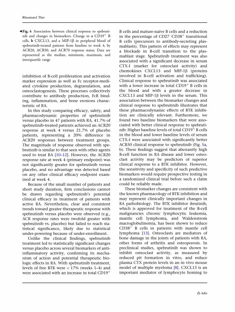
Association Between SpebrutinibPharmacodynamics and Clinical Response
Post hoc analyses aimed at comparing pharma-codynamic changes between baseline and week4 among spebrutinib clinical responders andnon-responders revealed the following associa-tions. Patients who achieved an ACR50 clinicalresponse with spebrutinib treatment experi-enced a lower median percent increase inCD19? B cell levels from baseline to week 4compared with ACR50 non-responders (? 15.4vs. ? 42.9%; P = 0.06) (Fig. 4a). Similar trendswere observed when comparing CD19? B cellmedian percent change from baseline to week 4in spebrutinib responders versus non-respon-ders, as measured using ACR20 or ACR70response criteria (Fig. 4a). There were no differ-ences between spebrutinib responders and non-responders for changes in mature-naıve B cellsor transitional B cells (data not shown). Forserum CXCL13, patients who achieved anACR50 clinical response with spebrutinib
experienced a greater median percent decreasefrom baseline to week 4 compared with ACR50non-responders (- 47.2 vs. - 14.8%; P = 0.04)(Fig. 4b). Similar trends were observed whencomparing CXCL13 percent change from base-line to week 4 in spebrutinib responders versusnon-responders, as measured using ACR20 orACR70 response criteria (Fig. 4b). For serumMIP-1b, patients who achieved an ACR20 clin-ical response with spebrutinib experienced agreater median percent decrease in MIP-1b levelfrom baseline to week 4 compared with ACR20non-responders (- 31.5 vs. - 8.7%; P = 0.10)(Fig. 4c). Similar trends were observed whencomparing median MIP-1b percent change frombaseline to week 4 in spebrutinib respondersversus non-responders, as measured usingACR50 or ACR70 response criteria (Fig. 4c).
Biomarkers for Prediction of ClinicalResponse to Spebrutinib
Post hoc analyses aimed at predicting spebruti-nib clinical response revealed the followingassociations. Patients who achieved an ACR50clinical response to spebrutinib treatment had asignificantly higher median CD19? B cell levelin the blood at baseline compared with ACR50non-responders (18.8 vs. 9.6%; P = 0.027)(Fig. 5a). A similar relationship was observedwhen comparing median baseline CD19? B cell
Fig. 1 Patients achieving ACR20, ACR50, and ACR70 responses at week 4. ACR American College of Rheumatologyresponse criteria
Rheumatol Ther

Table 3 Baseline patient demographics and disease characteristics by ACR20 response status
Characteristic Placebo (N = 23) Spebrutinib 375 mg/day (N = 24)
ACR20 non-responders (N = 18)
ACR20responders(N = 5)
ACR20 non-responders (N = 14)
ACR20responders(N = 10)
Age (years), mean (SD) 57.2 (12.4) 45.0 (12.5) 55.5 (17.4) 54.3 (10.3)
Race, n (%)
White 13.0 (72.2) 4.0 (80.0) 12.0 (85.7) 8.0 (80.0)
African American 5.0 (27.8) 1.0 (20.0) 2.0 (14.3) 2.0 (20.0)
Weight (kg), mean (SD) 84.7 (16.8) 69.9 (10.8) 79.4 (18.5) 87.3 (19.4)
Body mass index (kg/m2), mean
(SD)
32.0 (6.5) 26.6 (3.1) 30.1 (6.9) 33.0 (8.4)
Duration of RA (years), mean
(SD)
7.6 (8.0) 3.4 (1.7) 9.7 (12.1) 3.4 (3.8)
Swollen joint count (0–66),
mean (SD)
13.7 (6.9) 17.6 (9.5) 16.9 (9.6) 16.6 (8.3)
Tender joint count (0–68),
mean (SD)
26.9 (16.0) 23.8 (10.5) 26.5 (15.0) 26.5 (12.7)
hsCRP (mg/dl), mean (SD) 7.6 (8.9) 3.0 (2.1) 5.9 (7.1) 5.0 (2.5)
DAS-28, mean (SD) 5.3 (0.9) 4.9 (0.9) 5.3 (1.2) 5.5 (0.8)
HAQ-DI score, mean (SD) 1.2 (0.7) 1.7 (0.5) 1.3 (0.7) 1.5 (0.6)
Erythrocyte sedimentation rate
(mm/h), mean (SD)
29.0 (11.8) 43.2 (32.2) 23.4 (11.6) 37.3 (14.9)
Anti-CCP antibody positive,
n (%)
10.0 (55.6) 5.0 (100.0) 9.0 (64.3) 7.0 (70.0)
High rheumatoid factor, n (%) 10.0 (55.6) 5.0 (100.0) 11.0 (78.6) 5.0 (50.0)
Methotrexate dose (mg/week),
mean (SD)
15.1 (3.03) 17.0 (5.42) 15.9 (3.62) 17.3 (5.33)
NSAID use, n (%) 6.0 (33.3) 2.0 (40.0) 6.0 (42.9) 4.0 (40.0)
Oral corticosteroid use, n (%) 2.0 (11.1) 2.0 (40.0) 2.0 (14.3) 3.0 (30.0)
Hydroxychloroquine or
chloroquine, n (%)
1.0 (5.6) 1.0 (20.0) 2.0 (14.3) 0.0 (0.00)
Prior use of biologic DMARDs,
n (%)
4.0 (22.2) 1.0 (20.0) 2.0 (14.3) 3.0 (30.0)
CCP cyclic citrullinated peptide, DAS-28 28-joint count Disease Activity Score, DMARDs disease-modifying anti-rheumaticdrugs, hsCRP high-sensitivity C-reactive protein, NSAID non-steroidal anti-inflammatory drug, RA rheumatoid arthritis
Rheumatol Ther

levels in spebrutinib ACR70 responders versusnon-responders (20.8 vs. 11.0%; P = 0.048)(Fig. 5a). The median baseline CTX-I serumlevel was significantly lower among spebrutinibACR50 responders compared with ACR50 non-responders (0.39 vs. 0.49 lg/l; P = 0.032)(Fig. 5b). A similar trend was observed whencomparing spebrutinib ACR20 or ACR70responders with non-responders: the spebruti-nib responders had a lower median CTX-I serumlevel at baseline than the non-responders(Fig. 5b). There were no differences in median
baseline serum CXCL13 or MIP-1b levelsbetween spebrutinib responders and non-re-sponders (data not shown).
Safety
Spebrutinib was well tolerated over the 4-weektreatment phase. Most TEAEs were mild ormoderate in severity; one severe TEAE (stom-atitis) was reported with spebrutinib. The mostfrequently reported events (C 5% in eithertreatment group) were nausea, back pain,
Fig. 2 Free BTK to total BTK in PBMC and spebruti-nib’s effects on B-cell subsets. a Fraction of free BTK tototal BTK in PBMC and b–d effects of spebrutinib onB-cell subsets in circulation. The binding of spebrutinib toBTK in PBMC is shown in a as the ratio of free(unbound) BTK to total BTK. Flow cytometry analysis ofwhole blood from patients at the indicated times post-treatment are shown as b percent change from baseline for
CD19? total B cells, c CD19?CD27-CD38-IgD?
mature naive B cells, and d CD19?CD27-CD38? tran-sitional B cells at weeks 1, 2, and 4. Data are represented asmedian values ± 95% confidence interval of placebo (opencircles) and spebrutinib treatment (open triangles).*P\ 0.05; **P\ 0.01 versus collection date-matchedplacebo. BTK Bruton’s tyrosine kinase, PBMC peripheralblood mononuclear cells
Rheumatol Ther

diarrhea, cough, and migraine (Table 4); nopatients were discontinued from spebrutinibbecause of TEAEs, and no serious TEAEs werereported during the study. No treatment-relatedtrends were observed in hematology and serumchemistry results; few patients had markedlyabnormal hematology values (placebo: N = 4;spebrutinib: N = 3).
Blood pressure, vital signs, electrocardio-gram, and ophthalmologic findings were gen-erally unremarkable with no treatment-relatedtrends. TEAEs associated with electrocardiogramfindings were reported in two spebrutinib-trea-ted patients; however, neither event was
suspected to be related to treatment. TEAEsassociated with ophthalmologic findings werereported in one spebrutinib-treated patient andone placebo patient; neither event was sus-pected to be related to treatment.
DISCUSSION
Using primary human immune cells, the cova-lent BTK inhibitor spebrutinib inhibited cellularresponses associated with BTK signaling,including BCR, FccR, FceR, and Toll-like recep-tor 9 pathways. These effects included
Fig. 3 Changes in CTX-I expression and inflammation-associated proteins over time after administration ofspebrutinib. Plasma levels from subjects at the indicatedtimes post-treatment are shown as percent change frombaseline for a CTX-I, b CXCL13, c MIP-1b, andd haptoglobin at weeks 1, 2, and 4. Data are represented
as the median ± 95% confidence interval of placebo (opencircles) and spebrutinib treatment (open triangles).*P\ 0.05; **P\ 0.01; ***P\ 0.001 versus collectiondate-matched placebo. CTX-I carboxy-terminal collagencross-linking telopeptide, CXCL13 chemokine ligand 13,MIP-1b macrophage inflammatory protein-1b
Rheumatol Ther

Rheumatol Ther
inhibition of B-cell proliferation and activationmarker expression as well as Fc receptor-medi-ated cytokine production, degranulation, andosteoclastogenesis. These processes collectivelycontribute to antibody production, FcR bind-ing, inflammation, and bone erosions charac-teristic of RA.
In this study comparing efficacy, safety, andpharmacodynamic properties of spebrutinibversus placebo in 47 patients with RA, 41.7% ofspebrutinib-treated patients achieved an ACR20response at week 4 versus 21.7% of placebopatients, representing a 20% difference inACR20 responses between treatment groups.The magnitude of response observed with spe-brutinib is similar to that seen with other agentsused to treat RA [10–12]. However, the ACR20response rate at week 4 (primary endpoint) wasnot significantly greater for spebrutinib versusplacebo, and no advantage was detected basedon any other clinical efficacy endpoint exam-ined at week 4.
Because of the small number of patients andshort study duration, firm conclusions cannotbe drawn regarding spebrutinib’s potentialclinical efficacy in treatment of patients withactive RA. Nevertheless, clear and consistenttrends toward greater therapeutic response withspebrutinib versus placebo were observed (e.g.,ACR response rates were twofold greater withspebrutinib vs. placebo) but failed to reach sta-tistical significance, likely due to statisticalunder-powering because of under-enrollment.
Unlike the clinical findings, spebrutinibtreatment led to statistically significant changesversus placebo across several biomarkers of anti-inflammatory activity, confirming its mecha-nism of action and potential therapeutic bio-logic effects in RA. With spebrutinib treatment,levels of free BTK were\ 17% (weeks 1–4) andwere associated with an increase in total CD19?
B cells and mature-naıve B cells and a reductionin the percentage of CD27-CD38? transitionalB cells (precursors to antibody-secreting plas-mablasts). This pattern of effects may representa blockade in B-cell transition to the plas-mablast stage. Spebrutinib treatment was alsoassociated with a significant decrease in serumCTX-I (marker for osteoclast activity) andchemokines CXCL13 and MIP-1b (proteinsinvolved in B-cell activation and trafficking).Clinical response to spebrutinib was associatedwith a lower increase in total CD19? B cells inthe blood and with a greater decrease inCXCL13 and MIP-1b levels in the serum. Thisassociation between the biomarker changes andclinical response to spebrutinib illustrates thatthese pharmacodynamic effects of BTK inhibi-tion are clinically relevant. Furthermore, wefound two baseline biomarkers that were asso-ciated with better clinical response to spebruti-nib: Higher baseline levels of total CD19? B cellsin the blood and lower baseline levels of serumCTX-I were associated with significantly betterACR50 clinical response to spebrutinib (Fig. 5a,b). These findings suggest that aberrantly highB-cell function in RA disease and lower osteo-clast activity may be predictors of superiorclinical response to a BTK inhibitor. However,the sensitivity and specificity of such predictivebiomarkers would require prospective testing ina randomized clinical trial before such a claimcould be reliably made.
These biomarker changes are consistent withthe known pharmacology of BTK inhibition andmay represent clinically important changes inRA pathobiology. The BTK inhibitor ibrutinib,which is approved for treatment of the B-cellmalignancies chronic lymphocytic leukemia,mantle cell lymphoma, and Waldenstrommacroglobulinemia, has been shown to reduceCD38? B cells in patients with mantle celllymphoma [13]. Osteoclasts are mediators ofbone damage in the joints of patients with RA,other forms of arthritis and osteoporosis. Inpreclinical studies, spebrutinib was shown toinhibit osteoclast activity, as measured byreduced pit formation in vitro, and reduceplasma CTX protein levels in an in vivo mousemodel of multiple myeloma [8]. CXCL13 is animportant mediator of lymphocyte homing to
bFig. 4 Association between clinical response to spebruti-nib and changes in biomarkers. Change in a CD19? Bcells, b CXCL13, and c MIP-1b in peripheral blood ofspebrutinib-treated patients from baseline to week 4, byACR20, ACR50, and ACR70 response status. Data arerepresented as the median, minimum, maximum, andinterquartile range
Rheumatol Ther

Fig. 5 Association between clinical response to spebruti-nib and baseline biomarkers. Baseline levels of a CD19? Bcells and b CTX-I in peripheral blood of spebrutinib-
treated patients by ACR20, ACR50, and ACR70 responsestatus. Data are represented as the median, minimum,maximum, and interquartile range
Table 4 Adverse events reported during the 4-week placebo-controlled study period
Patients Placebo (N = 23) Spebrutinib 375 mg/day (N = 24)
Adverse events, n (%)
C 1 adverse event 11 (47.8) 14 (58.3)
C 1 serious adverse event 0 (0.0) 0 (0.0)
C 1 severe adverse event 0 (0.0) 1 (4.2)
Adverse event leading to drug withdrawal 0 (0.0) 0 (0.0)
Adverse events in any treatment group (C 5% of patients), n (%)
Nausea 2 (8.7) 5 (20.8)
Back pain 1 (4.3) 2 (8.3)
Diarrhea 1 (4.3) 2 (8.3)
Cough 0 (0.0) 2 (8.3)
Migraine 0 (0.0) 2 (8.3)
Rheumatol Ther

the lymph node, which occurs in a BTK-de-pendent manner [4]. Ibrutinib has been shownto directly inhibit CXCL13 production bymacrophages in vitro [14]. Ibrutinib was shownto reduce blood CXCL13 protein levels inpatients with mantle cell lymphoma [11] andWaldenstrom macroglobulinemia; highCXCL13 levels at baseline were associated withmajor clinical response in patients withWaldenstrom macroglobulinemia [15]. CXCL13is highly relevant in the pathogenesis of RA, ashigh CXCL13 expression levels in the RA syn-ovium have been associated with a lymphoidsynovial phenotype; CXCL13 is more fre-quently observed in patients with RA whoexpress anti-citrullinated protein antibodiesand is a marker of more severe disease [16].Furthermore, high CXCL13 serum levels havebeen reproducibly associated with poor clinicalresponse to anti-TNF biologic in patients withRA [17, 18]. Thus, the ability of a BTK inhibitorto reduce CD38? B cells, osteoclast-mediatedbone damage, and CXCL13-mediated lympho-cytic inflammation would be potentiallymeaningful pathobiologic changes in patientswith RA.
During the review of this report, it wasreported that the BTK inhibitor fenebrutinibsignificantly improved ACR50 clinical responserates in RA patients with an inadequateresponse to methotrexate or TNF inhibitors(Cohen S, et al. EULAR 2019 [abstract OP0025];12 June 2019; Madrid, Spain). The fenebrutinibstudy enrolled 480 patients, and the treatmentduration was 12 weeks. By comparison, thecurrent spebrutinib RA study was much smaller,enrolling only 47 patients, with a treatmentduration of only 4 weeks. Nonetheless, both thefenebrutinib study and the spebrutinib trialsshowed similar pharmacodynamic effects bythese two BTK inhibitors. Fenbrutinib, which isa reversible BTK inhibitor, significantlydecreased CXCL13 and MIP-1b (CCL4) (Mori-moto A, et al. EULAR 2019 [abstract FRI0129];14 June 2019; Madrid, Spain). Spebrutinib,which is an irreversible BTK inhibitor, likewisereduced CXCL13 and MIP-1b (CCL4) (Fig. 3b,c). Therefore, regardless of whether the BTKinhibitor binds in a reversible or irreversiblemanner, the pharmacodynamic effects on
downstream B-cell and myeloid chemokinesappear to be the same.
Spebrutinib was well tolerated, as mostTEAEs were mild or moderate in severity and nopatient experienced a TEAE resulting in studydiscontinuation or dose reduction. Nausea wasthe most common TEAE reported; no otherindividual events were experienced by morethan two patients in either group. Overall, therewere no remarkable findings or changes in lab-oratory evaluations, vital sign measurements,electrocardiogram readings, or ophthalmologicexaminations.
This study had several limitations. The studyduration was 4 weeks and low enrollmentunder-powered the study; therefore, a thera-peutic benefit of spebrutinib versus placebocould not be detected. Another limitation wasthe short-term treatment period of 4 weeks.Finally, the spebrutinib dose in this study waschosen based on a small phase 1 study in heal-thy volunteers [7]. Higher spebrutinib doses (upto 1000 mg/day) were administered safely in aphase 1 study in patients with relapsed/refrac-tory chronic lymphocytic leukemia [19]; there-fore, the current study might have been moreinformative if a higher range of spebrutinibdoses had been used.
CONCLUSIONS
In summary, although a clear conclusion on theclinical benefit of spebrutinib cannot be made,significant pharmacodynamic findings andnumerical trends in efficacy observed in spe-brutinib-treated patients suggest covalent BTKinhibition may have clinical potential for thetreatment of RA.
ACKNOWLEDGEMENTS
The authors thank the patients who partici-pated in this study and multiple study sites andstudy investigators.
Funding. This study and the Rapid ServiceFee were funded by Celgene Corporation.
Rheumatol Ther

Editorial Assistance. The authors receivededitorial support in the preparation of thisreport from Kristin Carlin, RPh, MBA, of Pelo-ton Advantage, LLC, an OPEN Health company,Parsippany, NJ, USA, sponsored by CelgeneCorporation, Summit, NJ, USA. The authors,however, directed and are fully responsible forall content and editorial decisions for thisreport.
Authorship. All named authors meet theInternational Committee of Medical JournalEditors (ICMJE) criteria for authorship for thisarticle, take responsibility for the integrity ofthe work as a whole, and have given theirapproval for this version to be published.
Authorship Contributions. All authors wereinvolved in the drafting and critical review ofthe manuscript and approved the final versionfor submission. PHS, AJK, JM, SK, DS, LL, AA, JK,MA, LC, EMH, DRH, and GER were involvedwith the conception or design of the work, thedevelopment of the statistical analysis plan, andthe acquisition of clinical data and participatedin the clinical study from which data arereported in the manuscript. All authors agree tobe accountable for all aspects of the work andattest to the accuracy and integrity of the work.
Disclosures. Peter H. Schafer is an employeeof and has stocks or stock options in CelgeneCorporation. Jianglin Ma is an employee of andhas stocks or stock options in Celgene Corpo-ration. Shimon Korish is an employee of andhas stocks or stock options in Celgene Corpo-ration. Donna Sutherland is an employee of andhas stocks or stock options in Celgene Corpo-ration. Li Li is an employee of and has stocks orstock options in Celgene Corporation. JolantaKosek is an employee of and has stocks or stockoptions in Celgene Corporation. Mary Adams isan employee of and has stocks or stock optionsin Celgene Corporation. Lori Capone is anemployee of and has stocks or stock options inCelgene Corporation. Eun Mi Hur is anemployee of and has stocks or stock options inCelgene Corporation. Garth E. Ringheim is anemployee of and has stocks or stock options inCelgene Corporation. Alan J. Kivitz has received
grant/research support and served as a consul-tant and speaker for Celgene Corporation. AdaAzaryan was employed by Celgene Corporationat the time of study conduct and is nowemployed by Genmab USA, Inc. Douglas R.Hough was an employee of Celgene Corpora-tion at the time of study conduct and is nowretired.
Compliance with Ethics Guidelines. Thestudy was done in accordance with the Inter-national Conference on Harmonisation E6requirements for Good Clinical Practice and inaccordance with the ethical principles outlinedin the Declaration of Helsinki. The study pro-tocol, amendments, and informed consent formwere approved by the institutional review boardat each investigational site or by a centralreview board, and all enrolled patients providedwritten informed consent before starting thestudy. Schulman Associates IRB in Cincinnati,OH (IRB #1 registration number 00000971) wasthe main ethics committee. The names of theindependent ethics committee and/or institu-tional review board at each investigational siteare listed in the Supplementary Material.
Data Availability. Celgene is committed toresponsible and transparent sharing of clinicaltrial data with patients, healthcare practition-ers, and independent researchers for the pur-pose of improving scientific and medicalknowledge as well as fostering innovativetreatment approaches. For more information,please visit: https://www.celgene.com/research-development/clinical-trials/clinical-trials-data-sharing/.
Open Access. This article is distributedunder the terms of the Creative CommonsAttribution-NonCommercial 4.0 InternationalLicense (http://creativecommons.org/licenses/by-nc/4.0/), which permits any noncommer-cial use, distribution, and reproduction in anymedium, provided you give appropriate creditto the original author(s) and the source, providea link to the Creative Commons license, andindicate if changes were made.
Rheumatol Ther

REFERENCES
1. Chang BY, Huang MM, Francesco M, Chen J,Sokolove J, Magadala P, et al. The Bruton tyrosinekinase inhibitor PCI-32765 ameliorates autoim-mune arthritis by inhibition of multiple effectorcells. Arthritis Res Ther. 2011;13(4):R115.
2. Niiro H, Clark EA. Regulation of B-cell fate byantigen-receptor signals. Nat Rev Immunol.2002;2(12):945–56.
3. Khan WN. Regulation of B lymphocyte develop-ment and activation by Bruton’s tyrosine kinase.Immunol Res. 2001;23(2–3):147–56.
4. de Gorter DJ, Beuling EA, Kersseboom R, Midden-dorp S, van Gils JM, Hendriks RW, et al. Bruton’styrosine kinase and phospholipase Cc2 mediatechemokine-controlled B cell migration and hom-ing. Immunity. 2007;26(1):93–104.
5. de Rooij MF, Kuil A, Geest CR, Eldering E, ChangBY, Buggy JJ, et al. The clinically active BTK inhi-bitor PCI-32765 targets B-cell receptor- and che-mokine-controlled adhesion and migration inchronic lymphocytic leukemia. Blood.2012;119(11):2590–4.
6. Kivitz A, Gupta R, Valenzuela G, Smith E, RehmanQ, El-Kadi H, et al. A phase 2a, 4-week double-blind,proof-of-concept efficacy and safety study of CC-292 versus placebo as co-therapy with methotrexatein active rheumatoid arthritis [poster 1587]. Pre-sented at: the Annual Meeting of the AmericanCollege of Rheumatology; November 12–16, 2016;Washington, DC.
7. Evans EK, Tester R, Aslanian S, Karp R, Sheets M,Labenski MT, et al. Inhibition of Btk with CC-292provides early pharmacodynamic assessment ofactivity in mice and humans. J Pharmacol Exp Ther.2013;346(2):219–28.
8. Eda H, Santo L, Cirstea DD, Yee AJ, Scullen TA,Nemani N, et al. A novel Bruton’s tyrosine kinaseinhibitor CC-292 in combination with the protea-some inhibitor carfilzomib impacts the bonemicroenvironment in a multiple myeloma modelwith resultant antimyeloma activity. Leukemia.2014;28(9):1892–901.
9. Nakayama Y, Kosek J, Capone L, Hur EM, SchaferPH, Ringheim GE. Aiolos overexpression in sys-temic lupus erythematosus B cell subtypes andBAFF-induced memory B cell differentiation arereduced by CC-220 modulation of cereblon activ-ity. J Immunol. 2017;199(7):2388–407.
10. Weinblatt ME, Fleischmann R, Huizinga TW, EmeryP, Pope J, Massarotti EM, et al. Efficacy and safety of
certolizumab pegol in a broad population ofpatients with active rheumatoid arthritis: resultsfrom the REALISTIC phase IIIb study. Rheumatol-ogy (Oxford). 2012;51(12):2204–14.
11. Hobbs K, Deodhar A, Wang B, Bitman B, NussbaumJ, Chung J, et al. Randomized, double-blind, pla-cebo-controlled study to evaluate the efficacy andsafety of etanercept in patients with moderatelyactive rheumatoid arthritis despite DMARD ther-apy. SpringerPlus. 2015;4:113.
12. van de Putte LB, Atkins C, Malaise M, Sany J, RussellAS, van Riel PL, et al. Efficacy and safety of adali-mumab as monotherapy in patients with rheuma-toid arthritis for whom previous disease-modifyingantirheumatic drug treatment has failed. AnnRheum Dis. 2004;63(5):508–16.
13. Chang BY, Francesco M, De Rooij MF, Magadala P,Steggerda SM, Huang MM, et al. Egress ofCD19(?)CD5(?) cells into peripheral blood fol-lowing treatment with the Bruton tyrosine kinaseinhibitor ibrutinib in mantle cell lymphomapatients. Blood. 2013;122(14):2412–24.
14. Ping L, Ding N, Shi Y, Feng L, Li J, Liu Y, et al. TheBruton’s tyrosine kinase inhibitor ibrutinib exertsimmunomodulatory effects through regulation oftumor-infiltrating macrophages. Oncotarget.2017;8(24):39218–29.
15. Vos JM, Tsakmaklis N, Patterson CJ, Meid K, Cas-tillo JJ, Brodsky P, et al. CXCL13 levels are elevatedin patients with Waldenstrom’s macroglobuline-mia, and are predictive of major response to ibru-tinib. Haematologica. 2017;102(11):e452–5.
16. Bugatti S, Manzo A, Vitolo B, Benaglio F, Binda E,Scarabelli M, et al. High expression levels of the Bcell chemoattractant CXCL13 in rheumatoid syn-ovium are a marker of severe disease. Rheumatology(Oxford). 2014;53(10):1886–95.
17. Dennis G Jr, Holweg CT, Kummerfeld SK, Choy DF,Setiadi AF, Hackney JA, et al. Synovial phenotypes inrheumatoid arthritis correlate with response to bio-logic therapeutics. Arthritis Res Ther. 2014;16(2):R90.
18. Folkersen L, Brynedal B, Diaz-Gallo LM, RamskoldD, Shchetynsky K, Westerlind H, et al. Integrationof known DNA, RNA and protein biomarkers pro-vides prediction of anti-TNF response in rheuma-toid arthritis: results from the COMBINE study. MolMed. 2016;22:322–8.
19. Brown JR, Harb WA, Hill BT, Gabrilove J, SharmanJP, Schreeder MT, et al. Phase I study of single-agentCC-292, a highly selective Bruton’s tyrosine kinaseinhibitor, in relapsed/refractory chronic lympho-cytic leukemia. Haematologica.2016;101(7):e295–8.
Rheumatol Ther