specific inhibition of β-secretase processing of the ... · article specific inhibition of...
TRANSCRIPT

Article
Specific Inhibition of b-Se
cretase Processing of theAlzheimer Disease Amyloid Precursor ProteinGraphical Abstract
Highlights
d The AD-linked protease BACE1 cleaves APP to produce toxic
b-amyloid peptides
d BACE1 also cleaves the non-amyloid substrates NRG1 and
L1
d BACE1 cleavage of NRG1 and L1 is endocytosis-
independent, unlike the cleavage of APP
d The endosomally targeted BACE1 inhibitor spares NRG1 and
L1 but inhibits APP processing
Ben Halima et al., 2016, Cell Reports 14, 2127–2141March 8, 2016 ª2016 The Authorshttp://dx.doi.org/10.1016/j.celrep.2016.01.076
Authors
Saoussen Ben Halima, Sabyashachi
Mishra, K. Muruga Poopathi Raja, ...,
Christian Haass, Amedeo Caflisch,
Lawrence Rajendran
In Brief
Ben Halima et al. demonstrate the
feasibility of designing drugs targeting
the Alzheimer-related enzyme BACE1
without affecting its physiological
function. Using structural, biochemical,
and cellular approaches, they show that
BACE1 inhibitors can be designed to
specifically inhibit its disease-causing
activity, enhancing their potential as
therapeutics without undesired side
effects.
Cell Reports
Article
Specific Inhibition of b-Secretase Processingof the Alzheimer Disease Amyloid Precursor ProteinSaoussen Ben Halima,1,2,3 Sabyashachi Mishra,4,12 K. Muruga Poopathi Raja,5 Michael Willem,6 Antonio Baici,4
Kai Simons,7 Oliver Br€ustle,8,9,10 Philipp Koch,8 Christian Haass,6,9,11 Amedeo Caflisch,4 and Lawrence Rajendran1,2,3,*1Systems and Cell Biology of Neurodegeneration, Institute of Regenerative Medicine, University of Zurich, Wagistrasse 12,
8952 Schlieren, Switzerland2Graduate Program in Neuroscience, Neuroscience Center Zurich, 8057 Zurich, Switzerland3Graduate Program of the Zurich Center for Integrative Human Physiology, University of Zurich, 8057 Zurich, Switzerland4Department of Biochemistry, University of Zurich, 8057 Zurich, Switzerland5Department of Physical Chemistry, School of Chemistry, Madurai Kamaraj University, Tamil Nadu 625002, Madurai, India6Biomedical Center, Ludwig-Maximilians-University, 81337 Munich, Germany7Max Planck Institute of Molecular Cell Biology and Genetics, 01307 Dresden, Germany8Institute of Reconstructive Neurobiology, University of Bonn, 53127 Bonn, Germany9German Center for Neurodegenerative Diseases, 53175 Bonn, Germany10Life & Brain, 53127 Bonn, Germany11Munich Cluster for Systems Neurology (SyNergy), 81377 Munich, Germany12Present address: Department of Chemistry, Indian Institute of Technology of Kharagpur, West Bengal 721302, Kharagpur, India
*Correspondence: [email protected]
http://dx.doi.org/10.1016/j.celrep.2016.01.076This is an open access article under the CC BY-NC-ND license (http://creativecommons.org/licenses/by-nc-nd/4.0/).
SUMMARY
Development of disease-modifying therapeutics isurgently needed for treating Alzheimer disease (AD).AD is characterized by toxic b-amyloid (Ab) peptidesproduced by b- and g-secretase-mediated cleavageof the amyloid precursor protein (APP). b-secretaseinhibitors reduce Ab levels, but mechanism-basedside effects arise because they also inhibit b-cleav-age of non-amyloid substrates like Neuregulin. Wereport thatb-secretase has ahigher affinity forNeure-gulin than it does for APP. Kinetic studies demon-strate that the affinities and catalytic efficiencies ofb-secretase are higher toward non-amyloid sub-strates than toward APP. We show that non-amyloidsubstrates are processed by b-secretase in anendocytosis-independent manner. Exploiting thiscompartmentalization of substrates, we specificallytarget the endosomalb-secretase by an endosomallytargeted b-secretase inhibitor, which blocked cleav-age of APP but not non-amyloid substrates in manycell systems, including induced pluripotent stemcell (iPSC)-derived neurons. b-secretase inhibitorscan be designed to specifically inhibit the Alzheimerprocess, enhancing their potential as AD therapeu-tics without undesired side effects.
INTRODUCTION
Alzheimer disease (AD) is associated with extracellular deposits
of b-amyloid (Ab) peptide (De Strooper, 2010; Hardy and
Higgins, 1992; Tanzi, 2005), which is generated by proteolytic
Ce
processing of the amyloid precursor protein (APP) by the b-sec-
retase BACE1 (Hussain et al., 1999; Sinha et al., 1999; Vassar
et al., 1999; Yan et al., 1999) and g-secretase. Mutations within
the b-cleavage sites of APP can increase the risk for familial
forms of AD (Citron et al., 1992; Thinakaran et al., 1996; Zhou
et al., 2011) or confer protection against cognitive decline in
the elderly (Jonsson et al., 2012), causatively linking BACE1 to
AD. Moreover, because of the failure in recent clinical trials of
g-secretase inhibition, mainly because of mechanism-based
side effects in humans, BACE1 is considered the preferred
drug target (Vassar and Kandalepas, 2011). BACE1 is an attrac-
tive therapeutic target for AD. However, complete abolishment
of BACE1 activity is associated with specific behavioral and
physiological alterations in mice (Cai et al., 2012; Cheret et al.,
2013; Hitt et al., 2012; Kim et al., 2007; Lahiri et al., 2014; Li
and S€udhof, 2004). These alterations may arise from the failure
to process some non-amyloid BACE1 substrates such as
NRG1, which is involved in axonal myelination of neurons (Hu
et al., 2006, 2010; Ma et al., 2007; Willem et al., 2006); immuno-
globulin (Ig)-containing b1 Nrg1 (IgNrg1b1) (Cheret et al., 2013);
the b2 subunit voltage-gated sodium channel (Nav1b2) (Kim
et al., 2007; Wong et al., 2005); and axon guidance molecules,
including CHL1 (Rajapaksha et al., 2011) and L1 (Zhou et al.,
2012), Jagged (Hu et al., 2013), b-galactoside a 2, 6-sialyltrans-
ferase (ST6GalI) (Kitazume et al., 2005; Sugimoto et al., 2007),
APLP1 and APLP2 (Li and S€udhof, 2004), lipoprotein receptor-
related protein (LRP) (von Arnim et al., 2005), interleukin 1 recep-
tor II (IL-1R2, but not tumor necrosis factor a [TNF-a]) (Kuhn
et al., 2007, 2012), and vascular endothelial growth factor recep-
tor 1 (VEGFR1) (Cai et al., 2012). Therefore, a general BACE1
inhibitor might block cleavage of non-amyloid substrates,
decreasing its value as an AD therapeutic drug (Cai et al.,
2012; Cheret et al., 2013; Hitt et al., 2012; Kim et al., 2007; Lahiri
et al., 2014; Li and S€udhof, 2004).
ll Reports 14, 2127–2141, March 8, 2016 ª2016 The Authors 2127
In the cell, proteins are distributed to multiple subcellular loca-
tions (Mellman and Nelson, 2008). Spatial distribution of proteins
is crucial for spatial cellular functions (Rajendran and Simons,
2005). In polarized cells such as neurons, cellular distribution
of proteins allows temporal and spatial control of diverse cellular
functions. Many cellular proteins show multiple subcellular loca-
tions, such as BACE1. BACE1 is located in the trans-Golgi
network (TGN) (Yan et al., 2001), plasma membrane (Walter
et al., 2001a, 2001b), and early endosomes (Rajendran et al.,
2006) and in polarized cells such as neurons because these cells
also display compartmentalization of BACE1 in axons versus so-
matodendritic compartments (Buggia-Prevot et al., 2014; Vassar
et al., 2014). BACE1 cleaves APP in early endosomes after endo-
cytosis (Rajendran et al., 2006). Both the enzyme and the sub-
strate undergo endocytosis, presumably through different
routes, and meet in early endosomes for processing (Rajendran
et al., 2006; Sannerud et al., 2011; Schneider et al., 2008).
Currently it is unknown whether non-amyloid substrates such
as NRG1 or L1 are processed in the endosomal compartment
or other compartments. Selectively inhibiting the activity of
BACE1 in a particular subcellular compartment, early endo-
somes, where it cleaves APP, could be an effective therapeutic
strategy provided the other substrates are cleaved in non-endo-
somal compartments. Therefore, in this work, we explored the
compartmentalization of BACE1 substrate processing to
address the feasibility of targeting BACE1 for inhibition in the
subcellular compartment, where it cleaves the APP substrate.
We studied the expression profiles of BACE1 substrates.
Indeed, RT-PCR analysis of many BACE1 substrates, including
APP and NRG1, revealed their expression throughout the mouse
lifespan (Figure S1A), suggesting that general BACE1 inhibition
could affect the processing of both amyloid and non-amyloid
substrates at all stages.Whether an inhibitor could be developed
to specifically target APP cleavage and, thereby, minimize non-
specific side effects is currently unclear. We explored the possi-
bility of specifically inhibiting b-cleavage of APP, but not that of
non-amyloid substrates, by assessing its distinct structural,
biochemical and, cellular requirements.
RESULTS
Molecular Dynamics Simulations Suggest that NRG1 Is aBetter Substrate Than WT APP for BACE1To characterize BACE1 interaction with non-amyloid versus am-
yloid substrates, we designed eight-residue P4-P40 peptides as
substrates based on the BACE1 binding regions from wild-type
(WT) APP, NRG1, and P-selectin glycoprotein ligand 1 (PSGL1,
[SELPLG1]) for structural and biochemical analyses (Figure S1B;
Hu et al., 2006; Lichtenthaler et al., 2003;Willem et al., 2006). The
corresponding peptide sequence derived from the Swedish
mutant of the amyloid precursor protein (swAPP), a familial mu-
tation that causes early-onset AD (Citron et al., 1992) and differs
by only two residues fromWT APP and binds BACE1 better, was
used as a positive control (Hong et al., 2000). The corresponding
substrate analog inhibitors were derived by replacing the scissile
peptide bond with an isostere moiety, which renders the peptide
bond non-cleavable.
2128 Cell Reports 14, 2127–2141, March 8, 2016 ª2016 The Authors
To this end, we first performed explicit solvent molecular dy-
namics (MD) simulations of the BACE1/substrate complexes
(Figures 1A and 1B; Figure S1C) and BACE1/substrate analog
inhibitors (Figure S2A) based on the BACE1-OM99 inhibitor
complex (Hong et al., 2000). The ensemble-averaged interaction
energies and their electrostatic and van der Waals contributions
were determined. We found that the overall plasticity of the
BACE1 protein was essentially the same in all MD runs irrespec-
tive of the substrate to which it was bound (Figure S2). As antic-
ipated, the interaction energy for BACE1 with swAPP was more
favorable than with WT APP (Barman et al., 2011; Hong et al.,
2000; Figure 1B).
Unexpectedly, MD simulations indicated that BACE1 interacts
more favorably with the non-amyloid substrate NRG1 than with
either amyloid substrate (Figure 1B). Furthermore, a higher affin-
ity of BACE1 for NRG1 than WT APP and swAPP was also
observed inMD simulations carried out with the substrate analog
inhibitors (Figure S2). The range of values of the electrostatic
contribution for the four different substrates was about an order
of magnitude larger than the van der Waals range (400 kcal/mol
versus 40 kcal/mol, respectively) and therefore dominated the
variation in the total interaction energy (which is the sum of these
two terms). The individual contributions of each of theP4-P40 res-idues of the substrates showed that the Glu residues at (P2) and
(P20) of NRG1 are involved in more favorable interactions with
BACE1 than the corresponding residues in WT APP and PSGL1
(Figures 1A and 1C; Figure S1C). We observed that this could
be due to salt bridge interactions of the P2 and P20 Glu residues
in NRG1 with Arg235 and Arg128 of BACE1, respectively (Fig-
ure 1A). This observation suggested that the replacement of the
acidic Glu to a basic amino acid in the substrate would be unfa-
vorable because this will inhibit the formation of the electrostatic
interaction between the substrate and the enzyme. Indeed, inWT
APP, a Lys residue is present at the (P2) position, which creates
an unfavorable interaction with BACE1, presumably because of
the proximity of the side chain of Arg235 (Figure 1A).
To ascertain the individual contributions of the P2 and P20 Glu
side chains of NRG1, simulations were carried out with mutants
containing Lys instead of Glu at both sites or only at (P2). The
interaction energy of the single (P2) mutant was weakened
almost as much as for the double mutant, which suggests that
the Glu at (P2) site alone contributes significantly to the interac-
tion with BACE1 andmuchmore than the Glu at (P20) (Figure 1C).
Further, we replaced the Lys at (P2) in WT APP with Glu and
found that this substitution conferred a higher affinity for
BACE1 (Figure 1B). Therefore, position (P2) strongly affects the
substrate affinity for BACE1 through the formation of favorable
(NRG1) or unfavorable (WT APP) interactions (Figure 1C). These
results also revealed additional information: it is the presence of
the (P2) Lys that renders WT APP a weaker substrate rather than
the acquisition of the dipeptide segment Asn-Leu at (P2)-(P1),
which makes the Swedish mutant of APP a better substrate (Fig-
ure 1C; Figure S2; Barman et al., 2011; Hong et al., 2000). Inter-
estingly, another non-amyloid substrate, L1, also harbors a
similar acidic amino acid (Glu) and may potentially interact with
the Arg235 in BACE1 (Zhou et al., 2012).
Therefore, MD simulations of BACE1 binding to amyloid as
well as the non-amyloid substrate NRG1 uncovered the
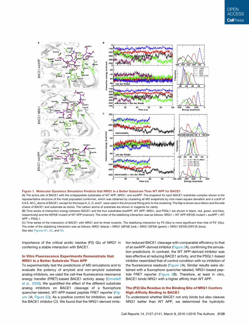
Figure 1. Molecular Dynamics Simulation Predicts that NRG1 Is a Better Substrate Than WT APP for BACE1
(A) The active site of BACE1 with the octapepetide substrates of WT APP, NRG1, and swAPP. The snapshot for each BACE1-substrate complex shown is the
representative structure of the most populated conformer, which was obtained by clustering all MD snapshots by root-mean-square deviation and a cutoff of
0.8 A. All Ca atoms of BACE1, except for the loops A, C, D, and F, were used in the structural fitting prior to the clustering. The flap is shown as a ribbon and the side
chains of BACE1 and substrate as sticks. The carbon atoms of substrate are shown in magenta for clarity.
(B) Time series of interaction energy between BACE1 and the four substrates (swAPP, WT APP, NRG1, and PSGL1 are shown in black, red, green, and blue,
respectively) and the K(P2)E mutant of WT APP (maroon). The order of the stabilizing interaction was as follows: NRG1 > WT APP K(P2)E mutant > swAPP > WT
APP > PSGL1.
(C) Time series of the interaction of BACE1 with NRG1 and its three mutants. The stabilizing interaction by P2 (Glu) is more significant than that of P20 (Glu).
The order of the stabilizing interaction was as follows: NRG1 (black) > NRG1 G(P4)E (red) > NRG1 E(P2)K (green) > NRG1 E(P2K) E(P20)K (blue).
See also Figures S1, S2, and S3.
importance of the critical acidic residue (P2) Glu of NRG1 in
conferring a stable interaction with BACE1.
In Vitro Fluorescence Experiments Demonstrate thatNRG1 Is a Better Substrate Than APPTo experimentally test the predictions of MD simulations and to
evaluate the potency of amyloid and non-amyloid substrate
analog inhibitors, we used the cell-free fluorescence resonance
energy transfer (FRET)-based BACE1 activity assay (Ermolieff
et al., 2000). We quantified the effect of the different substrate
analog inhibitors on BACE1 cleavage of a fluorophore
quencher-labeled, WT-APP-based peptide FRET reporter (Fig-
ure 2A; Figure S3). As a positive control for inhibition, we used
the BACE1 inhibitor C3. We found that the NRG1-derived inhib-
Ce
itor reduced BACE1 cleavage with comparable efficiency to that
of an swAPP-derived inhibitor (Figure 2A), confirming the simula-
tion predictions. In contrast, the WT APP-derived inhibitor was
less effective at reducing BACE1 activity, and the PSGL1-based
inhibitor resembled that of control condition with no inhibition of
the fluorescence readouts (Figure 2A). Similar results were ob-
tained with a fluorophore quencher-labeled, NRG1-based pep-
tide FRET reporter (Figure 2B). Therefore, at least in vitro,
BACE1 binds NRG1 with a higher affinity than WT APP.
The (P2) Glu Residue in theBinding Site of NRG1ConfersHigh-Affinity Binding to BACE1To understand whether BACE1 not only binds but also cleaves
NRG1 better than WT APP, we determined the hydrolytic
ll Reports 14, 2127–2141, March 8, 2016 ª2016 The Authors 2129
BA
ED
V Km Kcat Kcat/Km
( M S-1) ( M) (S-1) (M-1 S-1)
APP (5.0 ± 0.1) x 10-4 36 ± 3.6 (2.08 ± 0.04) x 10-3 57.8 ± 5.9
swAPP (6.0 ± 0.5) x 10-3 5.2 ± 1.2 01x)2.0±5.2( -2 0021±0084
NRG1 (2.7 ± 0.1) x 10-3 19.6 ± 2.8 01x)40.0±(1.12 -2 570 ± 80
C
0
100
200
300
400
500
600
PCDNA wtAPP K (P2)A K (P2)E K (P2)R
*
n.s*
F
APP mutants
Nor
mal
ized
ECL
cou
nts
(to
Cell
viab
ility
and
pcD
NA
)
NRG1-FL
NRG1-CTFs
GAPDH
98
62
38
kDa
PC
DN
A
wtN
RG
1
E(P
2)D
E(P
2)K
wtN
RG
1
E(P
2)D
E(P
2)K
NRG1 mutants + DMSO
NRG1mutants+ C3
G
A H
-2000
0
2000
4000
6000
8000
10000
12000
14000
16000
0 2000 4000 6000 8000
RFU
Time (sec)
S+E
S+E+C3
S+E+swAPP inhibitor
S+E+NRG1 inhibitor
S+E+wtAPP inhibitor
S+E+SELPLG inhibitor
Time (sec)
RFU
E + wtNRG1E + NRG1 E (P2) KE + wtNRG1 + C3
E + wtAPPE + APP K (P2) EE + wtAPP + C3
0
1000
2000
3000
4000
5000
6000
0 400 800 1200 1600
RFU
Time (sec)
0
5000
10000
15000
20000
25000
30000
35000
40000
0 400 800 1200 1600
0
5000
10000
15000
20000
25000
30000
35000
40000
0 2000 4000 6000 8000
RFU
Time (sec)
S+E
S+E+C3
S+E+swAPP
S+E+NRG1 inhibitor
S+E+wtAPP inhibitor
0
5000
10000
15000
20000
25000
0 400 800 1200 1600
RFU
Time (sec)
E+wt L1
E+wtL1+C3
E+L1E1087K
E+L1E1087K+C3
E+wtAPP
E+wtAPP+C3
wtAPP-based FRET reporter NRG1-based FRET reporter
L1/ mutant L1/ wtAPP-based FRET reporters
(legend on next page)
2130 Cell Reports 14, 2127–2141, March 8, 2016 ª2016 The Authors
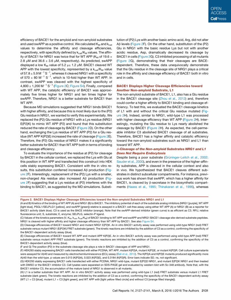
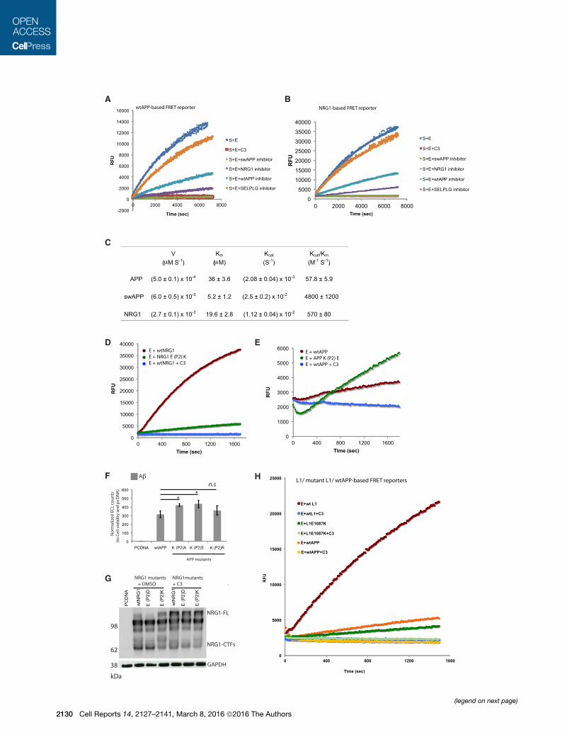
efficiency of BACE1 for the amyloid and non-amyloid substrates
and used swAPP as a positive control. We calculated KM and kcatvalues to determine the affinity and cleavage efficiencies,
respectively, with specificity defined by kcat/KM values. The affin-
ity of BACE1 for NRG1 was twice that for WT APP (KM of 19.6 ±
2.8 mM and 36.6 ± 3.6 mM, respectively). As predicted, swAPP
displayed a low KM value of 5.2 m± 1.2 mM. BACE1 cleaved WT
APP with the lowest specificity, represented by a kcat/KM value
of 57.8 ± 5.9M�1 S�1, whereas it cleaved NRG1with a specificity
of 570 ± 80 M�1 S�1, which is 10-fold higher than WT APP. In
contrast, swAPP was cleaved with the highest specificity of
4,800 ± 1,200 M�1 S�1 (Figure 2C; Figure S4). Finally, compared
with WT APP, the catalytic efficiency of BACE1 was approxi-
mately five times higher for NRG1 and ten times higher for
swAPP. Therefore, NRG1 is a better substrate for BACE1 than
WT APP.
Because MD simulations suggested that NRG1 binds BACE1
with higher affinity, and because this is most likely due to the (P2)
Glu residue in NRG1, wewanted to verify this experimentally. We
replaced the (P2) Glu residue of NRG1 with a Lys residue (NRG1
E(P2)K) to mimic WT APP (P2) and found that this substitution
reduced the rate of cleavage by BACE1 (Figure 2D). On the other
hand, exchanging the Lys residue of WT APP (P2) for a Glu res-
idue (WT APP K(P2)E) increased the rate of cleavage (Figure 2E).
Therefore, the (P2) Glu residue of NRG1 makes it a significantly
better substrate for BACE1 thanWTAPP both in terms of binding
and cleavage efficiency.
To evaluate the importance of the residue at (P2) for cleavage
by BACE1 in the cellular context, we replaced the Lys with Glu at
this position in WT APP and transfected this construct into HEK
cells stably expressing BACE1. Consistent with the in vitro re-
sults, this substitution conferred increased Ab production (Fig-
ure 2F). Interestingly, replacement of the (P2) Lys with a smaller,
non-charged Ala residue also increased Ab production (Fig-
ure 2F) suggesting that a Lys residue at (P2) interferes with the
binding to BACE1, as suggested by the MD simulations. Substi-
Figure 2. BACE1 Displays Higher Cleavage Efficiencies toward the No
(A and B) Kinetics of the binding of WT APP (A) and NRG1 (B) to BACE1. The inhibi
[light blue], PSGL1/SELPLG1 [yellow], and swAPP [green])-statine is assayed in a
BACE1 activity (dark blue). C3 is used as the BACE inhibitor (orange). Note that
fluorescence unit; S, substrate; E, enzyme; SELPLG, selectin-P ligand.
(C) Values of the kinetics parameters (V, KM, kcat, kcat/KM) of BACE1 binding to WT
NRG1 is cleaved with higher affinity and higher cleavage efficiency than WT APP
(D) Cleavage efficiencies of BACE1 toward NRG1 and mutant NRG1 E(P2)K. An
substrate versus mutant NRG1 (E(P2)K) FRET substrate (green). The kinetic reacti
the BACE1-dependent activity assay (blue).
(E) Cleavage efficiencies of BACE1 toward WT APP and mutant APP K(P2)E. An
substrate versus mutant APP FRET substrate (green). The kinetic reactions are
BACE1-dependent activity assay (blue).
(F and G) The position (P2) in the substrate cleavage site plays a role in BACE1 c
(F) HEK293 stably expressing BACE1 cells transfected with either PCDNA, WT AP
were assayed for Ab40 and measured by electrochemiluminescence (ECL) assay
Ab40 than the wild-type. p values are 0.013 (K(P2)A), 0.023 (K(P2)E), and 0.3184
(G) HEK293 stably expressing BACE1 cells transfected with either PCDNA, NRG
with DMSO or the BACE1 inhibitor C3. Cell lysates were separated on SDS-PAG
BACE1 inhibitor C3, the accumulation of the mature band of NRG1 is observed
(H) L1 is a better substrate than WT APP. An in vitro BACE1 activity assay was
substrate (dark green). The kinetic reactions are inhibited by the addition of C3 a
(WT L1 + C3 [blue], mutant L1 + C3 [light green], and WT APP with [light yellow fi
Ce
tution of (P2) Lys with another basic amino acid, Arg, did not alter
Ab levels (Figure 2F). On the other hand, substitution of the (P2)
Glu in NRG1 with the basic residue Lys but not with another
acidic residue, Asp, dramatically decreased its cleavage by
BACE1 in cells (Figure 2G). C3 inhibited processing of all mutants
(Figure 2G), demonstrating that their cleavages are BACE-
dependent. Therefore, these data unequivocally demonstrate
that the Glu residue in the cleavage site of NRG1 plays a critical
role in the affinity and cleavage efficiency of BACE1 both in vitro
and in cells.
BACE1 Displays Higher Cleavage Efficiencies towardAnother Non-amyloid Substrate, L1The non-amyloid substrate of BACE1, L1, also has a Glu residue
in the BACE1 cleavage site (Zhou et al., 2012) and, therefore
could confer a higher affinity to BACE1 binding and cleavage ef-
ficiency. To test this, we evaluated the BACE1 cleavage kinetics
of L1 with and without the critical Glu residue mutated (Fig-
ure 2H). Indeed, similar to NRG1, wild-type L1 was processed
with higher cleavage efficiency than WT APP (Figure 2H). Inter-
estingly, mutating the Glu residue to Lys nearly abolished the
cleavage by BACE1 (Figure 2H). As expected, the cell-perme-
able inhibitor C3 abolished BACE1 cleavage of all substrates.
Therefore, BACE1 has a higher affinity and catalytic efficiency
toward the non-amyloid substrates such as NRG1 and L1 than
toward WT APP.
b-Cleavage of the Non-amyloid Substrates NRG1 and L1
Does Not Require Endocytosis
Despite being a poor substrate (Gr€uninger-Leitch et al., 2002;
Sauder et al., 2000), and even in the presence of the higher-affin-
ity substrates, APP is cleaved in the cellular context and also
in vivo. We hypothesized that BACE1 cleaves different sub-
strates in distinct subcellular compartments. For instance, previ-
ous work has shown that swAPP, which has a higher affinity for
BACE1, is cleaved by b-secretase in the biosynthetic compart-
ments (Haass et al., 1995; Thinakaran et al., 1996), whereas
n-amyloid Substrates NRG1 and L1
tory potential of each of the substrate analog inhibitors (NRG1 [purple], WT APP
BACE1 cell-free assay using either WT APP (A) or NRG1 (B) as a reporter for
the swAPP-derived inhibitor (green curve) is as efficient as C3. RFU, relative
APP and swAPP and NRG1 BACE1 cleavage site-derived substrate peptides.
by BACE1. See also Figure S4.
in vitro BACE1 activity assay was performed using wild-type NRG1 (red) FRET
ons are inhibited by the addition of C3 as a control, confirming the specificity of
in vitro BACE1 activity assay was performed using wild-type APP (red) FRET
inhibited by the addition of C3 as a control, confirming the specificity of the
leavages of APP and NRG1.
P, mutant K(P2)A, mutant K(P2)E, or mutant K(P2)R. Cell culture supernatants
(Bali et al., 2012). The K(P2)A and K(P2)E mutants produced significantly more
(K(P2)R). Error bars indicate SD. ns, not significant.
1 wild-type, mutant E(P2)D NRG1, and mutant E(P2)K NRG1 and then treated
E gel and evaluated by western blot with Sc-348 antibody. Note that, with the
in all mutants.
performed using wild-type L1 (red) FRET substrate versus mutant L1 FRET
s a control, confirming the specificity of the BACE1-dependent activity assay
lled circle] and without C3 [orange filled triangle]).
ll Reports 14, 2127–2141, March 8, 2016 ª2016 The Authors 2131

cleavage of WT APP occurs in endosomes (Carey et al., 2005;
Koo and Squazzo, 1994; Rajendran and Annaert, 2012; Rajen-
dran et al., 2006; Udayar et al., 2013). Because our findings
show that NRG1 also has a higher affinity for BACE1 than WT
APP, we hypothesized that NRG1might also be processed inde-
pendent of endocytosis. To determine whether b-cleavage of
NRG1 is similar or different to that of WT APP in terms of its
requirement of endocytosis, we perturbed membrane trafficking
pathways and assessed the b-cleavage of NRG1. Expression of
dominant-negative dynamin (Dyn K44A), amutant of the GTPase
involved in fission of the endocytic vesicles that inhibits dyna-
min-dependent endocytosis, did not inhibit the cleavage of
NRG1 (Figure 3A). Similarly, pharmacological inhibition of cla-
thrin-dependent/clathrin-independent endocytosis using Pit-
stop2 (Dutta et al., 2012; Stahlschmidt et al., 2014; von Kleist
et al., 2011), an inhibitor that inhibits clathrin-mediated endocy-
tosis and clathrin-independent endocytosis, did not inhibit the
cleavage of NRG1 (Figure 3A). However, both treatments signif-
icantly inhibited b-cleavage of APP and Ab production (Fig-
ure 3B). Pitstop2 had a more pronounced effect on Ab than
secreted APPb (sAPPb), probably because of the inhibition of
g-secretase internalization as well as inhibition of BACE1 endo-
cytosis. Control experiments showed that, indeed, treatment of
cells with Pitstop2 (Figure 3C) or cells expressing Dynamin
K44A inhibited endocytosis of Transferrin (Figures 3C and 3D)
and epidermal growth factor (EGF) (Figure 3C), two clathrin/dy-
namin-dependent cargoes. Therefore, unlike APP, NRG1 does
not require endocytosis for cleavage by BACE1.
Consistent with these observations and similar to NRG1, L1
was also cleaved by BACE1 in an endocytosis-independent
manner (Figure 3E) suggesting that unlike APP, the other physi-
ologically relevant substrates may not require dynamin/clathrin
endocytosis for their b-cleavages and providing further support
to the idea that BACE1 cleaves these higher affinity substrates
in an endocytosis-independent manner.
The Endosomally Targeted, Sterol-Linked BACE1Inhibitor Inhibits Ab Production and Cleavage of APPwithout Affecting b-Cleavage of NRG1 and L1Exploiting the observation that different membrane trafficking
pathways differentially regulate b-cleavage of APP and NRG1
or L1, we tested whether inhibiting BACE1 activity specifically
in the endosomal compartment would inhibit b-cleavage of
APP and, thereby, spare the cleavage of the non-amyloid sub-
strates NRG1 and L1. We found that the cell-permeable pan in-
hibitor C3 substantially inhibited b-cleavage of NRG1 in cells,
whereas an endosomally targeted, sterol-linked BACE1 inhibitor
(Rajendran et al., 2008) showed no significant inhibition of NRG1
b-cleavage (Figures 4A and 4B). However, both treatments in-
hibited b-cleavage of WT APP and Ab production (Figure 4C).
Similar results were obtained in primary mouse neuronal cul-
tures, where an endosomally targeted, sterol-linked inhibitor of
b-secretase spared cleavage of NRG1 but not of APP (Figure S5)
under an endogenous BACE1 expression level.
Similar to NRG1, we then tested whether BACE1-mediated
cleavage of L1, which also did not require endocytosis, could
be spared by using the endosomally targeted, sterol-linked
BACE1 inhibitor. Treatment with the endosomally targeted, ste-
2132 Cell Reports 14, 2127–2141, March 8, 2016 ª2016 The Authors
rol-linked BACE1 inhibitor did not affect b-cleavage of L1,
whereas treatment with the general cell-permeable BACE1 in-
hibitor C3 abolished almost all BACE1 processing (Figures 4D
and 4E). In control experiments performed with APP, b-cleavage
of WT APP was inhibited completely by both the endosomally
targeted, sterol-linked BACE1 inhibitor and the general cell-
permeable inhibitor (Figure 4C).
The Endosomally Targeted, Sterol-Linked BACE1Inhibitor Inhibits NRG1 Processing When SubcellularCompartmentalization Is CompromisedTo test whether the endosomally targeted, sterol-linked BACE1
inhibitor can indeed inhibit NRG1 processing when the integrity
of subcellular compartmentalization is compromised, we
tested the effect of both BACE1 inhibitors: the endosomally
targeted, sterol-linked inhibitor and the cell-permeable pan in-
hibitor C3 on solubilized membranes and assessed inhibition
of NRG1 cleavage. Under these conditions, where cellular
compartmentalization is compromised, both the endosomally
targeted, sterol-linked BACE1 inhibitor and C3 significantly
blocked BACE1 cleavage of NRG1 (Figure 5A). These results
suggest that subcellular compartmentalization of different
substrates in endosomal and non-endosomal compartments
contributes to the differential processing of these substrates
by BACE1.
Among the non-endosomal compartments that harbor b-sec-
retase activity, the TGN and the plasma membrane have been
suggested to be places for b-secretase activity (Li and S€udhof,
2004; Prabhu et al., 2012). To investigate whether the processing
of NRG1 by BACE1 occurs at the cell surface, we used a cell-
impermeable BACE1 inhibitor, GL189 (Capell et al., 2002), and
tested its inhibitory potential on BACE1 cleavage of NRG1.
This inhibitor is a substrate analog transition state inhibitor that
has been demonstrated to bind to active BACE1 in solubilized
membrane fraction assays (Capell et al., 2002). Therefore, we hy-
pothesized that, if active BACE1 is present at the cell surface that
is competent to cleave NRG1, then this cell-impermeable transi-
tion state inhibitor should inhibit BACE1 cleavage of NRG1.
However, treatment of cells expressing NRG1 with GL189
did not inhibit the processing of NRG1 by BACE1 (Figure 5B).
However, the control C3, the cell-permeable BACE1 inhibitor,
almost abolished the processing of NRG1 by BACE1 (Figure 5B).
Similarly, endocytosis inhibition also did not inhibit BACE1 pro-
cessing of NRG1. However, addition of C3 under endocytosis in-
hibition inhibits NRG1 cleavage, demonstrating that NRG1
cleavage occurs neither at the plasma membrane nor in endo-
somes (Figure 5B). As additional controls, we also checked
whether BACE1 cleavage of APP was affected under similar
conditions and found that treatment of cells with the cell-imper-
meable inhibitor GL-189 had no effect on b-cleavage of APP.
However, treatment with the endocytosis inhibitor or cell-perme-
able inhibitor or both inhibited b-cleavage, showing conclusively
that b-cleavage of APP does not occur at the plasma membrane
but is dependent on endocytosis. Taken together, these results
indicate that BACE1 cleaves NRG1 neither at the plasma mem-
brane nor in endosomes but, most likely, at the TGN, the only
other low-pH, non-endo-lysosomal organelle that is conducive
for BACE1 activity.
A B
C
D E
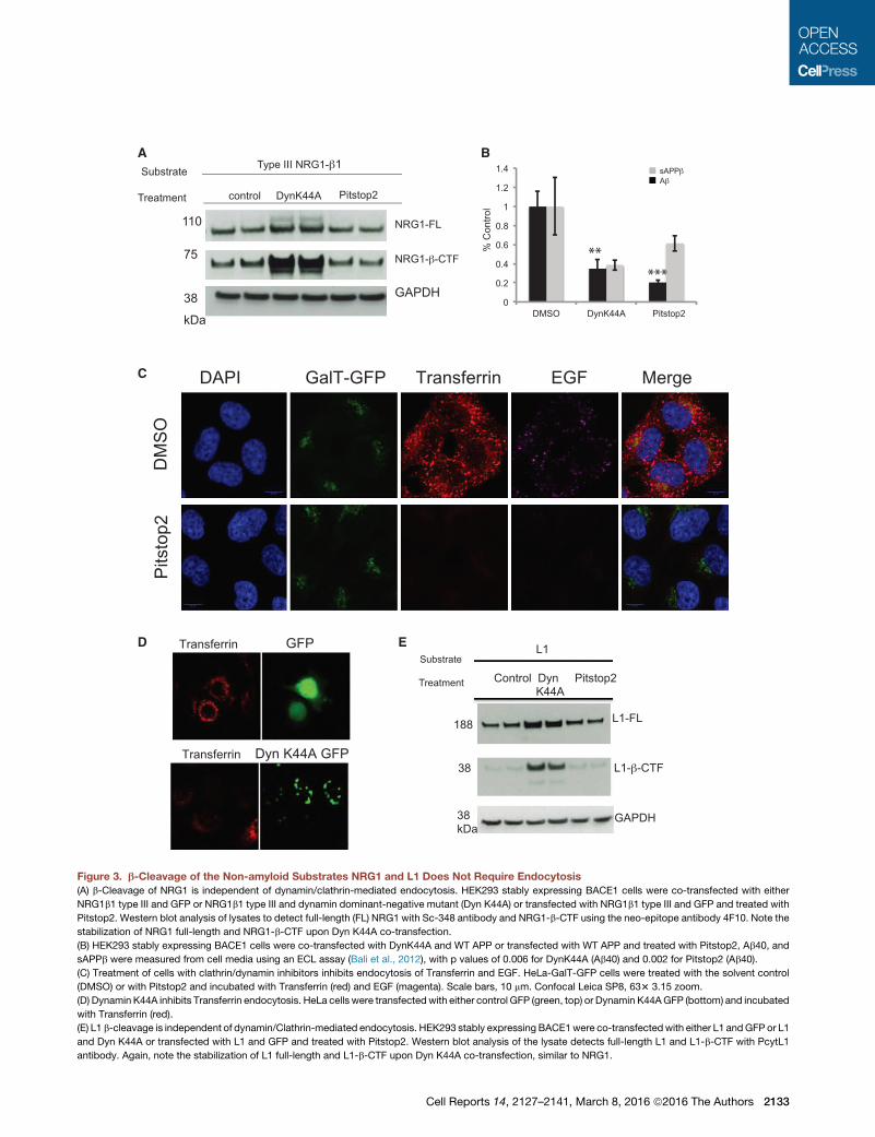
Figure 3. b-Cleavage of the Non-amyloid Substrates NRG1 and L1 Does Not Require Endocytosis
(A) b-Cleavage of NRG1 is independent of dynamin/clathrin-mediated endocytosis. HEK293 stably expressing BACE1 cells were co-transfected with either
NRG1b1 type III and GFP or NRG1b1 type III and dynamin dominant-negative mutant (Dyn K44A) or transfected with NRG1b1 type III and GFP and treated with
Pitstop2. Western blot analysis of lysates to detect full-length (FL) NRG1 with Sc-348 antibody and NRG1-b-CTF using the neo-epitope antibody 4F10. Note the
stabilization of NRG1 full-length and NRG1-b-CTF upon Dyn K44A co-transfection.
(B) HEK293 stably expressing BACE1 cells were co-transfected with DynK44A and WT APP or transfected with WT APP and treated with Pitstop2, Ab40, and
sAPPb were measured from cell media using an ECL assay (Bali et al., 2012), with p values of 0.006 for DynK44A (Ab40) and 0.002 for Pitstop2 (Ab40).
(C) Treatment of cells with clathrin/dynamin inhibitors inhibits endocytosis of Transferrin and EGF. HeLa-GalT-GFP cells were treated with the solvent control
(DMSO) or with Pitstop2 and incubated with Transferrin (red) and EGF (magenta). Scale bars, 10 mm. Confocal Leica SP8, 633 3.15 zoom.
(D) Dynamin K44A inhibits Transferrin endocytosis. HeLa cells were transfected with either control GFP (green, top) or Dynamin K44AGFP (bottom) and incubated
with Transferrin (red).
(E) L1 b-cleavage is independent of dynamin/Clathrin-mediated endocytosis. HEK293 stably expressing BACE1were co-transfectedwith either L1 andGFP or L1
and Dyn K44A or transfected with L1 and GFP and treated with Pitstop2. Western blot analysis of the lysate detects full-length L1 and L1-b-CTF with PcytL1
antibody. Again, note the stabilization of L1 full-length and L1-b-CTF upon Dyn K44A co-transfection, similar to NRG1.
Cell Reports 14, 2127–2141, March 8, 2016 ª2016 The Authors 2133

A B
C D E
Figure 4. The Endosomally Targeted, Sterol-Linked BACE1 Inhibitor Inhibits Ab Production and Cleavage of APPwithout Affecting Cleavage
of NRG1 and L1
(A) The endosomally targeted, sterol-linked inhibitor spares NRG1 processing in cells. HEK293 stably expressing BACE1 cells were transfected with NRG1 b1 type III
and treatedwithDMSOasacontrol,with thecell-permeableBACE1 inhibitorC3, orwith theendosomally targeted, sterol-linkedBACE1 inhibitor.Westernblot analysis
of lysates detects full-length NRG1 with Sc-348 antibody and NRG1-b-CTF using the neo-epitope antibody 4F10. A representative western blot is shown.
(B) Semiquantification of western blots. Data were mean ± S.E. (n = 3). Student’s t test was used to calculate p values: C3-FL, 0.427; C3-b-CTF (b-cleaved
C-terminal fragment), 0.0075; endosomally targeted, sterol-linked BACE1 inhibitor-FL, 0.882; endosomally targeted, sterol-linked BACE1 inhibitor-b-CTF, 0.488.
(C) The endosomally targeted, sterol-linked BACE1 inhibitor inhibited APP processing. HEK293 stably expressing BACE1 were transfected with WT APP and
treated with DMSO as a control, with the cell-permeable BACE1 inhibitor C3, or with the endosomally targeted, sterol-linked BACE1 inhibitor for 12 hr. Ab40
and sAPPb were measured from cell media using an ECL assay. The values represent experimental triplicates. p values for the sterol-linked inhibitor for sAPPb
and Ab40 are 0.0009 and 0.0021, respectively. P-values for the C3 inhibitor for sAPPb and Ab40 are 0.0006 and 0.0020, respectively. Error bars indicate SD.
(D) The endosomally targeted, sterol-linked inhibitor spares L1 processing in cells. HEK293 stably expressing BACE1 cells were transfected with L1 and treated
with DMSO as a control, with the cell-permeable BACE1 inhibitor C3, or with the endosomally targeted, sterol-linked BACE1 inhibitor. Western blot analysis of
lysates with PcytL1 antibody detects full-length L1 and L1-b-CTF.
(E) Semiquantification of western blots. Data were mean ± SE (n = 3). Student’s t test was used to calculate the p values: C3-FL, 0.0169; C3-b-CTF, 0.0022;
endosomally targeted, sterol-linked BACE1 inhibitor-FL, 0.4975; endosomally targeted, sterol-linked BACE1 inhibitor-b-CTF, 0.187.
The Endosomally Targeted, Sterol-Linked BACE1Inhibitor Inhibits APP Processing without AffectingNRG1 Processing in iPSC-Derived Human NeuronsTo validate the findings regarding the endosomally targeted, ste-
rol-linked BACE1 inhibitor sparing NRG1 cleavage in a more
relevant setting for potential treatment in AD patients, we treated
neurons generated from human induced pluripotent stem cells
(h-iPSCs) derived from healthy human donors (Figure 6A; Fig-
ure S6) with C3, the cell-permeable BACE1 inhibitor, or the endo-
somally targeted, sterol-linked BACE1 inhibitor and assayed for
BACE1 processing of NRG1 and APP (as a control) in those cells.
C3 BACE1 inhibitor treatment inhibited BACE1 processing of
NRG1 (Figures 6B and 6C). Treatment with the endosomally tar-
geted, sterol linked BACE1 inhibitor did not affect the processing
of NRG1 by BACE1 (Figures 6B and 6C). However, upon C3
BACE1 inhibitor and endosomally targeted, sterol-linked inhibi-
tor treatment, both Ab40 and sAPPb were reduced dramatically,
with sAPPa levels increased (Figure 6D).
2134 Cell Reports 14, 2127–2141, March 8, 2016 ª2016 The Authors
The Endosomally Targeted, Sterol-Linked BACE1Inhibitor Is Targeted to Endocytic Compartments butNot to the GolgiConsistent with these results, the fluorescently labeled, sterol-
linked inhibitor trafficked to endosomal compartments (Figures
7A and 7B) but not to the trans-Golgi network, where high-affinity
substrates could be cleaved (Haass et al., 1995; Thinakaran
et al., 1996), as judged by the colocalization experiments (Fig-
ure 7C). When incubated on cells, the fluorescently labeled, ste-
rol-linked inhibitor trafficked from the plasma membrane to
EEA1-positive early endosomes (Figure 7A), the compartments
shown previously to be important for b-cleavage of APP, and
then was transported retrogradely to late endosomal/lysosomal
compartments at longer periods of incubation, as demonstrated
by colocalization with Lamp1 (Figure 7B). The endosomally tar-
geted, sterol-linked inhibitor did not traffic to the TGN 30 min
to 1 hr after uptake, as shown by the absence of localization be-
tween the inhibitor and GalT-GFP, a Golgi-resident protein
A B
C D
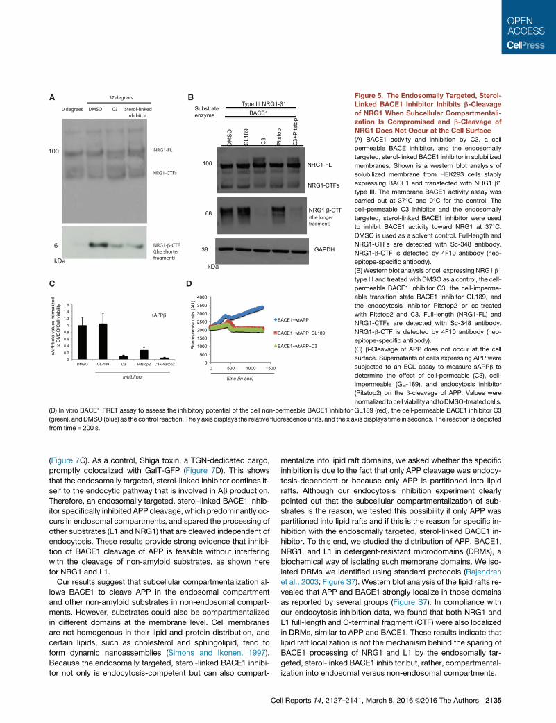
Figure 5. The Endosomally Targeted, Sterol-
Linked BACE1 Inhibitor Inhibits b-Cleavage
of NRG1 When Subcellular Compartmentali-
zation Is Compromised and b-Cleavage of
NRG1 Does Not Occur at the Cell Surface
(A) BACE1 activity and inhibition by C3, a cell
permeable BACE inhibitor, and the endosomally
targeted, sterol-linked BACE1 inhibitor in solubilized
membranes. Shown is a western blot analysis of
solubilized membrane from HEK293 cells stably
expressing BACE1 and transfected with NRG1 b1
type III. The membrane BACE1 activity assay was
carried out at 37�C and 0�C for the control. The
cell-permeable C3 inhibitor and the endosomally
targeted, sterol-linked BACE1 inhibitor were used
to inhibit BACE1 activity toward NRG1 at 37�C.DMSO is used as a solvent control. Full-length and
NRG1-CTFs are detected with Sc-348 antibody.
NRG1-b-CTF is detected by 4F10 antibody (neo-
epitope-specific antibody).
(B)Western blot analysis of cell expressing NRG1 b1
type III and treated with DMSO as a control, the cell-
permeable BACE1 inhibitor C3, the cell-imperme-
able transition state BACE1 inhibitor GL189, and
the endocytosis inhibitor Pitstop2 or co-treated
with Pitstop2 and C3. Full-length (NRG1-FL) and
NRG1-CTFs are detected with Sc-348 antibody.
NRG1-b-CTF is detected by 4F10 antibody (neo-
epitope-specific antibody).
(C) b-Cleavage of APP does not occur at the cell
surface. Supernatants of cells expressing APP were
subjected to an ECL assay to measure sAPPb to
determine the effect of cell-permeable (C3), cell-
impermeable (GL-189), and endocytosis inhibitor
(Pitstop2) on the b-cleavage of APP. Values were
normalized tocell viability and toDMSO-treatedcells.
(D) In vitro BACE1 FRET assay to assess the inhibitory potential of the cell non-permeable BACE1 inhibitor GL189 (red), the cell-permeable BACE1 inhibitor C3
(green), andDMSO (blue) as the control reaction. The y axis displays the relative fluorescence units, and the x axis displays time in seconds. The reaction is depicted
from time = 200 s.
(Figure 7C). As a control, Shiga toxin, a TGN-dedicated cargo,
promptly colocalized with GalT-GFP (Figure 7D). This shows
that the endosomally targeted, sterol-linked inhibitor confines it-
self to the endocytic pathway that is involved in Ab production.
Therefore, an endosomally targeted, sterol-linked BACE1 inhib-
itor specifically inhibited APP cleavage, which predominantly oc-
curs in endosomal compartments, and spared the processing of
other substrates (L1 and NRG1) that are cleaved independent of
endocytosis. These results provide strong evidence that inhibi-
tion of BACE1 cleavage of APP is feasible without interfering
with the cleavage of non-amyloid substrates, as shown here
for NRG1 and L1.
Our results suggest that subcellular compartmentalization al-
lows BACE1 to cleave APP in the endosomal compartment
and other non-amyloid substrates in non-endosomal compart-
ments. However, substrates could also be compartmentalized
in different domains at the membrane level. Cell membranes
are not homogenous in their lipid and protein distribution, and
certain lipids, such as cholesterol and sphingolipid, tend to
form dynamic nanoassemblies (Simons and Ikonen, 1997).
Because the endosomally targeted, sterol-linked BACE1 inhibi-
tor not only is endocytosis-competent but can also compart-
Ce
mentalize into lipid raft domains, we asked whether the specific
inhibition is due to the fact that only APP cleavage was endocy-
tosis-dependent or because only APP is partitioned into lipid
rafts. Although our endocytosis inhibition experiment clearly
pointed out that the subcellular compartmentalization of sub-
strates is the reason, we tested this possibility if only APP was
partitioned into lipid rafts and if this is the reason for specific in-
hibition with the endosomally targeted, sterol-linked BACE1 in-
hibitor. To this end, we studied the distribution of APP, BACE1,
NRG1, and L1 in detergent-resistant microdomains (DRMs), a
biochemical way of isolating such membrane domains. We iso-
lated DRMs we identified using standard protocols (Rajendran
et al., 2003; Figure S7). Western blot analysis of the lipid rafts re-
vealed that APP and BACE1 strongly localize in those domains
as reported by several groups (Figure S7). In compliance with
our endocytosis inhibition data, we found that both NRG1 and
L1 full-length and C-terminal fragment (CTF) were also localized
in DRMs, similar to APP and BACE1. These results indicate that
lipid raft localization is not the mechanism behind the sparing of
BACE1 processing of NRG1 and L1 by the endosomally tar-
geted, sterol-linked BACE1 inhibitor but, rather, compartmental-
ization into endosomal versus non-endosomal compartments.
ll Reports 14, 2127–2141, March 8, 2016 ª2016 The Authors 2135
A B
C D
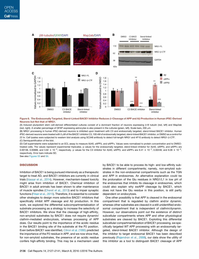
Figure 6. The Endosomally Targeted, Sterol-Linked BACE1 Inhibitor Reduces b-Cleavage of APP and Ab Production in Human iPSC-Derived
Neurons but Not that of NRG1.
(A) Induced pluripotent stem cell-derived differentiated cultures consist of a dominant fraction of neurons expressing b-III tubulin (red, left) and Map2ab
(red, right). A smaller percentage of GFAP-expressing astrocytes is also present in the cultures (green, left). Scale bars, 200 mm.
(B) NRG1 processing in human iPSC-derived neurons is inhibited upon treatment with C3 and endosomally targeted, sterol-linked BACE1 inhibitor. Human
iPSC-derived neurons were treated with 5 mMof the BACE1 inhibitor C3, 100 nMof endosomally targeted, sterol-linked BACE1 inhibitor, or DMSO as a control for
22 hr. Cell lysates were subjected to western blot analysis using SC348 antibody to detect full-length NRG1 and 4F10 antibody to detect NRG1-b-CTF.
(C) Semiquantification of the blot.
(D) Cell supernatants were subjected to an ECL assay to measure Ab40, sAPPb, and sAPPa. Values were normalized to protein concentration and to DMSO-
treated cells. The values represent experimental triplicates. p values for the endosomally targeted, sterol-linked inhibitor for Ab40, sAPPb, and sAPPa are
0.00138, 0.00888, and 3.82 3 10�6, respectively. p values for the C3 inhibitor for Ab40, sAPPb, and sAPPa are 9.41 3 10�5, 0.00248, and 8.06 3 10�6,
respectively. Error bars indicate SD.
See also Figures S5 and S6.
DISCUSSION
Inhibition of BACE1 is being pursued intensively as a therapeutic
target to treat AD, and BACE1 inhibitors are currently in clinical
trials (Vassar et al., 2014). However, mechanism-based toxicity
might arise from inhibition of BACE1. Chemical inhibition of
BACE1 in adult animals has been shown to alter maintenance
of muscle spindles (Cheret et al., 2013) and to impair synaptic
functions (Filser et al., 2015). Therefore, it is essential to consider
other strategies to design more selective BACE1 inhibitors that
specifically inhibit APP cleavage and Ab production. In this
work, we explored the differential subcompartmentalization of
substrate processing as a strategy to increase the selectivity of
BACE1 inhibitors. We show that the cleavage of high-affinity,
non-amyloid substrates by BACE1 does not require dynamin/
clathrin-mediated endocytosis, whereas processing of APP
does. Our results point to the importance of the acidic residue
in the BACE1 binding site of the substrate at the P2 position.
Even before BACE1 was identified, Citron et al. (1995) predicted
the importance of the P2 residue in APP, and we now show that,
for non-amyloid substrates, the presence of an acidic residue
confers high-affinity binding. This may be a mechanism used
2136 Cell Reports 14, 2127–2141, March 8, 2016 ª2016 The Authors
by BACE1 to be able to process its high- and low-affinity sub-
strates in different compartments; namely, non-amyloid sub-
strates in the non-endosomal compartments such as the TGN
and APP in endosomes. An alternative explanation could be
the protonation of the Glu residues in NRG1/L1 in low pH of
the endosomes that inhibits its cleavage in endosomes, which
could also explain why swAPP cleavage by BACE1, which
does not have the Glu residue in this position, is still partly
dependent on endocytosis.
One other possibility is that APP is cleaved in the endosomal
compartment that is regulated by clathrin and/or dynamin,
whereas other substrates are cleaved in a still unidentified endo-
somal compartment that is independent of clathrin/dynamin.
However, our observations point out the existence of distinct
subcellular compartments where APP and other physiological
substrates are cleaved by BACE1. Exploiting this differential
subcellular compartmentalization of BACE1 processing, we spe-
cifically targeted WT APP processing with an endosomally tar-
geted, sterol-linked BACE1 inhibitor. Although the design of
the inhibitor to target endosomal BACE1 has been described
previously (Rajendran et al., 2008), our data show the utility of
this inhibitor as a tool to distinguish BACE1 cleavage of APP
(legend on next page)
Cell Reports 14, 2127–2141, March 8, 2016 ª2016 The Authors 2137
and non-amyloid substrates because of the subcellular compart-
mentalization of these cleavages. An alternative possibility for
the mode of action of the endosomally targeted, sterol-linked
BACE1 inhibitor is that the endosomally targeted inhibitor,
because of its sterol linkage, targets APP cleavage that occurs
in lipid rafts of endosomes (Ehehalt et al., 2003) and, therefore,
spares non-amyloid substrates that could be localized in non-
raft domains in the endosomes. However, for this to occur,
NRG1 processing must be endocytosis-dependent, and its
processing should occur in non-raft domains of endosomes.
By using Pitstop2, an inhibitor of clathrin-dependent and -inde-
pendent endocytosis (Dutta et al., 2012; Stahlschmidt et al.,
2014; von Kleist et al., 2011), and also with dominant-negative
dynamin, we clearly demonstrate that, although APP processing
by BACE1 requires endocytosis, b-cleavage of the non-amyloid
substrates NRG1 and L1 does not require endocytosis. This sug-
gests that the NRG1/L1-sparing activity of the endosomally
targeted, sterol-linked BACE1 inhibitor is most likely due to en-
dosomal targeting and not due to differential localization of these
non-amyloid substrates in non-raft domains of the endosomes.
Moreover, in our experiments with isolated DRMs, all substrates
are localized in DRMs as well. Therefore, our results point out
that only APP b-cleavage occurs in endosomes and, therefore,
encourages the possibility that endosomally targeted BACE1
therapeutic substances can reduce Ab production without
affecting other BACE1 substrate cleavages. Endosomally tar-
geted BACE1 inhibitors are potential therapeutic substances
for the specific treatment of AD without adverse effects.
The limitations of our study include the lack of translation in an-
imal models because these are beyond the scope of this study,
but our experiments on human iPSCs provides hope. Clearly,
further research is needed to translate these findings in experi-
mental animals and also patients. Regardless, our work demon-
strates that inhibiting Ab production without affecting BACE1
processing of other substrates is possible and of important clin-
ical value for the specific treatment of AD.
EXPERIMENTAL PROCEDURES
MD Simulations
For the MD simulations, the initial structure of the BACE1-substrate complex
was obtained from the crystal structure of the ecotodomain of BACE1 bound
to the OM99-2 inhibitor (Glu-Val-Asn-Leu-4[CHOH-CH2]-Ala-Ala-Glu-Phe),
where 4[CHOH-CH2] represents a hydroxyethylene isostere of the peptide
bond (PDB: 1FKN) (Hong et al., 2000). The hydroxyethylene isostere was re-
placed by a carbonyl group to obtain the peptide bond and all MD simulations
were carried out with peptide substrates. TheOM99-2 inhibitor corresponds to
the swAPP sequence with the Asp(P10)Ala mutation. The original form of the
swAPP sequence was obtained by mutating Ala at P10 of OM99-2 to Asp.
From the structure of the swAPP substrate, the initial structures of WT APP
(Glu-Val-Lys-Met-Asp-Ala-Glu-Phe), NRG1 (Gly-Ile-Glu-Phe-Met-Glu-Ala-
Glu), and PSGL1 (Ala-Ser-Asn-Leu-Ser-Val-Asn-Tyr) substrates were ob-
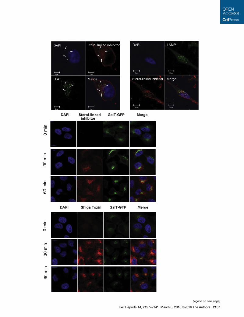
Figure 7. Trafficking of the Endosomally Targeted, Sterol-Linked BAC
(A and B) Uptake of the rhodamine-conjugated, endosomally targeted, sterol-link
zation (A) and with Lamp-1 (green) after 15 min of internalization (B) in HeLa cells
(C) HeLa-GalT-GFP cells were incubated with rhodamine-conjugated, endosom
different times (0, 30, and 60 min). Scale bar, 10 mm. Confocal Leica SP8, 633 3
(D) Similarly, HeLa-GalT-GFP cells were incubated with Shiga toxin (red) and GalT
Leica SP8, 633 3.15 zoom.
2138 Cell Reports 14, 2127–2141, March 8, 2016 ª2016 The Authors
tained by appropriate mutations. The BACE-substrate complexes were
immersed in a cubic water box of size 93 A. The initial structures were equili-
brated for 2-ns and 100-ns MD trajectories were calculated for each of the four
BACE-substrate complexes at constant temperature (300 K) and pressure
(1 atm) employing the NAMD program (Phillips et al., 2005) with the
CHARMM22 force field (MacKerell et al., 1998) and TIP3 potential for water
molecules (Jorgensen et al., 1983). The procedure was repeated for the three
mutants of NRG1, i.e., NRG1 G(P4)E, NRG1 E(P2)K, NRG1 E(P20 )K, and the
K(P2)E mutant of WT APP. For each these four mutants, 60 ns MD trajectories
were calculated. Similarly, we performed 60 ns of MD simulations for the four
BACE-inhibitor complexes where the hydroxyethylene isostere of the peptide
bond was retained (data not shown). Here we considered the Ala(P10) residueof OM99-2 for a direct comparison with in vitro assays. In addition, a 120-ns
MD run was performed for each of the complexes between BACE1 and the
statine-based (hydroxyethylene isostere) inhibitors. The P4-P40 residues
were extracted from the sequences of swAPP (OM99-2 inhibitor), WT APP,
NRG1, and PSGL1, except for the Ala at P10, which was kept as in OM99-2
for all four inhibitors.
Cell-free BACE1 Activity Assay
The cell-free BACE1 activity assay was performed in a final volume of 100 ml in
assay buffer: 50 mM sodium acetate (pH 4.5) and 0.1% Triton X-100. 0.24 mM
of recombinant human BACE1-ectodomain (rhBACE1) (Sigma S4195) was
mixed with 10 mM of each substrate in a flat-bottom black 96-wells plate
(Nunc 237105). The assay was performed at 37 ± 1�Cwith excitation and emis-
sion wavelengths set at 340 nm and 400 nm, respectively. For cell-free BACE1
activity assay with inhibtors, 0.24 mMof rhBACE1 was incubated with 10 mMof
each inhibitor for 10min at 37�C in the assay buffer prior to the addition of each
substrate.
Km and Kcat Measurements and Calculations
The specificity of the different substrates for rhBACE1 was kinetically assayed
bymeasuring theMichaelis constant Km and the limiting rate V, fromwhich the
specificity constant Kcat/Km was calculated. Instability of the enzyme during
assay was assessed with the Selwyn test (Selwyn, 1965). Fluorescence mea-
surements were performed in a final volume of 100 ml using flat bottomed black
96-well plates in a Tecan Infinite M1000Promicroplate reader thermostatted at
37 ± 1�C with excitation and emission wavelengths of 340 nm and 400 nm,
respectively. Readings were performed in kinetic mode for 5min, during which
time fluorescence increased linearly. For a constant 0.24 mM enzyme concen-
tration, 5 or 6 substrate concentrations covering approximately the range 0.2
to 5 Km, as determined in preliminary experiments, were used. Stock solutions
of the substrates were prepared in pure DMSO, but the concentration of the
organic solvent was kept constant at 1% v/v for all substrate concentrations.
Only for the swAPP substrate, the final DMSO concentration was 2% v/v. The
buffer was 50 mM sodium acetate (pH 4.5) with 0.1% Triton X-100 added. Km
and Vwere calculated by non-linear regression fitting of the Michaelis-Menten
equation.
Quantitative Measurements of Ab40, sAPPb and sAPPa
Human Ab40, human sAPPb, human sAPPa, and mouse Ab40 were assayed
from supernatants, after centrifugation at 1,000 rpm for 3 min, using the elec-
trochemiluminescence (ECL) assay as described in Bali et al., 2012.
Primary Neuronal Culture, Transduction and Inhibitors Treatments
Mixedcortical/hippocampal neuronswereprepared fromembryonicday15and
16mice. In brief, dissociated cells were plated onto 6-well poly-D-lysine-coated
E1 Inhibitor
ed inhibitor (red) and colocalization with EEA1 (green) after 5 min of internali-
.
ally targeted, sterol-linked BACE1 inhibitor (red) and GalT-GFP (green) for
.15 zoom.
-GFP (green) for different times (0, 30, and 60 min). Scale bar, 10 mm. Confocal

dishes at a density of �250.000/cm2 and cultured in Neurobasal medium (Invi-
trogen) with B27 supplements (Invitrogen), 2mM L-glutamine, and penicillin/
streptomycin. After 5 days, in vitro neuronal cultures were incubated with lenti-
viralNRG1b1 type III orGFPparticles for 8 hr. 48hr after transduction, cellswere
treated with DMSO or inhibitors (C3, TAPI-1 and the endosomally-targeted ste-
rol-linked BACE1 inhibitor) for 24 hr, as previously described.
iPSC-Derived Neuronal Cultures and Treatment
Human iPSC-derived long-term self-renewing neuroepithelial stem cells
(lt-NES cells) (Mertens et al., 2013) were maintained in DMEM/F12, 2 mM
L-glutamine, 1.6 g/l glucose, 0.1 mg/ml penicillin/streptomycin, N2 supple-
ment (Life Technologies), B27 (1 ml/ml; Life Technologies), and fibroblast
growth factor 2 (FGF2) and epidermal growth factor (EGF; both 10 ng/ml;
Cell Guidance Systems) on tissue culture plates coated with poly-L-orni-
thine/laminin (both Sigma), and passaged every 3 or 4 days. Neuronal differen-
tiation was induced by withdrawal of FGF2 and EGF in differentiation
media (MACS Neuro Medium supplemented with MACS NeuroBrew-21
(1:50; Miltenyi Biotec) and DMEM/ F12 supplemented with N2 mixed at a 1:1
ratio) that was exchanged every other day. C3 BACE1 inhibitor (5 mM), endo-
somally-targeted sterol-linked BACE1 inhibitor (100 nM), or DMSO solvent
control were added to neuronal cultures differentiated for 4 weeks. Cultures
were incubated for 22 hr before supernatant and protein lysates were
collected.
Statistical Analysis
All data are shown as mean ± SD. Two-tailed Student’s t test was used for
comparison of the means between two groups. *p < 0.05, **p < 0.005, and
***p < 0.0005.
SUPPLEMENTAL INFORMATION
Supplemental Information includes Supplemental Experimental Procedures
and seven figures and can be found with this article online at http://dx.doi.
org/10.1016/j.celrep.2016.01.076.
AUTHOR CONTRIBUTIONS
L.R. designed the research. S.B.H. and M.W. performed the biochemistry and
cell biological experiments. S.M. and A.C. analyzed the MD simulations. A.B.
helped with the kinetics experiments. K.M.P.R. synthesized the statine-based
inhibitors. All authors participated in designing the experiments and in data
analysis. L.R. and S.B.H. wrote the paper, and all authors participated in the
editing of the paper. P.K. and O.B. contributed to the iPSC experiments.
ACKNOWLEDGMENTS
We thank T. S€udhof, M. Schwab, A. Aguzzi, G. Jennings, H. Knoelker, and U.
Konietzko for their input. We gratefully acknowledge the help of Jado Technol-
ogies GmbH with synthesis of the sterol-linked BACE1 inhibitors. We thank
E. Schwarz and S. Hoey for critical reading of the manuscript. We thank
U. Konietzko for the APP-HA tag plasmids, B. de Strooper for the L1 plasmid,
P. Altevogt for the anti-L1 antibody, L. Johannes for the fluorescently labeled
Shiga toxin B subunit, G. Siegel for the preparation of primary mice neurons,
Z. Goodger and J. Ries for help with RT-PCR analysis, and H. Schwalbe for al-
lowing us to use the instrument facility. L.R. acknowledges financial support
from Swiss National Science Foundation grants, the Velux Foundation, the
Cure Alzheimer Fund, the Baugarten Stiftung, and the Bangerter Stiftung.
P.K. and O.B. acknowledge financial support from the German Federal Minis-
try for Education and Research (BioPharma-NeuroAllianz Grant 0315608A).
A.C. acknowledges financial support from the Swiss National Science
Foundation.
Received: April 15, 2013
Revised: November 9, 2015
Accepted: January 26, 2016
Published: February 25, 2016
Ce
REFERENCES
Bali, J., Gheinani, A.H., Zurbriggen, S., and Rajendran, L. (2012). Role of genes
linked to sporadic Alzheimer’s disease risk in the production of b-amyloid pep-
tides. Proc. Natl. Acad. Sci. USA 109, 15307–15311.
Barman, A., Sch€urer, S., and Prabhakar, R. (2011). Computational modeling of
substrate specificity and catalysis of the b-secretase (BACE1) enzyme.
Biochemistry 50, 4337–4349.
Buggia-Prevot, V., Fernandez, C.G., Riordan, S., Vetrivel, K.S., Roseman, J.,
Waters, J., Bindokas, V.P., Vassar, R., and Thinakaran, G. (2014). Axonal
BACE1 dynamics and targeting in hippocampal neurons: a role for Rab11
GTPase. Mol. Neurodegener. 9, 1.
Cai, J., Qi, X., Kociok, N., Skosyrski, S., Emilio, A., Ruan, Q., Han, S., Liu, L.,
Chen, Z., Bowes Rickman, C., et al. (2012). b-Secretase (BACE1) inhibition
causes retinal pathology by vascular dysregulation and accumulation of age
pigment. EMBO Mol. Med. 4, 980–991.
Capell, A., Meyn, L., Fluhrer, R., Teplow, D.B., Walter, J., and Haass, C. (2002).
Apical sorting of beta-secretase limits amyloid beta-peptide production.
J. Biol. Chem. 277, 5637–5643.
Carey, R.M., Balcz, B.A., Lopez-Coviella, I., and Slack, B.E. (2005). Inhibition of
dynamin-dependent endocytosis increases shedding of the amyloid precursor
protein ectodomain and reduces generation of amyloid beta protein. BMCCell
Biol. 6, 30.
Cheret, C., Willem, M., Fricker, F.R., Wende, H., Wulf-Goldenberg, A., Tahir-
ovic, S., Nave, K.A., Saftig, P., Haass, C., Garratt, A.N., et al. (2013). Bace1
and Neuregulin-1 cooperate to control formation and maintenance of muscle
spindles. EMBO J. 32, 2015–2028.
Citron, M., Oltersdorf, T., Haass, C., McConlogue, L., Hung, A.Y., Seubert, P.,
Vigo-Pelfrey, C., Lieberburg, I., and Selkoe, D.J. (1992). Mutation of the beta-
amyloid precursor protein in familial Alzheimer’s disease increases beta-pro-
tein production. Nature 360, 672–674.
Citron, M., Teplow, D.B., and Selkoe, D.J. (1995). Generation of amyloid beta
protein from its precursor is sequence specific. Neuron 14, 661–670.
De Strooper, B. (2010). Proteases and proteolysis in Alzheimer disease: a
multifactorial view on the disease process. Physiol. Rev. 90, 465–494.
Dutta, D.,Williamson, C.D., Cole, N.B., and Donaldson, J.G. (2012). Pitstop 2 is
a potent inhibitor of clathrin-independent endocytosis. PLoS ONE 7, e45799.
Ehehalt, R., Keller, P., Haass, C., Thiele, C., and Simons, K. (2003). Amyloido-
genic processing of the Alzheimer beta-amyloid precursor protein depends on
lipid rafts. J. Cell Biol. 160, 113–123.
Ermolieff, J., Loy, J.A., Koelsch, G., and Tang, J. (2000). Proteolytic activation
of recombinant pro-memapsin 2 (Pro-beta-secretase) studied with new fluoro-
genic substrates. Biochemistry 39, 16263.
Filser, S., Ovsepian, S.V., Masana, M., Blazquez-Llorca, L., Brandt Elvang, A.,
Volbracht, C., M€uller, M.B., Jung, C.K., and Herms, J. (2015). Pharmacological
inhibition of BACE1 impairs synaptic plasticity and cognitive functions. Biol.
Psychiatry 77, 729–739.
Gr€uninger-Leitch, F., Schlatter, D., K€ung, E., Nelbock, P., and Dobeli, H.
(2002). Substrate and inhibitor profile of BACE (beta-secretase) and compari-
son with other mammalian aspartic proteases. J. Biol. Chem. 277, 4687–4693.
Haass, C., Lemere, C.A., Capell, A., Citron, M., Seubert, P., Schenk, D., Lann-
felt, L., and Selkoe, D.J. (1995). The Swedish mutation causes early-onset Alz-
heimer’s disease by beta-secretase cleavage within the secretory pathway.
Nat. Med. 1, 1291–1296.
Hardy, J.A., and Higgins, G.A. (1992). Alzheimer’s disease: the amyloid
cascade hypothesis. Science 256, 184–185.
Hitt, B., Riordan, S., Kukreja, L., Eimer, W., Rajapaksha, T., and Vassar, R.
(2012). b-Site amyloid precursor protein (APP)-cleaving enzyme 1 (BACE1)-
deficient mice exhibit a close homolog of L1 (CHL1) loss-of-function pheno-
type involving axon guidance defects. J. Biol. Chem. 287, 38408–38425.
Hong, L., Koelsch, G., Lin, X., Wu, S., Terzyan, S., Ghosh, A.K., Zhang, X.C.,
and Tang, J. (2000). Structure of the protease domain of memapsin 2 (beta-
secretase) complexed with inhibitor. Science 290, 150–153.
ll Reports 14, 2127–2141, March 8, 2016 ª2016 The Authors 2139
Hu, X., Hicks, C.W., He, W., Wong, P., Macklin, W.B., Trapp, B.D., and Yan, R.
(2006). Bace1 modulates myelination in the central and peripheral nervous
system. Nat. Neurosci. 9, 1520–1525.
Hu, X., Zhou, X., He, W., Yang, J., Xiong, W., Wong, P., Wilson, C.G., and Yan,
R. (2010). BACE1 deficiency causes altered neuronal activity and neurodegen-
eration. J. Neurosci. 30, 8819–8829.
Hu, X., He, W., Luo, X., Tsubota, K.E., and Yan, R. (2013). BACE1 regulates
hippocampal astrogenesis via the Jagged1-Notch pathway. Cell Rep. 4,
40–49.
Hussain, I., Powell, D., Howlett, D.R., Tew, D.G., Meek, T.D., Chapman, C.,
Gloger, I.S., Murphy, K.E., Southan, C.D., Ryan, D.M., et al. (1999). Identifica-
tion of a novel aspartic protease (Asp 2) as beta-secretase. Mol. Cell. Neurosci.
14, 419–427.
Jonsson, T., Atwal, J.K., Steinberg, S., Snaedal, J., Jonsson, P.V., Bjornsson,
S., Stefansson, H., Sulem, P., Gudbjartsson, D., Maloney, J., et al. (2012). A
mutation in APP protects against Alzheimer’s disease and age-related cogni-
tive decline. Nature 488, 96–99.
Jorgensen, W.L., Chandrasekhar, J., Madura, J.D., Impey, R.W., and Klein,
M.L. (1983). Comparison of Simple Potential Functions for Simulating Liquid
Water. J. Chem. Phys. 79, 926–935.
Kim, D.Y., Carey, B.W., Wang, H., Ingano, L.A., Binshtok, A.M., Wertz, M.H.,
Pettingell, W.H., He, P., Lee, V.M., Woolf, C.J., and Kovacs, D.M. (2007).
BACE1 regulates voltage-gated sodium channels and neuronal activity. Nat.
Cell Biol. 9, 755–764.
Kitazume, S., Nakagawa, K., Oka, R., Tachida, Y., Ogawa, K., Luo, Y., Citron,
M., Shitara, H., Taya, C., Yonekawa, H., et al. (2005). In vivo cleavage of
alpha2,6-sialyltransferase by Alzheimer beta-secretase. J. Biol. Chem. 280,
8589–8595.
Koo, E.H., and Squazzo, S.L. (1994). Evidence that production and release of
amyloid beta-protein involves the endocytic pathway. J. Biol. Chem. 269,
17386–17389.
Kuhn, P.H., Marjaux, E., Imhof, A., De Strooper, B., Haass, C., and Lich-
tenthaler, S.F. (2007). Regulated intramembrane proteolysis of the inter-
leukin-1 receptor II by alpha-, beta-, and gamma-secretase. J. Biol. Chem.
282, 11982–11995.
Kuhn, P.H., Koroniak, K., Hogl, S., Colombo, A., Zeitschel, U., Willem, M., Vol-
bracht, C., Schepers, U., Imhof, A., Hoffmeister, A., et al. (2012). Secretome
protein enrichment identifies physiological BACE1 protease substrates in neu-
rons. EMBO J. 31, 3157–3168.
Lahiri, D.K., Maloney, B., Long, J.M., and Greig, N.H. (2014). Lessons from a
BACE1 inhibitor trial: off-site but not off base. Alzheimers Dement. 10 (5,
Suppl), S411–S419.
Li, Q., and S€udhof, T.C. (2004). Cleavage of amyloid-beta precursor protein
and amyloid-beta precursor-like protein by BACE 1. J. Biol. Chem. 279,
10542–10550.
Lichtenthaler, S.F., Dominguez, D.I., Westmeyer, G.G., Reiss, K., Haass, C.,
Saftig, P., De Strooper, B., and Seed, B. (2003). The cell adhesion protein P-
selectin glycoprotein ligand-1 is a substrate for the aspartyl protease
BACE1. J. Biol. Chem. 278, 48713–48719.
Ma, H., Lesne, S., Kotilinek, L., Steidl-Nichols, J.V., Sherman, M., Younkin, L.,
Younkin, S., Forster, C., Sergeant, N., Delacourte, A., et al. (2007). Involvement
of beta-site APP cleaving enzyme 1 (BACE1) in amyloid precursor protein-
mediated enhancement of memory and activity-dependent synaptic plasticity.
Proc. Natl. Acad. Sci. USA 104, 8167–8172.
MacKerell, A.D., Bashford, D., Bellott, M., Dunbrack, R.L., Evanseck, J.D.,
Field, M.J., Fischer, S., Gao, J., Guo, H., Ha, S., et al. (1998). All-atom empirical
potential for molecular modeling and dynamics studies of proteins. J. Phys.
Chem. B. 102, 3586–3616.
Mellman, I., and Nelson, W.J. (2008). Coordinated protein sorting, targeting
and distribution in polarized cells. Nat. Rev. Mol. Cell Biol. 9, 833–845.
Mertens, J., St€uber, K., Wunderlich, P., Ladewig, J., Kesavan, J.C., Vanden-
berghe, R., Vandenbulcke, M., van Damme, P., Walter, J., Br€ustle, O., and
Koch, P. (2013). APP processing in human pluripotent stem cell-derived neu-
2140 Cell Reports 14, 2127–2141, March 8, 2016 ª2016 The Authors
rons is resistant to NSAID-based g-secretase modulation. Stem Cell Reports
1, 491–498.
Phillips, J., Braun, R., Wang, W., Gumbart, J., Tajkhorshid, E., Villa, E., Chipot,
C., Skeel, R., Kale, L., and Schulten, K. (2005). Scalable molecular dynamics
with NAMD. J. Comput. Chem. 26, 1781–1802.
Prabhu, Y., Burgos, P.V., Schindler, C., Farıas, G.G., Magadan, J.G., and Bo-
nifacino, J.S. (2012). Adaptor protein 2-mediated endocytosis of the b-secre-
tase BACE1 is dispensable for amyloid precursor protein processing. Mol.
Biol. Cell 23, 2339–2351.
Rajapaksha, T.W., Eimer, W.A., Bozza, T.C., and Vassar, R. (2011). The Alz-
heimer’s b-secretase enzyme BACE1 is required for accurate axon guidance
of olfactory sensory neurons and normal glomerulus formation in the olfactory
bulb. Mol. Neurodegener. 6, 88.
Rajendran, L., and Annaert, W. (2012). Membrane trafficking pathways in Alz-
heimer’s disease. Traffic 13, 759–770.
Rajendran, L., and Simons, K. (2005). Lipid rafts and membrane dynamics.
J. Cell Sci. 118, 1099–1102.
Rajendran, L., Masilamani, M., Solomon, S., Tikkanen, R., Stuermer, C.A.,
Plattner, H., and Illges, H. (2003). Asymmetric localization of flotillins/reggies
in preassembled platforms confers inherent polarity to hematopoietic cells.
Proc. Natl. Acad. Sci. USA 100, 8241–8246.
Rajendran, L., Honsho, M., Zahn, T.R., Keller, P., Geiger, K.D., Verkade, P.,
and Simons, K. (2006). Alzheimer’s disease beta-amyloid peptides are
released in association with exosomes. Proc. Natl. Acad. Sci. USA 103,
11172–11177.
Rajendran, L., Schneider, A., Schlechtingen, G.,Weidlich, S., Ries, J., Braxme-
ier, T., Schwille, P., Schulz, J.B., Schroeder, C., Simons, M., et al. (2008).
Efficient inhibition of the Alzheimer’s disease beta-secretase by membrane
targeting. Science 320, 520–523.
Sannerud, R., Declerck, I., Peric, A., Raemaekers, T., Menendez, G., Zhou, L.,
Veerle, B., Coen, K., Munck, S., De Strooper, B., et al. (2011). ADP ribosylation
factor 6 (ARF6) controls amyloid precursor protein (APP) processing by medi-
ating the endosomal sorting of BACE1. Proc. Natl. Acad. Sci. USA 108, E559–
E568.
Sauder, J.M., Arthur, J.W., and Dunbrack, R.L., Jr. (2000). Modeling of sub-
strate specificity of the Alzheimer’s disease amyloid precursor protein beta-
secretase. J. Mol. Biol. 300, 241–248.
Schneider, A., Rajendran, L., Honsho, M., Gralle, M., Donnert, G., Wouters, F.,
Hell, S.W., and Simons, M. (2008). Flotillin-dependent clustering of the amyloid
precursor protein regulates its endocytosis and amyloidogenic processing in
neurons. J. Neurosci. 28, 2874–2882.
Selwyn, M.J. (1965). A simple test for inactivation of an enzyme during assay.
Biochim. Biophys. Acta 105, 193–195.
Simons, K., and Ikonen, E. (1997). Functional rafts in cell membranes. Nature
387, 569–572.
Sinha, S., Anderson, J.P., Barbour, R., Basi, G.S., Caccavello, R., Davis, D.,
Doan, M., Dovey, H.F., Frigon, N., Hong, J., et al. (1999). Purification and clon-
ing of amyloid precursor protein beta-secretase from human brain. Nature 402,
537–540.
Stahlschmidt, W., Robertson, M.J., Robinson, P.J., McCluskey, A., and
Haucke, V. (2014). Clathrin terminal domain-ligand interactions regulate sort-
ing of mannose 6-phosphate receptors mediated by AP-1 and GGA adaptors.
J. Biol. Chem. 289, 4906–4918.
Sugimoto, I., Futakawa, S., Oka, R., Ogawa, K., Marth, J.D., Miyoshi, E.,
Taniguchi, N., Hashimoto, Y., and Kitazume, S. (2007). Beta-galactoside
alpha2,6-sialyltransferase I cleavage by BACE1 enhances the sialylation of
soluble glycoproteins. A novel regulatory mechanism for alpha2,6-sialylation.
J. Biol. Chem. 282, 34896–34903.
Tanzi, R.E. (2005). The synaptic Abeta hypothesis of Alzheimer disease. Nat.
Neurosci. 8, 977–979.
Thinakaran, G., Teplow, D.B., Siman, R., Greenberg, B., and Sisodia, S.S.
(1996). Metabolism of the ‘‘Swedish’’ amyloid precursor protein variant in
neuro2a (N2a) cells. Evidence that cleavage at the ‘‘beta-secretase’’ site oc-
curs in the golgi apparatus. J. Biol. Chem. 271, 9390–9397.
Udayar, V., Buggia-Prevot, V., Guerreiro, R.L., Siegel, G., Rambabu, N., Soo-
hoo, A.L., Ponnusamy, M., Siegenthaler, B., Bali, J., Simons, M., et al.; AESG
(2013). A paired RNAi and RabGAP overexpression screen identifies Rab11 as
a regulator of b-amyloid production. Cell Rep. 5, 1536–1551.
Vassar, R., and Kandalepas, P.C. (2011). The b-secretase enzyme BACE1 as a
therapeutic target for Alzheimer’s disease. Alzheimers Res. Ther. 3, 20.
Vassar, R., Bennett, B.D., Babu-Khan, S., Kahn, S., Mendiaz, E.A., Denis, P.,
Teplow, D.B., Ross, S., Amarante, P., Loeloff, R., et al. (1999). Beta-secretase
cleavage of Alzheimer’s amyloid precursor protein by the transmembrane as-
partic protease BACE. Science 286, 735–741.
Vassar, R., Kuhn, P.H., Haass, C., Kennedy, M.E., Rajendran, L., Wong, P.C.,
and Lichtenthaler, S.F. (2014). Function, therapeutic potential and cell biology
of BACE proteases: current status and future prospects. J. Neurochem. 130,
4–28.
von Arnim, C.A., Kinoshita, A., Peltan, I.D., Tangredi, M.M., Herl, L., Lee, B.M.,
Spoelgen, R., Hshieh, T.T., Ranganathan, S., Battey, F.D., et al. (2005). The low
density lipoprotein receptor-related protein (LRP) is a novel beta-secretase
(BACE1) substrate. J. Biol. Chem. 280, 17777–17785.
von Kleist, L., Stahlschmidt, W., Bulut, H., Gromova, K., Puchkov, D., Robert-
son, M.J., MacGregor, K.A., Tomilin, N., Pechstein, A., Chau, N., et al. (2011).
Role of the clathrin terminal domain in regulating coated pit dynamics revealed
by small molecule inhibition. Cell 146, 471–484.
Walter, J., Fluhrer, R., Hartung, B., Willem, M., Kaether, C., Capell, A., Lam-
mich, S., Multhaup, G., and Haass, C. (2001a). Phosphorylation regulates
intracellular trafficking of beta-secretase. J. Biol. Chem. 276, 14634–14641.
Ce
Walter, J., Kaether, C., Steiner, H., and Haass, C. (2001b). The cell biology of
Alzheimer’s disease: uncovering the secrets of secretases. Curr. Opin. Neuro-
biol. 11, 585–590.
Willem, M., Garratt, A.N., Novak, B., Citron, M., Kaufmann, S., Rittger, A.,
DeStrooper, B., Saftig, P., Birchmeier, C., and Haass, C. (2006). Control of pe-
ripheral nerve myelination by the beta-secretase BACE1. Science 314,
664–666.
Wong, H.K., Sakurai, T., Oyama, F., Kaneko, K., Wada, K., Miyazaki, H., Kur-
osawa, M., De Strooper, B., Saftig, P., and Nukina, N. (2005). beta Subunits of
voltage-gated sodium channels are novel substrates of beta-site amyloid pre-
cursor protein-cleaving enzyme (BACE1) and gamma-secretase. J. Biol.
Chem. 280, 23009–23017.
Yan, R., Bienkowski, M.J., Shuck, M.E., Miao, H., Tory, M.C., Pauley, A.M.,
Brashier, J.R., Stratman, N.C., Mathews, W.R., Buhl, A.E., et al. (1999). Mem-
brane-anchored aspartyl protease with Alzheimer’s disease beta-secretase
activity. Nature 402, 533–537.
Yan, R., Han, P., Miao, H., Greengard, P., and Xu, H. (2001). The transmem-
brane domain of the Alzheimer’s beta-secretase (BACE1) determines its late
Golgi localization and access to beta -amyloid precursor protein (APP) sub-
strate. J. Biol. Chem. 276, 36788–36796.
Zhou, L., Brouwers, N., Benilova, I., Vandersteen, A., Mercken, M., Van Laere,
K., Van Damme, P., Demedts, D., Van Leuven, F., Sleegers, K., et al. (2011).
Amyloid precursor protein mutation E682K at the alternative b-secretase
cleavage b0-site increases Ab generation. EMBO Mol. Med. 3, 291–302.
Zhou, L., Barao, S., Laga, M., Bockstael, K., Borgers, M., Gijsen, H., Annaert,
W., Moechars, D., Mercken, M., Gevaert, K., and De Strooper, B. (2012). The
neural cell adhesion molecules L1 and CHL1 are cleaved by BACE1 protease
in vivo. J. Biol. Chem. 287, 25927–25940.
ll Reports 14, 2127–2141, March 8, 2016 ª2016 The Authors 2141