specific n-cadherin–dependent pathways drive human breast
TRANSCRIPT

Research Article
Specific N-cadherin–dependent pathways drive humanbreast cancer dormancy in bone marrowGarima Sinha1,2, Alejandra I Ferrer1,2, Seda Ayer2, Markos H El-Far1,2 , Sri Harika Pamarthi2, Yahaira Naaldijk2,Pradeep Barak4,5, Oleta A Sandiford2, Bernadette M Bibber1,2 , Ghassan Yehia3 , Steven J Greco2, Jie-Gen Jiang4,5,Margarette Bryan2, Rakesh Kumar6, Nicholas M Ponzio4,5, Jean-Pierre Etchegaray7, Pranela Rameshwar1,2
The challenge for treating breast cancer (BC) is partly due to long-term dormancy driven by cancer stem cells (CSCs) capable ofevading immune response and resist chemotherapy. BC cells showpreference for the BM, resulting in poor prognosis. CSCs useconnexin 43 (Cx43) to form gap junctional intercellular commu-nication with BM niche cells, fibroblasts, and mesenchymal stemcells (MSCs). However, Cx43 is an unlikely target to reverse BCdormancy because of its role as a hematopoietic regulator. Wefound N-cadherin (CDH2) and its associated pathways as potentialdrug targets. CDH2, highly expressed in CSCs, interacts intracel-lularly with Cx43, colocalizes with Cx43 in BC cells within BMbiopsies of patients, and is required for Cx43-mediated gapjunctional intercellular communication with BM niche cells. No-tably, CDH2 and anti-apoptotic pathwaysmaintained BC dormancy.We thereby propose these pathways as potential pharmacologicaltargets to prevent dormancy and chemosensitize resistant CSCs.
DOI 10.26508/lsa.202000969 | Received 20 November 2020 | Revised 19 April2021 | Accepted 20 May 2021 | Published online 2 June 2021
Introduction
Breast cancer (BC) remains a clinical problem despite the devel-opment of new drugs, improved screenings, and early intervention(Berry, 2013; Miller et al, 2016). One of the major challenges is theability of BC cells (BCCs) to survive for decades in a dormant state.Such a state enables the BCCs to exist in cycling quiescence, whichsupports immune evasion and resistance towards therapy (Braunet al, 2005, 2009; Gelao et al, 2013; Mao et al, 2014; Massague &Obenauf, 2016; Otvos et al, 2016; Tjensvoll et al, 2019). These dor-mant BCCs can eventually serve as the source of metastatic cancerdecades after remission (Talmadge, 2007; Tivari et al, 2018; DeAngelis et al, 2019). Shared functional and molecular similaritiesbetween dormant BCCs and healthy stem cells have led to the
designation of cancer stem cells (CSCs) (Carcereri de Prati et al,2017; De Angelis et al, 2019).
BCCs show preference for BM, resulting in poor prognosis (Braunet al, 2005; Tjensvoll et al, 2019). Once in the BM, BCCs evoke theresident BM niche cells (such as macrophages, fibroblasts, andmesenchymal stem cells [MSCs]) to facilitate their transition into adormant phase (Rao et al, 2004; Patel et al, 2012, 2014; Bliss et al,2016; Sandiford et al, 2021). Investigations to identify specificmarkers for CSCs revealed continuous phases of cellular plasticity,which include dedifferentiation of BCC progenitors into BCCs withstem cell–like properties (Naume et al, 2007; Plaks et al, 2015; Wanget al, 2017; Tjensvoll et al, 2019; Lynch-Sutherland et al, 2020). Thus, itcould be argued that CSCs may be defined as a cellular state that isdependent on the tissue microenvironment. Indeed, MSCs, whichconstitute the cellular niche within the BM, can induce BCCquiescence through gap junctional intercellular communication(GJIC) and, through their exosomal secretome (Patel et al, 2014;Bliss et al, 2016; Sandiford et al, 2021).
The connexin (Cx) family of proteins constitutes gap junction. Sixconnexins assemble to form connexon (hemichannel), which pairswith hemichannel on neighboring cell to form a pore-likestructure/gap junction. GJIC is among the mechanisms by whichBCCs directly interact with BM niche cells to allow for the exchangeof molecules to sustain dormancy (Lim et al, 2011). Expression ofCx26, Cx32, and Cx43 has been reported during different stages of BCdevelopment (Sinha et al, 2020). Specifically, CSCs exhibit highexpression of Cx26 and Cx43 in comparison to BC progenitors;however, only Cx43 was involved in gap junction formation (Patelet al, 2012). Interestingly, Cx26, independent of forming gap junction,can maintain CSC stemness by complexing with Nanog and Focaladhesion kinase (Thiagarajan et al, 2018).
Despite a critical role for Cx43 in BC dormancy within BM, itsubiquitous expression and involvement in key cellular processessuch as hematopoietic regulation make this an unlikely druggabletarget to reverse GJIC (Taniguchi Ishikawa et al, 2012; Martins-Marques
1Rutgers School of Graduate Studies at New Jersey Medical School, Newark, NJ, USA 2Department of Medicine, Hematology/Oncology, Rutgers New Jersey MedicineSchool, Newark, NJ, USA 3Genome Editing Shared Resource, Office of Research and Economic Development, Rutgers University, New Brunswick, NJ, USA 4Department ofPathology, Immunology and Laboratory Medicine, Rutgers New Jersey Medical School, Newark, NJ, USA 5ONI, Linacre House, Oxford, UK 6Department of Biotechnology,Rajiv Gandhi Centre for Biotechnology, Kerala, India 7Department of Biological Sciences, Rutgers University, Newark, NJ, USA
Correspondence: [email protected]
© 2021 Sinha et al. https://doi.org/10.26508/lsa.202000969 vol 4 | no 7 | e202000969 1 of 22
on 27 March, 2022life-science-alliance.org Downloaded from http://doi.org/10.26508/lsa.202000969Published Online: 2 June, 2021 | Supp Info:

et al, 2015). Therefore, it is imperative to identify new molecularmechanisms leading to GJIC between BM niche cells and CSCs thatwill not result in adverse effects upon targeting. Given that Cx43expression was highest in CSCs and such cells are difficult toeradicate, this study investigated the associated mechanism inCx43-mediated GJIC between CSCs and BM niche cells to maintainBC dormancy. The long-term goal is to discern a strategy to ef-fectively sensitize the dormant BCCs to chemotherapy and/or toidentify new treatments.
Studies in nonmalignant models reported on a role for CDH2(neuronal cadherin/CD325/Cadherin-2) to translocate Cx43 to thecell membrane (Wei et al, 2005; Matsuda et al, 2006). CDH2 belongsto the cadherin family and is involved in cell adhesion, asymmetriccell division, and pre-/postsynapsis in the central nervous system(Garcia-Castro et al, 2000). The role of CDH2 in cell migration andinvasion has been reported for several cancer cells, including BCCs(Kim et al, 2000). During epithelial mesenchymal transition (EMT),cancer cells can morphed in CSC-like phenotype with increasedCDH2 (Yang et al, 2017). CDH2 has five extracellular domains, atransmembrane region, and a conserved cytoplasmic domain(Zuppinger et al, 2000). The discussed role of Cx43 and CDH2 in CSCs,which are associated with a dormant phenotype, led us to inves-tigate how CDH2 contributes to Cx43-mediated gap junction for thepurpose of long-term cancer cell survival in the BM.
BCCs can acquire dormancy at secondary sites, including the BM(Boire et al, 2019). The microenvironment within an organ is crucialfor BC dormancy acquisition and maintenance. In this study, weexamined how BM microenvironmental cells interact with CSCs byapplying a previously reported BCC hierarchy (Patel et al, 2012). Thetwo key nonhematopoietic cells incorporated in the experimentalmodel are stromal fibroblasts and MSCs, both of which can es-tablish GJIC with CSCs for sustained dormancy (Lim et al, 2011; Patelet al, 2014).
We report on a specific role for CDH2 in forming GJIC betweenCSCs and BM niche cells. Interestingly, CDH2 and Cx43 expressionswere similar in the subset of BCCs with abundance CSCs. Using invivo and in vitro studies, we showed CDH2-associated pathways aspotential drug targets to reverse dormancy and thereby chemo-sensitize the otherwise resistant dormant CSCs. Importantly, CDH2knockdown blocked GJIC without altering Cx43, which is required forGJIC. Colocalization of CDH2 and Cx43 in BM biopsies of BC patientswas similar to the cell lines, underscoring the significance of thefindings. Overall, these findings advance our understanding of BCdormancy and highlight potential therapeutic strategies.
Results
Increased CDH2 protein in CSCs
The major question posed in this study is to determine themechanisms of BC dormancy, specifically pathways involved in GJICbetween CSCs and BM niche cells (Patel et al, 2012). We selectedCDH2 and its associated pathways because CDH2 is required forexpression of transmembrane protein Cx43, which occurs partly byintracellular interaction (Wei et al, 2005; Matsuda et al, 2006).
Because CSCs showed preference in GJIC with BM niche cells (Patelet al, 2012; Walker et al, 2019), we compared the relative expressionof CDH2 in different BCC subsets, including CSCs.
We first analyzed unsorted BCCs and two BM niche cells (MSCsand fibroblasts) for CDH2 and Cx43 (control) in Western blots (Patelet al, 2012) (Fig 1A). We characterized MSCs by morphology, phe-notype, and multilineage differentiation (Fig S1A–C). There werestrong bands for CDH2 and Cx43 in the BCCs with a weak band forCDH2 in stromal cells (Fig 1A). We verified the weak CDH2 bands inMSCs and stroma by immunoprecipitation with anti-CDH2 followedby blotting for Cx43 (Fig S1D).
We previously established an exhaustive model of BC dormancywithin BM stromal compartment, close to the endosteum (Patelet al, 2012). In this study, BCCs with high Oct4a expression dem-onstrated properties of dormancy, including stem cell function.There was independent validation of the model (Bliss et al, 2016;Walker et al, 2019). We acknowledge that other groups have re-ported on models of dormancy that uses the integrin family ofproteins, including dormancy in lung, as well as an orthotopicmouse model (Lu et al, 2011; Abravanel et al, 2015; Albrengues et al,2018). Our model recapitulates human dormancy within the cellularBM compartment, before bone invasion and dormancy establishedby gap junction with resident BM nonhematopoietic cells. Thus, ourmodel expands on understanding of BC dormancy.
Next, we compared different BCC subsets for CDH2 with MDA-MB-231-pOct4A-GFP, which was under the control of pOct4a regulatoryregion. The GFP was under the control of Oct4a regulatory region,thus intensity of GFP would be proportional to Oct4a expression, asreported (Patel et al, 2012). We sorted different cell subsets, basedon GFP intensity (Patel et al, 2012) (Fig 1B). qPCR indicated similar(P > 0.05) CDH2 mRNA among BCC subsets (Fig S1E). However, flowcytometry and Western blot indicated a direct correlation betweenmembrane and intracellular CDH2 with BCC maturity (Figs 1C and Dand S1E, respectively). In summary, CSCs (GFPhi) showed the highestCDH2 protein as compared with the other BCC subsets. Thus, theresults linked CDH2 to cellular stemness.
Role of CDH2 in GJIC between CSCs and BM niche cells
CDH2 was increased in CSCs, which show preference for GJIC withBM niche cells (Figs 1C and D and S1E). We therefore investigated arole for CDH2 in GJIC using loss and gain of function studies (Patelet al, 2012). We selected CSCs from MDA-MB-231-pOct4a-GFP,knockdown for CDH2 (red fluorescence protein, RFP), Cx43 (RFP), orscramble shRNA using the depicted gating scheme (Fig 1E). Amongthe four CDH2-shRNA clones, Western blot for CDH2 with unsortedand CSCs indicated that Clone B was most efficient (Fig S1G and H).We co-cultured CSCs with 7-amino-4-chloromethylcoumarin(CMAC) (blue) labeled stromal fibroblasts or MSCs for 72 h andthen examined the CSCs for CMAC transfer by fluorescence mi-croscopy (Fig 1F). The images (Figs 1G and H and S1I and J, top rowwith enlarged regions below) showing white areas indicate dyetransfer (yellow CSCs + blue CMAC = white) with scramble shRNA andvehicle. This transfer was blunted with GJIC inhibitor, 1-octanol, whichserved as a positive control. The blunting effect of 1-octanol mirroredthe results with Cx43 or CDH2 knockdown CSCs. Flow cytometricanalyses for dye transfer with deep red-labeled stromal fibroblasts
CDH2 and breast cancer dormancy Sinha et al. https://doi.org/10.26508/lsa.202000969 vol 4 | no 7 | e202000969 2 of 22

Figure 1. CDH2 in gap junctional intercellular communication (GJIC).(A) Top panel: Western blot for CDH2 and Cx43 with whole-cell extracts from BCCs, stromal fibroblast, and MCF12A. The normalized band densities are shown below.(B) Shown is the gating scheme to select different BCC subsets with MDA-MB-231 cells with stable pEGFP1-Oct4A. (C, D) Representative histogram for flow cytometry fortotal (intracellular + membrane) (C) and membrane (D) CDH2 (left). The mean fluorescence intensities are presented ±SD for five independent analyses (right). (E) Thegating scheme used to sort cancer stem cells (CSCs) (GFP), knockdown for CDH2 (RFP) or Cx43 (RFP). Lower left shows a representative image of the sorted cells (GFP +RFP = Yellow). (F) Diagram for the assay used to detect dye transfer from stromal fibroblast to CSCs. (G, H) MDA-MB-231-pOct4A-GFP were transfected with scramble-RFP
CDH2 and breast cancer dormancy Sinha et al. https://doi.org/10.26508/lsa.202000969 vol 4 | no 7 | e202000969 3 of 22

and MSCs showed the most immature BCC subsets (gating schemefor GFPhi+med BCCs shown in Fig 1I) with scrambled shRNA receiving95% and 75% dye from fibroblasts and MSCs, respectively (Fig 1J andK). Such transfer was significantly (P < 0.05) reduced with CSCs,knockdown for CDH2 or Cx43 (Fig 1J and K).
Gain-of-function studies replaced CDH2 and Cx43 in theknockdown BCCs using an inducible expression system (Fig S1K andL). This induced CDH2 allowed for stable expression despite theshRNA. The rescued CSCs, when co-cultured with CMAC-blue–labeled MSCs, restored CSC’s ability to establish GJIC with >80% dyetransfer (Fig 1K, two right panels).
Dye transfer occurred between Oct4ahi/med and BM niche cells,and also between Oct4ahi/CSCs and MSCs (Fig 1J–L). Rescue of CDH2and Cx43 in the knockdown BCCs resulted in dye transfer similar toscramble shRNA. Control with 1-octanol showed minimal dyetransfer. Overall, CDH2 was necessary and sufficient to form GJICconnecting CSCs with stromal fibroblasts and MSCs (Fig 1M).
Colocalization of CDH2 and Cx43 in BCC subsets
Other experimental system reported on intracellular colocalizationbetweenCx43 and CDH2 as a requirement for Cx43 to be expressed onthe cell membrane (Wei et al, 2005; Matsuda et al, 2006). Thus, weasked if CDH2 is in close proximity to CX43. Image stream of singleunsorted BCCs, labeled for intracellular CDH2 (PE) and Cx43 (AF488),showed colocalization with a bell-shape distribution (Fig 2A). Thissuggested that colocalized CDH2 and Cx43 within the unsorted BCCswere heterogenous. To narrow the BCC subset with prominencecolocalization, we selected CSCs with higher expression of CDH2 andrepeated the image stream analyses (Fig 1B–D). The results indicatedbright yellow fluorescence in the CSCs/Oct4a-GFPhi as comparedwiththe other BCC subsets, indicating that colocalization was most ef-ficient in CSCs (Fig 2B).
We validate CDH2-Cx43 colocalization using nanoscale imagingof individual BCCs (Fig 2C). MDA-MB-231 cells were colabeled withanti-Cx43-Alexa 647 (green) and anti–CDH2-PE (red) and then ex-amined with a Nanoimager. Isotype control showed no backgroundfluorescence whereas cells labeled with anti-Cx43 and CDH2 in-dicated intracellular and membrane (yellow) colocalization.
Next, we sought the regions of intracellular colocalization be-tween Cx43 and CDH2 by colabeling for anti–CDH2-PE (red), anti–Cx43-Alexa 488 (green), and the one of following organelle-specificantibodies: anti–GRP78-Alexa405 (blue) for ER or anti–GM130-Alexa405 (blue) for Golgi (G) (Fig 2D). Identifying these organelles will be
important to fully decipher how Cx43 and CDH2 interact to establishdormancy and therefore lead to avenues in developing treatment.The white regions indicated colocalized CDH2 and Cx43 in bothorganelles: blue for organelle, red for CDH2, and green for Cx43.Next, we asked if intracellular colocalized CDH2 and Cx43 wererelevant for Cx43 to reach the cell membrane. To address this, wepreincubated BCCs with 5 µg/ml brefeldin A for 24 h, followed bylabeling for Cx43 and CDH2 (Fig 2E). Brefedin A, which inhibitsprotein transfer from ER to Golgi, resulted in retention of bothproteins in the cytosol (inset) and undetectable Cx43 on the cellmembrane. In summary, these results demonstrated CDH2-Cx43colocalization in the ER, G, and the cell membrane (Fig 2F).
CDH2-Cx43 colocalization in BM biopsy of BC patients
We asked if BM biopsies of BC patients also show colocalized Cx43and CDH2. We analyzed BM sections from 22 available BC patients byimmunohistochemistry. The pathologist provided us with the linkeddemographics in the repository. Because some of the patients weretreated, analyses of these samples would be important to deter-mine if the residual BCCs have colocalized CDH2 and Cx43.
We performed triple labeling with anti–Cx43-AF-488 (green),anti–CDH2-PE (red), and anti–pan-cytokeratin-AF-405 (blue). Werandomly selected two images from BC patients and hematologicalmalignancy for Fig 3A with the remainder in Figs S2 and S3. Theisotype control is shown in Fig S3C. Shown are colocalized CDH2 andCx43 in cytokeratin+ cells (green + red + blue = white areas) as wellas in some sections from hematological malignancies (Fig S3B).However, the percentages of colocalized CDH2 and Cx43 in he-matological malignancies were significantly (P < 0.05) less than thenumber of slides with positive colocalized CDH2/Cx43 from BCpatients (Fig 3B). One slide from hematological malignancy (#34)expressed pan-cytokeratin, suggesting this subject might have anundiagnosed solid tumor (Fig S3B). Analyses of slides from benigntumors showed weak evidence of Cx43-CDH2 colocalization (FigS3A). The data indicate predominance colocalization of CDH2 andCx43 in the BM of BC patients but not in benign BC sections.
Interaction between CDH2 and Cx43 in BCCs
We asked of colocalized CDH2 and Cx43 interact. In silico analyseswith the Fast Fourier Transform based protein docking program,ZDOCK, using the X-ray crystallographic structure of CDH2 ectodo-main (grey) and Cx43 cytoplasmic domain indicated five interaction
(control), CDH2-shRNA-RFP, or Cx43-shRNA-RFP and then co-cultured) in four-well chamber slides at 1:1 ratio with BM stromal fibroblasts or mesenchymal stem cells(MSCs), respectively. The two latter cells were labeled with CMAC (blue). After 72 h, the cells were imaged for dye transfer using EVOS FL2 Auto 2 (200×). Parallel co-cultureswith scramble shRNA contained 300 µM 1-octanol. Dye transfer is shown in the white areas (arrows). Larger images of the top images are shown below. (G, H, I) Gatingscheme: the imaging studies for dye transfer in “(G, H)” were repeated by flow cytometry as follows: BM fibroblasts and MSCs were labeled with deep red dye. The livecells were gated based on FSC and SSC. After this, we selected RFPhi (shRNA) BCCs within the Oct4a-GFPhi/med subset then analyze for deep red dye transfer into theselected BCCs. (J) Flow cytometry representing the% dye transfer in fibroblast (included in the respective quadrant). (K) Flow cytometry representing the% dye transfer inMSCs (included in the respective quadrant). The last two panels show that CDH2 and Cx43 were rescued in the respective knockdown CSCs and then used for dyetransfer fromMSC into Oct4aGFPhi/med BCCs by flow cytometry. The% dye transfer is shown within the respective quadrant. (K, L) The rescue studies in (K) were repeated byimaging except for labeling MSCs with CMAC-blue dye. The cells showing dye transfer were imaged with the EVOS FL Auto 2, 200× magnification. The data were quantifiedwith imageJ and then presented asmean % CMAC ± SD/10 fields/experiment (n = 3). M) (1) CDH2–Cx43 complex in the cytosol shownmoving to the cell membrane whereGJIC is established with BM niche cells. (2) CDH2 knockdown prevents Cx43 membrane localization, blunting CSC to form GJIC with BM niche cells. (3) Cx43 knockdowndecreases GJIC on the membrane.Source data are available for this figure.
CDH2 and breast cancer dormancy Sinha et al. https://doi.org/10.26508/lsa.202000969 vol 4 | no 7 | e202000969 4 of 22

Figure 2 Colocalization of CDH2 and Cx43 in unsorted BC cells.(A) ImageStream analyses of single cells for colocalized CDH2 (PE) and Cx43 (AF610) in MDA-MB-231. Upper panel: top row, bright-field cells; lower rows, fluorescenceimaging of single cells; Lower panel: histogram of fluorescence distribution for the merged images. (A, B) Upper panel: ImageStream analyses was repeated as for “(A),”except with different BC cell subsets (Oct4hi, Octmed, and Oct4lo) from MDA-MB-231-Oct4a-GFP cells; lower panel: histogram of fluorescence distribution for the mergedimages. (C) Nanoscale imaging of MDA-MB-231, labeled with anti–CDH2-PE (red) and anti–Cx43-AF647 (green), actin with AF488 Phalloidin (white/grey) and nuclei withDAPI (Blue). Slides were imaged with the Nanoimager S Mark II, 1,000× magnification. Cells 1 and 2 represent images of colocalized CDH2 and Cx43 (yellow). Enlarged
CDH2 and breast cancer dormancy Sinha et al. https://doi.org/10.26508/lsa.202000969 vol 4 | no 7 | e202000969 5 of 22

sites (Fig 3C, colored regions). Such interaction appeared to beimportant for molecular signaling, based on STRING, which predictedthe incorporation of key proteins, linked to dormancy such as tightjunction protein 1 and β-catenin (CTNNB1) (Fig 3D) (Kim et al, 2017;Paquet-Fifield et al, 2018).
Next, we used protein extracts from unsorted BCCs to immu-noprecipitate Cx43 with anti-CDH2 and noted a faint band (Fig 3E).Similar immunoprecipitation with anti-Cx43 showed bright band forCHD2 (Fig 3F). In addition, immunoprecipitation with extracts fromBCCs with ectopic CDH2-Flag or Cx43-HA, using anti-Flag showed astrong band for HA, confirming CDH2-Cx43 interaction (Fig 3G).Whole cell extracts from CSCs, when immunoprecipitated with anti-Cx43, resulted a bright band for CDH2, indicating efficient CDH2-Cx43interaction in CSCs as compared with unsorted BCCs (Fig 3H). Thislatter observation is consistent with the imaging studies (Fig 2).
To validate CDH2-Cx43 interaction further, we randomly selectedBM biopsy from a patient with BC (Patient 3) for proximity ligationassay (PLA) (Fig 3I). The PLA probe would amplify only if CDH2 andCx43 are <40 nm apart, which implies direct interaction (Alam, 2018).Indeed, we observed bright amplification in the patient samplewhereas no signal was observed with isotype control. This findingcombined with the immunoprecipation strongly supports CDH2 andCx43 interaction.
CDH2 in CSC dormancy within the bone marrow
We validated the in vitro findings showing a role for CDH2 in BCdormancy using immune deficient mice. CSCs from the scramble andCDH2/Cx43 knockdownMDA-MB-231 were isolated as explained in Fig1E and then injected intravenously into the tail vein (Fig 4A). At day 10,postinjection, cells from mice femurs were analyzed for human andmouse GAPDH by real-time PCR. The results, human/total (mouse +human) GAPDH indicated significantly (P < 0.05) more human cellswith scrambled shRNA as compared with CDH2 or Cx43 knockdowncells, indicating reduced BCCs in femurs (Fig 4B lower graph). The toppanel (Fig 4B) showed baseline dormancy femurs of mice injectedwith CSCs carrying scramble shRNA–red fluorescence indicatescytokeratin+ cells; white arrows indicate relatively few Ki67+ cells(red/pan-cytokeratin + blue/Ki67 = purple/pink).
As compared to scramble shRNA, CDH2 knockdown led to re-duced human cells in mouse femurs (Fig 4C, last row). Lungs, brain,and liver are common metastatic site for human BC (Boire et al,2019). We therefore imaged sections from these tissues for meta-static BCCs. The number of CSCs (yellow = GFP/CSCs + RFP/shRNA) inthe lungs and brain were increased when CDH2 or Cx43 wasknockdown, relative to scramble shRNA (Fig 4C, top two rows, yellowrepresenting CSCs). We did not include the image for liver becauseof undetectable BCCs, regardless of the injected BCCs. We quan-tified the data from images of six mice, 10 fields/mouse. There was asignificant (P < 0.05) decrease in CSCs in the femurs as comparedwith scramble shRNA (Fig 4D). Furthermore, as the number of CSCs
decrease in femurs, we observed increases in lungs and brain (Fig4D). The CSCs entering the lungs and brain showed increased Ki67,indicating enhanced proliferation (Fig S4A).
M2 MΦs, a component of BM stroma, facilitate BC dormancy byestablishing GJIC with CSCs (Walker et al, 2019). Because CDH2knockdown CSCs retained Cx43, we asked if transplantation of M2MΦs in the non-obese diabetic (NOD) scid gamma mouse (NSG)immunedeficient mice would be able to reverse the ability of CDH2knockdown CSCs to adapt dormancy in femurs. We injected the M2MΦs with cell tracker blue to track their entry into BM (Fig S4B).Although M2 MΦs enter the femurs, they did not change the mi-gratory pattern of the CDH2 knockdown cells (Fig S4B and C). Insummary, we showed that CDH2 knockdown CSCs lose preferencefor the BM.
RNA-Seq analyses of CDH2 knockdown CSCs
We dissected the complex role of CDH2 in BC dormancy by sub-jecting the CSCs, knockdown for CDH2 or Cx43 or scramble shRNA toRNA-Seq. Principal component analysis indicated clustering amongthe groups (Fig 5A). There were 956 and 368 differentially expressedgenes in Cx43 and CDH2 knockdown versus scramble shRNA, re-spectively (Fig S5A and B). Hierarchical clustering showed distinctgene populations among the groups (Fig 5B). We selected theoverlapped genes between Cx43 and CDH2 knockdowns to deter-mine the common function when these two genes were knockdown.IPA analyses indicated up-regulated pathways with links to DNAmetabolism and apoptosis (Fig 5B/grey box). The output also in-dicated increases in pathways relevant to metastasis, for example,EMT regulation, cytokine (CXCR4 and TGFβ) signaling, cell cycleregulation, and inflammation (Fig 5C and D).
Gene set enrichment analyses (GSEA) indicated up-regulation ofG2M checkpoint and E2F targets in the knockdown cells, consistentwith increased cell proliferation (Figs 5E and S5C). The genes de-creased in both knockdown cells included those linked to EMT, suchas Zeb1, retinoblastoma (Rb), stem cell–associated and p53 (Figs 5Fand S5D). Because CSCs express EMT proteins (Mani et al, 2008), weverified these changes by Western blot with extracts from BCCs,unsorted, knockdown for CDH2 or Cx43, or scramble shRNA. Therewere negligible changes in the expression of metastatic-relatedgenes—vimentin, twist, slug, β-catenin, and Zeb1 (Fig 5G). We followup on Zeb 1 because of its role in maintaining CSCs (Perez et al,2021). This was addressed with CSC extracts, resulting in a faintdouble band in the scramble and knockdown cells (Fig 5H). Similaranalyses with CSC extracts indicated deceased bands for vimentin,β-catenin, and twist in Cx43 knockdown cells without any changewith CDH2 knockdown (Fig 5H). MDA-MB-231 is a mesenchymal typecells and express high levels of CDH2. Despite its knockdown,E-cadherin remained undetectable (Fig 5H).
MAPK pathway has been reported to regulate stem cell renewal(Niu et al, 2017). We therefore assessed proteins associated with this
sections, second from right column, with the respective channels at the far-right column. Isotype control is shown at the far-left column. (D) MDA-MB-231 cells werecolabeled with anti–CDH2-PE, anti–Cx43-AF488, and anti–GRP78-AF405 (ER) or anti–GM130 AF405 (Golgi G). The cells were imaged with EVOS FL2 Auto at 200× magnification.(E) MDA-MB-231 cells were treated with 5 μg/ml brefeldin-A. After 24 h, the cells were washed and then colabeled with anti–CDH2-PE and anti–Cx43-AF405 followed byimaging with EVOS FL2 Auto at 200× magnification. (F) Cartoon summaries the organelle labeling for colocalized CDH2 and Cx43 from ER to G and then to cell membrane.
CDH2 and breast cancer dormancy Sinha et al. https://doi.org/10.26508/lsa.202000969 vol 4 | no 7 | e202000969 6 of 22

Figure 3. CDH2-Cx43 interaction and colocalization in BM biopsies of BC patients.(A) Representative images of tissue sections from biopsies of BC patients (A and S2), hematological malignancy (S3), and benign tumor (S3). The slides were colabeledwith anti–CDH2-PE (red), anti–Cx43-AF488 (green), and anti–pan-cytokeratin-AF405 (blue). Images were acquired with EVOS FL Auto 2, 200× magnification. Black arrowsshow colocalized Cx43 and CDH2 in the white areas (red + green + blue). Inset, zoomed regions of colocalized proteins. (B) Table summarizes the total number of sectionspositive for colocalized CDH2, Cx43 in pan-cytokeratin + cells. ImageJ software was used to count the colocalized cells in 10 fields/slide. The last column shows thepercentages of colocalized CDH2-Cx43. See also Table S1 and Figs S2 and S3. (C, D) Computational and functional prediction of CDH2–Cx43 interaction using ZDOCK (C) and
CDH2 and breast cancer dormancy Sinha et al. https://doi.org/10.26508/lsa.202000969 vol 4 | no 7 | e202000969 7 of 22

pathway in the knockdown cells and scramble CSCs. Western blotshowed similar bands for p38 between knockdown and scrambleCSCs from MDA-MB-231 (Fig 5I). p-Erk1/2, which is associated withproliferation in the MAPK pathway, was increased the scrambleshRNA CSCs (Fig 5I) (Mebratu & Tesfaigzi, 2009). Together, the dataindicated that CDH2 knockdown leads to the loss of stemness withthe potential for CSCs to become sensitive to apoptosis and to bemore proliferative. Our conclusion is based on the following: in-creases in genes linked to apoptosis and DNA metabolism (Fig 5B,grey region), down-regulated stem cell—related pathways (Fig 5F),decreased p-ERK, and increased pathways to propagate cell cycle.
Increased proliferation and reduced stemness of CSCs in CDH2knockdown
TheRNA-Seqdata and follow-upWesternblots suggested that deficientCDH2 or Cx43 in CSCs might lead to loss of stemness, acquired pro-liferative capacity, and possible sensitivity to apoptosis. These pointswere deduced from the top pathways associated with stemness, forexample, mitochondrial dysfunction (Figs 5D and S5C and D). Therefore,we performed functional studies to validate these findings. First, wedetermined if there is reduced stemness by flow cytometry for GFP inthe knockdown CSCs. Because the CSCs expressed p-Oct4a-GFP, GFPintensity served as a surrogate of the relative cell maturity (Patel et al,2012; Bliss et al, 2018). Cx43 and CDH2 knockdown significantly (P < 0.05)reduced primitive BCCs (Oct4ahi and Oct4amed) and concomitantlyincreased Oct4aneg BCCs, suggesting differentiation with CDH2 and Cx43knockdown (Fig 5J, relative cell maturity within the diagram, Fig S5G andH). These phenotypic analyses correlated with reduced ability of CDH2or Cx43 knockdown CSCs to form tumorspheres (Fig 5J spheres).
The loss of stemness in the knockdown CSCs, combined withincreased expression of genes involved in cellular proliferation ledus to study the relative migration of CSCs, knockdown for Cx43 orCDH2, or transfected with scramble shRNA. Scratch assay showed asignificant (P < 0.05) increase in cell movement at 50 h, relative toscramble shRNA, indicating increased migration by the knockdowncells (Fig 5K and L). These results are consistent with the RNA-Seqdata pointing to increased cell cycle and DNA metabolism in theCDH2 or Cx43 knockdown BCCs (Figs 6A–F and S5E and F and TablesS2 and S3). Western blot analysis confirmed increase levels ofproteins linked to cell cycling progression (Fig 6G). Indeed, cell cycleanalyses indicated ~50% less of low RNA in the knockdown cells ascompared with scramble shRNA (Fig 6H and I). Further analysesusing a reporter gene system under the control of cyclin D1 59regulatory region showed a significant (P < 0.05) decrease in lu-ciferase activity in CDH2 and Cx43 knockdown BCCs, relative toscrambled shRNA (Fig 6J). Because cyclin D1 is required for G0/G1
transition, flow cytometric analyses in cells labeled with 7-AADshowed an increase in the S-phase in the knockdown cells (Fig 6K).In summary, CDH2/Cx43 knockdown caused BCCs to cycle, as notedwith the cells predominantly in the S-phase.
Increased apoptotic pathways in CDH2 knockdown CSCs
There were increased apoptotic genes in the CDH2 and Cx43knockdown CSCs (Fig 5). Further analyses for apoptotic genes in theRNA-Seq data verified their increases in CDH2 knockdown CSCs,relative to scrambled shRNA (Fig 7A). We corroborated thesestudies in Western blot for caspase 3 and 7 using protein extractsfrom CDH2 knockdown CSCs (Fig 7B, normalized band densities atright). Functional analyses for caspase activity indicated a sig-nificant (P < 0.05) increase in CDH2/Cx43 knockdown CSCs, relativeto scramble shRNA (Fig 7C). Caspase activity was further increasedwhen we treated the knockdown CSCs with 220 µg/ml carboplatin(Fig 7C). We selected this chemotherapy because this allowed us touse the same treatment for both triple negative and positive BCCs.The efficiency of chemotherapy varied because similar studieswith 1 μM doxorubicin resulted in reduced caspase activity ascompared with carboplatin (Fig 7C). The sensitivity of theknockdown cells was in line with reduced P-gp (Fig 7D). Thesefindings correlated with increased cell death (P < 0.05) with 220μg/ml carboplatin or 1 μM doxorubicin treatment, relative toscramble shRNA (Fig 7E and F). By 96 h, the treated cells showednegligible surviving CDH2 and Cx43 knockdown CSCs whereassimilar studies with scramble shRNA resulted in negligible celldeath (Fig 7E/zoomed section, Fig 7F).
We asked if the in vitro sensitivity of the knockdown CSCs tochemotherapy also occurs in vivo. We therefore injected NSG miceintravenously with 5 × 10⁵ CSCs, knockdown for Cx43 or CDH2, orscrambled shRNA and then treated twice with carboplatin (5 mg/kg) (Fig 7G). Sections of decalcified paraffin femurs were labeledwith anti–pan-cytokeratin (Texas Red) and anti-Ki67 (Blue). Car-boplatin reduced the number of BCCs in femurs when the micewere injected with CDH2 and Cx43 knockdown CSCs, as comparedwith vehicle (Fig 7H and I). BCCs, enumerated in 10 fields/femurfrom five mice ± SD, indicated a significantly (P < 0.05) reducedBCCs relative to scramble shRNA (Fig 7I). Carboplatin treatmentcleared CDH2/Cx43 knockdown CSCs in the lungs of mice, as in-dicated by a lack of yellow cells (GFP + RFP) (Fig S4D). This con-trasted increased number of BCCs in the lungs and brain anddecreased BCCs in femurs when mice, injected with CDH2/Cx43knockdown CSCs, were untreated (Fig 4). In summary, CDH2knockdown increased apoptotic pathways, independent of p53(Fig 5D), favoring responses to carboplatin (Fig 7J).
STRING (D), respectively. CDH2, Cadherin-2 (N-cadherin); TJP1, tight junction protein 1; CTNNB1, β catenin; GJA1, gap junction alpha protein 1(Cx43); JUP, junctionplakoglobin; NEDD4, neural precursor cell expressed, developmentally down-regulated 4. (E)Whole cell lysates fromMDA-MB-231 and T47D were immunoprecipitated withanti-CDH2 or IgG and then electrophoresed on 12% SDS–PAGE. The membranes were blotted with anti-Cx43 (light band at 43 kD). (F)Whole-cell lysated from MDA-MB-231cells were subjected to immunoprecipitation (IP) with anti-CDH2 or anti-Cx43. Themembrane was blotted with anti-CDH2. (G)MDA-MB-231 was transfected with pCMV2-CDH2 Flag and pcDNA 3.2-Cx43-HA. Protein lysates were immunoprecipitated with anti-Flag or anti-IgG and blotted with anti-Flag or anti-HA. (H) Lysates from cancer stemcells, isolated from MDA-MB-231 were immunoprecipitated with anti-IgG, anti-Cx43, or anti-CDH2. The samples were electrophoresed and then blotted with anti-CDH2.(I) BM biopsy from BC patient (#3) was subjected Proximity Ligation Assay with anti-CDH2 and anti-Cx43. The proximity of the two antibodies was determined using EVOSFL Auto 2, 600× magnification. Control slide was labeled with isotype control.Source data are available for this figure.
CDH2 and breast cancer dormancy Sinha et al. https://doi.org/10.26508/lsa.202000969 vol 4 | no 7 | e202000969 8 of 22

Discussion
We report on the mechanisms by which CDH2 facilitates GJIC be-tween CSCs and BM stromal cells. CDH2 and its associated pathwaysare responsible for Cx43-mediated GJIC, maintaining stemness anddrug resistance (Figs 5–7). There is a great necessity to reverse BC dor-mancy to achieve chemosensitivity and to prevent BCCs from entering
dormancy. Targeting dormant BCCs has been a clinical challenge due tothe dormant BCCs showing properties of CSCs (Carcereri de Prati et al,2017; De Angelis et al, 2019). Targeting themultipotent capacity of CSCswilllikely affect HSCs, resulting in BM toxicity. Similarly, Cx43, which is re-sponsible for GJIC between CSCs and BM niche cells, is also required fornormal hematopoiesis (Patel et al, 2012; Taniguchi Ishikawa et al, 2012).This study provides newly elucidated pathways to reverse BC dormancy
Figure 4. Organ localization of BC cells knockdown for CDH2, in vivo.(A) Cartoon summarizes study design. (B) 5 × 10⁵ cancer stem cells fromMDA-MB-231-Oct4-GFP, knockdown for CDH2 (RFP) or Cx43 (RFP), or control with scramble shRNA(RFP) were injected intravenously into NSG mice. At day 10, the lungs, brain, and femurs were harvested. Cells from one femur/mouse were used to isolate total RNA forreal time PCR with primers for human andmouse GAPDH. The results are presented as human GAPDH/Total GAPDH (human&murine) ±SD, n = 6. **P < 0.05 versus CDH2 andCx43 knockdown. Fluorescence image: yellow arrow represents pan-cytokeratin positive cells (Texas red); white arrows: Ki67-Alexa-AF405–positive cycling cells (blue +red = pink). (B, C) Paraffin embedded from “(B)”were imaged for cancer stem cells (RFP for knockdown vector + GFP for Oct4A = yellow) with EVOS FL Auto 2. Shown are 200×magnification for each image, representing six mice/group. (D, E) Quantitation by ImageJ of BC cells in the femur, lungs, and brain from “(E),”mean of 10 fields/slide for sixmice ± SD. ***P < 0.05 versus lungs or brain.
CDH2 and breast cancer dormancy Sinha et al. https://doi.org/10.26508/lsa.202000969 vol 4 | no 7 | e202000969 9 of 22
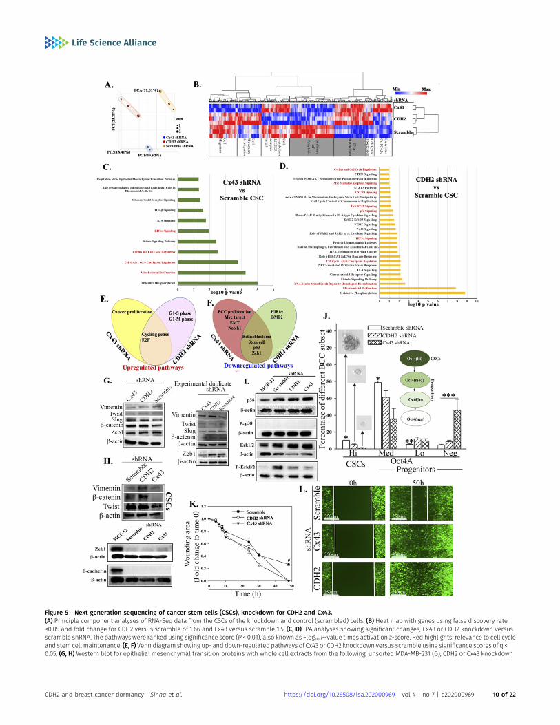
Figure 5 Next generation sequencing of cancer stem cells (CSCs), knockdown for CDH2 and Cx43.(A) Principle component analyses of RNA-Seq data from the CSCs of the knockdown and control (scrambled) cells. (B) Heat map with genes using false discovery rate<0.05 and fold change for CDH2 versus scramble of 1.66 and Cx43 versus scramble 1.5. (C, D) IPA analyses showing significant changes, Cx43 or CDH2 knockdown versusscramble shRNA. The pathways were ranked using significance score (P < 0.01), also known as −log10 P-value times activation z-score. Red highlights: relevance to cell cycleand stem cell maintenance. (E, F) Venn diagram showing up- and down-regulated pathways of Cx43 or CDH2 knockdown versus scramble using significance scores of q <0.05. (G, H) Western blot for epithelial mesenchymal transition proteins with whole cell extracts from the following: unsorted MDA-MB-231 (G); CDH2 or Cx43 knockdown
CDH2 and breast cancer dormancy Sinha et al. https://doi.org/10.26508/lsa.202000969 vol 4 | no 7 | e202000969 10 of 22

by rendering CSCs chemosensitive (Fig 8). Indeed, targeting CDH2 and itskeypathways trigger cell proliferation, lossof stemnessandactivatedpro-apoptotic pathways to chemosensitize resistant CSCs (Figs 4–7).
Knockdown of CDH2 and Cx43 led to reduced CSCs in femurs butconcomitant increased number of BCCs in the brain and lung (Fig4C). These findings strongly suggested that similar loss of stemnessin CSCs within femurs could reverse dormancy. The experimentalstudies indicated that the CDH2 knockdown cells entering the brainand lungs were chemosensitive, based on the clearance of BCCs inthese organs when the mice were treated (Fig S4D and 7H and I).These findings are relevant to treatment as the data provide in-formation to combine CDH2 inhibitor such as a γ-secretase inhibitorwith chemotherapy. Such combination will ensure targeting of theCSCs without allowing them to home to tertiary sites. CDH2 was highin T47D but Cx43 was increased in MDA-MB-231 (Fig 1A). Despitethese differences, CSCs from both cell lines show preference forGJIC with BM microenvironmental cells (Patel et al, 2012), indicatingrelevance to triple-negative and positive BCCs. This study em-phasized on MDA-MB-231 because of poor prognosis associatedwith triple-negative BCCs across the metropolitan areas, includingour inner city medial school oncological facility.
The loss- and gain-of-function studies on CDH2 in GJIC betweenCSCs and BM niche cells (Fig 1) indicate a central role for CDH2 thatexplains our finding on Cx43-mediated gap junction (Patel et al,2012). The studies did not address if deficient CDH2 could be re-sponsible for CSC exit from the BM, but we determined that itsdecrease caused migration of CSCs from the blood into brains andlungs. Because another report showed preference of CSCs for BMwithin 72 h, we deduced that CDH2 may have another role indirecting CSCs to BM (Patel et al, 2012). Going forward, it is importantto track the timeline movement of CDH2-deficient CSCs from BM toother organs. It is also important to identify the interacting sitesbetween CDH2 and Cx43, including orthogonal techniques toquantitate CDH2–Cx43 interaction in different organelle. Suchstudies will provide insights on the 3-dimensional structure andperhaps the effects on linked cell signaling pathways. Our in silicoanalyses show predicted interaction sites, which were validated bydifferent techniques, including PLA and nanoimaging to show Cx43-CDH2 colocalization (Fig 3). Thus, rather than inhibiting CDH2 alone,an exciting follow-up would be to focus on mapping the CDH2motifs that are specifically involved in the interaction with Cx43,which is up-regulated in CSCs. This would allow for the develop-ment of pharmacological agents to block CDH2-Cx43 interactions indormant chemoresistant CSCs.
The colocalized Cx43 and CDH2 in cancer cell lines were similar in22 BM biopsies from BC patients. Interestingly, there was colocalizedCx43/CDH2 (within very few cells) in BM biopsies of patients withhematologicalmalignancy. One slide fromhematologicalmalignancywas positive for cytokeratin, suggesting an undiagnosed epithelial
malignancy. Furthermore, using PLA with BM biopsy, we not that theaxis between CDH2 and Cx43 is not limited to colocalization; insteadthese two molecules for a complex with each other (Fig 3I). The datafrom patients, although limited, are exciting and warrant furtheranalyses. Nonetheless, the data support the idea that CDH2–Cx43interaction could be required for establishing and plausibly main-tainingmalignancy in BC patients. Because BC dormancy can occur atany time during the disease and after treatment, the continuedcolocalization between Cx43 and CDH2 in posttreatment samplesconfirmed the importance of such colocalization during dormancy.
The question is why there is a preferential effect of tumor cells toBM? This is a clinical observation with no clear answer. The CSCs withhigher CDH2 and Cx43 quickly home to the BM whereas cells withlower levels of thesemolecules remain at other sites such as lungs. Itis unclear if CDH2 andCx43 guide CSCs to BM.Wenow show that thesemolecules facilitate the cells’ survival in BM and possibly duringlong-term dormancy. Thus, this study begins to advance our un-derstanding on themechanisms bywhich BMniche cells maintain BCdormancy with the CDH2-Cx43 axis as a central theme for furtherstudies. This would lead to the discovery of new mechanisms toreverse and prevent BC dormancy for therapeutic targeting. Inter-estingly, M2 macrophages (MΦ), shown to mediate BC dormancy(Walker et al, 2019), could not replace CDH2 knockdown in CSCs withrespect to re-establishing dormancy in BM (Fig S4). Because M2 MΦcan also form GJIC with BM niche cells, it is possible that the loss ofCDH2 in CSCs also prevented dormancy with MΦ. The studies with M2MΦ taught us that perhaps the secretome fromM2MΦsmight be lessimportant and CDH2 warrants further studies.
The increased cell proliferation with enhancedmigration of BCCsin mouse brain and lungs following CHH2 knockdown resemblesreverse dormancy of BCCs when the dormancy cells becomemetastatic. We did not observe any change in EMT proteins withdecreased stemness in the CDH2 knockdown CSCs. This was not asurprise because EMT is not a decisive factor of stemness (Kalluri &Weinberg, 2009; Mitra et al, 2015). The identified pathways in thisstudy provide new avenues for the development of pharmaco-logical treatments to reverse BC dormancy without HSC toxicity.These findings could also serve as a platform for preclinical studies,perhaps by repurposing drugs. BM toxicity analyses are requiredbecause there are mixed information on the literature on the rolefor CDH2 in HCS maintenance (Kiel et al, 2009; Arai et al, 2012).
We immunoprecipitated CDH2 in MSCs and also showed theinteraction with Cx43 in MSCs (Fig S1D). This finding is significantbecause MSCs, which are also found in the perivascular niche(Ehninger & Trumpp, 2011), are important for immediate transitionto dormancy (Sandiford et al, 2021). In summary, we carried outknockdown of CDH2 in CSCs and demonstrated reduced cells infemurs. Because we previously demonstrated GJIC formation byCSCs and resident BM cells, the loss of stemness in CDH2
CSCs, CSCs with scrambled shRNA (H). (I) Western blot for p38, P-p38, Erk1/2, and p-Erk1/2 with whole cell extracts from MCF12A and MDA-MB-231 CSCs with or withoutCDH2/Cx43 knockdown. (J) BC cell subsets were quantitated by flow cytometry, based on GFP with MDA-MB-231–p-Oct4a-GFP, also knockdown for CDH2 or Cx43 or control/scramble shRNA. The cells were gated as for Fig 1B and % subset plotted as mean ± SD, n = 4. Each subset was analyzed for tumorsphere and representative sphere shownfor Oct4hi and Oct4med. No tumorsphere was formed with Oct4lo or neg. *P < 0.05 versus Oct4hi or Oct4med knockdowns; **P < 0.05 versus knockdowns; ***P < 0.05 versusscramble or knockdown. (K, L) Scratch assay with CSCs from MDA-MB-231, knockdown for CDH2 or Cx43 or control/scramble shRNA. The scratch areas were imaged atdifferent times with EVOS FL Auto 2. The timeline changes are presented as mean fold changes ± SD, n = 3. Fold change is calculated as experimental/time 0 h. #P < 0.05versus CDH2 and Cx43 knockdown. Scratch image (100×) is shown for the 50 h end point.Source data are available for this figure.
CDH2 and breast cancer dormancy Sinha et al. https://doi.org/10.26508/lsa.202000969 vol 4 | no 7 | e202000969 11 of 22

Figure 6. Cycling analyses of cancer stem cells (CSCs), knockdown for CDH2 or Cx43.(A, B) Gene set enrichment analyses hallmarks for CDH2 or Cx43 knockdown versus scramble CSCs. The red and blue bars indicate up-regulated hallmarks in CDH2knockdowns and control/scramble, respectively, with significance score of q < 0.05. (C, D) Representative gene set enrichment analyses graph showing top hallmarks, G2Mand E2F, which were enriched in CDH2 and Cx43 knockdown, respectively. (E, F) IPA output showing cell cycle progression, DNA metabolism, and replication pathways thatwere down-regulated in CSCs (scramble), relative to CDH2 or Cx43 knockdown cells. Significance score used P < 0.01. (G) Western blot for cycle proteins with wholeextracts from MDA-MB-231, knockdown for CDH2, and Cx43 or transfected with scramble shRNA. (H, I) The normalized band densities are shown below, mean ± SD, n = 3.
CDH2 and breast cancer dormancy Sinha et al. https://doi.org/10.26508/lsa.202000969 vol 4 | no 7 | e202000969 12 of 22

knockdown led us to deduce a reduction of GJIC. Based on theoverall data, it appears that CDH2 knockdown transitioned the CSCsinto increase in metastatic cycling cells/chemosensitive cells (FigS4A). CDH2 knockdown CSCs lost their ability to self-renew andshow increased apoptotic activity (Fig 7). The molecular changeswithin the CDH2 knockdown CSCs cause chemosensitization, indi-cating potential for therapeutic intervention. An interestingfinding is thenoted brain metastasis in both Cx43 and CDH2 knockdown CSCs (Fig 4CandD). In brain, we observed increase in Ki67 in the BCCs, which is in linewith other studies reporting on inflammatory-mediated propagation ofbrain metastasis (Chen et al, 2016). The involvement of cytokines issupported by the RNA-Seq analyses, which showed cytokine and STAT1signaling pathways in Cx43 and CDH2 knockdown CSCs, respectively (Fig5C and D). Overall, our data show that CDH2-dependent pathways in-volve interaction with Cx43, and the relationship between Cx43 andCDH2, are complex (Fig 8). As an example, the literature reported on afragment of Cx43 acting as a transcription factor to regulate CDH2 (Kotiniet al, 2018). This relationship is underscored by the RNA-Seq datashowingboth genes regulating commonpathwaysaswell asdecreaseofone gene when the other is knockdown.
Materials and Methods
Human subjects
Rutgers Institutional Review Board, Newark, NJ, approved the use ofparaffin sections from BM biopsy of BC patients, other hemato-logical malignancy, and benign sections of breast (Table S1). The IRBalso approved the use of BM aspirates and peripheral blood fromhealthy donors (18–35 y).
Reagents
DMEM, RPMI-1640, L-glutamine, penicillin–streptomycin, FCS,Ficoll–Hypaque, β-mercaptoethanol, PBS, doxycycline, hydrocor-tisone, polybrene, sodium chloride, magnesium chloride, NP-40,Triton X-100, bovine serum albumin, protease inhibitor, phos-phatase inhibitor, PureProteome Protein A Magnetic Bead,N,N,N9,N9-tetramethylethylenediamine (TEMED), ammonium per-sulfate, polyvinylidene difluoride membranes, 1-octonal, pyronin Y(PyY), Duolink in situ detection reagent red, and Duolink probemaker were purchased from Millipore Sigma; Tris, Tris–HCl, AlexaFluor 488 Phalloidin, and hyclone donor equine serum from ThermoFisher Scientific; α-MEM, accutase, 0.05% trypsin–EDTA, OptiMEM,Lipofectamine 3000, Lipofectamine RNAiMAX, Genetecin, 7-amino-actinomycin D (7-AAD), Platinum SYBR Green qPCR SuperMix-UDGKit, DAPI, Cell Tracker Blue CMAC Dye and Cell Tracker Deep Red dye,High-Capacity cDNA Reverse Transcription Kit, SuperSignal West
Femto Maximum Sensitivity Substrate, TRIzol, and glycerol werepurchased from Thermo Fisher Scientific; Tet-free FBS, Lenti-Xconcentrator, In-Fusion HD Cloning Kit, and p24 Rapid Titer Kitfrom Takara Bio; Bradford protein assay reagent, glycine, 2× loadingdye, and sodium dodecyl sulfate from Bio-Rad; TransIT-Lentitransfection reagent from Mirus; luciferase substrate, β--galactosidase assay kit, CellTiter-Blue Cell Viability Assay Kit andApo-ONE Homogeneous Caspase 3/7 assay from Promega; acryl/bissolution (30%) 37.5:1 was purchased from VWR; brefeldin-A fromInvivoGen and RNeasy Mini Kit, and plasmid miniprep kit fromQIAGEN. Carboplatin and doxorubicin were obtained from univer-sity hospital pharmacy.
Antibodies and cytokines
Mouse anti-human CDK4mAb (1/1,000 dilution), anti-human CDK6mAb(1/1,000 dilution), mouse anti-human cyclin E mAb (1/1,000 dilution),rabbit anti-human cyclin D1 mAb (1/1,000 dilution), rabbit anti-humanCx43 (1/1,000 dilution), rabbit anti-human Vimentin (1/1,000 dilution),rabbit anti-human Slug (1/1,000 dilution), rabbit anti-human β-catenin(1/1,000 dilution), rabbit anti-human Zeb1 (1/1,000 dilution), rabbit anti-human caspase 3 (1/1,000 dilution), rabbit anti-human caspase 7(1/1,000 dilution), rabbit anti-human MDR1 (1/1,000 dilution), rabbitanti-human E-cadherin (1/1,000 dilution), rabbit anti-human p38(1/1,000 dilution), rabbit anti-human p-p38 (1/1,000 dilution), rabbitanti-human Erk1/2 (1/1,000 dilution), rabbit anti-human p-Erk1/2(1/1,000 dilution), and normal anti-rabbit immunoglobulin G (IgG)were purchased from Cell Signaling Technology; rabbit polyclonal anti-human cyclin A (1/1,000 dilution), and goat anti-rabbit IgG-TR werepurchased from Santa Cruz Biotechnology; rabbit polyclonal anti-human CDH2 (1/1,000 dilution), rabbit polyclonal anti-human Ki67 (1/200 dilution), and rabbit anti-human vinculin mAb (1/1,000 dilution)from Abcam; rabbit anti-human Twist (1/1,000 dilution), mouse anti-human pan-cytokeratin (1/200 dilution), and mouse anti-humanβ-actin (1/2,000 dilution) from Millipore Sigma; goat polyclonal anti-rabbit IgG-HRP (1/5,000 dilution), goat polyclonal anti-murine IgG-HRP(1/5,000 dilution), goat anti-rabbit IgG–Alexa Fluor 405 (1/500 dilution),goat anti-mouse IgG–AlexaFluor 488 (1/500dilution), goatanti-rabbit IgG-Alexa Fluor 610 (1/500 dilution), goat anti-mouse Alexa 647 IgG (1/500dilution), rabbit anti-human HA tag (1/1,000 dilution), rabbit anti-humanFlag tag (1/1,000 dilution), goat polyclonal anti-rabbit IgG-AP (1/500 di-lution), rabbit anti-human GM130 (1/1,000 dilution), rabbit anti-humanGRP78 (1/1,000 dilution), and CD14+ Dynabeads Flow kit from ThermoFisher Scientific; HRP-mouse anti-rabbit IgG light chain–specific fromJackson Immuno Research, murine anti-CD206-PE (1/20 dilution), murineanti-HLA-DR-APC (1/20 dilution), murine anti-human CDH2-PE (1/20 dilu-tion), FITC murine anti-human CD73 (1/20 dilution), PE murine anti-humanCD29 (1/20 dilution), murine anti-human CD90−perCp/Cyanine 5.5 (1/20dilution) from BD Biosciences; and recombinant human M-CSF, and IL-4from R & D Systems.
*P < 0.05 (H, I) MDA-MB-231 (H) and T47D (I) were knockdown CDH2 and Cx43 with siRNA. Control cells used Risc free. The cells were labeled with PyY (RNA) and 7AAD(DNA). Cells with low RNA was gated within the low DNA area. The data were analyzed with BD Analyses software. (J) CDH2 or Cx43 knockdown MDA-MB-231-Oct4-GFP weretransfected with cyclin D1 reporter gene vector. The reporter gene activity (luciferase) was normalized with pβ–gal activity and the values presented as mean relativeluminescence unit ± SD, n = 4. (K) CDH2 or Cx43 knockdown MDA-MB-231 were labeled with 7AAD. The cells were analyzed for % cells in S-phase for each BC cell subsetusing ModFit software. The data are presented as mean S phase±SD, n = 3.Source data are available for this figure.
CDH2 and breast cancer dormancy Sinha et al. https://doi.org/10.26508/lsa.202000969 vol 4 | no 7 | e202000969 13 of 22

Figure 7. Apoptotic pathways in CDH2 knockdown cancer stem cells (CSCs).(A) IPA output shows down-regulation of apoptotic pathways in control (scramble) CSCs relative to CDH2 and Cx43 knockdown CSCs. (B)Western blot for caspase 3 and 7with whole cell extracts from CSCs with scramble shRNA or, CDH2 or Cx43 knockdown. The normalized band densities are shown at right. *P < 0.05 versus CDH2 or Cx43knockdown. (C) Apoptotic activity was analyzed with Apo-ONE Homogeneous caspase 3/7 assay kit and the relative fluorescence unit presented for MDA-MB-231,knockdown for CDH2 or Cx43, or scramble shRNA. In parallel, the assay was performed with cells, treated with carboplatin (220 mg/ml) or doxorubicin (1 mM) for 4 h. (D)Western blot for Pgp with whole-cell extracts from CSCs fromMDA-MB-231, knockdown for CDH2 or Cx43, or control/scramble shRNA. Bands were normalized with β-actin
CDH2 and breast cancer dormancy Sinha et al. https://doi.org/10.26508/lsa.202000969 vol 4 | no 7 | e202000969 14 of 22

Vectors and nucleotides
Human CDH2 and Cx43 siRNA and Risc free (control) were purchasedfrom Dharmacon; human cyclin D1 promoter in pGL3-basic (researchresource identifiers, RRID:Addgene_32726) and pcDNA 3.2 with Cx43-HA(RRID:Addgene_49851) were obtained from Addgene (which was adonation from Frank McCormick and Anne Brunet laboratory, respec-tively); pCMV2-CDH2-Flag from Sino Biologicals; and human shRNApRFP-C-RS with scramble sequence, CDH2-shRNA, or Cx43 shRNA fromOriGene Technologies.
Inducible CDH2 and Cx43 expression vectors were created with thelentiviral plasmid pLVX-TetOne-Puro (Takara Bio). The coding regionfor CDH2 and Cx43 was amplified from pCMV2-CDH2 Flag and pcDNA3.2-Cx43 HA, respectively, using RT-PCR with following primers: CDH2forward: 59-GCA GAG ATC TGG ATC CTC AGT CAT CAC CTC CAC CAT ACATG-39, CDH2 reverse: CCC TCG TAA AGA ATT CAT GTG CCG GAT AGC GGGAGC GCTG-39; Cx43 forward: 59-CCC TCG TAA AGA ATT CAT GGG TGA CTGGAG CGC C-39; Cx43 reverse: 59-GAG GTG GTC TGG ATC CCT AGA TCT CCAGGT CAT CAG GCC-39. The amplified region was inserted into pLVX-TetOne-Puro using In-Fusion HD Cloning Kit (Takara Bio). The frag-ment insertion was validated by DNA sequencing at Genewizusing
primer: GGATTAGGCAGTAGCTCTGACGGCCC. The complete vector ishereafter referred as pLVX-CDH2/GS and pLVX-Cx43/GS, respectively.
Viral propagation, quantitation, and induction
Lentiviral DNA for pLVX-CDH2/GS and pLVX-Cx43/GS was producedwith the 3rd Generation Packaging System mix (ABM Inc.). Thevectors were co-transfected into HEK293T cells using the packagingmix, lentiviral plasmids, and TransIT-Lenti transfection reagent. Thetransfection followed the manufacturer’s protocol. The trans-fectants were cultured in 10% DMEM with Tet-free FCS for 48 h. Afterthis, the supernatant containing the virus was collected and filteredthrough 0.45-µm membrane filtration system. The supernatant wasthen concentrated using Lenti-X Concentrator Takara Bio as per themanufacturer’s protocol. Quantification of viral particles used p24Rapid Titer Kit Takara Bio.
Ectopic expressions of CDH2 and Cx43 in MDA-MB-231 Oct4 GFP/CDH2-shRNA-RFP and MDA-MB-231 Oct4 GFP/Cx43-shRNA-RFP weredone by spinfection. We transduced pLVX-TetOne-CDH2/GS andpLVX-TetOne-Cx43/GS at MOI of 2:1, with 4 µg/ml of polybrene. Thecells were immediately centrifuged for 1 h at 1,200g at 32°C for 1 h.
and presented at right, n = 3. **P < 0.05 versus Cx43 or CDH2 knockdown. (E) Cell viability was determined for MDA-MB-231, knockdown for CDH2 or Cx43, or control with scrambleshRNA, treated with carboplatin (220mg/ml). The analyses were performed at 24 h intervals up to 120 h using cell titer blue. The data are presented as% viable cells ± SD, n = 3. Thepercentage of viable cells at 120 h is zoomed on the right. *P < 0.05 versus knockdown cells at the 120 h time point. (E, F) The studies in “(E)”were repeated, except with doxorubicin(1mM). *P < 0.05 versus Cx43 andCDH2 knockdown. (G)Protocol used to inject NSGmice, i.v. with 5 × 10⁵ CSCs isolated fromMDA-MB-231-Oct4-GFP, knockdown for CDH2 (RFP), Cx43(RFP), or scrambled shRNA (RFP). Mice were injected intraperitoneally with 5 mg/ml carboplatin at days 3 and 5. (H) At day 7, sections from paraffin embedded femurs were labeledwith anti–pan-cytokeratin-Texas Red (red) and anti–Ki67-AF405 (blue). Tissues were imaged with EVOS FL Auto 2 at magnification of 200×. Images represent six mice/group. (I) Thenumber of BC cells (red) in mouse femur from “(H)” was counted using ImageJ and presented as mean BC cells ± SD, n = 6. (J) Cartoon summarizes the data in this Figure:Decreased apoptotic pathways in CSCs impart chemoreistance. Cx43/CDH2 knockdown cells reversed the resistance to chemosensitization.Source data are available for this figure.
Figure 8. Summary.CDH2 is required for Cx43-mediated GJ between cancer stem cells (CSC) and BM stromal cells. CDH2 knockdown led to increase in apoptotic genes. CDH2 knockdownincrease cell cycle of CSCs and metastasis. CDH2/Cx43 knockdown chemosensitized CSCs.
CDH2 and breast cancer dormancy Sinha et al. https://doi.org/10.26508/lsa.202000969 vol 4 | no 7 | e202000969 15 of 22

After this, the cells were incubated at 37°C for 24 h. CDH2 expressionwas induced with 500 ng/ml Dox and Cx43 with 10 ng/ml Dox.
Cell lines
MDA-MB-231 (Cat. no. HTB-26), T47D (Cat. no. HTB-133), HEK-293T(Cat. no. CRL-3216), and MCF12A (Cat. no. CRL-10782) were purchasedfrom American Type Culture Collection (ATCC) and cultured as pertheir instruction. MDA-MB-231 is negative for estrogen, progester-one, and herceptin receptors (triple negative), whereas T47D istriple positive. MCF12A is a non-tumorigenic breast epithelial cellline.
We previously described MDA-MB-231 cells with stable trans-fection of pOct4a-GFP (Patel et al, 2012). Stably transfection ofpEGFP1-Oct3/4 allowed for selection of CSC based on GFP intensity(top 5% of total cells). Hi, Med, and Low GFP represent the relativeOct4a expression. The pOct4a-GFP transfectants were stably knock-down for CDH2 and Cx43 by transfecting with pRFP-C-RS-Scramble,-CDH2, and -Cx43 using Lipofectamine 3000 as per the manufacturer’sinstruction. The stable transfectants were selected with puromycin.The media also contained G418 to ensure stability of pOct4a-GFP.The surviving colonies of cells were verified for CDH2 or Cx43knockdown. Clone B was selected for CDH2 because of >80% knock-down efficiency (Fig S1F and G). We designated the stable knockdowncells as follows: M-Oct4-GFP/scramble-RFP, M-Oct4-GFP/CDH2-shRNA-RFP and M-Oct4-GFP/Cx43-shRNA-RFP.
CSC selection with CDH2 or Cx43 knockdown used the followinggating schemes: We first selected the knockdown cells within thetotal BCCs by gating on RFP (knockdown and vector control), usingforward (FSC-H), and side scatter (SSC-H). This was followed by asecond gating for CSCs, based on GFPhi. We further ensure theselection of CSCs with knockdown Cx43 and CDH2 by a secondgating of RFPhi cells.
BM stromal culture
Stroma cells were cultured from BM aspirates, as described(Corcoran et al, 2007). The aspirate was diluted in α-MEM containing12.5% FCS, 12.5% horse serum, 10−7 mol/l hydrocortisone, 10−4 mol/lβ-mercaptoethanol, and 1.6 mmol/l glutamine and then added toT25 tissue culture flasks. After 3 d, red blood cells and granulocyteswere removed by Ficoll–Hypaque density gradient centrifugation.The mononuclear fraction was replaced in the flasks and at weeklyintervals, 50% of media were replaced until confluence.
MSC culture
MSCs were cultured from BM aspirates, as described (Corcoran et al,2007). Briefly, BM aspirate was diluted in DMEM containing 10% FCSin vacuum gas plasma-treated 100mm petri dishes (BD Falcon). Theplates were incubated at 37°C with 5% CO2. At day 3, red blood cellsand granulocytes were removed by Ficoll–Hypaque density gra-dient centrifugation. The mononuclear fraction was replaced in thedishes. At weekly intervals, 50% of the media were replaced untilthe cells were ~80% confluent. At passage 3, the cells were char-acterized for phenotype and multipotency (Fig S1A–C).
Flow cytometry
Cell surface labeling for CDH2 was performed with cells, washedwith 1× PBS, and then fixed with 3.7% formaldehyde for at RT for15 min. This was followed by labeling with anti–CDH2-PE for 30 minat RT, in the dark. In the case of intracellular labeling for CDH2, thefixed cells were washed with 1× PBS for 5 min and then per-meabilized at 4°C with 0.2% Triton X-100 for 5 min. After this, thecells were washed again with 1× PBS and then labeled with anti–CDH2-PE or APC as for extracellular labeling. The cells were washedwith 1× PBS and then immediately analyzed on the FACSCalibur (BDBiosciences).
Sorting of BCC subsets
BCC subsets were sorted as described (Patel et al, 2012). Briefly,BCCs, stably transfected with pEGFP1-Oct3/4, were selected, basedon relative GFP expression using the FACSDiva (BD Biosciences). Thetop 5% GFP (Oct4hi) cells contained CSCs (Patel et al, 2012). Furthersorting was conducted for CSCs with specific knockdown, based onthe fluorescence-tagged shRNA, or scramble vector: GFPhi/RFPhi:MDA-MB-231 Oct4 GFP/scramble RFP, MDA-MB-231 Oct4 GFP/CDH2-shRNA-RFP, and MDA-MB-231 Oct4 GFP/Cx43-shRNA-RFP cells weresorted on the FACSDiva (BD Biosciences) to select GFPhi/RFPhi cells.
Dye transfer assay
GJIC between CSCs and stroma or MSCs were performed as de-scribed by Patel et al (2012). MSCs or stromal cells were labeled with2.5 mM of Cell Tracker Blue CMAC Dye (microscopy) or Cell TrackerDeep Red dye (flow cytometry). For labeling the cells, the dyes wereincubated for 30 min at 37°C in a CO2 incubator. After incubation,the cells were washed twice with 1× PBS. Co-cultures of dye-loadedMSCs or stromal fibroblasts and CSCs (yellow: Oct4A-GFPhi + shRNA/RFPhi) at 1:1 ratio were assessed for GJIC by imaging for dye transferinto CSCs. Control co-cultures used CSCs with scramble shRNA BCCswith vehicle or 1-octanol (300 µM). Dye transfer by flow cytometryoccurred after 72 h as follows: Cells were trypsinized, pelleted bycentrifugation, and then resuspended in 0.5 ml PBS. Fluorescencemicroscopy: Cells were imaged using the EVOS FL Auto 2. Thenumber of CSCs with dye were counted 10 fields per condition usingImageJ (Schneider et al, 2012) (n = 3) (https://imagej.net/ RRID:SCR_003070). Percent of dye transfer is number of CSCs with dyedivided by total number of cells within the field, times 100.
Immunocytochemistry
GFPhi/RFPhi sorted MDA-MB-231 were placed in four-well chamberslides (Nunc; Thermo Fisher Scientific). After 24 h, the cells adheredand were fixed with 3.7% formaldehyde for 15 min at RT followed bypermeabilization for 5 min using 0.2% Triton X-100 in PBS. The cellswere blocked with 1% BSA in PBS for 1 h. The wells were carefullywashed twice with 1X PBS and then incubated for 2 h at RT withthe following: anti-Cx43 (1/200), anti-CDH2-PE (1/20), anti-GM130(1/200), or anti-GRP78 (1/200). All antibodies were diluted in 1× PBS.After incubation, the wells were washed with 1× PBS and thenincubated with goat anti-mouse IgG–Alexa Fluor 488 and goat anti-
CDH2 and breast cancer dormancy Sinha et al. https://doi.org/10.26508/lsa.202000969 vol 4 | no 7 | e202000969 16 of 22

rabbit IgG–Alexa Fluor 405. The cells were imaged with the EVOS FLAuto 2.
Golgi disruption assayMDA-MB-231 were plated at 2 × 10⁴ in four-well chamber slides. Afterovernight adherence, the cells were treated with 5 μg/ml ofbrefeldin-A. After 24 h, the cells were washed and then fixed,followed by labeling with anti-CDH2 and anti-Cx43 as above.
Image stream multispectral imaging
Image stream multispectral analyses used ImageStreamX Mark IIImaging Flow Cytometer, as described (Ghazaryan et al, 2014).Briefly, MDA-MB-231 cells were trypsinized, pelleted, resuspendedfor fixing, and permeabilization with 3.7% formaldehyde for 15 minand 0.2% Triton X-100 in PBS for 5 min, respectively. The cells wereincubated with anti–CDH2-PE (1:20) for 30 min in dark followed bywashing with 1× PBS. After this, the cells were resuspended in 500 μlof 1% formaldehyde in 1× PBS. Cx43 was assessed by labeling withgoat anti-rabbit Cx43 (1:100) for 30 min at RT and secondary goatanti-rabbit Alexa 610 IgG for another 30 min in the dark. The cellswere then washed with 1× PBS and resuspended in 500 μl of 1%formaldehyde in 1× PBS. The cells were acquired with the Image-StreamX Mark II Imaging Flow Cytometer (Luminex).
Single cell imaging
Single cell imaging for colocalized proteins used the Nanoimager SMark II from ONI (Oxford Nanoimaging) with lasers 405 nm/150 m,488 nm/1 W, 561 nm/500 mW, and 640 nm/1 W. Dual channel ac-quisition was setup using 640-nm dichoric mirror paired with576–620-nm bandpass filter for PE and 685/40 nm emission filtersfor Alexa 647. Images were acquired with NimOS 1.3.7511 imagingsoftware at 1,000× magnification and 1.4 NA objective. Four colorwide-field images were acquired in a sequential mode with 100-msexposure time for each image. To show protein localization, all thechannels were reconstructed in Fiji.
Immunoprecipitation
Cells were lysed as for Western blot, and 15-µg protein extracts used toimmunoprecipitate CDH2 and Cx43, as described (Ghazaryan et al,2014). Briefly, the lysates were precleaned with magnetic beads boundto IgG. During this time, the extracts were incubated on ice for 30 min.After this, the beads weremagnetically removed and the extracts wereincubated overnight at 4°Cwith 2 µg/ml goat anti-rabbit Cx43 fromCellSignaling Technology, 2 µg/ml goat anti-rabbit CDH2, or 0.5 µg/mlrabbit anti-human Flag tag or normal anti-rabbit IgG. After 24 h, theextracts were incubated with magnetic beads bound to anti-rabbit IgGfor 4 h/4°C. The unbound protein was then removed using a magneticseparator. The beadboundproteinswere thenwashedfive times usinglysis buffer and then resuspended in 2× loading dye with reducingagent, β-mercaptoethanol. The immunoprecipitated lysates wereelectrophoresed on 12%SDS–PAGE and then developed for CDH2, Cx43,Flag, or HA with specific antibodies.
Molecular modeling
The molecular interaction between CDH2-Cx43 was predicted usingZDOCK (http://zdock.umassmed.edu/) and functional interactionusing STRING (v11) (https://string-db.org/ RRID:SCR_005223) (Pierceet al, 2014; Szklarczyk et al, 2019). ZDOCK used CDH2 (PDB ID: 3Q2W,ectodomain) and Cx43 (PDB ID: 2LL2, cytoplasmic domain) X-raycrystallographic database ID number as the protein of interest forinput. CDH2 protein sequence is evolutionarily conserved, hence,identical sequences in mouse and human. As such, the murinecrystal structure was used for ZDOCK analyses. The top five po-tential binding sites are shown in grey for CDH2 and blue, green,orange, pink, and red for Cx43 (Fig 3C). STRING used Cx43 (GJA1) asinput, resulting in CDH2 as one of the potential interacting proteins.
Monocyte/MΦ culture
M2 MΦ were isolated from monocytes as described (Walker et al,2019). Briefly, the monocytes were selected from human PBMCsusing human CD14+ Dynabeads Flow Kit (Thermo Fisher Scientific).The monocytes were incubated with 50 ng/ml M-CSF. After 48 h, wereplaced the media with fresh medium containing 20 ng/ml IL-4.After 3 d, the cells were positive for CD206 and negative for MHC-II.
In vivo studies
The Institutional Animal Care and Use Committee of Rutgers,Newark Campus, approved the use of mice. Female (6 wk) NOD/SCID(NSG) mice were purchased from Jackson Laboratories and housedat an AAALAC-accredited facility, Rutgers Comparative MedicineResource, New Jersey Medical School, Newark, NJ.
BC dormancyWe established dormancy in female nude and NOD/SCID (NSG)BALB/c mice, as described (Patel et al, 2012; Bliss et al, 2016; Walkeret al, 2019). Briefly, we sorted CSCs from BCCs with stable pOct4a-GFP with or without Cx43/CDH2-shRNA. Control cells expressedpOct4a and scramble shRNA. We previously reported on self-renewal of the Oct4ahi BCCs using in vitro and in vivo studies, in-cluding serial passaging (Patel et al, 2012). We injected 5 × 10⁵ Oct4hi
sorted BCCs with the highest GFP (Oct4ahi) intravenously via the tailvein of mice. After sorting to injecting of the CSCs, it is possible thatthere could be differentiation of the CSCs, resulting in proliferatingcells. Thus, to ensure elimination of these cells, we injected themice at days 3 and 5 with low-dose carboplatin (2 mg/kg), intra-peritoneally. The additional chemotherapy did not change theCDH2, Cx43, or Oct4a expression on the CSCs. These genes were thehighest in CSCs, relative to the other BC subsets (Fig 1). Dormancy inmice femurs was sustained even without the low dose chemo-therapy, which was used to eliminate proliferating cells.
CDH2 and Cx43 knockdown BCCs
NSG mice were injected with 5 × 10⁵ Oct4hi MDA-MB-231 Oct4 GFP/scramble RFP, MDA-MB-231 Oct4 GFP/CDH2-shRNA-RFP, and MDA-MB-231 Oct4 GFP/Cx43-shRNA-RFP CSCs intravenously (tail vein). Atday 10, mice were euthanized and embedded in parafilm for
CDH2 and breast cancer dormancy Sinha et al. https://doi.org/10.26508/lsa.202000969 vol 4 | no 7 | e202000969 17 of 22

processing, as described above. The experiments with NSG micewere repeated by co-injecting 106 CMAC (blue)-labeled M2Φ and thefollowing CSCs isolated from MDA-MB-231-Oct4-GFP: scramble-RFP,CDH2-shRNA-RFP, or Cx43-shRNA-RFP. At day 10, the mice wereeuthanized and tissues processed as for nude mice.
Immunohistochemistry
Antigens were retrieved from parafilm-embedded mouse femursand human BM biopsies by overnight incubation at 56°C. After this,the slides were dewaxed with xylene and ethanol and thendehydrated, followed by rehydration using deionized water. Thecells were permeabilized with 0.1% Triton X-100. The slides werethen washed with 1X PBS and then incubated overnight at 37°C in ahumidified chamber with primary antibodies, each at 1/250 finaldilution. The slides were washed thrice with 1× PBS then incubatedfor 2 h at RT in a humidified chamber in the dark with fluorescence-tagged secondary antibodies (1/500 final dilution). After washingthe slides with 1× PBS, the tissue was immediately analyzed on theEVOS FL Auto 2 Imaging System. The results of immunohisto-chemical analyses of slides from mice and human sections werequantified with ImageJ. We counted BCCs in 10 fields/slide. Im-munohistochemistry for human pan-cytokeratin and Ki67 wasperformed in murine femurs and CDH2, Cx43, and pan-cytokeratinfor human BM biopsies.
PLA
Interaction between CDH2 and Cx43 was confirmed in human BM BCbiopsy (randomly selected patient 3) using PLA as per manufac-turers instruction (Alam, 2018). Briefly, the antigen surface ofparafilm-embedded biopsy was exposed, as explained before inimmunohistochemistry. Anti-CDH2 and anti-Cx43 PLA probe wasfirst created followed by ligation and signal amplification usingTexas red probe. The signal was amplified only when the twoproteins are <40 nm apart. The slides were imaged using EVOS FLAuto 2 at 600× magnification. Isotype was negative control probecreated as per manufacturer’s instruction.
RNA sequencing (Seq)
CSCs from MDA-MB-231 Oct4 GFP/scramble RFP, MDA-MB-231 Oct4GFP/CDH2-shRNA-RFP, and MDA-MB-231 Oct4 GFP/Cx43-\shRNA-RFP were sorted as outlined in Fig 1E (Patel et al, 2012). The cellswere allowed to recover from stress by incubating overnight at 37°C,5% CO2. Total RNA was isolated with RNeasy Mini Kit (QIAGEN) andthen submitted to the Genomics Center at Rutgers New JerseyMedical School. Ribosomal RNA was depleted with Ribo-Zero goldfrom Illumina and then used to prepare libraries for next gener-ation sequencing. The libraries were prepared with NEB Ultra IILibrary Preparation kit and NEBNext Multiplex Oligos for Illumina(Dual Index Primers Set 1) (New England BioLabs). Upon qualitycontrol of the libraries with Qubit instrument and a high-sensitivityKit from Thermo Fisher Scientific as well as TapeStation 2200 in-strument and D1000 ScreenTapes from Agilent, the cDNA librarieswere diluted to 2 nM. The libraries were denatured as per Illu-mina’s protocol and then sequenced on Illumina’s NextSeq 500
[NIH-1S10OD018206-01A1] using 1X75 cycle high output kit. The BCLfiles from sequencing were converted to FastQ files usingBCL2FASTQ software from Illumina (RRID:SCR_015058).
Data analyses
RNA-SeqPartek Flow software (Version 7) was used to convert fastq.gz to txtfiles (Partek Inc.). Reads were then aligned using STAR 3.6.1 to hg38with Partek’s optimization of the expectation–maximization algo-rithm. One low-quality sample containingmore than 50% intergenicand intronic reads was removed and the remainder of the datawere normalized using log counts permillion +1. Batch effect amongthree replicates of sequencing was removed via Partek log normalmodel with shrinkage and low count reads were removed (maxi-mum sample <1.5).
Partek GSA analysis was used to interrogate the effect of the twoknockdowns versus scramble (control). Genes with a false discoveryrate < 0.05 and a fold change of 1.66 in shRNA CDH2 versus Scrambleand a fold change of 1.5 in shRNA Cx43 versus Scramble wereretained. Differentially expressed genes were visualized in a heatmap with hierarchical clustering in Morpheus (Broad Institute:https://software.broadinstitute.org/morpheus). Common dendro-gram nodes were queried for enriched gene ontology pathways andsubsequently annotated.
GSEA analyses were also used on all genes that passed qualitycontrol. Hallmark and C2 gene sets were used for analysis. Sig-nificant results, q < 0.05, were retained. Ingenuity Pathway Analysis(IPA; QIAGEN Inc.) of differentially expressed genes was used tointerrogate the effect on canonical pathways and cell functions, aswell as to generate networks of selected cell functions. Thepathways passing P < 0.01 were ranked using −log10 P-value timesactivation z-score.
Real-time PCR
Real-time PCR for CDH2 was performed as described (Patel et al,2012). Briefly, RNA was extracted from samples using TRIzol as perthe manufacturer’s instruction. RNA was quantified with QIAxpertand then converted to cDNA with the High-Capacity cDNA ReverseTranscription Kit. Real-time PCR used Platinum SYBR Green qPCRSuperMix-UDG Kit and the 7300 Real-Time PCR System (ThermoFisher Scientific). The following primers were used in the PCR mix:CDH2 forward, 59-CAG TGG CCA CCT ACA AAG-39; CDH2 reverse, 59-AAATGA AAC CGG GCT ATC-39; β-actin forward, 59-GCC CTA TAA AAC CCAGCG GC-39; β-actin reverse, 59-AGA GGC GTA CAG GGA TAG CA-39;Human GAPDH forward, 59-CAG AAG ACT GTG GAT GGC C-39; HumanGAPDH reverse, 59-CCA CCT TCT TGA TGT CAT C-39; Human andmouseGAPDH, forward 59-AGT CCC CCA CCA CAC CAC TGA AT-39 and humanand mouse GAPDH reverse, 59-TTG ATG GTA CAT GAC AAG GTG C-39.
Western blot analyses
The proteins were isolated using a cell lysis buffer (50 mM Tris–HCl[pH 7.4], 100 mM NaCl, 2 mM MgCl2, 10% glycerol, and 1% NP-40) asexplained in Ghazaryan et al (2014). The extracts (15 μg) wereelectrophoresed on 12% SDS–PAGE gel. Proteins were transferred
CDH2 and breast cancer dormancy Sinha et al. https://doi.org/10.26508/lsa.202000969 vol 4 | no 7 | e202000969 18 of 22

onto polyvinylidene difluoride (PVDF) membranes (Perkin Elmer).The membranes were incubated overnight at 4°C on rocker withanti-Cx43, anti-CDH2, anti-Vimentin, anti-Twist, anti-Slug, anti-β−catenin, anti-Zeb1, anti-caspase 3, anti-caspase-7, anti-MDR1, or anti-β-actin at a dilution mentioned above in 1% nonfat milk dissolved in1× PBS Tween. Next day, the membranes were incubated withspecies-specific HRP-tagged IgG in 1% milk 1× PBS Tween for 2 h at4°C. The membranes were developed chemiluminescence usingSuperSignal West Femto Maximum Sensitivity Substrate. The banddensities were normalized using UN-SCAN-IT densitometry soft-ware (RRID:SCR_017291; Silk Scientific).
Tumorsphere assay and in vitro serial passage
Tumorsphere assay used CSCs from MDA-MB-231-Oct4-GFP withscramble-RFP, CDH2-shRNA-RFP or Cx43-shRNA-RFP, as described (Patelet al, 2012). Cells were seeded 1 cell/well in 96-well low adhesion plate(Costar). After 10 d, tumorsphereswere dissociatedwith trypsin and thenpassed through a 40 mm mesh (BD cell strainer cap tube) and thetumorsphere assay repeated five times (serial passaging).
Wound healing (scratch) assay
MDA-MB-231-pOct4A-GFP, knockdown for Cx43 or CDH2 or transfectedwith scrambled shRNA,were seededat 5 × 10⁵ cells/well in six-well plates.The plates were incubated overnight at 37°C and 5% CO2. A scratch wasformed across the confluent cells with a sterile 200 µl tip. After this, themedia were replaced with fresh media. The scratch area was imagedusing the EVOS Auto FL2 at times ranging between 1 and 50 h.
The wound size was quantified by measuring the distance be-tween the scratch. Time 0 wound was considered to be 100% gap andwas given the value of 1. Wound was calculated bymeasuring the gapat a given time point/the original size of gap at time 0. The distancebetween the scratch was measured using ImageJ software. The ex-periment was repeated three times (n = 3). The data were analyzedusing t test; P-value ≤ 0.05 was considered to be significant.
Cell cycle analyses
Cell cycle analyses colabeled cells with 7AAD and PyY, as described(Bliss et al, 2016). Because PyY and RFP from shRNA used samechannel on the flow cytometry, we transiently transfected the cellswith siRNA using MDA-MB-231 and T47D. The cells were transfectedwith CDH2 siRNA or Cx43 siRNA or Risc free (control) siRNA usingLipofectamine RNAimax. After 24 h, the cells were labeled with PyYfollowed by 7AAD staining. The cells were labeled with 7-AAD and PyYand gated for low DNA based on 7-AAD and then selected the labeledRNA, based on PyY incorporation for assessing the G0/G1 population.
Cell viability
Cell viability was performed as described (Patel et al, 2012). M-Oct4-GFP/scramble-RFP, M-Oct4-GFP/CDH2-shRNA-RFP, and M-Oct4GFP/Cx43-shRNA-RFP cells were treated with carboplatin (220 µg/ml) or doxorubicin (1 µM). The cell viability was assessed every 24 hwith CellTiter-Blue as per the manufacturer’s instruction. The fluores-cence was taken at 560(20)Ex/590(10)Em.
The absorbance of each well at 570 and 600 nm wavelength wasused to calculate viability as follows:
Treated ðAbsorbance570−Absorbance600Þ × 100Average of Untreated ðAbsorbance570−Absorbance600Þ
The data were analyzed using t test; P-value ≤ 0.05 was con-sidered to be significant.
Reporter gene assay
Reporter gene assays were performed as described (Qian et al,2001). M-Oct4 GFP cells with CDH2 or Cx43 knockdown or withscrambled shRNA were transfected with human cyclin D1 pro-moter in pGL3-basic. The cells were transfected with Lipofect-amine 3000, as per the manufacturer’s instruction. The cellswere co-transfected with pβgal as described (Qian et al, 2001).After 24 h, cell lysates were prepared using 1× Promega lysisbuffer. Protein concentration was determined using the Brad-ford protein assay reagent. Luciferase and β-galactosidaselevels were quantified using kits from Promega. For calculatingrelative luminescence unit, the luciferase values were normal-ized with β-galactosidase.
Apoptosis assay
We quantitated apoptotic activity with Apo-ONE Homogeneous Caspase3/7 assay kit. Themethod followed themanufacturer’s instruction. Briefly,M-Oct4-GFP, knockdown for CDH2 or Cx43 or scramble shRNA wereadded to 96-well black plates. Each experimental point used sixreplicates. Apo-ONE caspase 3/7 reagent was added to each well.The plate was incubated for 4 h at RT, followed by fluorescencemeasurement at 499Ex/521Em. The relative fluorescence unit wascalculated by subtracting relative fluorescence unit blank fromthe samples. The same assay was repeated with carboplatin (220µg/ml) and doxorubicin (1 µM) treated BCCs.
Data Availability
The data from Next Generation Sequencing are available in the GeneExpression Omnibus database: GSE138434 (https://www.ncbi.nlm.nih.gov/geo/query/acc.cgi?acc=GSE138434).
Supplementary Information
Supplementary Information is available at https://doi.org/10.26508/lsa.202000969.
Acknowledgements
This work was supported in part by a grant awarded by the New JerseyCommission of Health and Metavivor Foundation. This work is in partialfulfillment for a doctoral thesis for G Sinha.
CDH2 and breast cancer dormancy Sinha et al. https://doi.org/10.26508/lsa.202000969 vol 4 | no 7 | e202000969 19 of 22

Author Contributions
G Sinha: conceptualization, data curation, software, validation,methodology, and writing—original draft.AI Ferrer: software, methodology, and writing—review and editing.S Ayer: software, formal analysis, methodology, and writing—reviewand editing.MH El-Far: software, validation, methodology, and writing—reviewand editing.SH Pamarthi: validation, methodology, and writing—review and editing.Y Naaldijk: data curation, software, methodology, and writing—reviewand editing.P Barak: resources, software, investigation, visualization, and writing—review and editing.OA Sandiford: resources, validation, methodology, and writing—review and editing.BM Bibber: conceptualization, data curation, methodology, andwriting—review and editing.G Yehia: validation, methodology, and writing—review and editing.SJ Greco: resources, formal analysis, validation, methodology, andwriting—review and editing.J-G Jiang: data curation, formal analysis, visualization, and writing—review and editing.M Bryan: resources, data curation, investigation, and writing—reviewand editing.R Kumar: data curation, formal analysis, methodology, and writing—review and editing.NM Ponzio: formal analysis, supervision, methodology, and writing—review and editing.J-P Etchegaray: validation, methodology, and writing—review andediting.P Rameshwar: conceptualization, data curation, formal analysis,supervision, funding acquisition, project administration, and writing—review and editing.
Conflict of Interest Statement
The authors declare that they have no conflict of interest.
References
Abravanel DL, Belka GK, Pan TC, Pant DK, Collins MA, Sterner CJ, Chodosh LA(2015) Notch promotes recurrence of dormant tumor cells followingHER2/neu-targeted therapy. J Clin Invest 125: 2484–2496. doi:10.1172/JCI74883
Alam MS (2018) Proximity ligation assay (PLA). Curr Protoc Immunol 123: e58.doi:10.1002/cpim.58
Albrengues J, Shields MA, Ng D, Park CG, Ambrico A, Poindexter ME, UpadhyayP, Uyeminami DL, Pommier A, Küttner V, et al (2018) Neutrophilextracellular traps produced during inflammation awaken dormantcancer cells in mice. Science 361: eaao4227. doi:10.1126/science.aao4227
Arai F, Hosokawa K, Toyama H, Matsumoto Y, Suda T (2012) Role of N-cadherinin the regulation of hematopoietic stem cells in the bone marrowniche. Ann N Y Acad Sci 1266: 72–77. doi:10.1111/j.1749-6632.2012.06576.x
Berry DA (2013) Breast cancer screening: Controversy of impact. Breast 22:S73–S76. doi:10.1016/j.breast.2013.07.013
Bliss SA, Paul S, Pobiarzyn PW, Ayer S, Sinha G, Pant S, Hilton H, Sharma N,Cunha MF, Engelberth DJ, et al (2018) Evaluation of a developmentalhierarchy for breast cancer cells to assess risk-based patientselection for targeted treatment. Sci Rep 8: 367. doi:10.1038/s41598-017-18834-5
Bliss SA, Sinha G, Sandiford OA, Williams LM, Engelberth DJ, Guiro K,Isenalumhe LL, Greco SJ, Ayer S, Bryan M, et al (2016) Mesenchymalstem cell-derived exosomes stimulate cycling quiescence and earlybreast cancer dormancy in bone marrow. Cancer Res 76: 5832–5844.doi:10.1158/0008-5472.CAN-16-1092
Boire A, Coffelt SB, Quezada SA, Vander Heiden MG, Weeraratna AT (2019)Tumour dormancy and reawakening: Opportunities and challenges.Trends Cancer 5: 762–765. doi:10.1016/j.trecan.2019.10.010
Braun S, Auer D, Marth C (2009) The prognostic impact of bone marrowmicrometastases in women with breast cancer. Cancer Invest 27:598–603. doi:10.1080/07357900802574496
Braun S, Vogl FD, Naume B, Janni W, Osborne MP, Coombes RC, Schlimok G,Diel IJ, Gerber B, Gebauer G, et al (2005) A pooled analysis of bonemarrow micrometastasis in breast cancer. N Engl J Med 353: 793–802.doi:10.1056/NEJMoa050434
Carcereri de Prati A, Butturini E, Rigo A, Oppici E, Rossin M, Boriero D, MariottoS (2017) Metastatic breast cancer cells enter into dormant state andexpress cancer stem cells phenotype under chronic hypoxia. J CellBiochem 118: 3237–3248. doi:10.1002/jcb.25972
Chen Q, Boire A, Jin X, Valiente M, Er EE, Lopez-Soto A, Jacob L, Patwa R, Shah H,Xu K, et al (2016) Carcinoma-astrocyte gap junctions promote brainmetastasis by cGAMP transfer. Nature 533: 493–498. doi:10.1038/nature18268
Corcoran KE, Trzaska KA, Fernandes H, Bryan M, Taborga M, Srinivas V,Packman K, Patel PS, Rameshwar P (2007) Mesenchymal stem cells inearly entry of breast cancer into bone marrow. PLoS One 3: e2563.doi:10.1371/journal.pone.0002563
De Angelis ML, Francescangeli F, Zeuner A (2019) Breast cancer stem cells asdrivers of tumor chemoresistance, dormancy and relapse: Newchallenges and therapeutic opportunities. Cancers 11: 1569.doi:10.3390/cancers11101569
Ehninger A, Trumpp A (2011) The bone marrow stem cell niche grows up:Mesenchymal stem cells and macrophages move in. J Exp Med 208:421–428. doi:10.1084/jem.20110132
Garcıa-Castro MI, Vielmetter E, Bronner-Fraser M (2000) N-Cadherin, a celladhesion molecule involved in establishment of embryonic left-rightasymmetry. Science 288: 1047–1051. doi:10.1126/science.288.5468.1047
Gelao L, Criscitiello C, Fumagalli L, Locatelli M, Manunta S, Esposito A,Minchella I, Goldhirsch A, Curigliano G (2013) Tumour dormancy andclinical implications in breast cancer. Ecancermedicalscience 7: 320.doi:10.3332/ecancer.2013.320
Ghazaryan S, Sy C, Hu T, An X, Mohandas N, Fu H, Aladjem MI, Chang VT,Opavsky R, Wu L (2014) Inactivation of Rb and E2f8 synergizes to triggerstressed DNA replication during erythroid terminal differentiation.Mol Cell Biol 34: 2833–2847. doi:10.1128/MCB.01651-13
Kalluri R, Weinberg RA (2009) The basics of epithelial-mesenchymaltransition. J Clin Invest 119: 1420–1428. doi:10.1172/JCI39104
Kiel MJ, Acar M, Radice GL, Morrison SJ (2009) Hematopoietic stem cells do notdepend on N-cadherin to regulate their maintenance. Cell Stem Cell 4:170–179. doi:10.1016/j.stem.2008.10.005
Kim JB, Islam S, Kim YJ, Prudoff RS, Sass KM, Wheelock MJ, Johnson KR (2000)N-Cadherin extracellular repeat 4 mediates epithelial tomesenchymal transition and increased motility. J Cell Biol 151:1193–1206. doi:10.1083/jcb.151.6.1193
Kim JH, Park SY, Jun Y, Kim JY, Nam JS (2017) Roles of Wnt target genes in thejourney of cancer stem cells. Int J Mol Sci 18: 1604. doi:10.3390/ijms18081604
CDH2 and breast cancer dormancy Sinha et al. https://doi.org/10.26508/lsa.202000969 vol 4 | no 7 | e202000969 20 of 22

Kotini M, Barriga EH, Leslie J, Gentzel M, Rauschenberger V, Schambony A,Mayor R (2018) Author correction: Gap junction protein Connexin-43 isa direct transcriptional regulator of N-cadherin in vivo. Nat Commun9: 4633. doi:10.1038/s41467-018-07167-0
Lim PK, Bliss SA, Patel SA, Taborga M, Dave MA, Gregory LA, Greco SJ, Bryan M,Patel PS, Rameshwar P (2011) Gap junction-mediated import ofMicroRNA from bone marrow stromal cells can elicit cell cyclequiescence in breast cancer cells. Cancer Res 71: 1550–1560.doi:10.1158/0008-5472.CAN-10-2372
Lu X, Mu E, Wei Y, Riethdorf S, Yang Q, Yuan M, Yan J, Hua Y, Tiede BJ, Lu X, et al(2011) VCAM-1 promotes osteolytic expansion of indolent bonemicrometastasis of breast cancer by engaging α4β1-positiveosteoclast progenitors. Cancer Cell 20: 701–714. doi:10.1016/j.ccr.2011.11.002
Lynch-Sutherland CF, Chatterjee A, Stockwell PA, Eccles MR, Macaulay EC(2020) Reawakening the developmental origins of cancer throughtransposable elements. Front Oncol 10: 468. doi:10.3389/fonc.2020.00468
Mani SA, Guo W, Liao MJ, Eaton EN, Ayyanan A, Zhou AY, Brooks M, Reinhard F,Zhang CC, Shipitsin M, et al (2008) The epithelial-mesenchymaltransition generates cells with properties of stem cells. Cell 133:704–715. doi:10.1016/j.cell.2008.03.027
Mao CP, Wu T, Song KH, Kim TW (2014) Immune-mediated tumor evolution:Nanog links the emergence of a stem like cancer cell state andimmune evasion. Oncoimmunology 3: e947871. doi:10.4161/21624011.2014.947871
Martins-Marques T, Anjo SI, Pereira P, Manadas B, Girão H (2015) Interactingnetwork of the gap junction (GJ) protein Connexin43 (Cx43) ismodulated by ischemia and reperfusion in the heart. Mol CellProteomics 14: 3040–3055. doi:10.1074/mcp.M115.052894
Massague J, Obenauf AC (2016) Metastatic colonization by circulating tumourcells. Nature 529: 298–306. doi:10.1038/nature17038
Matsuda T, Fujio Y, Nariai T, Ito T, Yamane M, Takatani T, Takahashi K, Azuma J(2006) N-cadherin signals through Rac1 determine the localization ofconnexin 43 in cardiac myocytes. J Mol Cell Cardiol 40: 495–502.doi:10.1016/j.yjmcc.2005.12.010
Mebratu Y, Tesfaigzi Y (2009) How ERK1/2 activation controls cellproliferation and cell death: Is subcellular localization the answer?Cell Cycle 8: 1168–1175. doi:10.4161/cc.8.8.8147
Miller KD, Siegel RL, Lin CC, Mariotto AB, Kramer JL, Rowland JH, Stein KD, AlteriR, Jemal A (2016) Cancer treatment and survivorship statistics, 2016. CACancer J Clin 66: 271–289. doi:10.3322/caac.21349
Mitra A, Mishra L, Li S (2015) EMT, CTCs and CSCs in tumor relapse and drug-resistance. Oncotarget 6: 10697–10711. doi:10.18632/oncotarget.4037
Naume B, Zhao X, Synnestvedt M, Borgen E, Russnes HG, Lingjaerde OC,Strømberg M, Wiedswang G, Kvalheim G, Karesen R, et al (2007)Presence of bone marrow micrometastasis is associated withdifferent recurrence risk within molecular subtypes of breast cancer.Mol Oncol 1: 160–171. doi:10.1016/j.molonc.2007.03.004
Niu Z, Mu H, Zhu H, Wu J, Hua J (2017) p38 MAPK pathway is essential for self-renewal of mouse male germline stem cells (mGSCs). Cell Prolif 50:e12314. doi:10.1111/cpr.12314
Otvos B, Silver DJ, Mulkearns-Hubert EE, Alvarado AG, Turaga SM, SorensenMD, Rayman P, Flavahan WA, Hale JS, Stoltz K, et al (2016) Cancer stemcell-secreted macrophage migration inhibitory factor stimulatesmyeloid derived suppressor cell function and facilitates glioblastomaimmune evasion. Stem Cells 34: 2026–2039. doi:10.1002/stem.2393
Paquet-Fifield S, Koh SL, Cheng L, Beyit LM, Shembrey C, Mølck C,Behrenbruch C, Papin M, Gironella M, Guelfi S, et al (2018) Tightjunction protein claudin-2 promotes self-renewal of humancolorectal cancer stem-like cells. Cancer Res 78: 2925–2938.doi:10.1158/0008-5472.CAN-17-1869
Patel SA, Dave MA, Bliss SA, Giec-Ujda AB, Bryan M, Pliner LF, Rameshwar P(2014) Treg/Th17 polarization by distinct subsets of breast cancer cellsis dictated by the interaction with mesenchymal stem cells. J CancerStem Cell Res 2014: e1003. doi:10.14343/JCSCR.2014.2e1003
Patel SA, Ramkissoon SH, Bryan M, Pliner LF, Dontu G, Patel PS, Amiri S, PineSR, Rameshwar P (2012) Delineation of breast cancer cell hierarchyidentifies the subset responsible for dormancy. Sci Rep 2: 906.doi:10.1038/srep00906
Perez G, López-Moncada F, Indo S, Torres MJ, Castellón EA, Contreras HR(2021) Knockdown of ZEB1 reverses cancer stem cell properties inprostate cancer cells. Oncol Rep 45: 58. doi:10.3892/or.2021.8009
Pierce BG, Wiehe K, Hwang H, Kim BH, Vreven T, Weng Z (2014) ZDOCK server:Interactive docking prediction of protein-protein complexes andsymmetric multimers. Bioinformatics 30: 1771–1773. doi:10.1093/bioinformatics/btu097
Plaks V, Kong N, Werb Z (2015) The cancer stem cell niche: How essential is theniche in regulating stemness of tumor cells? Cell Stem Cell 16: 225–238.doi:10.1016/j.stem.2015.02.015
Qian J, Yehia G, Molina C, Fernandes A, Donnelly R, Anjaria D, Gascon P,Rameshwar P (2001) Cloning of human preprotachykinin-I promoterand the role of cyclic adenosine 5’-monophosphate responseelements in its expression by IL-1 and stem cell factor. J Immunol 166:2553–2561. doi:10.4049/jimmunol.166.4.2553
Rao G, Patel PS, Idler SP, Maloof P, Gascon P, Potian JA, Rameshwar P (2004)Facilitating role of preprotachykinin-I gene in the integration ofbreast cancer cells within the stromal compartment of the bonemarrow: A model of early cancer progression. Cancer Res 64:2874–2881. doi:10.1158/0008-5472.can-03-3121
Sandiford OA, Donnelly RJ, El-Far MH, Burgmeyer LM, Sinha G, Pamarthi SH,Sherman LS, Ferrer AI, DeVore DE, Patel SA, et al (2021) Mesenchymalstem cell-secreted extracellular vesicles instruct stepwisededifferentiation of breast cancer cells into dormancy at the bonemarrow perivascular region. Cancer Res 81: 1567–1582. doi:10.1158/0008-5472.CAN-20-2434
Schneider CA, Rasband WS, Eliceiri KW (2012) NIH image to ImageJ: 25 years ofimage analysis. Nat Methods 9: 671–675. doi:10.1038/nmeth.2089
Sinha G, Ferrer AI, Moore CA, Naaldijk Y, Rameshwar P (2020) Gap junctionsand breast cancer dormancy. Trends Cancer 6: 348–357. doi:10.1016/j.trecan.2020.01.013
Szklarczyk D, Gable AL, Lyon D, Junge A, Wyder S, Huerta-Cepas J, Simonovic M,Doncheva NT, Morris JH, Bork P, et al (2019) STRING v11: Protein-proteinassociation networks with increased coverage, supporting functionaldiscovery in genome-wide experimental datasets. Nucleic Acids Res47: D607–D613. doi:10.1093/nar/gky1131
Talmadge JE (2007) Clonal selection of metastasis within the life history of atumor. Cancer Res 67: 11471–11475. doi:10.1158/0008-5472.CAN-07-2496
Taniguchi Ishikawa E, Gonzalez-Nieto D, Ghiaur G, Dunn SK, Ficker AM, MuraliB, Madhu M, Gutstein DE, Fishman GI, Barrio LC, et al (2012) Connexin-43 prevents hematopoietic stem cell senescence through transfer ofreactive oxygen species to bone marrow stromal cells. Proc Natl AcadSci U S A 109: 9071–9076. doi:10.1073/pnas.1120358109
Thiagarajan PS, Sinyuk M, Turaga SM, Mulkearns-Hubert EE, Hale JS, Rao V,Demelash A, Saygin C, China A, Alban TJ, et al (2018) Cx26 drives self-renewal in triple-negative breast cancer via interact ion with NANOGand focal adhesion kinase. Nat Commun 9: 578. doi:10.1038/s41467-018-02938-1
Tivari S, Lu H, Dasgupta T, De Lorenzo MS, Wieder R (2018) Reawakening ofdormant estrogen-dependent human breast cancer cells by bonemarrow stroma secretory senescence. Cell Commun Signal 16: 48.doi:10.1186/s12964-018-0259-5
Tjensvoll K, Nordgard O, Skjaeveland M, Oltedal S, Janssen EAM, Gilje B (2019)Detection of disseminated tumor cells in bone marrow predict late
CDH2 and breast cancer dormancy Sinha et al. https://doi.org/10.26508/lsa.202000969 vol 4 | no 7 | e202000969 21 of 22

recurrences in operable breast cancer patients. BMC Cancer 19: 1131.doi:10.1186/s12885-019-6268-y
Walker ND, Elias M, Guiro K, Bhatia R, Greco SJ, Bryan M, Gergues M, Sandiford OA,Ponzio NM, Leibovich SJ, et al (2019) Exosomes from differentially activatedmacrophages influencedormancyor resurgenceof breast cancer cellswithinbone marrow stroma. Cell Death Dis 10: 59. doi:10.1038/s41419-019-1304-z
Wang P, Wan WW, Xiong SL, Feng H, Wu N (2017) Cancer stem-like cells can beinduced through dedifferentiation under hypoxic conditions inglioma, hepatoma and lung cancer. Cell Death Discov 3: 16105.doi:10.1038/cddiscovery.2016.105
Wei CJ, Francis R, Xu X, Lo CW (2005) Connexin43 associated with an N-cadherin-containing multiprotein complex is required for gapjunction formation in NIH3T3 cells. J Biol Chem 280: 19925–19936.doi:10.1074/jbc.M412921200
Yang C, Zhao X, Cui N, Liang Y (2017) Cadherins associate with distinct stemcell-related transcription factors to coordinate the maintenance ofstemness in triple-negative breast cancer. Stem Cells Int 2017: 5091541.doi:10.1155/2017/5091541
Zuppinger C, Eppenberger-Eberhardt M, Eppenberger HM (2000) N-cadherin:Structure, function and importance in the formation of newintercalated disc-like cell contacts in cardiomyocytes. Heart Fail Rev 5:251–257. doi:10.1023/A:1009809520194
License: This article is available under a CreativeCommons License (Attribution 4.0 International, asdescribed at https://creativecommons.org/licenses/by/4.0/).
CDH2 and breast cancer dormancy Sinha et al. https://doi.org/10.26508/lsa.202000969 vol 4 | no 7 | e202000969 22 of 22