spectral studies on the interaction of cationic dye...
TRANSCRIPT

A
boppbg©
K
1
oh[ebwnKvpt7cTc
0d
Colloids and Surfaces A: Physicochem. Eng. Aspects 302 (2007) 17–23
Spectral studies on the interaction of cationic dye/surfactantswith Klebsiella K28 capsular polysaccharide
S. Dasgupta a, R.K. Nath a, S. Biswas a, J. Hossain a, A. Mitra b, A.K. Panda c,∗a Department of Chemistry, Tripura University, Suryamaninagar, Tripura 799130, India
b Department of Chemistry, N.S. Mahavidyalaya, Udaipur, Tripura 799120, Indiac University of North Bengal, Department of Chemistry, P.O. North Bengal University, DT. Darjeeling, West Bengal 734430, India
Received 21 March 2006; received in revised form 29 November 2006; accepted 29 January 2007Available online 3 February 2007
bstract
Spectral studies on the interaction of acidic capsular polysaccharide (SPS), isolated from Klebsiella K28, with cationic dye and surfactants haveeen reported. The polymer induced strong metachromasy (∼100 nm blue shift) in the cyanine dye pinacyanol bromide (PCYB). The SPS consistsf d-glucose, d-galactose, d-mannose and d-glucuronic acid (2:1:2:1 molar ratio) in hexasaccharide repeating units. Glucouronic acid acted asotential anionic binding sites. 1:1 stoichiometry of the SPS–dye complex indicated the association of every potential anionic binding site of the
olymer and its stacking conformation. Addition of different cosolvents resulted reversal of metachromasy. Oppositely charged surfactant–SPSinding was evaluated by dye incorporation technique. Cationic surfactants replaced bound dye molecules, thus the original band intensity of dyeot increased. From the spectral data, binding constants of polymer–surfactant aggregates were calculated. 2007 Elsevier B.V. All rights reserved.; Surf
Sata[ptroruKbei
eywords: Capsular polysaccharide; Dye–polymer interaction; Metachromasy
. Introduction
The Gram-negative bacteria Klebsiella belongs to Enter-bacteriaceae family, and are the causative factors for severaluman diseases which result significant morbidity and mortality1–3]. The severe diseases include liver abscesses [4] septicndophthalamitis [5,6], infections in urinary tract, pneumonia,acteremia [1], meningities [3,7]. Less virulent diseases includeound infection. Klebsiella born diseases are transfectedosocomically and as community-acquired infections [7,8].lebsiella is considered as an extra cellular pathogen whoseirulence is associated with the production of an encapsulatedolysaccharide. The polysaccharide capsule provides pro-ection against host defense mechanism [9]. There are about7 serological strains of Klebsiella and certain serotypes are
ausative factors of certain type of infections/diseases [10].hus, a detailed study on the structure and conformation of theapsular polysaccharides are assumed to be highly important.∗ Corresponding author. Tel.: +91 3532581425; fax: +91 3532581546.E-mail address: [email protected] (A.K. Panda).
totcs
a
927-7757/$ – see front matter © 2007 Elsevier B.V. All rights reserved.oi:10.1016/j.colsurfa.2007.01.035
actant
uch studies would be beneficial to develop prototype vaccinesgainst the bacterium [11]. Moreover, it has also been foundhat Klebsiella capsular antigens are safe in human and thesentigenic poloysaccharides are now used as human vaccines3,12–14]. Primary structures of different Klebsiella capsularolysaccharides (SPS) are now known and it has been foundhat most of the polysaccharides are composed of definiteepeating units, which may vary from 3 to 7. Primary structuref Klebsiella K28 reveals the presence of hexasaccharideepeating units where one glucouronic acid in every repeatingnit makes it a unique anionic polyelectrolyte [15]. Klebsiella28 is one of the virulent serotypes which specially infectslood, urinary tract and respiratory tract [3]. This furtherncouraged the authors to study the solution behavior of SPSsolated from this particular serotype. During last two decades,he present authors have been engaged in the characterizationf SPS isolated from different serotypes of Klebsiella [16]. Stillhe results could be considered to be only fragmentary in the
ontext of all the different serotypes of Klebsiella family. Thus,uch studies were assumed to be significant scientific activities.Studies on the interaction of biomacromolecules with dyesre considered to be significant as such studies are beneficial in

1 : Phy
uitUtitmict[wipetdmcitAcpicpm
bcpdcsdohmWidaTt(f[[aPbtcat
hptmtpldaicplsaipatttt
icptptsparoeab
oacc(dwpstot
2
8 S. Dasgupta et al. / Colloids and Surfaces A
nderstanding the physiology, biochemistry and physical chem-stry of macromolecules. Such studies are usually done by spec-roscopic techniques, as very dilute solutions of dyes are needed.sually, the spectra of dye–polymer complex are different from
he spectra of the pure aqueous dye solution in case of effectiventeraction. Complexation of the dye may be mediated by elec-rical charges or through hydrophobic interactions. As already
entioned that bacterial polysaccharides are usually poloyan-onic in nature, these polysaccharides can act as chromotrope toationic cyanine dyes. A blue shift in the spectrum of the dyeakes place, the phenomenon, which is known as metachromasy16–19]. Metachromasy is related to interaction of cationic dyesith polyanions where a single individual complex compound
s formed by interaction of dye cation and the chromotropeolyanionic polymer. Several physiochemical parameters can bevaluated/assessed from dye polymer interaction. These includehe molecular weight of each repeating unit, stoichiometry ofye–polymer complex, binding constant and other related ther-odynamic parameters like free energy, enthalpy and entropy
hanges. Biological activity of a macromolecule depends onts tertiary conformation. Conformation of the polyanion con-rols the induction of metachromasy of aqueous dye solution.lthough there are several reports on metachromasy of various
lasses of acidic polysaccharides [16–19] and different syntheticolyanions [20,21] with different cationic dyes, but similar stud-es using bacterial polysaccharides are not plenty. Such studiesould help in understanding the conformation of the bacterialolysaccharides in aqueous solution, and hence can be used asodels for drug–biopolymer interaction [16].Cyanine dyes, which are cationic in nature, have widely
een used to probe biological systems such as the heli-al structure of DNA [17], tertiary conformation of bacterialolysaccharides [16–18] and other polymers [19–21]. As theseyes have high light absorptivity, they can be used as opti-al probes in studying membranes, micelles and other hostystems [22,23–25]. The cationic dye pinacyanol bromide (1,1′-iethyl-2,2′-carbocyanine bromide, PCYB) belongs to the classf conjugated cyanine dye, which is amphipathic in nature andas a tendency to aggregate in aqueous media. Local environ-ent surrounding PCYB strongly controls its absorption spectra.hen it interacts with negatively charged species, a blue shift
n the spectra takes place [16–21]. PCYB, like other cyanineyes, shows multibanded spectra. In water, the maxima appeart 600 and 550 nm with a broad hump at ∼520 nm [23–25].he band at 600 nm corresponds to the J-aggregates according
o the name of the inventor [24]. Existence of multiple bandsat 600 and 550 nm) corresponds to the monomer and dimmerorms of the dye [25]. Upon addition of anionic polyelectrolytes16–21], or when the dye gets adsorbed onto a colloidal surface26,27], a new band appears at ∼500 nm (known as H-band)t the cost of the 600 and 550 nm bands. This unique feature ofCYB has made the dye a potential marker for studying aqueousiomacromolecular solution. Absorption spectra help in quan-
itative determination of the binding constant of dye–polymeromplex, biomacromolecular conformation in aqueous mediand other physicochemical parameters [16–21], as already men-ioned earlier.p[
sicochem. Eng. Aspects 302 (2007) 17–23
Studies on polymer–surfactant interaction in aqueous mediaave been attracting widespread attention due to multifariousractical uses in biology [28–30] and their associated proper-ies [31]. Such studies are also assumed to be important as theixed systems/aggregates can give rise to advanced functions
hat are unobtainable from a single component [32]. Severalhysicochemical properties of macromolecule-surfactant (oripid) are quite relevant in this context. Formulation proce-ures based on a suitable mixture may have many appealingpplications [29,33–35]. Surfactant–polymer interaction studiesnclude synthetic polymers like polydiallyldimethylammoniumhloride [32,36], copolymers of maleic acid [37], polyvinylyrrolidone [38], polyethylene oxide [29], etc. The more bio-ogically relevant systems include triptophan dipeptides [39],erum albumin [30], polypeptides [40], plasmid DNA [41],nd carbohydrate based polymers [42–45]. But literature stud-es reveal the non-abundance of plenty reports on bacterialolysaccharide–surfactant interaction. Such studies, besides theforementioned objectives, would shed light in understandinghe complex lipid–protein and/or lipid–polysaccharides interac-ion prevalent in cellular membranes [46–48]. It has been notedhat oppositely charged surfactant bind to the polymer matriceshrough both electrostatic and hydrophobic interaction [49].
There are various ways by which polymer–surfactantnteractions can be studied. These include turbidimetry, vis-ometry, light scattering, small angle neutron/X-ray scattering,otentiometry, conductometry [42,50] and dye incorporationechnique [50,51], etc. Among different techniques, dye incor-oration has been found to be convenient due to its simplicity andime consumption. Spectral pattern of a dye–polymer–surfactantystem are different from the spectra of pure dye or even inresence of polymer. When a cationic surfactant is added ton anionic polymer-cationic dye aggregate, the bound dye getseleased/dislodged from the polymer matrix. Thus, the intensityf the dye at its original band increases. This principle has beenmployed to study the polymer–surfactant interaction by theuthors previously [50]. But no quantitative explanation coulde provided.
The present investigation deals with visible spectral studiesn interaction of Klebsiella K28 bacterial polysaccharide withcationic dye pinacyanol bromide (PCYB) and four different
ationic surfactants, benzyl dimethyl-(-n-)hexadecylammoniumhloride (BDHAC), N,N,N-cetyltrimethylammonium bromideCTAB), cetylpyridinium chloride (CPC) and dodecylpyri-inium chloride (DPC). Initially, the dye polymer interactionas studied to characterize the SPS and some physicochemicalarameters for the dye–SPS interaction. Then the said cationicurfactants were added to dye–SPS complex, thereby increasinghe original band intensity of pure dye. The binding constantf SPS–surfactant complexes was determined from the data ofhese reverse phenomena of dye polymer interaction.
. Materials and methods
General experimental details regarding the isolation andurification of bacterial polysaccharides can be found elsewhere16–18,52]. Briefly describing, the serological test strain for

A: Ph
KSGsmi
IBfwmDStdt
mCamatuamtswopmswtea
tpottTplsc
iab1
c(
go
3
gawhcs
t
jugated cyanine dye. Being amphipathic in nature, the moleculehas a tendency to aggregate. The dye shows two absorptionmaxima: one at 600 nm and another at 550 nm, respectively(Fig. 1). Although reported by others [23–25], in the present
S. Dasgupta et al. / Colloids and Surfaces
lebsiella K28 capsular antigens was kindly supplied by Dr.chlecht of Max-Planck Institute for Immunobiology, Freiburg,ermany. The stain was checked for agglutination in Difco type-
pecific antisera. The bacterial cells were grown in nutrient agaredium, harvested, dried and capsular polysaccharides were
solated and purified by phenol–water–cetavlon method.The dye PCYB was purchased from Sigma Chemicals, USA.
t was 99% pure and was used as received. The surfactantsDHAC, CTAB, CPC and DPC and solvents were products
rom E. Merck, Germany. They were recrystallized from ethanolater mixture and purities were checked with thin layer chro-atography. Other reagents were purchased from SRL, India.ouble distilled water was used throughout the experiments.pectral measurements were done at 400–700 nm using a Mil-
on Roy Spectronic 21D spectrophotometer. Concentration ofye, surfactants and the polymers used were in the range 10−3
o 10−5 mol dm−3.Stoichiometry of the metachromatic compounds was deter-
ined using MacIntosh (isolation) [16–18] as well asentrifugation method. This method has recently been modified,s method of continuous variation, by Konors [53]. In MacIntoshethod, increasing amounts of the K28 polymer solution were
dded to a fixed quantity of PCYB dye solution (10−5 mol dm−3)aken in different test tubes. It was thoroughly homogenizedsing a vortex mixer. Five milliliter petroleum ether was thendded to each test tube and shaken for another 15 min. Theetachromatic polymer–dye complex was thus transferred into
he organic layer. The uncomplexed aqueous dye solution waseparated out using a separating funnel and the concentrationas measured colorimetrically at 600 nm (the original J-bandf PCYB). Stoichiometry was obtained from the intersectionoint of the plots of complexed dye concentration versus poly-er concentration. In centrifugation method the metachromatic
olutions were centrifuged at 11,000 rpm (23,000 g) for 15 min,here the metachromatic compounds were sedimented. From
he supernatant, the concentration of the uncomplexed dye wasstimated and hence stoichiometry was determined graphicallys in earlier case.
By metachromatic titration the volume of the polymer solu-ion required for the equivalence of the anionic groups of theolymer and the dye cation was measured. Increasing amountf the polymer solution (0.1–10 ml, 10−4 mol dm−3) was addedo a fixed quantity of dye solution (10−5 mol dm−3) in differentest tubes. Absorbance of the solutions was measured at 600 nm.hese absorbance values were plotted against volumes of theolymer solutions added which gave two intersecting straightines. From the point of intersection, the volume of the polymerolution required for the equivalent consumption of the dye wasalculated.
The reversal of metachromasy was investigated by measur-ng the absorbance of the pure dye and dye–polymer solutiont 600 nm (J-band for the pure dye) as well as at 500 nm (H-and) upon addition of different solvent like ethanol, methanol,
-propanol, DMF, DMSO and urea.For studying the micellar effect on the bacterial polysac-haride, to a fixed concentration of dye–polymer mixture[Dye] = 10−5 mol dm−3, P/D = 30), increasing amounts of sin-
Ft1
ysicochem. Eng. Aspects 302 (2007) 17–23 19
le cationic surfactants were added and spectral measurementsf each solution was measured at 450–650 nm.
. Results and discussion
Klebsiella K28 capsular polysaccharide consists of d-lucose, d-galactose, d-mannose and d-glucuronic acid in thepproximate molar ratios of (2:1:2:1). The equivalent weightas found to be 980. The primary structure of the polysaccharideas been published by Curval et al. [15]. The polysaccharide isomposed of hexasaccharide repeating unit having the followingtructure:
The presence of glucuronic acid in every repeating unit makeshe polymer a unique polyelectrolyte.
As already mentioned that PCYB belongs to the class of con-
ig. 1. Effect of Klebsiella K28 capsular polysaccharide on the absorption spec-ra of aqueous 10−5 mol dm−3 PCYB at 298 K. Curves 1 → 7: P/D 0, 1, 3, 5,0, 20 and 30.
20 S. Dasgupta et al. / Colloids and Surfaces A: Physicochem. Eng. Aspects 302 (2007) 17–23
FP
sctvswbdvdit1amclaamftgTtKbbtoteaitotrRdic
Fig. 3. Effect of ethanol (40%, v/v) on the absorption spectra of Klebsiella K28polysaccharide–PCYB complex. [PCYB] = 10−5 mol dm−3 at 298 K. Curves:(P
satamAtmoc
tstp(tapiteltsteotpotsmcD
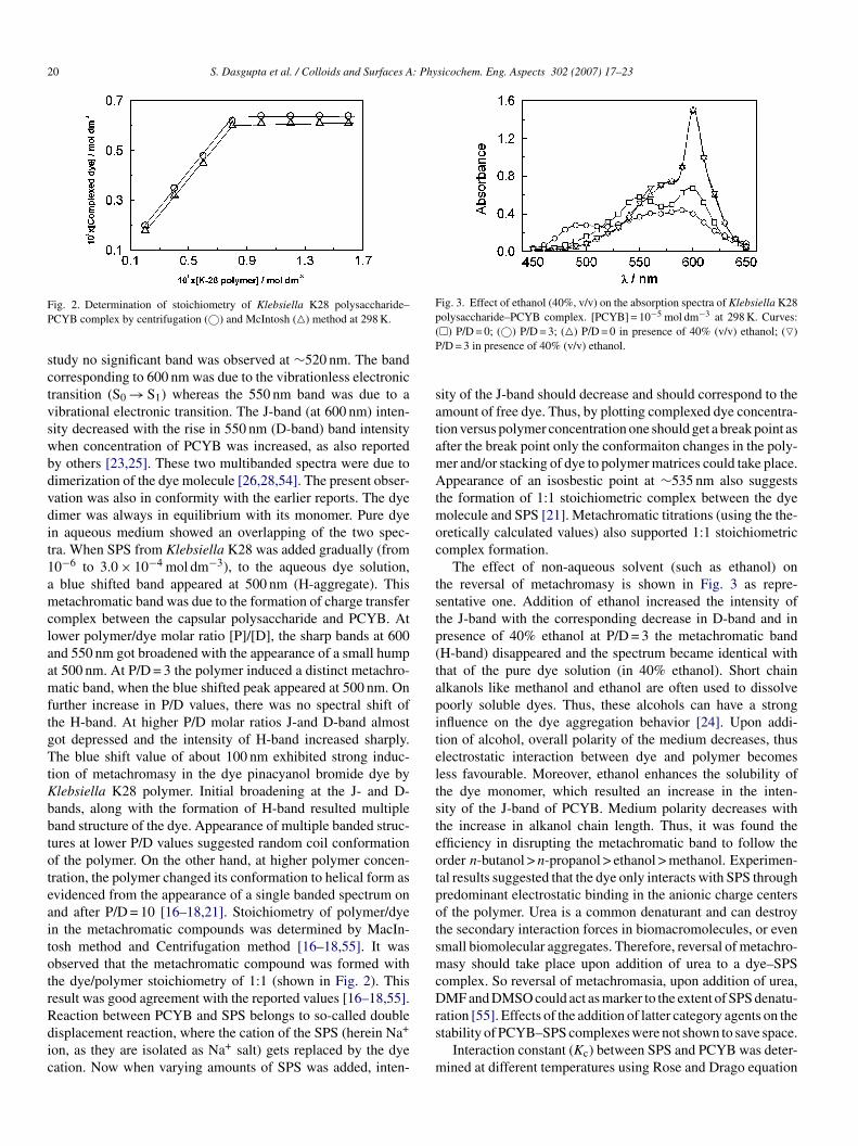
ig. 2. Determination of stoichiometry of Klebsiella K28 polysaccharide–CYB complex by centrifugation (©) and McIntosh (�) method at 298 K.
tudy no significant band was observed at ∼520 nm. The bandorresponding to 600 nm was due to the vibrationless electronicransition (S0 → S1) whereas the 550 nm band was due to aibrational electronic transition. The J-band (at 600 nm) inten-ity decreased with the rise in 550 nm (D-band) band intensityhen concentration of PCYB was increased, as also reportedy others [23,25]. These two multibanded spectra were due toimerization of the dye molecule [26,28,54]. The present obser-ation was also in conformity with the earlier reports. The dyeimer was always in equilibrium with its monomer. Pure dyen aqueous medium showed an overlapping of the two spec-ra. When SPS from Klebsiella K28 was added gradually (from0−6 to 3.0 × 10⁻⁴ mol dm−3), to the aqueous dye solution,blue shifted band appeared at 500 nm (H-aggregate). Thisetachromatic band was due to the formation of charge transfer
omplex between the capsular polysaccharide and PCYB. Atower polymer/dye molar ratio [P]/[D], the sharp bands at 600nd 550 nm got broadened with the appearance of a small humpt 500 nm. At P/D = 3 the polymer induced a distinct metachro-atic band, when the blue shifted peak appeared at 500 nm. On
urther increase in P/D values, there was no spectral shift ofhe H-band. At higher P/D molar ratios J-and D-band almostot depressed and the intensity of H-band increased sharply.he blue shift value of about 100 nm exhibited strong induc-
ion of metachromasy in the dye pinacyanol bromide dye bylebsiella K28 polymer. Initial broadening at the J- and D-ands, along with the formation of H-band resulted multipleand structure of the dye. Appearance of multiple banded struc-ures at lower P/D values suggested random coil conformationf the polymer. On the other hand, at higher polymer concen-ration, the polymer changed its conformation to helical form asvidenced from the appearance of a single banded spectrum onnd after P/D = 10 [16–18,21]. Stoichiometry of polymer/dyen the metachromatic compounds was determined by MacIn-osh method and Centrifugation method [16–18,55]. It wasbserved that the metachromatic compound was formed withhe dye/polymer stoichiometry of 1:1 (shown in Fig. 2). Thisesult was good agreement with the reported values [16–18,55].
eaction between PCYB and SPS belongs to so-called doubleisplacement reaction, where the cation of the SPS (herein Na+on, as they are isolated as Na+ salt) gets replaced by the dyeation. Now when varying amounts of SPS was added, inten-
rs
m
�) P/D = 0; (©) P/D = 3; (�) P/D = 0 in presence of 40% (v/v) ethanol; (�)/D = 3 in presence of 40% (v/v) ethanol.
ity of the J-band should decrease and should correspond to themount of free dye. Thus, by plotting complexed dye concentra-ion versus polymer concentration one should get a break point asfter the break point only the conformaiton changes in the poly-er and/or stacking of dye to polymer matrices could take place.ppearance of an isosbestic point at ∼535 nm also suggests
he formation of 1:1 stoichiometric complex between the dyeolecule and SPS [21]. Metachromatic titrations (using the the-
retically calculated values) also supported 1:1 stoichiometricomplex formation.
The effect of non-aqueous solvent (such as ethanol) onhe reversal of metachromasy is shown in Fig. 3 as repre-entative one. Addition of ethanol increased the intensity ofhe J-band with the corresponding decrease in D-band and inresence of 40% ethanol at P/D = 3 the metachromatic bandH-band) disappeared and the spectrum became identical withhat of the pure dye solution (in 40% ethanol). Short chainlkanols like methanol and ethanol are often used to dissolveoorly soluble dyes. Thus, these alcohols can have a strongnfluence on the dye aggregation behavior [24]. Upon addi-ion of alcohol, overall polarity of the medium decreases, thuslectrostatic interaction between dye and polymer becomesess favourable. Moreover, ethanol enhances the solubility ofhe dye monomer, which resulted an increase in the inten-ity of the J-band of PCYB. Medium polarity decreases withhe increase in alkanol chain length. Thus, it was found thefficiency in disrupting the metachromatic band to follow therder n-butanol > n-propanol > ethanol > methanol. Experimen-al results suggested that the dye only interacts with SPS throughredominant electrostatic binding in the anionic charge centersf the polymer. Urea is a common denaturant and can destroyhe secondary interaction forces in biomacromolecules, or evenmall biomolecular aggregates. Therefore, reversal of metachro-asy should take place upon addition of urea to a dye–SPS
omplex. So reversal of metachromasia, upon addition of urea,MF and DMSO could act as marker to the extent of SPS denatu-
ation [55]. Effects of the addition of latter category agents on the
tability of PCYB–SPS complexes were not shown to save space.Interaction constant (Kc) between SPS and PCYB was deter-ined at different temperatures using Rose and Drago equation

S. Dasgupta et al. / Colloids and Surfaces A: Physicochem. Eng. Aspects 302 (2007) 17–23 21
Table 1Thermodynamic parameters for the interaction of Klebsiella K28 capsular polysaccharide–pinacyanol bromide in aqueous media at 298 K
Temperature (K) 10−3 × Kc (mol−1 dm3) �G (kcal mol−1) �H (kcal mol−1) �S (cal mol−1 K−1)
303 2.40 −4.723 03 83 7
[wKt[�
ctcbor
tTcpffvcadfserad
oa
FK(
mcfitotsotf
pts[acb[cf
wcdd
08 2.06 −4.718 1.56 −4.623 1.37 −4.6
16–18]. Results are summarized in Table 1. Binding constantas found to be comparable with earlier findings [16–18].c values were found to decrease with the rise in tempera-
ure, revealing the binding process to be exothermic in nature18]. Free energy changes were calculated from the relationG = −RT ln Kc while the enthalpy and entropy changes were
alculated from the graphical plots of �G versus T accordingo the relation �G = �H − T�S. Higher values of free energyhange indicated the dye–polymer aggregation to be controlledoth by electrostatic and hydrophobic interactions [18]. Valuesf the enthalpy and entropy changes were within the range of aeversible biological process.
Interaction between the capsular polysaccharide and surfac-ants were studied by the dye incorporation technique [50].he absorbance of the dye pinacyanol bromide decreasedonsiderably at 600 nm, when it interacted with the capsularolysaccharide K28. At a definite P/D ratio of 30, cationic sur-actant of increasing concentration was added separately. It wasound that upon addition of cationic surfactants the absorbancealues of the dye–polymer complex at its J-band increasedonsiderably. It indicated that the surfactant molecules inter-cted with the capsular polysaccharide by replacing the cationicye molecules. In other words, the cationic dye molecules gotree from the polymer matrix with the addition of cationicurfactants. The increase in absorbance was considered to bequivalent to the extent of surfactant bound to the polysaccha-ide. The increase in absorbance can also be correlated with thebility of the surfactant in freeing the dye molecules from theye–polymer complex.
The effect of different cationic surfactants on the absorbancef pinacyanol bromide and Klebsiella K28 polymer at 600 nmre shown in Fig. 4. In all the cases it was observed that the
ig. 4. Effect of cationic surfactants on the absorbance of Klebsiella28–PCYB complex at 298 K. [PCYB] = 10−5 mol dm−3, P/D = 30. Surfactants
1.0 m mol dm−3): (©) none; (�) DPC; (�) CPC; (x) CTAB and (�) BDHAC.
fa
a(laTC
TVc
S
BCCD
−5.73 −2.54
etachromatic band at 500 nm of the dye-K28 capsular polysac-haride was depleted with the addition of surfactants. From thegures, it was clear that the increase in the absorbance value at
he J-band upon addition of different surfactants followed therder BDHAC > CTAB > CPCl > DPCl. Also it was found thathe concentration of surfactants required to reach to the corre-ponding increased absorbance value followed the descendingrder BDHAC < CTAB < CPCl < DPCl. Thus, it could be saidhat the ability in freeing the dye from the dye–polymer complexollowed the order BDHAC > CTAB > CPCl > DPCl.
Surfactant–polyelctrolyte interactions are electrostaticallyrevalent when they are oppositely charged. In syn-hetic polymer–surfactant systems polyelectrolytes and oppo-itely charged surfactants produce the strongest association36,46–48,50,51]. Usually, such kind of interactions are initi-ted by the saltlike electrostatic interactions among oppositelyharged polyelectrolytes and surfactant and then get stabilizedy the hydrophobic interactions of the bound surfactant tails46]. The binding constant between the anionic polymer and theationic surfactants were calculated by Rose and Drago equationrom the absorbance results. The equation is given below:
(CDCS)
(A − A0)= 1
[KC LεDS − εD)]+ CS
[(εDS − εD)L]− 1
here CD is the concentration of the polymer added, CS theoncentration of the surfactant added, A0 the absorbance of theye–polymer solution at 600 nm and A is the absorbance of theye–polymer solution at 600 nm when CS concentration of sur-actant added. KC is the binding constant between the polymernd surfactant, ε is the molar absorption coefficient.
The values of (CDCS)/(A–A0) was calculated for surfactantnd then the values of (CDCS)/(A–A0) were plotted against CSshown in Fig. 5). From the slope and the intercept of the straight
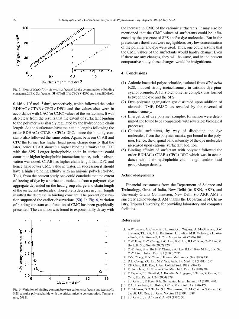
ine, the binding constant values between the K28 polymernd the surfactant were calculated. Results are summarized inable 2. The binding constant values in case of BDHAC, CTAB,PCl and DPCl were 1.67 × 10⁵, 0.55 × 10⁵, 0.245 × 10⁵ andable 2alues of binding constants of different cationic surfactants with Klebsiella K28apsular polysaccharide and their CMC values
urfactant CMC (m mol dm−3a) 10−5 Bindingconstant (mol−1 dm3)
DHAC 0.042 1.67TAB 0.80 0.55PCl 0.90 0.245PCl 14.70 0.146
a Results were adopted from Refs. [47–52].
22 S. Dasgupta et al. / Colloids and Surfaces A: Phy
Fc
0BaatlosClwcvhhToaortop
FKt
tmepotic
4
(
(
(
(
(
A
T
ig. 5. Plots of (CDCS/(A − A0) vs. [surfactant] for the determination of bindingonstant at 298 K. Surfactants: (�) CTAB (©) CPC (�) DPC and inset: BDHAC.
.146 × 105 mol−1 dm3, respectively, which followed the orderDHAC > CTAB > CPCl > DPCl and the values also were inccordance with CAC (or CMC) values of the surfactants. It waslso clear from the results that the extent of surfactant bindingo the polymer was sharply regulated by the hydrophobic chainength. As the surfactants have their chain lengths following therder BDHAC > CTAB ∼ CPC > DPC, hence the binding con-tants also followed the same order. Again, between CTAB andPC the former has higher head group charge density that the
ater, hence CTAB showed a higher binding affinity than CPCith the SPS. Longer hydrophobic chain in surfactant could
ontribute higher hydrophobic interaction; hence, such an obser-ation was noted. CTAB has higher chain length than DPC andence have lower CMC value in water. In succession it shouldave a higher binding affinity with an anionic polyelectrolyte.hus, from the present study one could conclude that the extentf freeing of dye by a surfactant molecule from a polymer–dyeggregate depended on the head group charge and chain lengthf the surfactant molecules. Therefore, a decrease in chain length
esulted the decrease in binding constant. The present observa-ion supported the earlier observations [50]. In Fig. 6, variationf binding constant as a function of CMC has been graphicallyresented. The variation was found to exponentially decay withig. 6. Variation of binding constant between cationic surfactant and Klebsiella28 capsular polysaccharide with the critical micelle concentration. Tempera-
ure, 298 K.
Usif
R
[[
[
sicochem. Eng. Aspects 302 (2007) 17–23
he increase in CMC of the cationic surfactants. It may also beentioned that the CMC values of surfactants could be influ-
nced by the presence of SPS and/or dye molecules. But in theresent case the effects were negligible as very low concentrationf the polymer and dye were used. Thus, one could assume thathe CMC values of the surfactants would hardly change. Evenf there are any changes, they will be same, and in the presentomparative study, these changes would be insignificant.
. Conclusions
1) Anionic bacterial polysaccharide, isolated from KlebsiellaK28, induced strong metachromasy in cationic dye pina-cyanol bromide. A 1:1 stoichiometric complex was formedbetween the dye and the SPS.
2) Dye–polymer aggregation got disrupted upon addition ofalcohols, DMF, DMSO, as revealed by the reversal ofmetachromasy.
3) Energetics of dye polymer complex formation were deter-mined and found to be comparable with reversible biologicalprocesses.
4) Cationic surfactants, by way of displacing the dyemolecules, from the polymer matrix, got bound to the poly-mer. Hence, the original band intensity of the dye moleculesincreased upon cationic surfactant addition.
5) Binding affinity of surfactant with polymer followed theorder BDHAC > CTAB > CPC > DPC which was in accor-dance with their hydrophobic chain length and/or headgroup charge density.
cknowledgements
Financial assistances from the Department of Science andechnology, Govt. of India, New Delhi (to RKN, AKP), andniversity Grants Commission, New Delhi (to AKP, AM) is
incerely acknowledged. AM thanks the Department of Chem-stry, Tripura University, for providing laboratory and computeracilities.
eferences
[1] A.W. Jenney, A. Clements, J.L. Arn, O.L. Wijburg, A. McGlinchey, D.W.Spelman, T.L. Pitt, M.E. Kaufmann, L. Liolios, M.B. Moloney, S.L. Wes-selingh, R.A. Strugnell, J. Clin. Microbiol. 44 (2006) 102.
[2] C.-P. Fung, F.-Y. Chang, S.-C. Lee, B.-S. Hu, B.I.-T. Kuo, C.-Y. Liu, M.Ho, L.K. Siu, Gut 50 (2002) 420.
[3] C.-P. Fung, B.-S. Hu, F.-Y. Chang, S.-C. Lee, B.I.-T. Kuo, M. Ho, L.K. Siu,C.-Y. Liu, J. Infect. Dis. 181 (2000) 2075.
[4] F.-Y. Chang, M.Y. Chou, J. Fomos. Med. Assoc. 94 (1995) 232.[5] D.L. Cheng, Y.C. Liu, M.Y. Yen, Arch. Int. Med. 151 (1991) 1557.[6] F.F. Chou, H.K. Kou, J. Am. Colloid Surf. 182 (1996) 33.[7] R. Podschun, U. Ullmann, Clin. Microbiol. Rev. 11 (1998) 589.[8] F. Paganin, F. Lillienthal, A. Bourdin, N. Lugagne, F. Tixier, R. Genin, J.L.
Yvin, Eur. Respir. J. 24 (2004) 779.
[9] S.J. Cryz Jr., F. Furer, R.E. Germanier, Infect. Immun. 43 (1984) 440.10] E.A. Blanchette, S.J. Rubin, J. Clin. Micribiol. 11 (1980) 474.11] R. Edelman, D.N. Taylor, S.S. Wasserman, J.B. McClain, A.S. Cross, J.C.Sadoff, J.U. Que, S.J. Cryz, Vaccine 12 (1994) 1288.12] S.J. Cryz Jr., S. African Z. A. 478 (1986) 33.

A: Ph
[
[[[
[[
[[[[
[
[[
[
[[
[[
[[[[[
[[
[[[
[[[
[
[[
[
[
[
[[
[
S. Dasgupta et al. / Colloids and Surfaces
13] S.J. Cryz Jr., A.S. Cross, G.C. Sadoff, J.U. Que, Eur. J. Immunol. 18 (1988)2073.
14] S.J. Cryz Jr., Adv. Biotechnol. Processes 13 (1990) 87.15] M. Curval, B. Lindberg, J. Lonngreen, Carbohydr. Res. 42 (1975) 95.16] A. Mitra, R.K. Nath, S. Biswas, A.K. Chakraborty, A.K. Panda, J. Pho-
tochem. Photobiol. A: Chem. 178 (2006) 98 (and references therein).17] A. Mitra, A.K. Chakraborty, Ind. J. Chem. 37A (1998) 418.18] A.K. Panda, A.K. Chakraborty, J. Photochem. Photobiol. A: Chem. 111
(1997) 157.19] R.W. Horbin, Biochemie. Biochem. 77 (2002) 3.20] J.A. Bergeron, M. Siger, J. Biophys. Biochem. Cytol. 4 (1958) 433.21] M.K. Pal, B.K. Ghosh, Mikromol. Chem. 181 (1980) 1459.22] B. Norden, M. Kubista, in: B. Samori, E.W. Thulstrup (Eds.), Polarized
Spectroscopy of Ordered Systems, vol. 242, Kluwer Academic Publisher,Dordrecht, Holland, 1988.
23] R. Sabate, J. Estelrich, J. Phys. Chem. B 107 (2003) 4137 (and referencestherein).
24] H. von Berlepsch, S. Kirstein, C. Bottcher, Langmuir 18 (2002) 7699.25] R. Sabate, M. Gallardo, A. De la Maza, J. Estelrich, Langmuir 17 (2001)
6433 (and references therein).26] S. Barazzouk, H. Lee, S. Hotchandani, P.V. Kumar, J. Phys. Chem. B 104
(2000) 3616.27] M.L. Horng, E.L. Quitevis, J. Chem. Educ. 77 (2000) 637.28] J.-F. Berret, G. Cristobal, P. Herv’e1, J. Oberdisse, I. Grillo, Eur. Phys. J.
E9 (2002) 301.29] R. Meszaos, I. Varga, T. Gilanyi, J. Phys. Chem. B 109 (2005) 13538.30] C. Monteux, C.E. Williams, J. Meunier, O. Anthony, V. Bergeron, Langmuir
20 (2004) 57;C. Honda, H. Kamizono, K. Matsumoto, K. Endo, J. Colloid Interface Sci.278 (2004) 310;B. Shweitzer, D. Zanette, R. Itri, J. Colloid Interface Sci. 277 (2004) 285.
31] S.P. Moulik, S. Gupta, A.R. Das, Makromol. Chem. 181 (1980) 1459.
32] J. Lee, Y. Moroi, J. Colloid Interface Sci. 273 (2004) 645.33] C.L. Mesa, J. Colloid Interface Sci. 286 (2005) 148.34] R. Konradi, J. Ruhe, Macromolecules 38 (2005) 6140.35] M.A. Villetti, R. Borsali, J.S. Crespo, V. Soldi, K. Fukada, Macromol.Chem. Phys. 205 (2004) 907.
[
[[
ysicochem. Eng. Aspects 302 (2007) 17–23 23
36] F.D. Anghel, S. Saito, A. Baran, A. Iovesw, Langmuir 14 (1998) 5352.37] A. Malovikova, K. Hayokawa, J.C.T. Kwak, J. Phys. Chem. 88 (1984)
1930.38] A.L. Carvalho, A.M. Carmona-Ribeiro, Langmuir 14 (1998) 6077.39] T. Imamura, K. Konishi, J. Colloid Interface Sci. 198 (1998) 300.40] H. Sjogren, C.A. Ericsson, J. Evenas, S. Ulvenlund, Biophys. J. 89 (2005)
4219.41] D.-M. Zhu, R.K. Evans, Langmuir 22 (2006) 3735.42] G. Wang, G. Olofsson, J. Phys. Chem. 99 (1995) 5588.43] J. Mata, J. Patel, N. Jain, G. Ghosh, P. Bahadur, J. Colloid Interface Sci.
297 (2006) 797.44] G. Bai, M. Nichifor, A. Lopes, M. Bastos, J. Phys. Chem. B 109 (2005)
518.45] K. Hayakawa, J.P. Santerre, J.C.T. Kwak, Macromolecules 16 (1983) 1642.46] E.D. Goddard, K.P. Ananthapadmanabhan, Interaction of Surfactants with
Polymers and Proteins, CRC Press, Boca Raton, FL, 1993.47] J.C.T. Kwak, J.C.T. Kwak (Eds.), Polymer–Surfactant Systems, Surfactant
Science Series, vol. 77, Dekker, New York, 1998.48] P. Linse, L. Picullel, P. Hansson, in: J.C.T. Kwak (Ed.), Polymer Surfactant
Systems, Surfactant Science Series, vol. 77, Dekker, New York, 1998, p.193 (Chapter 5).
49] J. Fundin, P. Hansson, W. Brown, I. Lidegran, Macromolecules 30 (1997)1118.
50] A.K. Panda, A.K. Chakraborty, J. Colloid Interface Sci. 203 (1998) 260.51] K. Thalberg, J.V. Stam, C. Lindblad, M. Almgren, B. Lindman, J. Phys.
Chem. 95 (1991) 8975;R. Zana, P. Lianos, J. Lang, J. Phys. Chem. 89 (1985) 41;P. Chandar, P. Somosundaram, N. Turro, Macromolecules 21 (1988) 950;C. Zhao, M.A. Winnik, G. Riess, M. Croucher, Langmuir 6 (1990) 514;V.A. De Oliveira, M.J. Tiera, M.G. Neumann, Langmuir 12 (1996) 607;P. Hansson, M. Almgren, J. Phys. Chem. 99 (1995) 16684.
52] A.K. Chakraborty, H. Friebolin, S. Strim, J. Bacteriol. 112 (1980) 635.
53] K.A. Connors, Binding Constants: The Measurements of Molecular Com-plex Stability, John Wiley, New York, 1987.54] R. Sabate, L. Freire, J. Estelrich, J. Chem. Educ. 78 (2001) 243.55] S. Zhang, N. Li, F. Zhao, K. Li, S. Tong, Spectrochim. Acta A 58 (2002)
273.

LDIS #427132, VOL 31, ISS 11
Spectral Studies on the Binding Behavior of CationicDyes and Surfactants with Bacterial Polysaccharideof Klebsiella K43
R. K. Nath, S. Dasgupta, S. Ghosh, A. Mitra, and A. K. Panda
QUERY SHEET
This page lists questions we have about your paper. The numbers displayed at left can be found in the text of the paper for reference. Inaddition, please review your paper as a whole for correctness.
Q1: Au: Shortened, running title ok as provided?
TABLE OF CONTENTS LISTING
The table of contents for the journal will list your paper exactly as it appears below:
Spectral Studies on the Binding Behavior of Cationic Dyes and Surfactants with Bacterial Polysaccharide of Klebsiella K43
R. K. Nath, S. Dasgupta, S. Ghosh, A. Mitra, and A. K. Panda

Spectral Studies on the Binding Behavior of CationicDyes and Surfactants with Bacterial Polysaccharideof Klebsiella K43
R. K. Nath,1 S. Dasgupta,1 S. Ghosh,1 A. Mitra,2 and A. K. Panda3
51Department of Chemistry, Tripura University, Suryamaninagar, Tripura, India2Department of Chemistry, M. B. B. College, Agatala, Tripura, India3Department of Chemistry, University of North Bengal, P. O. North Bengal University,West Bengal, India
The binding behavior of acidic capsular polysaccharide (SPS), isolated from Klebsiella serotype10 K43, with oppositely charged dyes and surfactants have been studied by way of absorbance and
emission spectroscopic measurements. Each repeating unit of the SPS consists of threeD-mannose, one D-galactose, and one D-glucuronic acid residue. The anionic polysaccharideexhibited chromotropic character and induced strong metachromasy in the cationic dye, pinacya-nol chloride (PCYN) through the formation of a 1:1 stoichiometric complex. Evaluation of
15 thermodynamic parameters, viz., changes in free energy (DG), enthalpy (DH), and entropy(DS), for the formation of dye-polymer complex and studies on the effect of different cosolventswere also evaluated to shed light on the binding nature as well as the extent of stability of thedye-polymer complex. Fluorescence of the cationic dye acridine orange (AO) was quenched withthe progressive addition of SPS, which was found to be of Stern-Volmer type. Cationic surfactants
20 in their pure form as well as in the mixed state with nonionic surfactant (Tween-20), replaced thedye bound to the polymer matrices; thus the original band intensities of the dyes could be reverted.Such studies revealed the involvement of both electrostatic as well as hydrophobic interactionsbetween the dye-polymer as well as surfactant-polymer aggregates.
25 Keywords Bacterial polysaccharide, binding constant, dyes, metachromasy, quenching,surfactants
1. INTRODUCTION
Klebsiella is a Gram-negative bacterium which belongsto Enterobacteriaceae family. They are the causative factors
30 for several human diseases[1–3] like liver abscess,[4] septicendophthalamitis,[5,6] infection in urinary tract, pneumonia,and meningitis,[1,7] etc.
Most of these Gram-negative bacteria form capsularpolysaccharides. Until now, about 82 serologically distinct
35 K-serotypes[8,9] have been found in the Gram-negativebacteria Klebsiella, which can produce acidic hetero poly-saccharides with wide structural variations. These bacterial
polysaccharides also have antigenic properties.[1] Presenceof definite repeating units ranging from tri- to hepta-
40saccharides, containing glucuronic acid and=or galacturo-nic acid, along with other neutral sugars, contribute to theirunique nature than other conventional polysaccharides.The existence of potential anionic site in the biopolymersis due to the presence of acidic sugars in every repeating
45unit which, in turn, also provide polyelectrolytic nature.The primary structures of most of the capsular polysac-charides are now known through chemical analyses.[10]
The primary structures of capsular polysaccharides andconformational factors are responsible for their serological
50specificity.[11] The biophysical properties of these polysac-charides are due to their secondary structures. Extracellularpolysaccharides are produced by Klebsiella bacteria, whichsurround the bacterium as an additional outer layer as cap-sule. Since Klebsiella capsular antigens have been found to
55be safe in human so these antigenic polysaccharides are alsobeing used as human vaccines.[12] Due to potential uses ofthe bacterial polysaccharides in immunological and vaccinepreparations, primary structural studies and conformationalanalysis, as well as studies on various physicochemical
Received 2 June 2009; accepted 12 June 2009.The authors are thankful to the Department of Chemistry,
Tripura University for providing laboratory and computationalfacilities. Financial assistance (to R. K. N. and A. K. P.) fromthe Department of Science and Technology, Govt. of India, NewDelhi and University Grants Commission (to A. K. P.), NewDelhi, India is sincerely acknowledged.
Address correspondence to R. K. Nath, Department ofChemistry, Tripura University, Suryamaninagar, Tripura-799130,India. E-mail: [email protected]
Journal of Dispersion Science and Technology, 31:1–9, 2010
Copyright # Taylor & Francis Group, LLC
ISSN: 0193-2691 print=1532-2351 online
DOI: 10.1080/01932690903269586
3b2 Version Number : 7.51c/W (Jun 11 2001)File path : P:/Santype/Journals/TandF_Production/LDIS/v31n11/LDIS427132/LDIS427132.3dDate and Time : 04/10/10 and 15:24
1
60 properties of these biopolymers are gaining more and moreimportance with the discovery of newer serotypes. Thus, thephysicochemical characterizations of these bacterial poly-saccharides are also becoming necessary. Although thepresent research group is associated in such studies since last
65 three decades still the results are considered to be fragmen-tary in nature.[13]
Klebsiella K43 consists of D-mannose, D-galactose, andD-glucuronic acid residues in the ratio 3:1:1. The term SPSarises from sauer polysaccharides (sauer means sour in
70 German). Presence of one glucuronic acid in each repeatingunit enables it to interact with positively charged dyes andsurfactants.[14,15] The authors have been carrying outdye-SPS interaction studies through spectroscopic mea-surements in characterizing different SPS isolated from
75 various serotypes of Klebsiella.[13,15–18] Spectral studieson the dye-polymer interaction could shed light on thedetailed structural aspects of the polysaccharides.[13]
Metachromasy is a well-known phenomenon in case ofdye-polymer interaction and is generally applied to the
80 aggregation of cationic dye on anionic polymers andcolloids.[19] The phenomena have also been observed infew cases of cationic polyelectrolyte and anionic dyesystems. Reports dealing with studies on dye-polymer inter-action inducing metachromasy in different cationic dyes by
85 different synthetic polyanions, DNA, and naturally occur-ring plant polyelectrolytes are also available. A clear ideaabout the mechanism of polyion induced metachromasyand the correlations between the dye structures and meta-chromatic behaviors could be obtained through such
90 studies, for example, interaction of heparins with azuredyes.[20–22] Dye-protein interaction study could also revealthe conformational aspects of protein.[23]
Different techniques have been reported for the isolationand stability determination of metachromatic complexes.[13]
95 The concept of reversal of metachromasy can be used todetermine the stability of the metachromatic complex.Reversal of metachromasy may be attained with theaddition of urea, alcohol, electrolytes, and also withincreasing the temperature of the dye-polymer aggregate.[24]
100 Evaluation of different thermodynamic parameters likechanges in free energy (DG), enthalpy (DH), and entropy(DS) could explain the nature of interaction in the forma-tion of metachromatic complex and can also predict aboutthe optimum conditions for the interaction between the
105 oppositely charged dye and macromolecules.Fluorescence quenching is a process of deactivation of
the excited state which results in a decrease in fluorescenceintensity. Acridine orange (AO) could be used as a photo-physical probe under the influence of different polymer
110 matrices.[13,15,24]
Polymer-surfactant interaction is a very convincing fieldof research. Several studies dealing with polyelectrolyte andoppositely charged surfactants[25–27] have been done for
both fundamental and applied aspects.[28] Surfactant115molecules can bind to the oppositely charged polyelectro-
lytes, forming the so called polymer and=or surfactantcomplexes. Additionally, hydrophobic interaction mayalso promote=favor surfactant binding to the polymermatrix.[29,30] A comprehensive study using a series of catio-
120nic surfactants with anionic polyelectrolytes such ascarboxymethylcellulose (CMC), dextran sulfate (DX),polyacrylic acid (PPA), poly(methacrylic acid) (PMAA),etc., have been done using various techniques.[25,27,30–35]
Surfactant-polymer interaction studies also include biologi-125cally associated systems like polypeptides,[33] proteins,[30]
DNA,[31,35] and carbohydrate-based polymers.[36] Differentmethods to study polymer-surfactant interaction includeturbidity measurement, viscosity measurements, lightscattering techniques, calorimetry, conductivity measure-
130ment, electromagnetic field (EMF) measurements, dye-incorporation techniques, etc. Such experimental methodsallow in gathering lot of information on the nature ofinteraction in different polymer-surfactant aggregates. Aquantitative understanding on the interaction in surfac-
135tant-biopolymer systems could be achieved by such studies.The present investigation deals with the studies on
interaction between anionic polysaccharide isolated fromKlebsiella K43 and cationic dye pinacyanol chloride andacridine orange in aqueous medium also associated with
140oppositely charged surfactant system. Spectrophoto-=fluorimetric techniques were used for detailed study ofdye-polymer interaction. Thermodynamic parameters ofinteraction and effects of different cosolvents wereevaluated. Interaction of Klebsiella K43 with different
145cationic surfactants, viz., benzyl dimethyl-(-n-) hexadecylammonium chloride (BDHAC), cetyltrimethylammoniumbromide (CTAB), cetylpyridinium chloride (CPC) anddodecylpyridinium chloride (DPC)in their pure form, aswell as in their mixed states with a nonionic surfactant
150Tween-20 were explored through spectrophoto-=fluori-metric measurements using dye probing techniques.Binding constant of biopolymer-surfactant aggregates wereevaluated by suitably analyzing the spectral data.
2. MATERIALS AND METHODS
1552.1. Materials
The serological strain of bacterial polysaccharideKlebsiella K43 was kindly supplied by Dr. S. Schlesht ofMax Plank Institute for Immunolobiology, Freiburg,Germany. Cationic dyes pinacyanol chloride (PCYN) and
160acridine orange (AO) from Sigma Chemicals (USA) were>99% pure and were used as such. HPLC grade alcohols,from E. Merck (Germany) were used. Cationic surfactantsbenzyldimethyl-n-hexadecyl ammonium chloride (BDHAC),cetyltrimethyl amminium bromide (CTAB), cetylpyridinium
2 R. K. NATH ET AL.
165 chloride (CPC), dodecylpyridinium chloride (DPC), andnonionic surfactant polyoxyethylene sorbitan monolaurate(Tween-20) were the products from E. Merck (Germany).They were used after purification[17] per the standard pro-cedure. Purities were checked through their conductance=
170 surface tension values.[37] Freshly prepared double distilledwater with a resistivity of 18mX cm�1 was used for all themeasurements.
2.2. Methods
In absorbance study, a 10 mmol dm�3 aqueous PCYN175 was used. Absorption spectra were recorded in the wave-
length range of 400–700 nm with a LS-55 spectrophot-ometer (Perkin Elmer, USA). The average molar mass ofone repeating unit of the polymer containing one anioniccharge site was referred as one mole of polymer. The
180 absorption spectra at different polymer=dye ([P]=[D])molar ratios, by addition of the capsular polysaccharidesto the aqueous solution of 10 mmol dm�3dye were recorded.Spectrofluorimetric measurements were done with the helpof a RF 5000 spectrofluorimeter (Shimadzu, Japan) by the
185 excitation at 475 nm of the AO dye in aqueous solution,using quartz cell of 1 cm path.
Stoichiometry of the dye-polymer complex was determ-ined by isolating the dye-polymer complex according toMcIntosh method,[14,17] which was modified by Kornors
190 et al.[38] Briefly, dye, bound to polymer (hereafter, dye-polymer complex), was separated out from the aqueouslayer by vortexing the mixture with benzine. The uncom-plexed dye in water was quantified by measuring the absor-bance at 600 nm; thus, the concentration of the complexed
195 dye could be calculated. Finally, from the point of inter-section obtained by plotting the values of complexed dyeconcentration against polymer concentration, the stoichi-ometry of the dye-polymer complex was determined.Centrifugation method[15] was also used to determine the
200 stoichiometry, by which the metachromatic complex wascentrifuged at 11,000 rpm (23,000 g) for 20 minutes andwhereby the dye-polymer complex was sedimented. Thesupernatant was analyzed colorimetrically for the free dyeand stoichiometry was calculated as in the previous
205 method.Metachromatic titration was performed by measuring
the absorbance of the complexed dye at 600 nm, fromwhich the volume of the polymer solution required forthe equivalent consumption of the dye was estimated.
210 The description of the procedure of metachromatictitration is available in literature.[13,15,24,39–43]
Effects of different cosolvents like methanol, ethanol,n-propanol, on the reversal of metachromasy was studiedby measuring absorbance of metachromatic complex at a
215 [P]=[D]¼ 5. The extent of reversal of metachromasy wasalso compared by studying the absorbance of pure dyesolution in presence and absence of the cosolvents.
Thermodynamic parameters of the interaction betweenPCYN and the experimental polymer were determined
220using Rose and Drago equation.[44] Charge transfer inter-action theory could represent the interaction between thecationic dye and polyanions. The following Rose andDrago equation was then employed for determining theinteraction constant between the dye and polymer.
CD � CS
A� AO¼ 1
KC � LðeDS � eDÞþ CS
LðeDS � eDÞ; ½1�
where, CD¼ Initial molar concentration of PCYN,CS¼molar concentration of capsular polysaccharideKlebsiella K43, 'D¼molar absorption coefficient ofPCYN, 'DS¼molar absorption coefficient of the PCYN-
230polymer complex, KC¼ interaction constant between thedye and polymer, AO¼ absorbance of the pure dye PCYNat 500 nm (metachromatic band of the dye-polymercomplex), A¼ absorbance of the dye-polymer solution at500 nm at a particular polymer concentration, and
235L=Length of light path.Detailed procedure for determining the thermodynamic
parameters for dye-polymer interaction as per Rose andDrago and other associated equations are availableelsewhere.[15,39–43]
240In fluorescence quenching experiments, 10 mmol dm�3
aqueous solution of AO was used as the probe. The fluor-escence of the dye solution as well as of the dye-polymercomplex (at different P=D) was measured. The excitationwavelength was at 475 nm and the emission spectra were
245recorded in the range 450 to 650 nm.The quenching of fluorescence in acridine orange dye by
K43 polymer were treated with Stern-Volmer equation[45]
as used in our earlier works,[15,39–43] then from the slopeof the linear plot, the Stern-Volmer constant (KSV; herein,
250the binding constant between the dye and polymer) wasevaluated.
The effects of different cationic as well as cationic-Tween-20 mixed surfactant systems on the dye-polymercomplex were studied spectrophoto-=fluorimetrically. In
255studying the surfactant effects on bacterial polysaccharide,to a fixed dye-polymer ratio (P=D¼ 5), increasing amountssurfactant was added and absorbance of each solution wasmeasured at 600 nm (at J band) of PCYN.
Finally, the binding constant between the SPS and260oppositely charged cationic as well as cationic-nonionic
mixed surfactants were evaluated by Rose and Dragoequation[44] by suitably processing the absorption spectraldata. The terms involved in Rose and Drago equation asused in dye-polymer interaction study, bear separate
265meaning. In the present case, the term Cp indicates theconcentration of the polymer, AO is the absorbance ofthe dye-polymer solution at 600 nm, A is the absorbance
CATIONIC DYES AND SURFACTANTS WITH KLEBSIELLA K43Q1 3
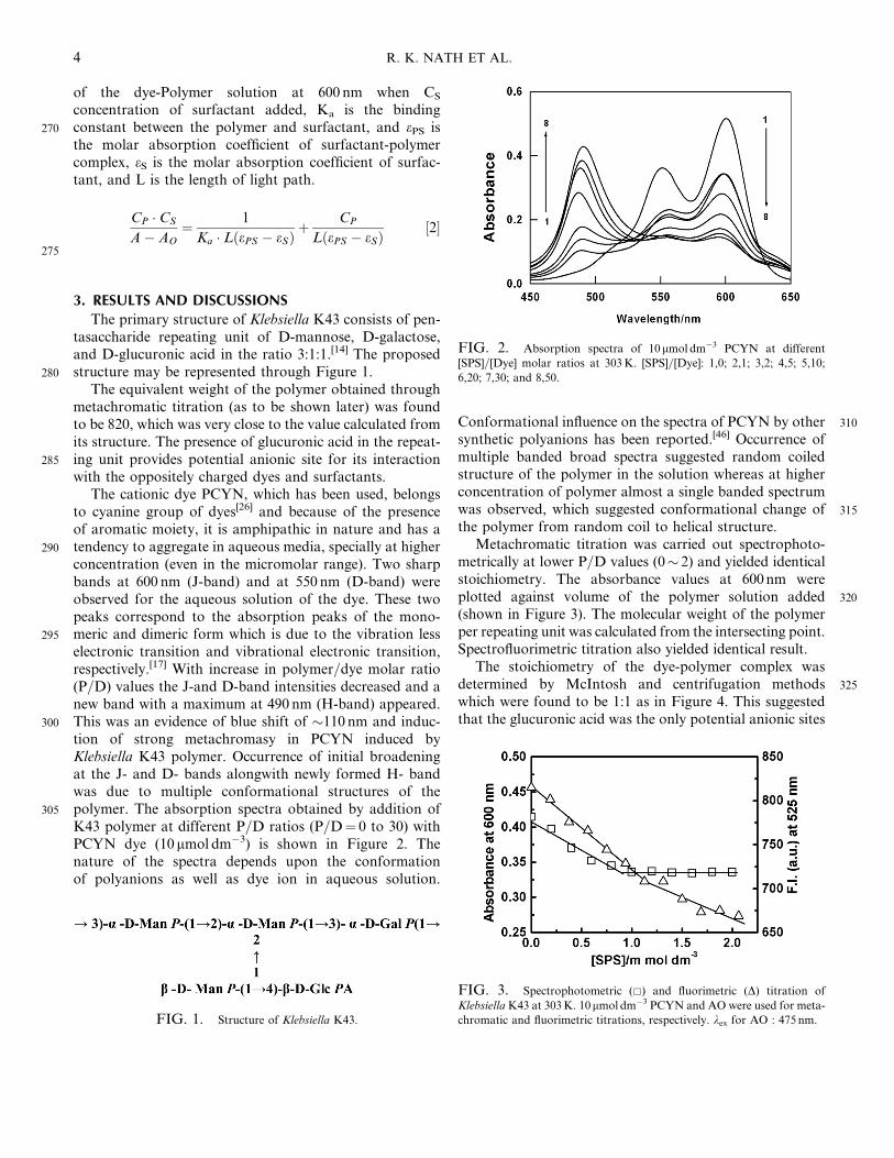
of the dye-Polymer solution at 600 nm when CS
concentration of surfactant added, Ka is the binding270 constant between the polymer and surfactant, and ePS is
the molar absorption coefficient of surfactant-polymercomplex, eS is the molar absorption coefficient of surfac-tant, and L is the length of light path.
CP � CS
A� AO¼ 1
Ka � LðePS � eSÞþ CP
LðePS � eSÞ½2�
275
3. RESULTS AND DISCUSSIONS
The primary structure of Klebsiella K43 consists of pen-tasaccharide repeating unit of D-mannose, D-galactose,and D-glucuronic acid in the ratio 3:1:1.[14] The proposed
280 structure may be represented through Figure 1.The equivalent weight of the polymer obtained through
metachromatic titration (as to be shown later) was foundto be 820, which was very close to the value calculated fromits structure. The presence of glucuronic acid in the repeat-
285 ing unit provides potential anionic site for its interactionwith the oppositely charged dyes and surfactants.
The cationic dye PCYN, which has been used, belongsto cyanine group of dyes[26] and because of the presenceof aromatic moiety, it is amphipathic in nature and has a
290 tendency to aggregate in aqueous media, specially at higherconcentration (even in the micromolar range). Two sharpbands at 600 nm (J-band) and at 550 nm (D-band) wereobserved for the aqueous solution of the dye. These twopeaks correspond to the absorption peaks of the mono-
295 meric and dimeric form which is due to the vibration lesselectronic transition and vibrational electronic transition,respectively.[17] With increase in polymer=dye molar ratio(P=D) values the J-and D-band intensities decreased and anew band with a maximum at 490 nm (H-band) appeared.
300 This was an evidence of blue shift of �110 nm and induc-tion of strong metachromasy in PCYN induced byKlebsiella K43 polymer. Occurrence of initial broadeningat the J- and D- bands alongwith newly formed H- bandwas due to multiple conformational structures of the
305 polymer. The absorption spectra obtained by addition ofK43 polymer at different P=D ratios (P=D¼ 0 to 30) withPCYN dye (10 mmol dm�3) is shown in Figure 2. Thenature of the spectra depends upon the conformationof polyanions as well as dye ion in aqueous solution.
310Conformational influence on the spectra of PCYN by othersynthetic polyanions has been reported.[46] Occurrence ofmultiple banded broad spectra suggested random coiledstructure of the polymer in the solution whereas at higherconcentration of polymer almost a single banded spectrum
315was observed, which suggested conformational change ofthe polymer from random coil to helical structure.
Metachromatic titration was carried out spectrophoto-metrically at lower P=D values (0� 2) and yielded identicalstoichiometry. The absorbance values at 600nm were
320plotted against volume of the polymer solution added(shown in Figure 3). The molecular weight of the polymerper repeating unit was calculated from the intersecting point.Spectrofluorimetric titration also yielded identical result.
The stoichiometry of the dye-polymer complex was325determined by McIntosh and centrifugation methods
which were found to be 1:1 as in Figure 4. This suggestedthat the glucuronic acid was the only potential anionic sites
FIG. 1. Structure of Klebsiella K43.
FIG. 2. Absorption spectra of 10 mmol dm�3 PCYN at different
[SPS]=[Dye] molar ratios at 303K. [SPS]=[Dye]: 1,0; 2,1; 3,2; 4,5; 5,10;
6,20; 7,30; and 8,50.
FIG. 3. Spectrophotometric (&) and fluorimetric (D) titration of
KlebsiellaK43 at 303K. 10mmol dm�3 PCYN and AO were used for meta-
chromatic and fluorimetric titrations, respectively. kex for AO : 475 nm.
4 R. K. NATH ET AL.
for interaction with the cationic dye PCYN and thussuggested a stair case stacking arrangement of the dye
330 molecules to polymer matrices. Appearance of isosbesticpoint at �535 nm in the absorption spectra of PCYN alsoconfirms the formation of 1:1 stoichiometric complexbetween dye and polymer.
Reversal of metachromasy is a phenomenon of destruc-335 tion of charge-transfer complex formation (i.e., formation
of metachromatic complex). The reversal of metachromasywas studied by measuring absorbance both at J- and H-band upon addition of different cosolvents like methanol,ethanol, and n-propanol. The destruction of metachro-
340 matic compound with the progressive addition of alcoholshas been attributed to the involvement of hydrophobicbonds in the induction of metachromasy. The reversal ofmetachromasy taking n-propanol as representative case is
shown in Figure 5. With gradual addition of ethanol, the345intensity of the J-band gradually increased with the
decrease in intensity of D-band. Finally, in presence of30% propanol at [P]=[D]¼ 5 the metachromatic band(H-band) completely disappeared and the spectra becameidentical with that of the pure dye solution (in 30%
350n-propanol). Experimental results suggested that the dyeonly interacts with SPS through predominant electrostaticbinding in the anionic charge centers of the polymer.
Rose and Drago equation was used to evaluate thethermodynamic parameters.[44] The parameters Kc, DH,
FIG. 4. Determination of stoichiometry of Klebsiella K43 SPS-PCYN
complex at 303K. &¼McIntosh method and �¼Centrifugation method.
FIG. 5. Reversal of metachromasy induced by propanol in Klebsiella
K43 SPS-PCYN complex: 1¼ dye-polymer complex; 2¼pure dye; 3¼dye-polymer complex in 30% propanol, and 4¼dye in 30% propanol.
FIG. 6. Plot of CD �CS=(A�AO) versus CS to determine the PCYN-
Klebsiella K43 SPS binding constant at different temperatures. [PCYN]
¼ 10 mmol dm�3. Temperature (K): &¼ 303; �¼ 308; D¼ 313;
4¼ 318,
and �¼ 323.
TABLE 1Thermodynamic parameters for the interaction of
pinacyanol chloride (PCYN) with Klebsiella 43 capsularpolysaccharides
Temp.=K
10�4KC
(dm3
mol�1)a
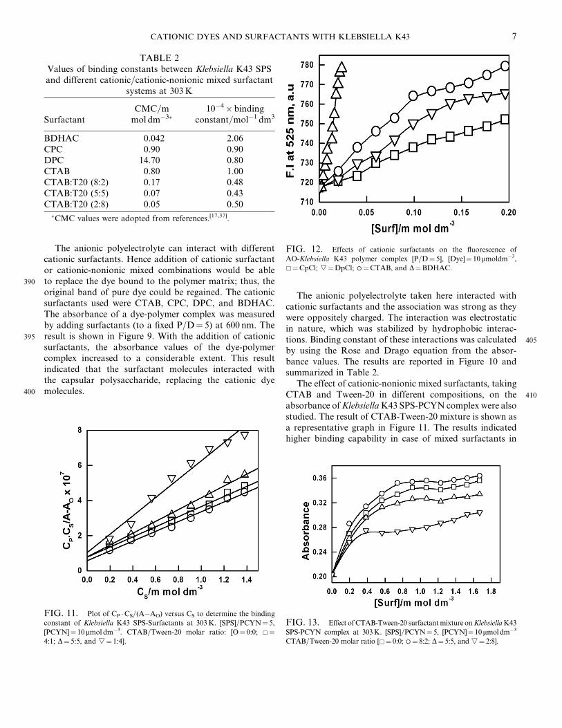
DG(kCalmol�1)b
DH(kCalmol�1)c
DS(Cal mol�1
deg�1)c
303 0.12 �4.29308 0.10 �4.26313 0.08 �4.19 �6.54� 0.98 7.45� 0.51318 0.07 �4.18323 0.06 �4.14
[PCYN] ¼ 10mmol dm�3.aCalculated from Figure 6, according to Rose and Drago
equation [43].bCalculated from the thermodynamic relation DG¼� RT ln KC.cCalculated from the graphical plot of DG vs T according to the
relation DG¼DH–TDS.
CATIONIC DYES AND SURFACTANTS WITH KLEBSIELLA K43Q1 5
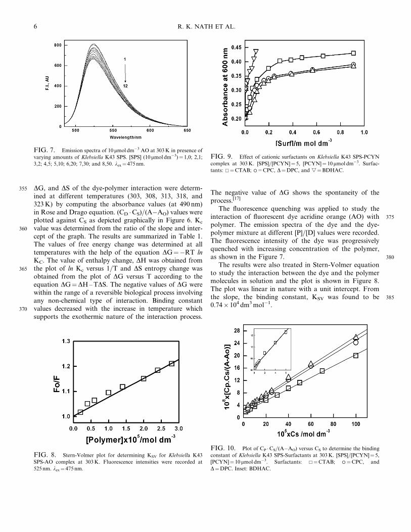
355 DG, and DS of the dye-polymer interaction were determ-ined at different temperatures (303, 308, 313, 318, and323K) by computing the absorbance values (at 490 nm)in Rose and Drago equation. (CD �CS)=(A�AO) values wereplotted against CS as depicted graphically in Figure 6. Kc
360 value was determined from the ratio of the slope and inter-cept of the graph. The results are summarized in Table 1.The values of free energy change was determined at alltemperatures with the help of the equation DG¼�RT lnKC. The value of enthalpy change, DH was obtained from
365 the plot of ln Kc versus 1=T and DS entropy change wasobtained from the plot of DG versus T according to theequation DG¼DH–TDS. The negative values of DG werewithin the range of a reversible biological process involvingany non-chemical type of interaction. Binding constant
370 values decreased with the increase in temperature whichsupports the exothermic nature of the interaction process.
The negative value of DG shows the spontaneity of theprocess.[17]
The fluorescence quenching was applied to study the375interaction of fluorescent dye acridine orange (AO) with
polymer. The emission spectra of the dye and the dye-polymer mixture at different [P]=[D] values were recorded.The fluorescence intensity of the dye was progressivelyquenched with increasing concentration of the polymer,
380as shown in the Figure 7.The results were also treated in Stern-Volmer equation
to study the interaction between the dye and the polymermolecules in solution and the plot is shown in Figure 8.The plot was linear in nature with a unit intercept. From
385the slope, the binding constant, KSV was found to be0.74� 104 dm3mol�1.
FIG. 7. Emission spectra of 10 mmol dm�3 AO at 303K in presence of
varying amounts of Klebsiella K43 SPS. [SPS] (10mmol dm�3)¼ 1,0; 2,1;
3,2; 4,5; 5,10; 6,20; 7,30; and 8,50. kex¼ 475 nm.
FIG. 8. Stern-Volmer plot for determining KSV for Klebsiella K43
SPS-AO complex at 303K. Fluorescence intensities were recorded at
525 nm. kex¼ 475nm.
FIG. 9. Effect of cationic surfactants on Klebsiella K43 SPS-PCYN
complex at 303K. [SPS]=[PCYN]¼ 5, [PCYN]¼ 10mmol dm�3. Surfac-
tants: &¼CTAB; �=CPC, D¼DPC, and
4¼BDHAC.
FIG. 10. Plot of CP �CS=(A�AO) versus CS to determine the binding
constant of Klebsiella K43 SPS-Surfactants at 303K. [SPS]=[PCYN]¼ 5,
[PCYN]¼ 10mmol dm�3. Surfactants: &¼CTAB; �¼CPC, and
D¼DPC. Inset: BDHAC.
6 R. K. NATH ET AL.
The anionic polyelectrolyte can interact with differentcationic surfactants. Hence addition of cationic surfactantor cationic-nonionic mixed combinations would be able
390 to replace the dye bound to the polymer matrix; thus, theoriginal band of pure dye could be regained. The cationicsurfactants used were CTAB, CPC, DPC, and BDHAC.The absorbance of a dye-polymer complex was measuredby adding surfactants (to a fixed P=D¼ 5) at 600 nm. The
395 result is shown in Figure 9. With the addition of cationicsurfactants, the absorbance values of the dye-polymercomplex increased to a considerable extent. This resultindicated that the surfactant molecules interacted withthe capsular polysaccharide, replacing the cationic dye
400 molecules.
The anionic polyelectrolyte taken here interacted withcationic surfactants and the association was strong as theywere oppositely charged. The interaction was electrostaticin nature, which was stabilized by hydrophobic interac-
405tions. Binding constant of these interactions was calculatedby using the Rose and Drago equation from the absor-bance values. The results are reported in Figure 10 andsummarized in Table 2.
The effect of cationic-nonionic mixed surfactants, taking410CTAB and Tween-20 in different compositions, on the
absorbance ofKlebsiellaK43 SPS-PCYN complex were alsostudied. The result of CTAB-Tween-20 mixture is shown asa representative graph in Figure 11. The results indicatedhigher binding capability in case of mixed surfactants in
TABLE 2Values of binding constants between Klebsiella K43 SPSand different cationic=cationic-nonionic mixed surfactant
systems at 303K
SurfactantCMC=mmol dm�3
10�4� bindingconstant=mol�1 dm3
BDHAC 0.042 2.06CPC 0.90 0.90DPC 14.70 0.80CTAB 0.80 1.00CTAB:T20 (8:2) 0.17 0.48CTAB:T20 (5:5) 0.07 0.43CTAB:T20 (2:8) 0.05 0.50
CMC values were adopted from references.[17,37].
FIG. 11. Plot of CP �CS=(A�AO) versus CS to determine the binding
constant of Klebsiella K43 SPS-Surfactants at 303K. [SPS]=PCYN¼ 5,
[PCYN]¼ 10 mmol dm�3. CTAB=Tween-20 molar ratio: [O¼ 0:0; &¼4:1; D¼ 5:5, and
4¼ 1:4].
FIG. 12. Effects of cationic surfactants on the fluorescence of
AO-Klebsiella K43 polymer complex [P=D¼ 5], [Dye]¼ 10mmoldm�3,&¼CpCl;
4¼DpCl; �¼CTAB, and D¼BDHAC.
FIG. 13. Effect of CTAB-Tween-20 surfactantmixture onKlebsiellaK43
SPS-PCYN complex at 303K. [SPS]=PCYN¼ 5, [PCYN]¼ 10mmoldm�3
CTAB=Tween-20 molar ratio [&¼ 0:0; �¼ 8:2; D¼ 5:5, and
4¼ 2:8].
CATIONIC DYES AND SURFACTANTS WITH KLEBSIELLA K43 7

415 comparison with pure cationic surfactants.[47–51] Additionof nonionic surfactant could lower down the critical micelleconcentration; hence, hydrophobicity would be enhanced.
Fluorescence studies of polymer-surfactant interactionobtained by using single as well as mixed surfactants are
420 shown in Figures 12 and 13. The results also revealed ident-ical binding effect between surfactant and SPS by absor-bance studies.
4. CONCLUSION
Studies on the interaction of K43 SPS with PCYN and425 AO were performed spectrophoto-=fluorimetrically. K43
SPS was found to be acidic in nature with definite repeatingsugar unit. Thermodynamic parameters for thedye-polymer interactions were found to be comparablewith the reversible biological processes. The polymer
430 quenched the fluorescence of AO. The present set ofphysicochemical studies thus could shade light on the ter-tiary conformation of the SPS in aqueous solutions. Theeffects of surfactants, pure as well as mixed, were also stud-ied and the results indicated higher binding capacity in case
435 of mixed surfactants in comparison with pure cationic sur-factants. The present studies strongly revealed that the twoforces, viz., electrostatic and hydrophobic forces are essen-tially involved during the aggregation process ofpolymer-dye and polymer-surfactant. However, further
440 studies like light scattering, zeta potential measurementsare essential to understand the complete solution behaviorof such polymeric materials.
REFERENCES
[1] Fung, C.P., Hu, B.S., Chang, F.Y., Lee, S.C., Kuo, B.I.T.,445 Ho, M., Siu, L.K., and Liu, C.Y. (2000) J. Infect. Dis.,
181: 2075–2079.[2] Fung, C.P., Chang, F.Y., Lee, S.C., Hu, B.S., Kuo, B.I.T.,
Liu, C.Y., Ho, M., and Siu, L.K. (2002) Gut., 50: 420–424.[3] Jenney, A.W., Clements, A., Farn, J.L., Wijburg, O.L.,
450 McGlinchey, A., Spelman, D.W., Pitt, T.L., Kaufmann, M.E.,Liolios, L., Moloney, M.B., Wesselingh, S.L., and Strugnell,R.A. (2006) J. Clin. Microbiol., 44: 102–107.
[4] Chang, F.Y. and Chou, M.Y. (1995) J. Form. Med. Assoc.Taiwan, 94: 232–237.
455 [5] Cheng, D.L., Liu, Y.C., Yen, M.Y., Liu, C.Y., andWang, R.S. (1991) Arch. Int. Med., 151: 1557–1559.
[6] Chou, F.F. and Kou, H.K. (1996) J. Am. Coll. Surg., 182:33–36.
[7] Podschun, R. and Ullmann, U. (1998) Clin. Microbiol. Rev.,460 11: 589–603.
[8] Nimmich, W. (1971) Uber die spezifischen Polysaccharide(K-Antigene) der Klebsiella-Typen K73-K80, 26: 397–403.
[9] Nimmich, W. and Muenter, W. (1975) Ein Neuer KlebsiellaSerotyp K81, 15: 127–129.
465[10] Aspinall, G.O. (1982) The Polysaccharides; New York:
Academic Press.[11] Jenings, H.J. (1983) Capsular Polysaccharides as Human
Vaccines, Advances in Carbohydrate Chemistry and Biochem-istry; New York: Academic Press.
470[12] Cryz Jr, S.J., Cross, A.S., Sadoff, G.C., and Que, J.U. (1988)Eur. J. Immunol., 18: 2073–2075.
[13] Mitra, A., Nath, R.K., Biswas, S., Chakraborty, A.K., andPanda, A.K. (2006) J. Photochem. Photobiol. A: Chem.,178: 98–105.
475[14] Aereboe, M., Parolis, H., and Parolis, L.A.S. (1993) Carbo-hydr. Res., 248: 213–223.
[15] Mitra, A., Nath, R.K., and Chakraborty, A.K. (1993)Colloid Polym. Sci., 271: 1042–1048.
[16] Chakrabarti, A., Nath, R.K., and Chakraborty, A.K. (1989)480Ind. J. Biochem. Biophys., 26: 74–79.
[17] Dasgupta, S., Nath, R.K., Biswas, S., Hossain, J., Mitra, A.,and Panda, A.K. (2007) Colloids Surf. A, 302: 17–23.
[18] Panda, A.K. and Chakraborty, A.K. (1998) J. ColloidInterface Sci., 203: 260–264.
485[19] Sabate, R., Gallardo, M., de la Maza, A., and Estelrich, J.(2001) Langmuir, 17: 6433–6437.
[20] Ortona, O., Costantino, L., and Vitagliano, V. (1989) J. Mol.Liq., 40: 17–24.
[21] Pal, M.K. and Ghosh, T.C. (1990) Ind. J. Biochem. Biophys.,49027: 176–178.
[22] Pal, M.K., Ghosh, T.C., and Ghosh, J.K. (1990) Bio-polymers, 30: 273–277.
[23] Sharma, Y., Rao, C.M., Rao, S.C., Krishna, A.G.,Somasundaram, T., and Balasubramanian, D. (1989) J. Biol.
495Chem., 264: 20923–20927.[24] Panda, A.K. and Chakraborty, A.K. (1997) J. Photochem.
Photobiol. A: Chem., 111: 157–162.[25] Bakshi, M.S. and Sachar, S. (2005) Colloid Polym. Sci., 283:
671–676.500[26] Jain, N., Trabelsi, S., Guillot, S., McLoughlin, D.,
Langevin, D., Letellier, P., and Turmine, M. (2004)Langmuir, 20: 8496–8503.
[27] Konradi, R. and Ruhe, J. (2005) Macromolecules, 38:6140–6151.
505[28] Meszaros, R., Varga, I., and Gilanyi, T. (2005) J. Phys.Chem. B., 109: 13538–13544.
[29] Goddard, E.D. and Ananthapadmanabhan, K.P. (1993)Interactions of Surfactants with Polymers and Proteins; Bocaraton, FL: CRC Press.
510[30] La Mesa, C. (2005) J. Colloid Interface Sci., 286: 148–157.[31] Chatterjee, A., Moulik, S.P., Majhi, P.R., and Sanyal, S.K.
(2002) Biophys. Chem., 98: 313–327.[32] Romani, A.P., Gehlen, M.H., and Itri, R. (2005) Langmuir,
21: 127–133.515[33] Sjogren, H., Ericsson, C.A., Evenas, J., and Ulvenlund, S.
(2005) Biophys. J., 89: 4219–4233.[34] Wang, G. and Olofsson, G. (1995) J. Phys. Chem., 99:
5588–5596.[35] Zhu, D.M. and Evans, R.K. (2006) Langmuir, 22:
5203735–3743.
[36] Mata, J., Patel, J., Jain, N., Ghosh, G., and Bahadur, P.(2006) J. Colloid Interface Sci., 297: 797–804.
8 R. K. NATH ET AL.

[37] Clint, J.H. (1992) Surfactant Aggregation; New York:
Chapman and Hall.525 [38] Connors, K.A. (1987) Binding Constants: TheMeasurement of
Molecular Complex Stability; New York: John Wiley & Sons.
[39] Mitra,A., Chakrabarti,A.,Nath,R.K., andChakraborty,A.K.(1990) Ind. J. Biochem. Biophys., 27: 291–294.
[40] Mitra, A. and Chakraborty, A.K. (1992) Ind. J. Biochem.530 Biophys., 29: 291–295.
[41] Mitra, A., Nath, R.K., and Chakraborty, A.K. (1992) Ind. J.Biochem. Biophys., 29: 411–414.
[42] Mitra, A. and Chakraborty, A.K. (1998) Ind. J. Chem., 37A:
418–422.535 [43] Mitra, A. and Chakraborty, A.K. (1998) Ind. J. Biochem.
Biophys., 35: 241–246.
[44] Rose, N.J. and Drago, R.S. (1959) J. Am. Chem Soc., 81:6138–6141.
[45] Carraway, E.R. and Demas, J.N. (1991) Anal. Chem., 63:540332–336.
[46] Pal, M.K. and Schubert, M. (1962) J. Am. Chem. Soc., 84:4384–4393.
[47] Goddard, E.D. (1986) Colloids Surf., 19: 255–300.[48] Goddard, E.D. (1986) Colloids Surf., 19: 301–329.
545[49] Goddard, E.D. (1990) J. Soc. Cosm. Chem., 41: 23–49.[50] Goddard, E.D. (1976) Interactions of Surfactants with
Polymers and Proteins, 123–169.[51] Goddard, E.D. and Hannan, R.B. (1976) J. Colloid Interface
Sci., 55: 73–79.
CATIONIC DYES AND SURFACTANTS WITH KLEBSIELLA K43 9