spectroscopic investigations of some ...doctorat.ubbcluj.ro/sustinerea_publica/rezumate/2012/...the...
TRANSCRIPT
Babes-Bolyai University
Faculty of Physics
SPECTROSCOPIC INVESTIGATIONS
OF SOME COMPLEXES WITH
BIOLOGICAL AND PHARMACEUTICAL
INTEREST
PhD thesis summary
Scientific supervisor:
Prof. Dr. Leontin. David
PhD student:
Csilla Nagy
Cluj-Napoca
2011
2
THE CONTENTS OF THE THESIS
Introduction
I. Coordinative compounds of transitional metals
I.1 The biological role of the transitional metals
I.2 The geometry of the coordination compounds of the transitional metals
I.3 The action of the ligand fields on the symmetry of the metallic complexes
I.4 The molecular orbital theory for high symmetry metallic compounds
II. Structural investigations of metallic complexes by spectroscopical methods
II.1 Vibrational specroscopy
II.2 Ultraviolet and visible spectroscopy (UV- VIS)
II.3 Electron spin resonance (ESR)
III. Dinuclear clusters incorporated in polyoxotungstates
III.1 Molecular complexes of heteropolyoxometalate type
III.2 Investigation of the heteropolyoxometalates’ structure by spectroscopic methods
III.3 Spectroscopic investigation of the dinuclear clusters incorporated in
polyoxotungstates
IV. Copper (II) complexes with theophylline ligand contaning ethylenediamine
derivatives as coligands
IV.1. The complexes of the theophylline with transitional ions
IV.2. IR and ESR studies of the copper (II) complexes with theophylline containing
ethylenediamine derivatives as ligand
V. Metallic complexes with aminoacids
V.1 The aminoacids’ complexes with biometals
V.2 Spectroscopic investigations of the Cu(II), Co (II) and Zn (II) complexes having
the leucine as ligand
V.3 Spectroscopic investigations of the Cu(II), Co (II) and Zn (II) complexes having
the phenylalanine as ligand
V.4 Spectroscopic investigations of the Cu(II), Co (II) and Zn (II) complexes having
the methionine as ligand
VI. Structural investigation of the ranitidine molecule by vibrational spectroscopy and
theoretical methods
VI.1 Spectroscopical and theoretical investigation methods of the ranitidine molecule
VI. 2 Theoretical and experimental study of the ranitidine hydrochloride
Conclusions
Bibliography
3
INTRODUCTION
The scientific research focusing on the synthesis and the characterization of biological
compounds of the metallic ions has a special interest due to its applicability in pharmacy,
medicine, agronomy and nutrition. The studies made on complexes of the transitional metals
having as ligands molecules with biological interest have evidenced an increase of their
biological activity in comparison with the pure ligands.
The introduction of the transitional metals into the human organism is indicated in the
form of complexes, frequently as chelate complexes. The efficiency and the action of some
well-known substances in pharmacy, such as the theophylline and the ranitidine, may be
improved by the formation of some complexes with ions of major biological interest, like the
Ca, Mg, or the ions of the transitional metals.
The compounds of the transitional metals with different molecules of biological interest
are strongly implied in catalytic, functional and structural processes in living organisms. The
biological activity of the complexes depend on the local structure around the metallic ions, on
the type and strength of the chemical bonds.
The present work, entitled SPECTROSCOPIC INVESTIGATIONS OF SOME
COMPLEXES WITH BIOLOGICAL AND PHARMACEUTICAL INTEREST presents the
results of some spectroscopical investigations done on Cu, Co and Zn complexes, having as
ligands the leucine, the theophylline or the sandwich-like Keggin structure formed by hetero-
polyoxotungstates. Finally, the thesis deals with the study of the rantidine molecule using
vibrational spectroscopy.
For the optimal characterization of these complexes both physico-chemical methods
(elemental anaysis, atomic mass absorbtion, differential calorimetric analysis) and
spectroscopic techniques, such as infrared spectroscopy (FT-IR), ultiviolet and visible (UV-
VIS) and electron spin resonance (ESR), were used.
The obtained data provided us with information about the coordination mode of the
metallic ion, about the participating atoms at the bonds between the central ion and ligands,
about chelating mode and the coordination number of the central ion, about the symmetry of
the molecule, the nature of the chromophore and the character of the bonds in the complex.
Finally, based on the data, a formula for the structure and the geometry of the molecule is
proposed.
The first chapter, entitled Coordinative compounds of transitional metals presents
some examples of compounds of the transitional metals implied in biochemical processes in
live systems, emphasizing the importance of the Cu, Zn and Co ions contained in
biomolecules in the functioning of animal and vegetal organisms.
4
The coordinative compounds of the transitional metals are symmetric structures,
influence by the action of the ligand field (crystal field).
The second chapter, entitled Structural investigations of metallic complexes by
spectroscopical methods, presents how different spectroscopic methods (FT-IR, FT-
Raman, UV-VIS, ESR), completed with chemical analysis, lead to the identification of the
molecular structure.
The third chapter, entitled Dinuclear clusters incorporated in polyoxotungstates,
presents the synthesis and characterization by spectroscopical methods (FT-IR, UV-VIS,
ESR) of four new metallic dimer complexes, having as ligand a sandwich-type complex
formed by two B-Keggin trilacunar structures: K10[M2Bi2W20O70]xH2O (M = Mn(II)(1), Co(II)
(2), Ni(II) (3), Cu(II) (4)).
The fourth chapter, entitled Copper (II) complexes with theophylline ligand contaning
ethylenediamine derivatives as coligands, presents the case of coordination of the
copper ion aside with the deprotonated theophylline molecule and of some coligands
derivated derivated from ethilendiamine, considered to be N,N donor chelating agents. The
IR and ESR studies of the [Cu(th)2(dmen)]·2H2O (1), [Cu(th)2(tmeda)(H2O)] (2) and
[Cu(th)2(pen)(H2O)]·5H2O (3) compounds indicates the monodentate coordination of the
theophylline molecule to the copper ion through the nitrogen atom N(7). The coligands, such
as dmen, tmeda, dpheda, act as bidentate ligands, bonding to the central ion through two
nitrogen atoms.
Chapter five, entitled Metallic complexes and aminoacids presents the investigations
on new combinations of of some transitional metals of major biological interest (Cu, Co, Zn)
with three α-aminoacids: leucine, phenilalanine and histidine.
Havind at least two functional groups with donor potential (NH3, COOH), the aminoacids
may show different coordination modes to the metallice cetres, forming strong chelates with
high thermodinamical stability. The divalent metals, such as Zn(II), Cu(II) and Co(II) may
form coordinative compounds, in which the metal links two aminoacid molecules through
nitrogen and oxygen, resulting in a ring-like structure of chelate, obtaining this way the
[Cu(L)2]·H2O, [Co(L)2]·2H2O and the [Zn(L)2]·H2O complexes
The last chapter, entitled Structural investigation of the ranitidine molecule by
vibrational spectroscopy and theoretical methods, presents the methods for the
determination of the geometrical, energetical and vibrational characteristics of the rantitidine
hydrochloride molecule by DFT (density functional theory) calculations and experiments: FT-
IR, Raman, Raman SERS, opening the way to the study of some metalic complexes, having
the ranididine as ligand.
The obtained results are useful to the studies of the metallic complexes’ applicability in
biochemistry and in the pharmaceutical industry.
5
Keywords: microelements, transitional metals, coordinative compounds, metallic
compounds, FT-IR spectroscopy, UV-VIS spectroscopy, ESR spectroscopy, Keggin-type
polyoxometalate, dinuclear clusters, sandwich-type complex, theophylline, N,N – dimethyl
ethylenediamine, meso-1,2 diphenyl-ethilenediamine, N,N,N’N’- tetramethyl-ethilenediamine,
leucine, phenylalanine, methionine, ranitidine, Raman-SERS spectroscopy.
I. COORDINATIVE COMPOUNDS OF THE TRANSITIONAL METALS
I.1 The biological role of the transitional metals
In general, metallic ions act in living organisms included in complexes. The biologically
active complexes of the microelements are implied in electron transfer, metal and oxygen
transport, cell redox reactions, enery transfer, the fixation of the nitrogen during
photosynthesis, the synthesis and the degradation of fundamental biomolecules, blocking
and the substitution of some functional groups. Beside the role of catalytic-enzymatic
centres, the microelements have an important role in many metabolic processes.
I.2 The geometry of the coordination compounds of the transitional metals
The electronic structure of transitional metals (n-1)d1-10 ns2 determins through a variety of
oxidation states their disponibility of forming compounds with a large spectrum of ligands,
such as complex cationic and anionics combinations, mono- or polynuclear, and chelate
complexes. In general the metallic ion is tied to the ligands through donor atoms: nitrogen,
oxygen or sulphur. In biological systems the transitional metallic ions usully participate in
compounds with the coordination numbers 4, 5 or 6.
The geometry of the bioinorganic combinations, including the coordinative compound,
obeys the principles of the hybridization theory for the central metallic ion’s atomic orbitals
(L. Pauling) and the principle of the repulsion of the electron pairs from the valence band
(Gillespie).
I.3 The action of the ligand fields on the symmetry of the metallic complexes
The application of the group theory in molecular physics, in order to determine the
energy levels and electronic transitions for some molecules, consists in performing a
representation of the group, to which belongs the investigated molecule.
According to the theory of the ligand field, the molecular complex is modelled by a
central metallic ion surrounded by a three-dimensional ligand arrangement. The cristalline
field and the coordination field theories consider the effect of neighbours as a small
perturbation upon the energy levels of the free ion. If on the spherical field of the ion is
superimposed a field of different symmetry due to the ligands, the symmetry of the ion
reduces. In the electrostatic field of the ligand the split of the 5-fold degenerated (n-1)d level
is produced, without the essential contribution of the ligand to the formation of the molecular
6
orbitals. Th five d orbitals are considered as basis for the ireducible representation of the
point groups corresponding to the system.
Due to the Jahn-Teller effect the molecule will be deformed in a way to reduce the
symmetry of the crystalline field and to lift the degeneration of the fundamental energy level.
This deformation appears through the elongation or comprimation of the metal-ligand bond.
II. STRUCTURAL INVESTIGATIONS OF METALllC COMPLEXES BY
SPECTROSCOPICAL METHODS
II.1 Vibrational spectroscopy
The spectroscopy in the infrared domain (IR) is based on the absorbtion
phenomenon of the infrared radiation by molecules, resulting in the change of the
interatomic bonds’ vibrational energies. This is considered to be the most proper method for
the identification of the functional groups from the structure of the molecules of organic
compounds. For the structural analysis the strict infrared domain (characterized by the wave
number between 4000-400 cm-1) is used.
In the IR domain only those vibrations are observed which assume a change in the
dipole momentum of the molecule.
The Raman spectroscopy differs significantly from the IR one, because it is based on
the inelastic scattering of the incident radiation’s photons on the molecule, resulting in the
change of the energy, and consequently the wavelength of the photon. The Raman
scattering occurs only if the polarizability of the molecule changes during its vibration.
The surface enhanced Raman spectroscopy – SERS is based on the electromagnetic
amplification of the Raman effect, which assumes the attachement of the molecules to the
surface of some metallic nanoparticles (gold, silver, copper) with dimentions between 20 –
300nm. This technique is very sensitive, it can evidence even nanomolar concentrations.
II.2 Visible and ultraviolet spectroscopy (UV- VIS)
The spectroscopy in the ultraviolet and visible domain implies the absorbtion of the close
UV (200-400 nm) or visible (400-800 nm) radiations by the molecules of the substance,
resulting in the transition of the electrons (being on bonding orbitals σ, π or on nonbonding
orbitals n) from a low energy state (most often the ground state, the most populated at
normal temperatures) to an excited state with higher energy. Because electronic transitions
occur, the spectra obtained by the absorption of these radiations are called electronic
spectra. The functional group which participates at the electronic transitions is called
chromophore.
7
II.3 Electron spin resonance
Electron spin resonance (ESR) is a branch of magnetic resonance spectroscopy based
on the absorbtion of the electromagnetic radiation in the microwave domain by the
paramagnetic molecular systems placed in a static homogenous magnetic field.
The phenomenon of the electron spin resonance requires the presence of an angular
momentum in the studied probe. The spin angular momentum is due to the unpaired
electrons from the p, d of f orbitals of the atoms in gasous phase or of some molecules.
The ESR spectrum contains four types of informations: intensity, line width, the value
of the giromagnetic factor (g) and the multiplet stucture (defined by the coupling constant A).
These parameters provide informations about the concentration of the complex, dynamic
processes, spin-spin interactions, the energies of the spin states and about the interactions
with the neighboring nuclei.
III. DINUCLEAR CLUSTERS INCORPORATED IN POLYOXOTUNGSTATES
III. 1 Introduction
Heteropolyoxometalates (HPOM) are metal oxide clusters with nanosize and abundant
topologies of compounds, which have been studied very extensively for their potential
applications in the fields of catalysis, biology, medicine, and various material sciences .
The HPOMs contain elements of the 5th group as heteroatoms, such as (X) As3+, Sb3+,
and Bi3+. These molecules display interesting structures due to the stereochemical effect of
the lone-pair electrons at the heteroatom bonded to three oxygen atoms constituting the XO3
trigonal pyramid.
We have investigated the new K10[M2Bi2W20O70]H2O (M = Mn(II)(1), Co(II) (2), Ni(II) (3),
Cu(II) (4)) sandwich-type complex by spectroscopic (FT-IR, UVVIS, EPR) methods. The
main goal was to obtain information about the structure of the M2W2 dinuclear cluster
encapsulated between two trilacunary B--Keggin units, as well as about metal ions
coordination to the trilacunary ligands, the local symmetry around the metal ions and the
presence of possible metal–metal couplings.
In Figure 1 is depicted the polyhedral plot of this type of compounds. Two -B-XW9
trilacunary Keggin-type subunits are linked together by two corner-sharing WO6 octahedra.
Two transition-metal atoms are bonded through two oxygen atoms of one unit and one
oxygen atom of the other unit to the tungsten-oxygen framework.
This unusual formation leads to three free coordination sites at the transition-metal atoms
that are completed by water molecules.
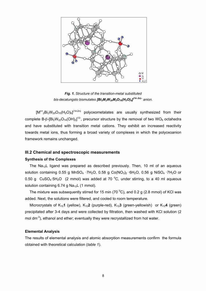
Fig. 1. Structure of the transition-metal substituted
bis-decatungsto bismutates [Bi2M2W20M2O70(H2O)6](14–2n)– anion.
[Mn+2Bi2W20O70(H2O)6]
(14-2n) polyoxometalates are usually synthesized from their
complete B--[Bi2W22O74(OH)2]12-, precursor structure by the removal of two WO6 octahedra
and have substituted with transition metal cations. They exhibit an increased reactivity
towards metal ions, thus forming a broad variety of complexes in which the polyoxoanion
framework remains unchanged.
III.2 Chemical and spectroscopic measurements
Synthesis of the Complexes
The Na12L ligand was prepared as described previously. Then, 10 ml of an aqueous
solution containing 0.55 g MnSO4 ·7H2O, 0.58 g Co(NO3)2 ·6H2O, 0.56 g NiSO4 ·7H2O or
0.50 g CuSO4·5H2O (2 mmol) was added at 70 0C, under stirring, to a 40 ml aqueous
solution containing 6.74 g Na12L (1 mmol).
The mixture was subsequently stirred for 15 min (70 0C), and 0.2 g (2.8 mmol) of KCl was
added. Next, the solutions were filtered, and cooled to room temperature.
Microcrystals of K101 (yellow), K102 (purple-red), K103 (green-yellowish) or K104 (green)
precipitated after 3-4 days and were collected by filtration, then washed with KCl solution (2
mol dm-3), ethanol and ether; eventually they were recrystallized from hot water.
Elemental Analysis
The results of elemental analysis and atomic absorption measurements confirm the formula
obtained with theoretical calculation (table 1).
8
% Calculated / (Experimental) Complex Color Yield
%
K Bi W M H2O
Crist.
H2O
Coord.
1 Yellow 62 6.03 (6.08)
6.76 (6.70)
59.46 (59.30)
1.78 (1.82)
5.82 (5.85)
1.75 (1.77)
2 Purple-red
68 6.08 (6.12)
6.50 (6.45)
57.22 (57.18)
1.83 (1.85)
9.25 (9.28)
1.68 (1.70)
3 Green-yellow
65 6.28 (6.30)
6.71 (6.58)
59.04 (58.96)
1.89 (1.92)
6.36 (6.40)
1.74 (1.76)
4 green 64 6.32 6.35
6.62 6.48
58.28 58.14
2.01 2.06
7.43 7.46
1.71 1.74
Table 1. Results of elemental analysis for the studied complexes
FT-IR Spectroscopy
FT-IR spectra of polyoxometalates generally exhibit contributions of the polyoxoanion
framework.
The characteristic IR vibration bands of 1, 2, 3 and 4 complexes, compared to those of
the Na12 [Bi2W22O74(OH)2] ligand, are displayed in Table 2.
Vibration L 1 2 3 4
as (W=Ot) 945 945 948 945 949
as(Bi-Oi) 832 825 826 828 836
as(W-Oc-W) 794
749
882
760
886
762
889
763
885
763
s (W-Oe-W) 649 668 669 669 664
(W-Oc,e-W) 508 515 510
505
512 505
as (OH) 3407 3431 3419 3420 3439
(HOH) 1624 1626 1616 1625 1621
Table 2. Main vibration bands observed in the FT-IR spectra (cm1)
All asymmetric bonds frequencies involving tungsten atoms are shifted towards higher or
lower frequencies (5-80 cm-1) in the complexes spectra compared to the ligand spectrum.
The tiny shift of the as (W=Ot) stretching vibration in the spectra of the complexes, compared
to the ligand, can be explained by the fact that terminal Ot atoms are not involved in the
coordination of metal ions. The vibrational frequency of the Bi-Oi bond at ~ 830 cm-1 are not
involved in the coordination of metal ions, too.
9
On the other hand, the larger shift of the as(W-Oc,e-W) stretching vibrations, for the bonds
from the belt and cap areas, indicates the coordination of metal ions by Oc and Oe oxygen
atoms from corner-sharing and edge- sharing octahedra. The shift of the frequencies for the
tri-centric bonds in the FT-IR spectrum of the complex compared to the ligands suggests the
coordination of the metal ions in the lacunary regions of the Keggin polyoxoanion structure.
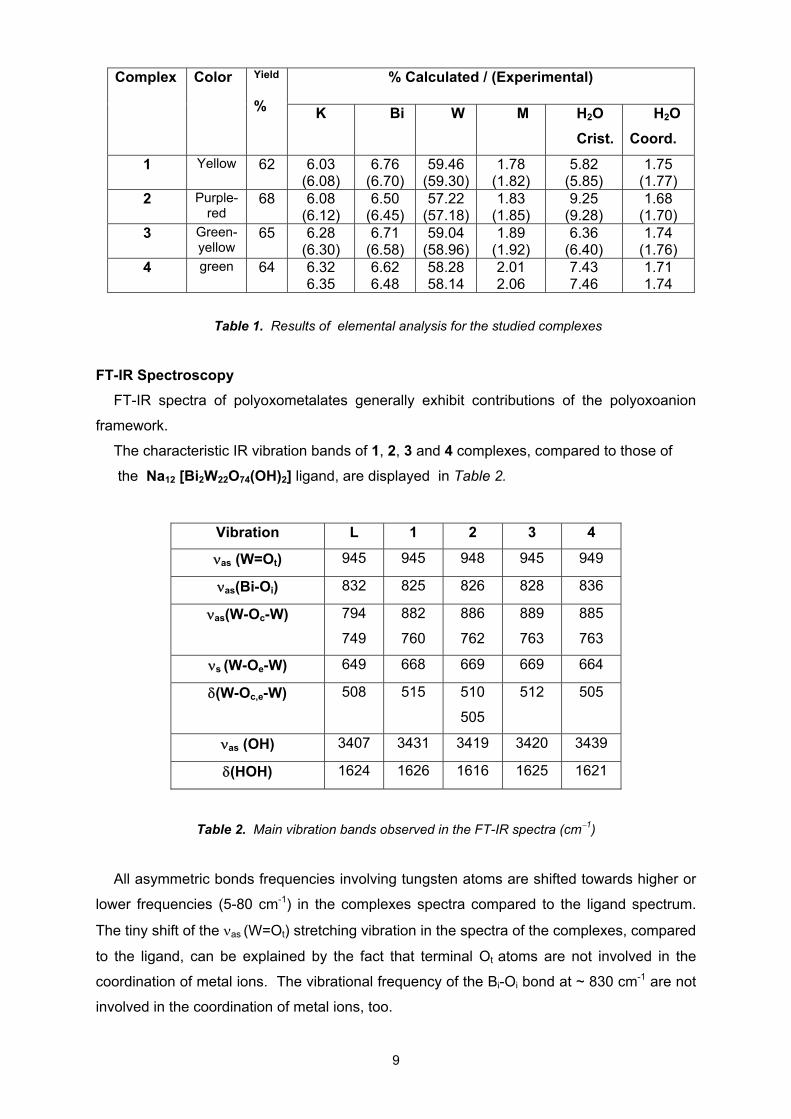
Electronic Spectroscopy
The UV electronic spectra (Figure 2) are characteristic to the polyoxometalates and
similar to the ligand. The lower energy band (1) at 256 nm, split into two bands in both
ligand and complexes, due to the d-p-d electronic transitions from the tricentric W-Oc,e-W
bonds, was shifted to lower frequencies in complexes spectra compared to ligand and is
due to the decrease of the symmetry as well as to the distortion of the WO6 octahedra
through complexation, which influences the electronic transfer from these bonds.
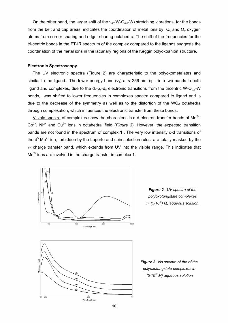
Visible spectra of complexes show the characteristic d-d electron transfer bands of Mn2+,
Co2+, Ni2+ and Cu2+ ions in octahedral field (Figure 3). However, the expected transition
bands are not found in the spectrum of complex 1 . The very low intensity d-d transitions of
the d5 Mn2+ ion, forbidden by the Laporte and spin selection rules, are totally masked by the
5 charge transfer band, which extends from UV into the visible range. This indicates that
Mn2+ ions are involved in the charge transfer in complex 1.
Figure 2. UV spectra of the
polyoxotungstate complexes
in (510-5) M) aqueous solution.
Figure 3. Vis spectra of the of the
polyoxotungstate complexes in
(510-3 M) aqueous solution
10
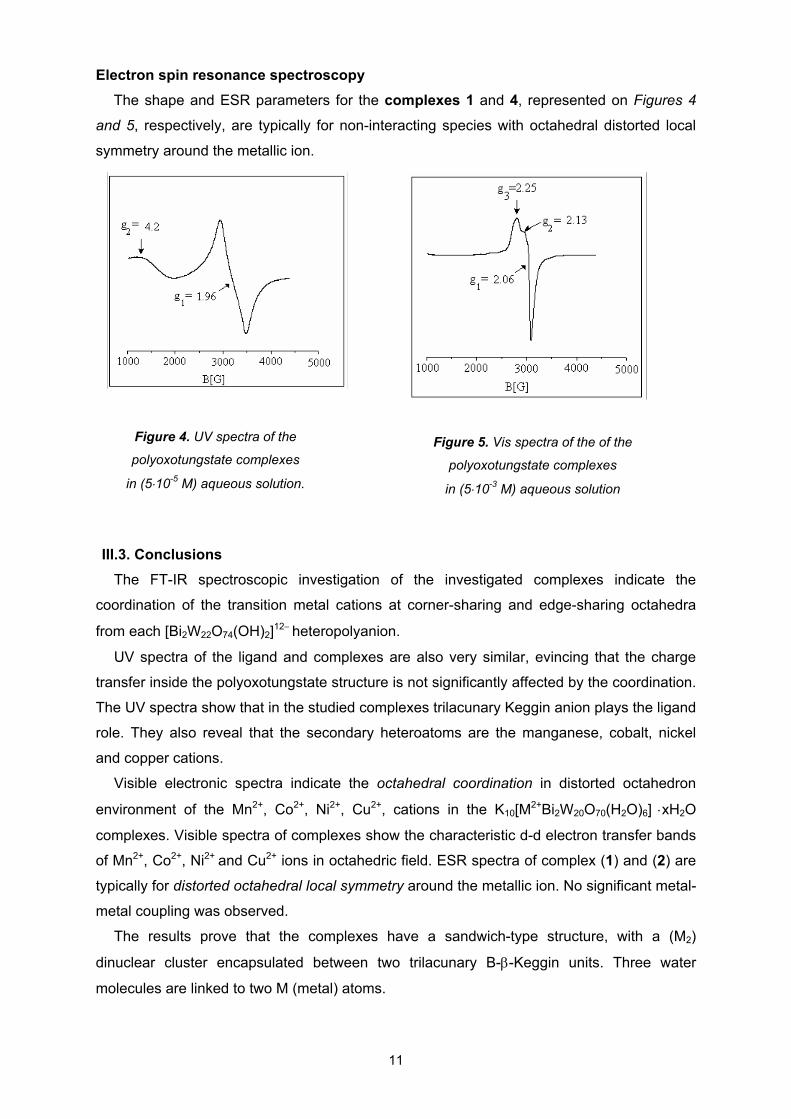
Electron spin resonance spectroscopy
The shape and ESR parameters for the complexes 1 and 4, represented on Figures 4
and 5, respectively, are typically for non-interacting species with octahedral distorted local
symmetry around the metallic ion.
11
Figure 4. UV spectra of the
polyoxotungstate complexes
in (510-5 M) aqueous solution.
Figure 5. Vis spectra of the of the
polyoxotungstate complexes
in (510-3 M) aqueous solution
III.3. Conclusions
The FT-IR spectroscopic investigation of the investigated complexes indicate the
coordination of the transition metal cations at corner-sharing and edge-sharing octahedra
from each [Bi2W22O74(OH)2]12 heteropolyanion.
UV spectra of the ligand and complexes are also very similar, evincing that the charge
transfer inside the polyoxotungstate structure is not significantly affected by the coordination.
The UV spectra show that in the studied complexes trilacunary Keggin anion plays the ligand
role. They also reveal that the secondary heteroatoms are the manganese, cobalt, nickel
and copper cations.
Visible electronic spectra indicate the octahedral coordination in distorted octahedron
environment of the Mn2+, Co2+, Ni2+, Cu2+, cations in the K10[M2+Bi2W20O70(H2O)6]
xH2O
complexes. Visible spectra of complexes show the characteristic d-d electron transfer bands
of Mn2+, Co2+, Ni2+ and Cu2+ ions in octahedric field. ESR spectra of complex (1) and (2) are
typically for distorted octahedral local symmetry around the metallic ion. No significant metal-
metal coupling was observed.
The results prove that the complexes have a sandwich-type structure, with a (M2)
dinuclear cluster encapsulated between two trilacunary B--Keggin units. Three water
molecules are linked to two M (metal) atoms.
IV. COOPER (II) COMPLEXES WITH THEOPHYLLINE LIGAND CONTANING
ETHILENDIAMINE DERIVATES AS COLIGANDS
III.1 Introduction
Derivatives of nucleobases belonging to the xanthine group, like theophylline, have been
known for a long time, and commonly used for their biologic effects. The coordination
compounds of these molecules, may serve as model compounds for the interaction of metal
ions with molecules of biologic interest.
The theophylline, i.e., 1,3-dimethyl-2,6-dioxo-purine, in neutral or basic medial act as
monodentate ligand and coordinates through the N7 atom which is the preferred binding site
in 6-oxopurines The deprotonated theophylline may act as bidentate ligand forming N7/O6
chelates.
A series of novel mixed-ligand theophylline (th) complexes, [Cu(th)2(dmen)]·2H2O·(1),
[Cu(th)2(tmeda)(H2O)]·0.5H2O (2) and [Cu(th)2(dpen)(H2O)]·5H2O (3), were synthesized
and investigated by means of infrared and EPR spectroscopic methods. As co-ligands the
following ethylenediamine derivatives were used: N,N-dimethyl-ethylenediamine (dmen),
N,N,N’,N’-tetramethyl-ethylenediamine (tmeda) and meso-1,2-diphenyl-ethylenediamine
(dpen). The geometry of complexes was optimized at B3LYP/LANL2DZ level of theory.
Figure 6.
Deprotonated theophylline structural formula
III.2 Chemical and spectroscopic measurement
Synthesis
The complexes [Cu(th)2(dmen)]·2H2O (1), [Cu(th)2(tmeda)(H2O)]·0.5H2O (2) and
[Cu(th)2(dpen)(H2O)]·5H2O (3) were synthesised from theophylline and appropriate
copper(II) complexes in basic media according to published methods. Compounds 13 were
isolated in good yield as microcrystalline solids and were characterized by elemental
analyses, IR and ESR spectroscopy.
Elemental analysis
The theoretically calculated and experimentally obtained elemental analysis data are
shown in table 3.
12
13
Complex
1 2 3
C18H30CuN10O6 C20H32CuN10O5 C28H42CuN10O10
MW (g·mol1) 545.78 556.08 742.25
Yield (%)
Experimental (calculated)
81 62 41
C (%)
39.58 (39.56)
30.78 (43.20)
45.33 (45.31)
H (%)
5.54 (5.45)
3.98 (5.80)
5.18 (5.70)
N (%)
25.66 (25.41) 20.44 (25.19) 18.63 (18.87)
Table 3. Physico-chemical properties and elemental analysis data of the metal complexes (13)
FT-IR spectra
Spectra of all copper(II) complexes show the two strong bands of theophylline assigned
to the stretching vibration of carbonyl groups shifted towards lower wavenumbers, due to
the deprotonation of theophylline and participation of C(6)=O and C(2)=O groups in intra- or
intermolecular hydrogen bond formation (table 4). The C=N vibrations of the theophylline
are shifted to lower wave numbers in complexes suggesting that the ligand coordinates
through one of the imidazole's nitrogen atoms, acting as monodentate ligand.
In all spectra of complexes the symmetric and antisymmetric stretching vibrations of
coordinated NH2 groups can be assigned in the 32853154 cm-1 region. There are
significant changes in the bands assigned to NH vibrations as consequence of the
deprotonation of theophylline at N(7) atom and coordination of the amine type ligands. The
amine ligands are coordinated as bidentate ligands through N atoms to the metal center.
Vibration
theophylline
1 [Cu(th)2(dmen)2]
2 [Cu(th)2(tmeda)
(H2O)]
3 [Cu(th)2(dpen)
(H2O)]
ν(NH) 3122 3285m
3154m 3102m
3462m 3245m 3161m
ν(C=O) 1717 1669
1686vs 1642vs
1690vs 1636vs
1690vs 1641vs
ν(C=C)
1610 1580m 1528m 1591m
ν(C=N)
1568 1527m 1528m 1530m
Table 4. Some IR absorption bands (cm-1) for the theophylline and the metal complexes 13
( m – medium, vs- very strong)
The νCH vibrations of ligands appear at 28512954 cm-1 and 30273066 cm-1 for aliphatic
CH2 and aromatic CH, respectively.
The presence of strong broad bands in FTIR spectra of the complexes at 35003200
cm1 may be assigned to various types of hydrogen bonds.
Electronic spin resonance spectroscopy
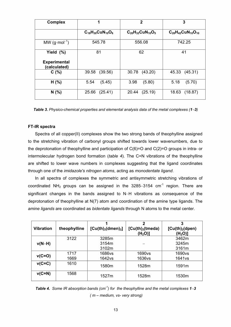
Figure 7. Powder ESR spectra of
Cu(th)2(dmen)]·2H2O (1)
Figure 9. Powder ESR spectra of
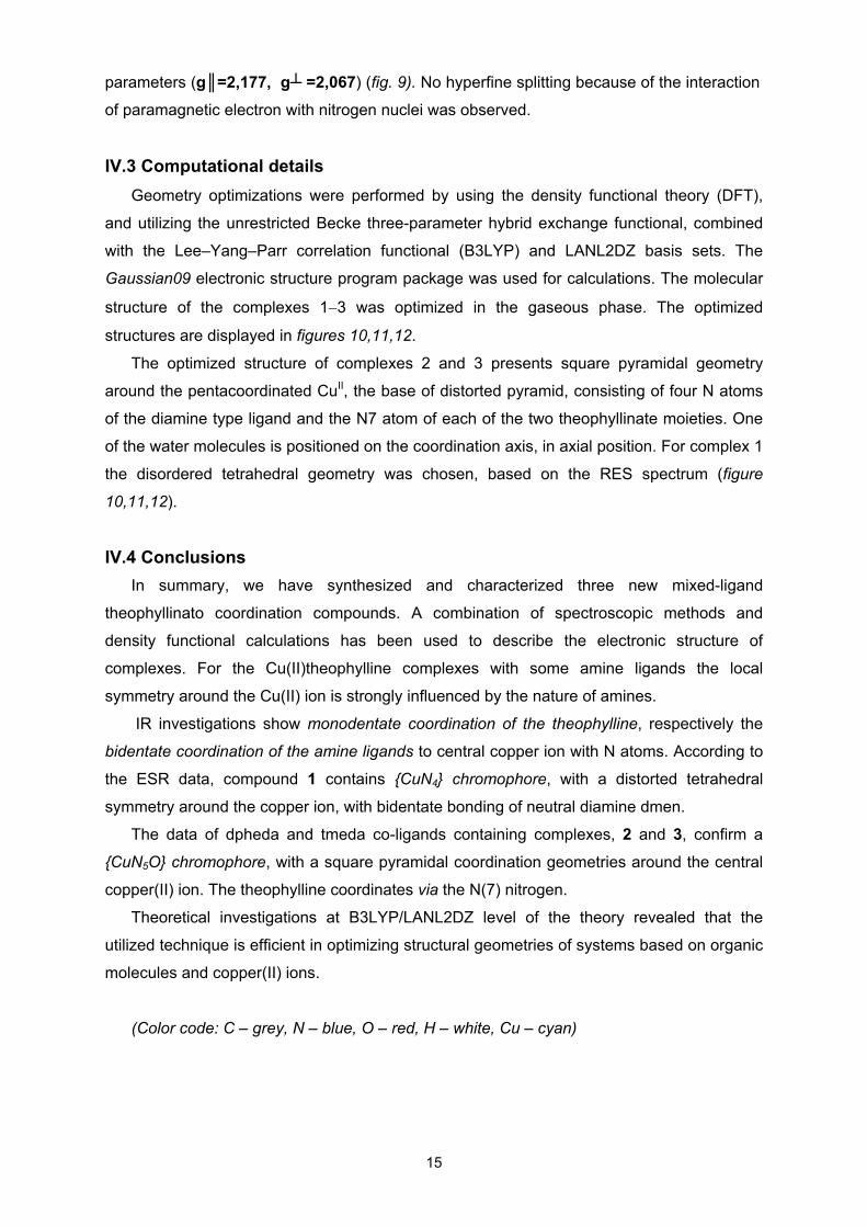
[Cu(th)2(dpen)(H2O)]·5H2O (3)
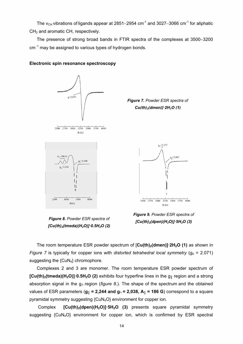
Figure 8. Powder ESR spectra of
[Cu(th)2(tmeda)(H2O)]·0.5H2O (2)
The room temperature ESR powder spectrum of [Cu(th)2(dmen)]·2H2O (1) as shown in
Figure 7 is typically for copper ions with distorted tetrahedral local symmetry (g0 = 2.071)
suggesting the {CuN4} chromophore.
Complexes 2 and 3 are monomer. The room temperature ESR powder spectrum of
[Cu(th)2(tmeda)(H2O)]·0.5H2O (2) exhibits four hyperfine lines in the g║ region and a strong
absorption signal in the g┴ region (figure 8.). The shape of the spectrum and the obtained
values of ESR parameters (g║ = 2,244 and g┴ = 2,038, A║ = 186 G) correspond to a square
pyramidal symmetry suggesting {CuN4O} environment for copper ion.
Complex [Cu(th)2(dpen)(H2O)]·5H2O (3) presents square pyramidal symmetry
suggesting {CuN4O} environment for copper ion, which is confirmed by ESR spectral
14
15
parameters (g║=2,177, g┴ =2,067) (fig. 9). No hyperfine splitting because of the interaction
of paramagnetic electron with nitrogen nuclei was observed.
IV.3 Computational details
Geometry optimizations were performed by using the density functional theory (DFT),
and utilizing the unrestricted Becke three-parameter hybrid exchange functional, combined
with the Lee–Yang–Parr correlation functional (B3LYP) and LANL2DZ basis sets. The
Gaussian09 electronic structure program package was used for calculations. The molecular
structure of the complexes 13 was optimized in the gaseous phase. The optimized
structures are displayed in figures 10,11,12.
The optimized structure of complexes 2 and 3 presents square pyramidal geometry
around the pentacoordinated CuII, the base of distorted pyramid, consisting of four N atoms
of the diamine type ligand and the N7 atom of each of the two theophyllinate moieties. One
of the water molecules is positioned on the coordination axis, in axial position. For complex 1
the disordered tetrahedral geometry was chosen, based on the RES spectrum (figure
10,11,12).
IV.4 Conclusions
In summary, we have synthesized and characterized three new mixed-ligand
theophyllinato coordination compounds. A combination of spectroscopic methods and
density functional calculations has been used to describe the electronic structure of
complexes. For the Cu(II)theophylline complexes with some amine ligands the local
symmetry around the Cu(II) ion is strongly influenced by the nature of amines.
IR investigations show monodentate coordination of the theophylline, respectively the
bidentate coordination of the amine ligands to central copper ion with N atoms. According to
the ESR data, compound 1 contains {CuN4} chromophore, with a distorted tetrahedral
symmetry around the copper ion, with bidentate bonding of neutral diamine dmen.
The data of dpheda and tmeda co-ligands containing complexes, 2 and 3, confirm a
{CuN5O} chromophore, with a square pyramidal coordination geometries around the central
copper(II) ion. The theophylline coordinates via the N(7) nitrogen.
Theoretical investigations at B3LYP/LANL2DZ level of the theory revealed that the
utilized technique is efficient in optimizing structural geometries of systems based on organic
molecules and copper(II) ions.
(Color code: C – grey, N – blue, O – red, H – white, Cu – cyan)
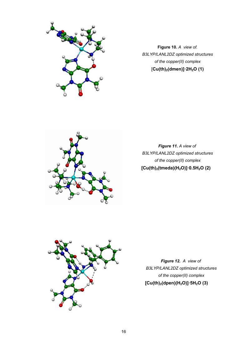
Figure 10. A view of.
B3LYP/LANL2DZ optimized structures
of the copper(II) complex
[Cu(th)2(dmen)]·2H2O (1)
Figure 11. A view of
B3LYP/LANL2DZ optimized structures
of the copper(II) complex
[Cu(th)2(tmeda)(H2O)]·0.5H2O (2)
Figure 12. A view of
B3LYP/LANL2DZ optimized structures
of the copper(II) complex
[Cu(th)2(dpen)(H2O)]·5H2O (3)
16
V. METALLIC COMPLEXES WITH AMINOACIDS
V.1 Introduction
Amino acids are the “building blocks” of the body. Besides building cells and repairing
tissue, they form antibodies to combat invading bacteria and viruses; they are part of the
enzyme and hormonal system, they build nucleoproteins and carry oxygen throughout the
body and participate in muscle activity.
A simple amino acid anion is a potential bidentate ligand which may coordinate to a
transition metal ion through the amino lone pair of electrons and the carboxylate oxygen lone
pair of electrons. The metallic complexes with amino acids as ligands were deeply
investigated due to their capacities of forming chelates, which are used in various domains
like: medicine, chemistry, pharmacy, biology, nutrition and physics.
Complexes of transition metals with amino acids in proteins and peptides are utilized in
numerous biological processes, such as oxygen conveyer, electron transfer and oxidation.
Metal (II) complexes present antibacterial and antifungal activity.
The metallic complexes with amino acids as ligands were deeply investigated due to
their capacities of forming chelates, which are used in various domains like: medicine,
chemistry, pharmacy, biology, nutrition and physics.
New complexes of α-aminoacids, [Cu(L)2]·H2O (1), [Co(L)2]·2H2O (2) and [Zn(L)2]·H2O
(3) have been investigated by physical-chemical methods. Here we present some
spectroscopic methods of molecular structure investigation, like FTIR, UV-Vis and ESR. (L
represents the aminoacid ligands: leucine, phenylalanine, methionine).
V.2 Spectroscopic investigation of metallic Cu(II), Co(II), Zn(II) complexes with
leucine as ligand
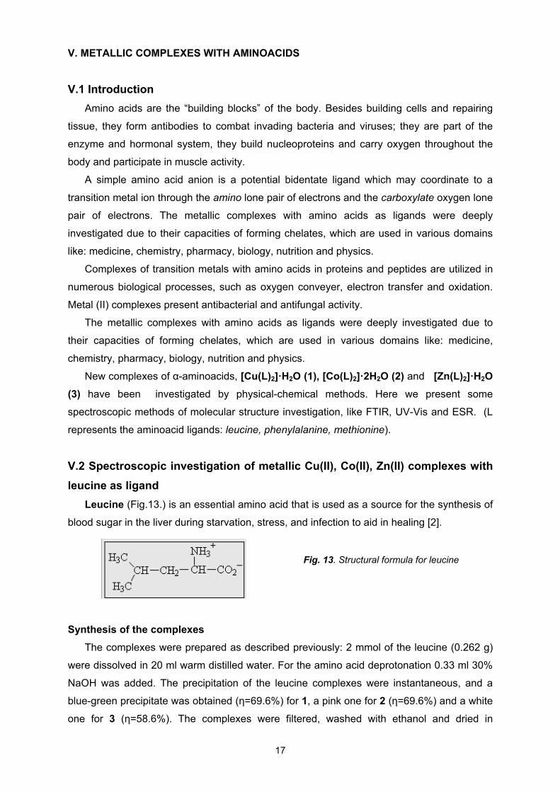
Leucine (Fig.13.) is an essential amino acid that is used as a source for the synthesis of
blood sugar in the liver during starvation, stress, and infection to aid in healing [2].
17
Fig. 13. Structural formula for leucine
Synthesis of the complexes
The complexes were prepared as described previously: 2 mmol of the leucine (0.262 g)
were dissolved in 20 ml warm distilled water. For the amino acid deprotonation 0.33 ml 30%
NaOH was added. The precipitation of the leucine complexes were instantaneous, and a
blue-green precipitate was obtained (η=69.6%) for 1, a pink one for 2 (η=69.6%) and a white
one for 3 (η=58.6%). The complexes were filtered, washed with ethanol and dried in
desiccators under P4O10. Afterward, the complexes were recrystallized on methanol, dried
and weight to establish the percent of complexation.
Elemental analysis and atomic absorption spectroscopy
The elemental analysis and atomic absorption spectroscopy measurements confirm
the metal ion: leucine ratio of 1:2 for composition of the complexes.
18
% Calculated/ Found
Complex Molecular weight
C H N Metal
[Cu(L)2]H2O 323.5 43.51 (42.71)
8.65 (9.22)
7.41 (6.38)
19.40 (19.36)
[Co(L)2]2H2O 319 44.14 (43.2)
8.77 (9.32)
7.52 (8.25)
21.35 (22.15)
[Zn(L)2]H2O 325 44.25 (43.75)
8.60 (9.10)
7.37 (6.80)
18.00 (17.98)
Table 5. Elemental analysis results of the metal-leucine complexes
FT-IR spectra
Complex ν(O-H ) ν(N-H) ν(C=O) δ(N-H)
Leu - 3052 1608 1577 1511
Cu- Leu 3421 3319 3245
1619 1561
Co- Leu 3475 3223 3107
1639 1578
Zn- Leu - 3325 3268
1654 1608
Table 6. FT-IR spectral data (cm-1) metal-leucine complexes
In the spectrum of the ligand, the s(N-H) stretching vibration appears at 3052 cm-1 and
is shifted in the complexes spectra at: 3319 cm-1, 3245 cm-1 (1), 3223 cm-1, 3107 cm-1 (2)
3325 cm-1 3268 cm-1 (3) proving the involvement of the –NH2− group in the complex
formation.
The absorption band at 1608 cm-1 was attributed to the (C=O) stretching vibration in the
spectrum of the ligand and appears to be shifted toward higher wave numbers with 11 cm-1,
31 cm-1 and 46 cm-1 in the spectra of 1, 2 respectively 3 proving the involvement of the
carboxylic group in the covalent bonding to the metal ion.
The (O–H) stretching vibrations does not appear in the spectra of the ligand and
complex 3 but emerge in spectra of complexes (1) and (2) at 3421 cm-1 and 3475 cm-1,
respectively, suggesting the presence of the crystal water in these compounds[11]. Due to
the δ(N-H) bending vibration shifting in the complexes spectra the involvement of the –NH2−
group to the metal bonding formation was confirmed.
Electronic spectra
Compound n→ π* transition d-d transition
Leucine 277 nm -
Cu-leu 270 nm 600-650 nm
Co-leu 276 nm 450-540 nm
Zn-leu 268 nm -
Table 7. UV and visible spectral data
The n→ π* characteristic band assigned to the C=O bond appears at 277 nm in the
ligand spectrum and is shifted toward UV domain with 7 nm, 1nm and 11 nm in the complex
1, 2 and 3 spectra , proving the presence of the ligand within the complex and the covalent
nature of the metal-ligand bond.
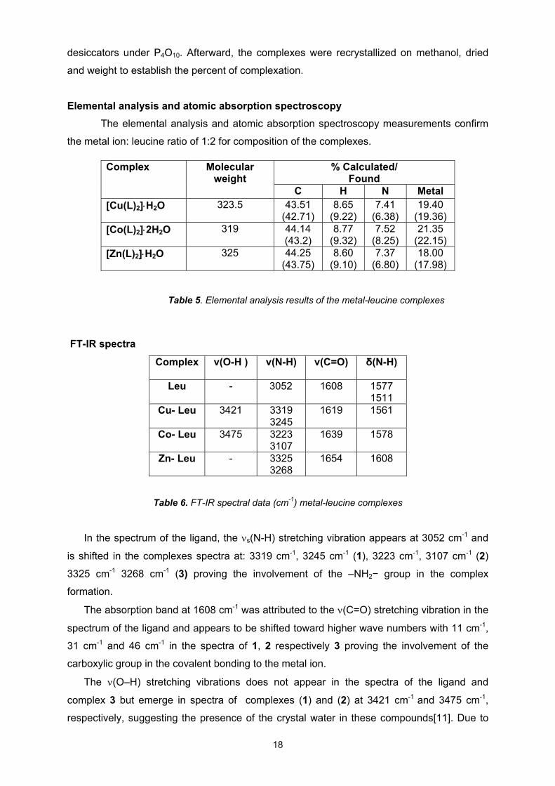
Fig. 15. Powder EPR spectrum
of [Cu(L)2]H2O (1) at room temperature
Fig. 14. Visible spectrum
of [Cu(L)2]H2O (1) in DMSO (10-3M)
The metal-ligand charge transfer band appears below at 250nm in the spectrum of
complex 2 and between 290- 320 nm in the spectrum of complex 3.
In the visible domain the spectrum of complex 1 (Fig.14) show a large shoulder at 620
nm, assigned to the 2T2g → 2Eg transition, specific to Cu (II) complexes with tetragonal
distortion due to the Jahn-Teller effect. Yet again in visible domain, the spectrum of complex
2 illustrates two bands at 360 nm and 525 nm, also ascribed to the d-d transition, the last
band within the spectrum was assigned to the 4T1g (P)→ 4T1g(F) transition, expected for an
octahedral symmetry of cobalt ion.
19
ESR Spectra
Powder ESR spectrum of complex 1 (Fig. 15) measured at room temperature is quasi-
isotropic (g= 2.178) and is characteristic for pseudotetrahedral symmetry around the copper
ion. The shape and the value of the g tensor correspond to a {CuN2O2} chromophore.
The powder ESR spectrum of complex 2 revealed the presence of monomeric
compounds, with octahedral symmetry around the cobalt ion, the g tensor value is g=2.195.
V.3 Spectroscopic investigation of metallic Cu(II), Co(II), Zn(II) complexes with
phenylalanine as ligand
Phenylalanine (Fig.16) is used by the brain to produce norepinephrine, a chemical that
transmits signals between nerve cells and the brain; keeps you awake and alert; reduces
hunger pains; functions as an antidepressant and helps improve memory.
Figure 16. Structure formula for
phenylalanine
Synthesis of the complexes: the complexes were prepared as described previously.
Elemental analysis
The elemental analysis and atomic absorption spectroscopy measurements confirm the
metal ion: phenylalanine ratio of 1:2 for the composition of the complexes.
%C %H %N Symbolic
formula
Molecular
weigth Meas. Calc. Meas. Calc. Meas. Calc.
Cu(L)2 391.5 54.79 55.1 7.31 7.66 6.92 7.15
Co(L)2 389.4 51.42 55.46 8.59 8.2 8.28 7.59
Zn(L)2 395 51.85 54.69 9.07 8.10 6.57 7.08
Table 8. Elemental analysis results for metal- phenylalanine complexes
FT-IR spectroscopy
Information about the metal ions coordination was obtained by comparing the IR
frequencies of the ligand with those of the copper, cobalt and zinc complexes.
The ν(OH) stretching vibration does not appear in the spectra of the ligand and complex
3, but emerges in the spectra of complexes 1 and 2 complexes at 3454 cm-1 respectively
3453 cm-1 and 3359 cm-1 suggesting the presence the crystal water in these compounds.
20
Compound ν(O-H)
(cm-1) ν(N-H) (cm-1)
ν(C=O) (cm-1)
δ(N-H) (cm-1)
Phenylalanine - 3078
3030 1623 1557
Cu-phe 3454 3320 3256
1629 1567
Co-phe 3453 3359
3220 1633 1586
Zn-phe - 3334 3256
1614 1531
Table 9. FT-IR spectral data (cm-1) for metal- phenylalanine complexes
Electronic spectroscopy
Information about local symmetry of metal ions was obtained by comparing the ligand
spectra with those of complexes with amino acids.
Compound n→ π* π→ π*(arom) d-d
Phenylalanine 231 nm 260 nm -
Cu-phe 225 nm 275 nm 600-650 nm
Co-phe 235 nm 280 nm 500-540 nm
Zn-phe 220 nm 260-270 nm -
Table 10. UV and visible spectral data for metal- phenylalanine complexes
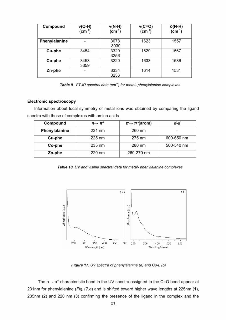
Figure 17. UV spectra of phenylalanine (a) and Cu-L (b)
The n→ π* characteristic band in the UV spectra assigned to the C=O bond appear at
231nm for phenylalanine (Fig.17.a) and is shifted toward higher wave lengths at 225nm (1),
235nm (2) and 220 nm (3) confirming the presence of the ligand in the complex and the
21
covalent nature of the metal-ligand bond. In the UV spectrum of the ligand the π→π*
characteristic band appears at 260nm, and is shifted in the complexes spectra with 15nm
(1), 20nm (2) and 7nm (3), being assigned to conjugated systems.
In the visible domain (Fig.17) a d–d transition points out at 615 nm in the copper complex
spectrum and was assigned to the 2T2g → 2Eg transition, specific for Cu (II) complexes with
tetragonal distortion owing to the Jahn–Teller effect. In the visible domain, the cobalt
complex spectrum shows a band at 540nm attributed to the d–d transition of cobalt
electrons.
Electron spin resonance spectroscopy
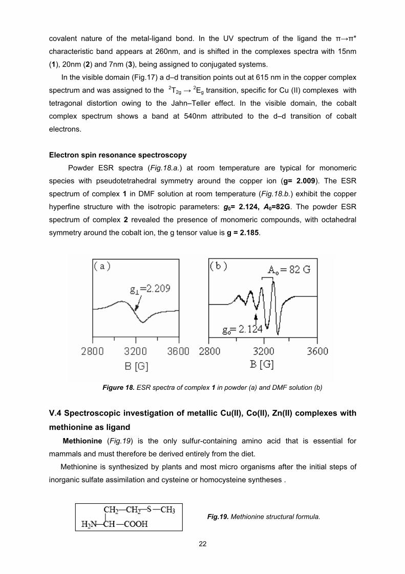
Powder ESR spectra (Fig.18.a.) at room temperature are typical for monomeric
species with pseudotetrahedral symmetry around the copper ion (g= 2.009). The ESR
spectrum of complex 1 in DMF solution at room temperature (Fig.18.b.) exhibit the copper
hyperfine structure with the isotropic parameters: g0= 2.124, A0=82G. The powder ESR
spectrum of complex 2 revealed the presence of monomeric compounds, with octahedral
symmetry around the cobalt ion, the g tensor value is g = 2.185.
Figure 18. ESR spectra of complex 1 in powder (a) and DMF solution (b)
V.4 Spectroscopic investigation of metallic Cu(II), Co(II), Zn(II) complexes with
methionine as ligand
Methionine (Fig.19) is the only sulfur-containing amino acid that is essential for
mammals and must therefore be derived entirely from the diet.
Methionine is synthesized by plants and most micro organisms after the initial steps of
inorganic sulfate assimilation and cysteine or homocysteine syntheses .
Fig.19. Methionine structural formula.
22
Synthesis of the complexes: the complexes were prepared as described previously.
FT-IR spectroscopy
The CH2-S and CH3-S stretching vibrations appear as a sharp band at 2915 cm-1 in the
ligand spectrum and are insignificantly shifted in the spectra of the complexes confirming the
non involvement of these groups in the coordination.
Compond ν(O-H ) ν(N-H) ν(C=O) δ(N-H) ν(C-C)
Met - 3146 1610 1580 1563 1508
1352 1316
Cu- Met - 3229 1649 1568 1616
1334
Co- Met 3419 3175 1640 1584 Zn- Met 3383 3170 1586 1502
1558 1385
Table 11. FT-IR spectral data (cm-1) for metal-methionine complexes
Electronic spectroscopy
Compound n→ π* transition d-d transition
Methionine 267 nm -
Cu-Met 275 nm 600-650 nm
Co-Met 274 nm 450-550 nm
Zn-Met 265 nm -
Table 12. UV and visible spectral data for metal- methionine complexes
The n→ π* characteristic band assigned to the C=O bond appears at 267 nm in the
ligand spectrum and is shifted toward UV domain with 8 nm, 7nm in the complex 1 and 2
spectra , proving the presence of the ligand within the complex and the covalent nature of
the metal-ligand bond.
In the visible domain the spectrum of complex 1 show a large shoulder at 625 nm,
assigned to the 2T2g → 2Eg transition, specific to Cu (II) complexes with tetragonal distortion
due to the Jahn-Teller effect. Yet again in visible domain, the spectrum of complex 2
illustrates a band at 512 nm, also ascribed to the d-d transition.
The position of the band maxima and respective assignment for Co(II) complex with
methionine is typical for octahedral geometries.
23
Electron spin resonance spectroscopy
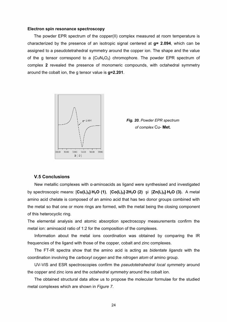
The powder EPR spectrum of the copper(II) complex measured at room temperature is
characterized by the presence of an isotropic signal centered at g= 2.094, which can be
assigned to a pseudotetrahedral symmetry around the copper ion. The shape and the value
of the g tensor correspond to a {CuN2O2} chromophore. The powder EPR spectrum of
complex 2 revealed the presence of monomeric compounds, with octahedral symmetry
around the cobalt ion, the g tensor value is g=2.201.
Fig. 20. Powder EPR spectrum
of complex Cu- Met.
V.5 Conclusions
New metallic complexes with α-aminoacids as ligand were synthesised and investigated
by spectroscopic means: [Cu(L)2]H2O (1), [Co(L)2]2H2O (2) şi [Zn(L)2]H2O (3). A metal
amino acid chelate is composed of an amino acid that has two donor groups combined with
the metal so that one or more rings are formed, with the metal being the closing component
of this heterocyclic ring.
The elemental analysis and atomic absorption spectroscopy measurements confirm the
metal ion: aminoacid ratio of 1:2 for the composition of the complexes.
Information about the metal ions coordination was obtained by comparing the IR
frequencies of the ligand with those of the copper, cobalt and zinc complexes.
The FT-IR spectra show that the amino acid is acting as bidentate ligands with the
coordination involving the carboxyl oxygen and the nitrogen atom of amino group.
UV-VIS and ESR spectroscopies confirm the pseudotetrahedral local symmetry around
the copper and zinc ions and the octahedral symmetry around the cobalt ion.
The obtained structural data allow us to propose the molecular formulae for the studied
metal complexes which are shown in Figure 7.
24
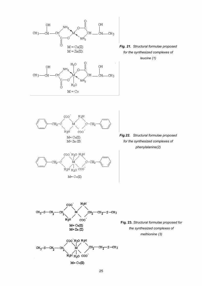
Fig. 21. Structural formulae proposed
for the synthesized complexes of
leucine (1)
Fig.22. Structural formulae proposed
for the synthesized complexes of
phenylalanine(2)
25
Fig. 23. Structural formulae proposed for
the synthesized complexes of
methionine (3)
VI. STRUCTURAL INVESTIGATION OF RANITIDINE MOLECULE BY
VIBRATIONAL SPECTROSCOPIC METHODS AND THEORETICAL
CALCULATION
VI.1 Introduction
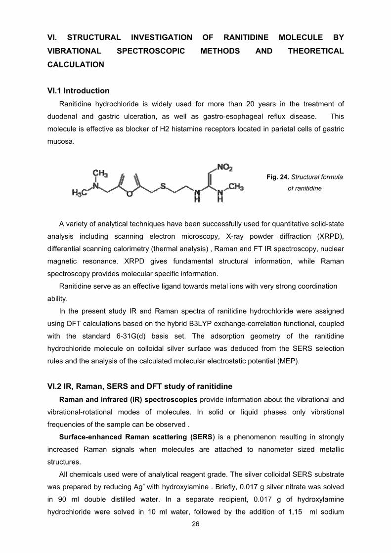
Ranitidine hydrochloride is widely used for more than 20 years in the treatment of
duodenal and gastric ulceration, as well as gastro-esophageal reflux disease. This
molecule is effective as blocker of H2 histamine receptors located in parietal cells of gastric
mucosa.
Fig. 24. Structural formula
of ranitidine
A variety of analytical techniques have been successfully used for quantitative solid-state
analysis including scanning electron microscopy, X-ray powder diffraction (XRPD),
differential scanning calorimetry (thermal analysis) , Raman and FT IR spectroscopy, nuclear
magnetic resonance. XRPD gives fundamental structural information, while Raman
spectroscopy provides molecular specific information.
Ranitidine serve as an effective ligand towards metal ions with very strong coordination
ability.
In the present study IR and Raman spectra of ranitidine hydrochloride were assigned
using DFT calculations based on the hybrid B3LYP exchange-correlation functional, coupled
with the standard 6-31G(d) basis set. The adsorption geometry of the ranitidine
hydrochloride molecule on colloidal silver surface was deduced from the SERS selection
rules and the analysis of the calculated molecular electrostatic potential (MEP).
VI.2 IR, Raman, SERS and DFT study of ranitidine
Raman and infrared (IR) spectroscopies provide information about the vibrational and
vibrational-rotational modes of molecules. In solid or liquid phases only vibrational
frequencies of the sample can be observed .
Surface-enhanced Raman scattering (SERS) is a phenomenon resulting in strongly
increased Raman signals when molecules are attached to nanometer sized metallic
structures.
All chemicals used were of analytical reagent grade. The silver colloidal SERS substrate
was prepared by reducing Ag+ with hydroxylamine . Briefly, 0.017 g silver nitrate was solved
in 90 ml double distilled water. In a separate recipient, 0.017 g of hydroxylamine
hydrochloride were solved in 10 ml water, followed by the addition of 1,15 ml sodium
26
27
hydroxide solution, 1%(v) to OH. The hydroxylamine/sodium hydroxide solution was then
added rapidly to the silver nitrate solution under vigorous stirring. After few seconds a grey-
brown colloidal solution resulted and it was further stirred for 10 min. The pH value of the
silver colloid, measured immediately after preparation, was found to be 8.5.
Computational details
The molecular geometry optimization, molecular electrostatic potential (MEP) and
vibrational spectra calculations were performed with the Gaussian 03W software package
by using density functional theory (DFT) methods with B3LYP hybrid exchange-correlation
functional and the standard 6-31G(d) basis set. No symmetry restriction was applied during
geometry optimization.
After geometric optimization, vibrational frequencies were calculated for ranitidine
hydrochloride.
The assignment of the experimental frequencies is based on the observed band
frequencies and intensity pattern of the Raman spectra and confirmed by establishing a one
to one correlation between the observed and theoretical calculated frequencies.
To aid in mode assignment, we based on the direct comparison between the
experimental and calculated spectra by considering both, the frequency sequence and
intensity pattern, and by comparisons with vibrational spectra of similar compounds.
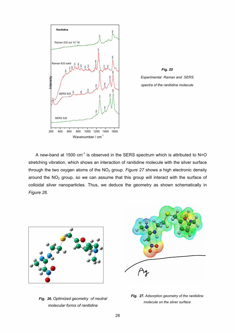
As deduced from calculations of pKa values, of interest are two molecular forms of
ranitidine, the neutral and protonated form of the N11 atom. Thus, molecular geometry
optimization was carried out of these two forms using B3LYP functional with 6-31G basis set
(d) (Fig.26). When comparing the experimental FTIR spectrum with the calculated spectra it
has to be taken in consideration that the theoretical spectra are calculated for the gas phase
of the substance and the experimental data were recorded for solid samples.
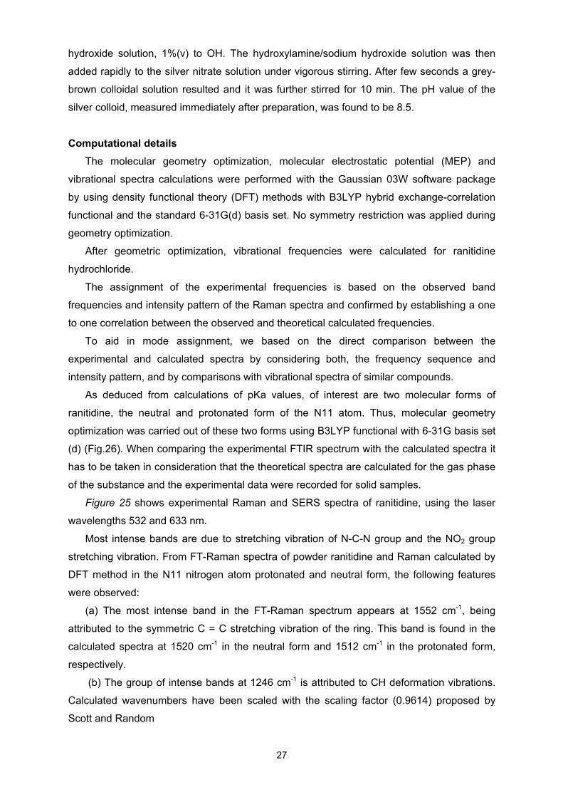
Figure 25 shows experimental Raman and SERS spectra of ranitidine, using the laser
wavelengths 532 and 633 nm.
Most intense bands are due to stretching vibration of N-C-N group and the NO2 group
stretching vibration. From FT-Raman spectra of powder ranitidine and Raman calculated by
DFT method in the N11 nitrogen atom protonated and neutral form, the following features
were observed:
(a) The most intense band in the FT-Raman spectrum appears at 1552 cm-1, being
attributed to the symmetric C = C stretching vibration of the ring. This band is found in the
calculated spectra at 1520 cm-1 in the neutral form and 1512 cm-1 in the protonated form,
respectively.
(b) The group of intense bands at 1246 cm-1 is attributed to CH deformation vibrations.
Calculated wavenumbers have been scaled with the scaling factor (0.9614) proposed by
Scott and Random
525
613
664
724
804
863
953
1023
1186
1247
1377
1452
1559
245
692
866 10
19
1196
1274
1386
1500
1566
1198
1276
1387
1502
1568
1274
1558
200 400 600 800 1000 1200 1400 1600
Inte
nsi
ty
Wavenumber / cm-1
Raman 633 solid
Ranitidine
SERS 633
SERS 532
Raman 532 sol 10-1 M
Fig. 25
Experimental Raman and SERS
spectra of the ranitidine molecule
A new-band at 1500 cm-1 is observed in the SERS spectrum which is attributed to N=O
stretching vibration, which shows an interaction of ranitidine molecule with the silver surface
through the two oxygen atoms of the NO2 group. Figure 27 shows a high electronic density
around the NO2 group, so we can assume that this group will interact with the surface of
colloidal silver nanoparticles. Thus, we deduce the geometry as shown schematically in
Figure 26.
Fig. 27. Adsorption geometry of the ranitidine
molecule on the silver surface Fig. 26. Optimized geometry of neutral
molecular forms of ranitidine
28
29
FINAL CONCLUSIONS
Three classes of metallic complexes have synthesised and characterised, having as
ligands polioxotungstates, theophyline, ethylendiamine derivatives and amino acids.
The goal of the metal–amino-acide compounds’ synthesis is their follow-up investigation
regarding their antibacterian, antifungal and anticarcinogen effect. The hetero-
plyoxometalates may form complex combinations as ligands, with interesting stuctures and
useful in the foundation of a stuctural physico-chemical theory. The research done in the
metallic ions’ complexes of pharmaceutical interest, like the theophylline and ranitidine, has
a clear perspective regarding the growing of the efficiency and improving the action of some
already known medications.
The structural characterization of these compounds with biological and pharmaceutical
interest was realized by infrared spectroscopy (FT-IR), Raman spectroscopy, surface
enhanced Raman spectroscopy, electronic spectroscopy in the ultraviolet and visible domain
(UV-VIS) and by electron spin resonance (ESR). As confirmed both by the shape of the ESR
spectra and by the values of the g parameters, we can state the followings:
The cations Mn2+, Co2+, Ni2+, Cu2+ are octahedrally coordinated in a distorted
octahedral crystalline field of the K10[M2+
2 Bi2W20O70(H2O)6] xH2O complex.
The [Cu(th)2(dmen)]·2H2O (1) compound contains the {CuN4} chromophore with a
distorted tetrahedral symmetry around the copper ion.
The [Cu(th)2(tmeda)(H2O)]·0.5H2O (2) and [Cu(th)2(dpheda)(H2O)]·5H2O (3)
present a pyramidal tetragonal symmetry for the {CuN5O} chromophore.
Pseudo-tetrahedral symmetry was observed around the Cu(II) and Zn(II) for ML2, L
being leucine, phenylalanine or methionine.
Octahedral surrounding of the Co(II) was observed in case of the complexes with
leucine, phenylalanine or methionine amino-acids.
The structure of the ranitidine molecule was studied by the FT-IR, FT-Raman and
Raman-SERS methods. Theoretical calculations lead to different geometries for the two
molecular forms of the ranitidine:neutral and protonated. From the analysis of the molecular
electrostatic potential (MEP) is observed, that the negative charge is concentrated mainly on
the oxygen atoms from the NO2 group, indicating the adsorption of the molecule to the
metallic surface through the oxygen atoms.
The obtained results ecourage the continuation of the present research, mainly regarding
the synthesis and the structural analysis of the theophylline’s complexes with other
transitional ions, and also of the ranitidine’s complexes with Cu or Co, focusing on the
improvement of the biological activity of the metallic complexes in comparison to the action
of the ligand.
30
SELECTIVE BIBLIOGRAPHY
1. G. Marcu, Chimia modernă a elementelor metalice, Editura Tehnicã, Bucureşti, 1993,
2. L. Ghizdavu, Chimie bioanorganică, Ed. Poliam, Cluj-Napoca, 2000.
3. C. Popa, A. Popescu, E. Trutia, V. Dinu, Tratat de biochimie medicală, Ed. Medicala, Bucureşti,
1991.
4. P.R. Gregoire, Biochimie patologique, Academic Press, Libr.Maloine, Paris, 1971, p. 30.
5. V. Chiş, O. Cozar, L. David, Simetrie moleculară, Editura Napoca Star, 2007.
6. B.H. Brandsen, C.J. Joachain, Fizica atomului și a moleculei, Editura Tehnică București 1998.
7. O. Cozar, V. Grecu, Z. Znamirovschi, Rezonanţa electronică de spin pe complecşi metalici,
Editura Academiei Române, Bucureşti, 2001.
8. L. David, C. Cristea, O. Cozar, L. Gaina, Identificarea structurii moleculare prin metode
spectroscopice, Presa Universitara Clujeana, Cluj-Napoca, 2004.
9. M. Avram, Gh.D. Mateescu, Spectroscopia în infraroşu, aplicaţii în chimia organică, Ed. Tehnică,
Bucureşti, 1966.
10. G. Socrates, Infrared and Raman Characteristic Group Frequencies: Tables and Charts, third
edition, Wiley, Chichester ,2001.
11. A.B.P. Lever, Inorganic Electronic Spectroscopy, ed. a 2-a, Elsevier, New York, 1984.
12. L. David, C. Crăciun, V. Chiş, O. Cozar, Rezonanţă Electronica de Spin - Probleme - Casa
Cărţii de Stiinţă, Cluj-Napoca, 2000.
13. J.A. Weil, J.R. Bolton, J.E. Wertz, Electron Paramagnetic Resonance Theory and Pactical
Applications, Wiley , New York, 1994.
14. I.Ursu, Rezonanta electronica de spin , Ed.Academiei RSR, Bucuresti, 1965.
15. L. David, O. Cozar. C. Crăciun, V. Chiș, Rezonanță Electronică de spini, Presa Universitară
Clujeană, Cluj-Napoca, 2001.
16. D. Rusu, C. Crăciun, Cercetări fizico-chimice în domeniul polioxometalaţilor complecşi, Ed. Casa
Cărţii de Ştiinţă, Cluj-Napoca, 2006.
17. Gh. Marcu, M. Rusu, Chimia Polioxometalaţilor, Ed. Tehnică, 1997.
18. M.T. Pope, Heteropoly and Isopoly Oxometalates, Springer-Verlag, Berlin, Heidelberg, 1983.
19. M. Bösing, I. Loose, H. Pohlmann, B. Krebs, Chem. Eur. J., 7 (3) 1232 (1999).
20. C. Roşu, M. Rusu, N. Casãn-Pastor, C. Jose Gömez-Garcia, Synth. React. Inorg. Met.-Org., 30,
369 (2000).
21. C. Nagy, D. Rusu, C. Somesan, S. Filip, M. Rusu, L. David, Structural Investigation of
Dinuclear Clusters Incorporated in Polyoxotungstates, AIP Conference Proceedings 1387 (2011)
288-293.
22. T.J. Kirstenmacher, D.J. Szalada, C.C. Chiang, M. Rossi, L.G. Manzilli, Inorg. Chem. 17 (1978)
2582.
23. S.B. Howell (Ed.), Platinum and Other Metal CoordinationCompounds in Cancer Chemotherapy,
Plenum Press, New York, 1991.
24. A. Romerosa, J. Suarez-Varela, M. A. Hidalgo, J. C. Avila-Roson, E. Colacio, Inorg. Chem.,
1997, 36, 3784.
25. W.J. Birdsall, Inorganica Chimica Acta, 99 (1985) 59-62 59.
26. P. Bombicz, J. Madarász, E. Forizs, I. Foch, Polyhedron, 1997, 16, 3601–3607.
31
27. S. Gál, J. Madarász, E. Forizs, I. Labádi, V. Izvekov, G. Pokol, J. Therm. Anal. Cal., 1998, 53,
343–354.
28. B. Mihály, E. Forizs, A.-Z. Kun, I. Silaghi-Dumitrescu, Acta Cryst., E65, m579, 2009.
29. F.E. Mabbs, D. Collinson, Electron Paramagnetic Resonanceof Transition Metal Compounds,
Elsevier, 1992.
30. Csilla Nagy, Cristina Someşan, Attila-Zsolt Kun, Béla Mihály, Edit Forizs, Leontin David,
Spectral Investigations and DFT Study of Mixed Theophyllline-N,N-Chelating Ligand Copper(II)
Complexes, Studia UBB Chemia, LVI, 3 (2011) 265-272.
31. G.C. Barett, D.T. Elmore, Amino Acids and Peptides, Cambridge University Press, 1998.
32. R. Bentley, Biochemistry and Molecular Biology Education, 33 ( 4) (2005) 274.
33. B. L.Silva, P. T. C. Freire, F. E. A Melo, I. Guedes, Araújo Silva, Mendes Filho, A. J. D
Moreno, Brazilian Journal of Physics, 28 (1998) 19.
34. A. Stanila, A. Marcu, D. Rusu, M. Rusu, L. David, Journal of Molecular Structure, 834-836 (2007)
364.
35. L.J. Bellamy, The Infra-red Spectra of Complex Molecules, Wiley, New York, 1975.
36. A. Marcu, A. Stanila, D. Rusu, M. Rusu, O.Cozar, L. David, Journal of Optoelectronics and
Advanced Materials, 9 (3) (2007) 741.
37. A. Stanila, Cs. Nagy, A. Marcu, D. Cozma, D. Rusu, L. David, Spectroscopic investigations of
new metallic complexes with leucine as ligand, Nuclear Instruments & Methods in Physics
Research, Section B 267 (2009) 419-421.
38. A. Bebu, I. B. Cozar, L. Mogonea, D. Cozma, C. Nagy , L. David, Spectroscopic Studies of
some Metallic Complexes with Phenylalanine as Ligand, Studia Universitatis Babes –Bolyai,
PHYSICA, Categ CNCSIS B+, 2, 2009, P.23 – 33.
39. A. Marcu, A. Stanila, Cs.Nagy, D.Cozma, L. David, Synthesis, thermic and spectroscopic
studies on bivalent copper, cobalt and zinc complexes of methionine, Journal of
Optoelectronics and Advanced Materials – Symposia, Vol. 2, 1 (2010) p. 98-10
40. http://wwwchem.uwimona.edu.jm/labmanuals/c31lex1.html
41. N. Mortazavi, H. N. Beidokhti, A. Saboury, A. Nasehsadeh, FEBS Journal, 272 (2005)
42. G. Mohamed, N. El-Gamel, Spectrochimica Acta Part A: Molecular and Biomolecular
Spectroscopy
43. M.S. Masond, O. H. Abd El-Hamid, Transition Metal Chemistry, 14(3), 233, (1989).
44. N. Leopold, Surface-enhanced Raman Spectroscopy, ed. Napoca Star, 2009.
45. J. R. Ferraro and K. Nakamoto, Introductory Raman Spectroscopy, Academic Press, San Diego,
1994
46. A. Pîrnău, Corelări teoretico-experimentale în analiza unor compuși de interes biomedical, ed.
Presa Universitară Clujeană, 2007.
47. L. Szabo, V. Chis, A. Pirnău, N. Leopold, O. Cozar, Sz. Orosz, Vib. Spectrosc. (2008) 297.
48. V. Chis, A. Pirnău, M. Vasilescu, R.A. Varga, O.Oniga, J. Mol. Struct. (Theochem) 831 (2008) 63.
49. N. Leopold, Spectroscopie de absorpție în infraroșu, Cluj-Napoca, 2010
50. Andreea Bebu , Laszlo Szabo, Nicolae Leopold, Catalin Berindean, Leontin David, Journal of
Molecular Structure, 993, (2011), 1-3, 52-56.
32
Mulțumiri
Elaborarea acestei teze nu ar fi fost posibilă fără ajutorul și îndrumarea profesională
acordată de domnul profesor dr. Leontin David, coordonatorul meu științific. Îi mulțumesc
din tot suflet pentru răbdarea și atenția, cu care a urmărit toate activitățile mele, precum și
pentru încurajarea continuă de-a lungul acestor șapte ani decisivi pentru cariera mea
profesională.
Sunt profund recunoscătoare doamnei profesoare dr. Forizs Edit pentru tot sprijinul
oferit în munca mea de cercetare, precum și în redactarea unor articole și a unor capitole
din teză.
Îi mulțumesc cu cea mai mare recunoștiință domnului conf. dr. Nicolae Leopold pentru
ajutorul substanțial acordat în efectuarea unor măsurători și în interpretarea acestora,
contribuind la finalizarea lucrării mele.
Aduc de asemenea cele mai sincere mulțumiri domnului prof.dr. Grigore Damian și
domnului prof. dr.Vasile Chiș pentru ajutorul acordat în domeniul experimental și în
domeniul calculelor.
Doresc să-mi exprim bucuria pentru a avea colegi ca Camelia și Dora, care, de câte ori
aveam nevoie, mi-au oferit atât ajutor profesional cât și suport moral.
Mulțumesc „băieților” din grupul de cercetare al d-lui profesor dr. Nagy Ladislau, care
au sărit în ajutorul meu, oricând era nevoie.
Mulțumesc din toată inima soțului meu pentru răbdarea și sprijinul acordat atât pe plan
științific, cât și pe plan personal, precum și fetelor mele dragi, care au înțeles importanța
acestei lucrări și împreună cu tatăl lor m-au ajutat enorm pentru a menține buna funcționare
a familiei noastre în acești șapte ani grei.