spencer’s pathology of the lung - cambridge university...
TRANSCRIPT

Chapter
1The normal lung: histology, embryology,development, aging and functionNeil Sahasrabudhe, John R. Gosney and Philip Hasleton
IntroductionKnowledge of normal lung anatomy and function is importantfor the interpretation of lung biopsies and resections.An understanding of different cell structures and functionsallows for a greater appreciation of disease states. In additionbasic pulmonary embryology explains congenital pulmonarydefects. At the other extreme of life, knowledge of how the lungages is important, not only for consideration of other diseases,such as hiatus hernias with co-existent aspiration, but alsobecause of the world’s increasing elderly population.
DevelopmentThe key events of pulmonary embryogenesis and postnataldevelopment are discussed in this chapter. For a more detailedaccount, the reader is referred to two monographs1,2 andseveral review articles.3–7
The events of human lung growth are divided into fivecontinuous stages, based on anatomic and histological character-istics.8 These are the embryonic, pseudoglandular, canalicular,saccular and alveolar stages (Table 1) (Figures 1 and 2). Airwayand vascular development are closely related. The conductingairways are formed in the embryonic and pseudoglandularstages, while gas exchange units characterized by vascularizationand reduction of mesenchyme are formed in the canalicular,saccular and alveolar stages.6
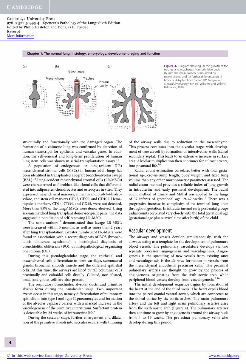
Airway and airspace developmentThe lung first appears as a diverticulum or bud from theventral wall of the foregut 22 to 26 days after fertilization.This bud grows caudally, with the epithelial cells from theforegut endoderm invading the surrounding mesenchyme toform the trachea (Figure 3). During the fourth week of gesta-tion, the caudal end of the trachea divides into two bronchialbuds, each proceeding to form the right and left main bronchi.By 32 to 35 days, the lobar bronchial buds form and up to 10days later, the segmental and subsegmental bronchial budsdevelop.1 Further dichotomous branching continues and by14 weeks 70% of the total airway is formed.9 At the end of the
pseudoglandular stage (17 weeks), the development of con-ducting airways up to the terminal bronchioles is complete.10
Human lung contains undifferentiated lung stem cells,nested in niches in the distal airways. These cells are self-renewing, clonogenic, and multipotent in vitro. After injectioninto damaged mouse lung in vivo, human lung stem cells formhuman bronchioles, alveoli and pulmonary vessels integrated
Table 1 Stages of lung growth
Stage Time Main events
Embryonic 0–7 weeks Formation of trachea, rightand left main bronchi,segmental bronchi, andvasculogenesis aroundairway buds
Pseudoglandular 7–17 weeks Differentiation of epithelialcells, formation ofconduction airways andterminal bronchioles,formation of pulmonaryarteries and veins
Canalicular 17–27 weeks Formation of respiratorybronchioles, alveolar ductsand primitive alveoli,differentiation of type Iand type II pneumocytesand formation of alveolarcapillary barrier
Saccular 27–36 weeks Increment in gas exchangeareas, further differentiationof type I and type II cells
Alveolar 36 weeks –2 yearsUp to 18–22years
Septation andmultiplication of alveoliEnlargement of terminalbronchioles and alveoli
Reprinted from Joshi S, Kotecha S. Lung growth and development. Early HumDev 2007;83:789–794. With permission from Elsevier.
Spencer’s Pathology of the Lung, Sixth Edition, ed. Philip Hasleton and Douglas B. Flieder. Published by Cambridge University Press.© Cambridge University Press 2013.
1
www.cambridge.org© in this web service Cambridge University Press
Cambridge University Press978-0-521-50995-4 - Spencer’s Pathology of the Lung: Sixth EditionEdited by Philip Hasleton and Douglas B. FliederExcerptMore information
(a) (b)
(c) (d)
(e) (f)
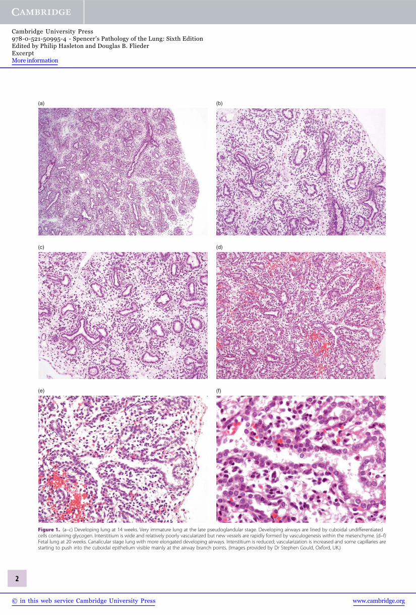
Figure 1. (a–c) Developing lung at 14 weeks. Very immature lung at the late pseudoglandular stage. Developing airways are lined by cuboidal undifferentiatedcells containing glycogen. Interstitium is wide and relatively poorly vascularized but new vessels are rapidly formed by vasculogenesis within the mesenchyme. (d–f)Fetal lung at 20 weeks. Canalicular stage lung with more elongated developing airways. Interstitium is reduced; vascularization is increased and some capillaries arestarting to push into the cuboidal epithelium visible mainly at the airway branch points. (Images provided by Dr Stephen Gould, Oxford, UK.)
2
www.cambridge.org© in this web service Cambridge University Press
Cambridge University Press978-0-521-50995-4 - Spencer’s Pathology of the Lung: Sixth EditionEdited by Philip Hasleton and Douglas B. FliederExcerptMore information
(a) (b)
(c) (d)
(e) (f)
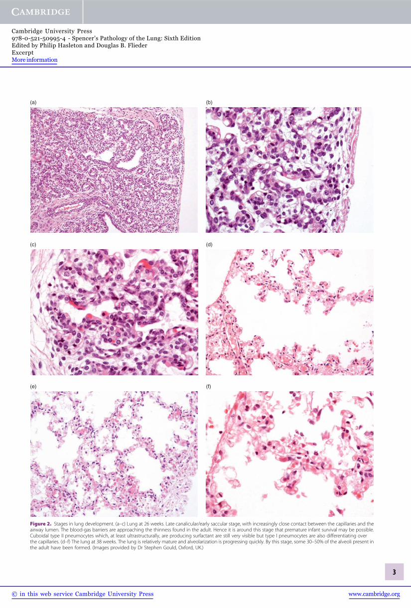
Figure 2. Stages in lung development. (a–c) Lung at 26 weeks. Late canalicular/early saccular stage, with increasingly close contact between the capillaries and theairway lumen. The blood-gas barriers are approaching the thinness found in the adult. Hence it is around this stage that premature infant survival may be possible.Cuboidal type II pneumocytes which, at least ultrastructurally, are producing surfactant are still very visible but type I pneumocytes are also differentiating overthe capillaries. (d–f) The lung at 38 weeks. The lung is relatively mature and alveolarization is progressing quickly. By this stage, some 30–50% of the alveoli present inthe adult have been formed. (Images provided by Dr Stephen Gould, Oxford, UK.)
3
www.cambridge.org© in this web service Cambridge University Press
Cambridge University Press978-0-521-50995-4 - Spencer’s Pathology of the Lung: Sixth EditionEdited by Philip Hasleton and Douglas B. FliederExcerptMore information
structurally and functionally with the damaged organ. Theformation of a chimeric lung was confirmed by detection ofhuman transcripts for epithelial and vascular genes. In addi-tion, the self-renewal and long-term proliferation of humanlung stem cells was shown in serial-transplantation assays.11
A population of endogenous or lung-resident (LR)mesenchymal stromal cells (MSCs) in human adult lungs hasbeen identified in transplanted allograft bronchoalveolar lavage(BAL).12 Lung-resident mesenchymal stromal cells (LR-MSCs)were characterized as fibroblast-like clonal cells that differenti-ated into adipocytes, chondrocytes and osteocytes in vitro. Theyexpressed mesenchymal markers, vimentin and prolyl-4-hydro-xylase, and stem cell markers CD73, CD90, and CD105. Hema-topoietic markers, CD14, CD34, and CD45, were not detected.More than 95% of the lungs’ MSCs were donor-derived. Usingsex-mismatched lung transplant donor-recipient pairs, the datasuggested a population of self-renewing LR-MSCs.
The same authors13 demonstrated that lavage LR-MSCswere increased within 3 months, as well as more than 2 yearsafter lung transplantation. Greater numbers of LR-MSCs werefound in association with a recent diagnosis of BOS (bronch-iolitis obliterans syndrome), a histological diagnosis ofbronchiolitis obliterans (BO), or histopathological organizingpneumonia (OP).
During this pseudoglandular stage, the epithelial andmesenchymal cells differentiate to form cartilage, submucosalglands, bronchial smooth muscle and the different epithelialcells. At this time, the airways are lined by tall columnar cellsproximally and cuboidal cells distally. Ciliated, non-ciliated,basal, and goblet cells are also present.
The respiratory bronchioles, alveolar ducts, and primitivealveoli form during the canalicular stage. Two importantevents occur in this stage, namely differentiation of pulmonaryepithelium into type I and type II pneumocytes and formationof the alveolar capillary barrier with a marked increase in thevasculogenesis of the primitive interstitium. Surfactant proteinis detectable by 24 weeks of intrauterine life.4
During the saccular stage, further enlargement and dilata-tion of the primitive alveoli into saccules occurs, with thinning
of the airway walls due to reduction in the mesenchyme.This process continues into the alveolar stage, with develop-ment of true alveoli by formation of interalveolar walls (calledsecondary septa). This leads to an extensive increase in surfacearea. Alveolar multiplication then continues for at least 2 years,into postnatal life.14
Radial count estimation correlates better with total gesta-tional age, crown-rump length, body weight, and fixed lungvolume than any other morphometric parameter assessed. Theradial count method provides a reliable index of lung growthin intrauterine and early postnatal development. The radialcount method of Emery and Mithal was applied to the lungsof 37 infants of gestational age 19–42 weeks.15 There was aprogressive increase in complexity of the terminal lung unitsthroughout gestation. In intrauterine and early post-natal groupsradial counts correlated very closely with the total gestational age(gestational age plus survival time after birth) of the child.
Vascular developmentThe airways and vessels develop simultaneously, with theairways acting as a template for the development of pulmonaryblood vessels. The pulmonary vasculature develops via twoseparate processes, angiogenesis and vasculogenesis. Angio-genesis is the sprouting of new vessels from existing onesand vasculogenesis is the de novo formation of vessels fromthe mesenchymal endothelial precursor cells.3 The proximalpulmonary arteries are thought to grow by the process ofangiogenesis, originating from the sixth aortic arch, whileperipheral blood vessels develop from vasculogenesis.5,16
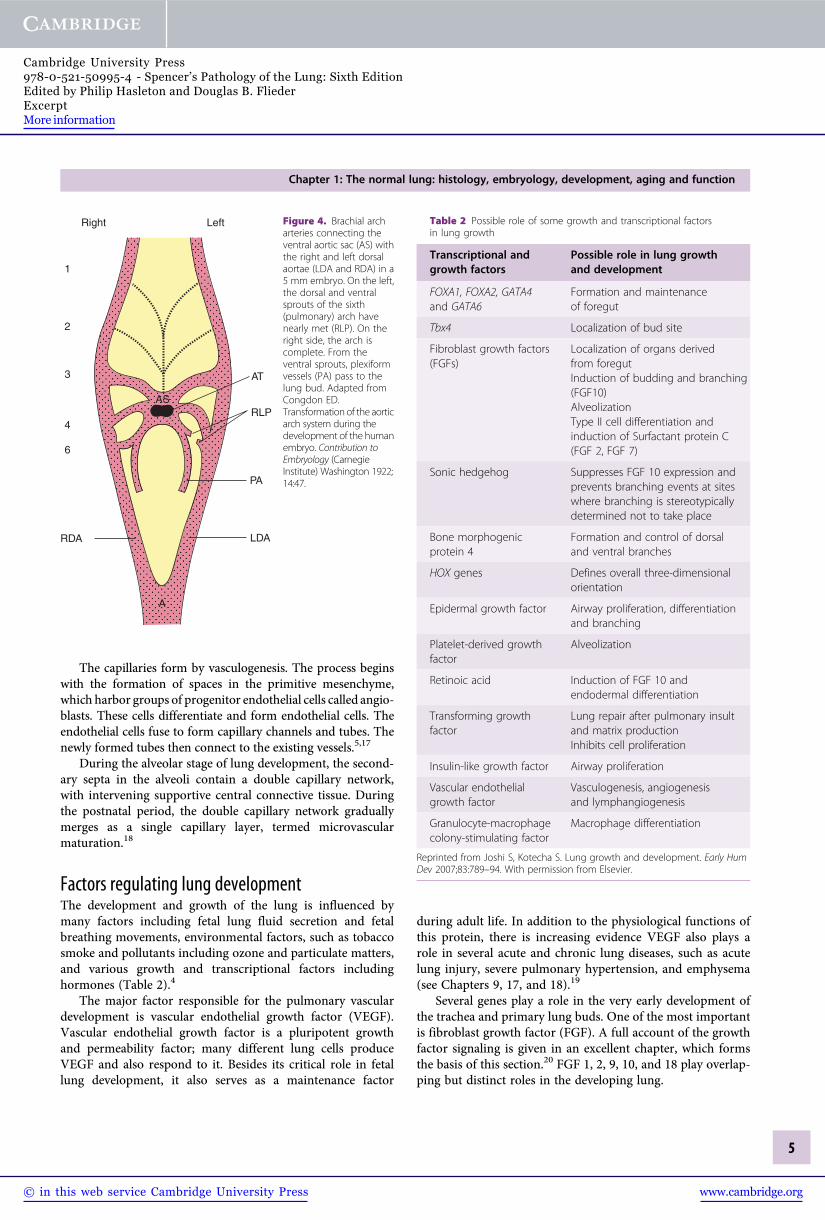
The initial development sequence begins by formation ofthe heart at the end of the third week. The heart expels bloodinto the paired cranial ventral aortae, which are connected tothe dorsal aortae by six aortic arches. The main pulmonaryartery and the left and right main pulmonary arteries arisefrom the sixth aortic arch (Figure 4). The pulmonary arteriesthen continue to grow by angiogenesis around the airway budsfrom 4 to 16 weeks. The pre-acinar pulmonary veins alsodevelop during this period.
Esophagus
Mesenchyme
(a) (b) (c)
TracheaPleuralcavity
Figure 3. Diagram showing (a) the growth of thetrachea and esophagus from primitive buds,(b) into the main bronchi surrounded bymesenchyme and (c) further differentiation ofbronchi. Adapted from Sadler TW. Langman’sMedical Embryology, 6th ed. Williams and Wilkins,Baltimore, 1990.
Chapter 1: The normal lung: histology, embryology, development, aging and function
4
www.cambridge.org© in this web service Cambridge University Press
Cambridge University Press978-0-521-50995-4 - Spencer’s Pathology of the Lung: Sixth EditionEdited by Philip Hasleton and Douglas B. FliederExcerptMore information
The capillaries form by vasculogenesis. The process beginswith the formation of spaces in the primitive mesenchyme,which harbor groups of progenitor endothelial cells called angio-blasts. These cells differentiate and form endothelial cells. Theendothelial cells fuse to form capillary channels and tubes. Thenewly formed tubes then connect to the existing vessels.5,17
During the alveolar stage of lung development, the second-ary septa in the alveoli contain a double capillary network,with intervening supportive central connective tissue. Duringthe postnatal period, the double capillary network graduallymerges as a single capillary layer, termed microvascularmaturation.18
Factors regulating lung developmentThe development and growth of the lung is influenced bymany factors including fetal lung fluid secretion and fetalbreathing movements, environmental factors, such as tobaccosmoke and pollutants including ozone and particulate matters,and various growth and transcriptional factors includinghormones (Table 2).4
The major factor responsible for the pulmonary vasculardevelopment is vascular endothelial growth factor (VEGF).Vascular endothelial growth factor is a pluripotent growthand permeability factor; many different lung cells produceVEGF and also respond to it. Besides its critical role in fetallung development, it also serves as a maintenance factor
during adult life. In addition to the physiological functions ofthis protein, there is increasing evidence VEGF also plays arole in several acute and chronic lung diseases, such as acutelung injury, severe pulmonary hypertension, and emphysema(see Chapters 9, 17, and 18).19
Several genes play a role in the very early development ofthe trachea and primary lung buds. One of the most importantis fibroblast growth factor (FGF). A full account of the growthfactor signaling is given in an excellent chapter, which formsthe basis of this section.20 FGF 1, 2, 9, 10, and 18 play overlap-ping but distinct roles in the developing lung.
Right
1
2
3
4
6
RDA LDA
PA
RLP
AT
AS
A
Left Figure 4. Brachial archarteries connecting theventral aortic sac (AS) withthe right and left dorsalaortae (LDA and RDA) in a5 mm embryo. On the left,the dorsal and ventralsprouts of the sixth(pulmonary) arch havenearly met (RLP). On theright side, the arch iscomplete. From theventral sprouts, plexiformvessels (PA) pass to thelung bud. Adapted fromCongdon ED.Transformation of the aorticarch system during thedevelopment of the humanembryo. Contribution toEmbryology (CarnegieInstitute) Washington 1922;14:47.
Table 2 Possible role of some growth and transcriptional factorsin lung growth
Transcriptional andgrowth factors
Possible role in lung growthand development
FOXA1, FOXA2, GATA4and GATA6
Formation and maintenanceof foregut
Tbx4 Localization of bud site
Fibroblast growth factors(FGFs)
Localization of organs derivedfrom foregutInduction of budding and branching(FGF10)AlveolizationType II cell differentiation andinduction of Surfactant protein C(FGF 2, FGF 7)
Sonic hedgehog Suppresses FGF 10 expression andprevents branching events at siteswhere branching is stereotypicallydetermined not to take place
Bone morphogenicprotein 4
Formation and control of dorsaland ventral branches
HOX genes Defines overall three-dimensionalorientation
Epidermal growth factor Airway proliferation, differentiationand branching
Platelet-derived growthfactor
Alveolization
Retinoic acid Induction of FGF 10 andendodermal differentiation
Transforming growthfactor
Lung repair after pulmonary insultand matrix productionInhibits cell proliferation
Insulin-like growth factor Airway proliferation
Vascular endothelialgrowth factor
Vasculogenesis, angiogenesisand lymphangiogenesis
Granulocyte-macrophagecolony-stimulating factor
Macrophage differentiation
Reprinted from Joshi S, Kotecha S. Lung growth and development. Early HumDev 2007;83:789–94. With permission from Elsevier.
Chapter 1: The normal lung: histology, embryology, development, aging and function
5
www.cambridge.org© in this web service Cambridge University Press
Cambridge University Press978-0-521-50995-4 - Spencer’s Pathology of the Lung: Sixth EditionEdited by Philip Hasleton and Douglas B. FliederExcerptMore information
FGF 10 has been linked with mesenchymal-epithelial inter-action, especially during branching. It is expressed focally inthe distal mesenchyme, adjacent to stereotypically determinedbranching sites. It is hypothesized that FGF 10 governs thedirectional outgrowth of lung buds during branching by trig-gering chemotaxis and proliferation of the adjacent epithe-lium. This causes unidirectional growth of the primary lungbuds. The chemotactic response of the lung endoderm involvesthe coordinated movement of the entire epithelial tip, with allits cells, towards an FGF 10 source.
An equally important factor in determining the specificityof the FGF signaling response may be the presence or absenceof key downstream intermediate genes. One such example istyrosine protein phosphatase Shp2, present in embryonic lungbranch tips. This gene is essential for FGF transduction.
In addition FGF 10 controls the differentiation of epithe-lium by inducing surfactant protein C (SP-C) expression andupregulating expression of bone morphogenetic protein 4(BMP4), a regulator of lung epithelial differentiation.
Transforming growth factor-p signaling, mediated bytransforming growth factor-p type II receptor, plays distinctroles in developing mouse lung epithelium. The integratedfunctions of this receptor are very important in embryoniclung branching morphogenesis and development of alveoliin the post-natal lung. The developmental immaturity oflung structure and function, resulting from loss-of-functionmutations in transforming growth factor-b signaling pathwaycomponents, may contribute to early post-natal respiratoryproblems, such as bronchopulmonary dysplasia (see Chapter 3).It may also increase the susceptibility to respiratory diseaseslater in life, including emphysema.21 Overexpression of TGFb1in the developing fetal monkey lung causes severe progressivepulmonary and pleural fibrosis, as well as pulmonary hypo-plasia.22 TGFb1 overexpression triggered mesenchymal cellproliferation that appeared to continue after the overexpressionof exogenous TGFb1 was discontinued.
Recessive mutations in latent transforming growthfactor-b binding protein 4 (LTBP4) gene leading to disruptedpulmonary, gastrointestinal, urinary, musculoskeletal, cranio-facial, and dermal development have been described.23 Patientshave severe respiratory distress, with cystic and atelectaticchanges in the lungs, complicated by tracheomalacia and dia-phragmatic hernia. Respiratory failure is the usual cause ofdeath. Impaired synthesis and deficient deposition of LTBP4into the extracellular matrix appears responsible for defectiveelastic fiber assembly in all tissues affected by the disease.
Abrogation of TGF-p type II receptor (TpRII) in mouselung epithelium causes retardation of post-natal lung alveolar-ization, with markedly decreased numbers of type I alveolarepithelial cells. No abnormalities in prenatal lung developmentare observed. In contrast, blockade of TpRII in mesoderm-derived tissues, including lung mesenchyme, results in mildlyabnormal lung branching and reduced cell proliferation aftermid-gestation. This is accompanied by multiple defects inother organs, including diaphragmatic hernia.
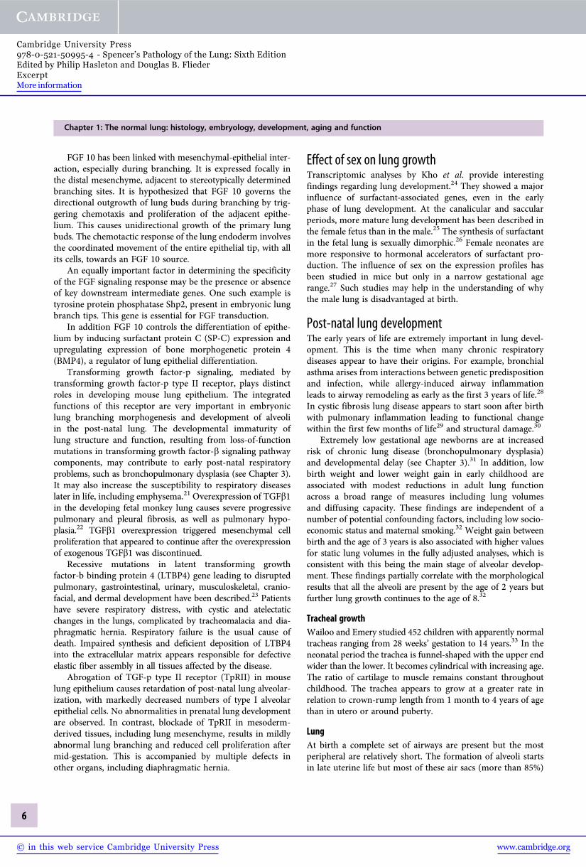
Effect of sex on lung growthTranscriptomic analyses by Kho et al. provide interestingfindings regarding lung development.24 They showed a majorinfluence of surfactant-associated genes, even in the earlyphase of lung development. At the canalicular and saccularperiods, more mature lung development has been described inthe female fetus than in the male.25 The synthesis of surfactantin the fetal lung is sexually dimorphic.26 Female neonates aremore responsive to hormonal accelerators of surfactant pro-duction. The influence of sex on the expression profiles hasbeen studied in mice but only in a narrow gestational agerange.27 Such studies may help in the understanding of whythe male lung is disadvantaged at birth.
Post-natal lung developmentThe early years of life are extremely important in lung devel-opment. This is the time when many chronic respiratorydiseases appear to have their origins. For example, bronchialasthma arises from interactions between genetic predispositionand infection, while allergy-induced airway inflammationleads to airway remodeling as early as the first 3 years of life.28
In cystic fibrosis lung disease appears to start soon after birthwith pulmonary inflammation leading to functional changewithin the first few months of life29 and structural damage.30
Extremely low gestational age newborns are at increasedrisk of chronic lung disease (bronchopulmonary dysplasia)and developmental delay (see Chapter 3).31 In addition, lowbirth weight and lower weight gain in early childhood areassociated with modest reductions in adult lung functionacross a broad range of measures including lung volumesand diffusing capacity. These findings are independent of anumber of potential confounding factors, including low socio-economic status and maternal smoking.32 Weight gain betweenbirth and the age of 3 years is also associated with higher valuesfor static lung volumes in the fully adjusted analyses, which isconsistent with this being the main stage of alveolar develop-ment. These findings partially correlate with the morphologicalresults that all the alveoli are present by the age of 2 years butfurther lung growth continues to the age of 8.32
Tracheal growthWailoo and Emery studied 452 children with apparently normaltracheas ranging from 28 weeks’ gestation to 14 years.33 In theneonatal period the trachea is funnel-shaped with the upper endwider than the lower. It becomes cylindrical with increasing age.The ratio of cartilage to muscle remains constant throughoutchildhood. The trachea appears to grow at a greater rate inrelation to crown-rump length from 1 month to 4 years of agethan in utero or around puberty.
LungAt birth a complete set of airways are present but the mostperipheral are relatively short. The formation of alveoli startsin late uterine life but most of these air sacs (more than 85%)
Chapter 1: The normal lung: histology, embryology, development, aging and function
6
www.cambridge.org© in this web service Cambridge University Press
Cambridge University Press978-0-521-50995-4 - Spencer’s Pathology of the Lung: Sixth EditionEdited by Philip Hasleton and Douglas B. FliederExcerptMore information
are formed after birth.34 The alveoli are formed from tissueridges on the existing primary septa. This produces a largenumber of small buds along the primary septa. Myofibroblasts,elastic fibers and collagen fibrils are the probable driving forcesfor septation. Inside the pre-existing septa platelet-derivedgrowth factor (PDGF) receptor-positive smooth muscle pre-cursors proliferate and move to locations where the new septaare to be formed. Alveolarization does not occur in PDGF-A-deficient mice.6
Alveolarization of the acinus primarily occurs between birthand 2 years; significant but slower growth is seen up to 8 years.
The ratio of pulmonary diffusion to alveolar volumeremains constant in the first 2 years of life. This is consistentwith lung growth in this age group occurring because of anincrease in the number of alveoli rather than an expansionof the same number of alveoli.35 These findings are in keepingwith morphometric data on number of alveoli per unit volumeand mean linear intercept in the post-natal period.36 After thistime, values plateau, suggesting alveolarization is complete.Radial counts correlate well with the chronological age of thechild.37
Pulmonary diffusing capacity and alveolar volume, meas-ured by DLCO and alveolar volume, increase with age andsomatic size among infants and toddlers. Sex differences areprimarily related to somatic size. There are no sex differenceswhen pulmonary diffusing capacity is related to alveolarvolume. However, in a morphometric study, individual lungunits, alveolar dimensions, and number of alveoli per unit areaand volume did not differ between boys and girls, but boys hadbigger lungs than girls for the same stature. This resulted in alarger total number of alveoli and a larger alveolar surface areain boys than in girls for a given age and stature. Boys may havemore respiratory units than girls.36
Stem/progenitor cells in the lungThis is a “hot” area for research, as stem cells offer new toolsfor the investigation of pathogenetic and developmental path-ways. It is likely that many stem cell populations exist in thelung with distinct lineage potential. The ability to purify andfunctionally assay these populations requires consistent use ofwell-defined protocols for isolation, culturing, and functionalassays.38 This area is still in its infancy and, while continuously“offering” new possible treatments, has yet to provide a proventherapeutic return.
An unanswered question is whether adult lung epithelialstem cells are a distinct population or whether some multi-potential embryonic progenitors persist into adult life. Evi-dence suggests that in liver and pancreas, the embryonic cellsthat build tissue are different from those that repair andmaintain it.39 In the lung evidence suggests lung embryonicprogenitors and adult stem cells are separate, although lineage-linked, populations.40
The pools of epithelial stem/progenitor cells are widelydistributed over the alveolar surface.41 They are located in
the basal layer of the upper airways, within or near neuro-endocrine cell rests, at the bronchoalveolar junctions, as well aswithin the alveolar epithelial surface.42 The most important ofthese are the alveolar epithelial cells, which have a large surfacearea. Either alveolar epithelial cells contain a subpopulation ofprogenitor cells or most alveolar epithelial cells can reactivateinto a progenitor-like state in response to injury.
Another subset of Club (Clara) cells has been identified,based on their location at the bronchiolar-alveolar junction.They co-express secretoglobin 1a1 (Scgb1a1, also known asCC10 or CCSP), and an alveolar type II cell marker surfactantprotein C (SftpC or SpC). These cells proliferate following lunginjury. Based on their in vitro differentiation potential, ithas been proposed that they are bronchioalveolar stem cells(BASCs) that give rise to both bronchiolar and alveolar cellsin vivo.43
Normal organizationAirwaysFor convenience of anatomical description, the airways aredivided into the upper and lower respiratory systems. Theupper respiratory system comprises the nasal cavity, paranasalsinuses, and pharynx, while the larynx, trachea, bronchi, andbronchioles are the lower respiratory tract. The nose andparanasal sinuses act as a first line of defense against bacteriaand inhaled particles, through the filtering function of thenasal hairs, as well as the irregular structure of the nasal bonesand mucosa. In addition these structures warm and humidifythe inspired air. Thus in a patient with a tracheostomy, thisfirst line of defense is bypassed. The mechanics of the upperairways, important in obstructive and sleep apnea, Cheyne-Stokes respiration, and the obesity hypoventilation syndrome,are often neglected by the histopathologist. An excellent reviewarticle is available.44
The trachea divides into the right and left primary extra-pulmonary or mainstem bronchi. Each of these then gives riseto secondary (lobar) bronchi, which supply the lung lobes.These further ramify into tertiary (segmental) bronchi, whichsupply the segments of each lobe. The bronchi branch progres-sively into bronchioles. The smallest are terminal bronchiolesthat constitute the most distal component of the conductingpart of the airways. The terminal bronchioles give rise to theacinus, which is the part of the respiratory system involved ingas exchange and comprises respiratory bronchioles, alveolarducts, alveolar sacs, and the alveoli.
The tracheaThe trachea begins anterior to the sixth cervical vertebral body,where it is attached to the inferior portion of the cricoidcartilage of the larynx. It ends in the mediastinum at the levelof the fifth thoracic vertebral body, where it branches toform the right and left primary bronchi. The total length anddiameter are approximately 11 cm and 2.5 cm, respectively.The trachea is formed of 15–20 C-shaped cartilage rings, the
Chapter 1: The normal lung: histology, embryology, development, aging and function
7
www.cambridge.org© in this web service Cambridge University Press
Cambridge University Press978-0-521-50995-4 - Spencer’s Pathology of the Lung: Sixth EditionEdited by Philip Hasleton and Douglas B. FliederExcerptMore information

open sides facing posteriorly, where the trachealis musclecompletes the circumference. These rings protect the tracheafrom frontal injury and also prevent collapse during the nega-tive intrathoracic pressure associated with respiration.
The primary bronchiThe trachea branches into the right and left primary bronchi,which are separated by an internal ridge called the carina. Thestructure of these primary bronchi is similar to that of thetrachea, being formed of C-shaped cartilage rings. The rightprimary bronchus is larger in diameter than the left. It closelyfollows the general direction of the trachea, whereas the leftdiverges at a greater angle, especially in females. For years itwas thought that such anatomy resulted in greater right lungaspiration but recent studies call this into question.45
Each primary bronchus enters the corresponding lung atthe pulmonary hilum or root, a groove along the medialsurface of each lung, which also provides entry to pulmonaryarteries and veins, nerves and lymphatics. All these structuresat the root of the lung are surrounded by connective tissue.The relationship of the pulmonary artery, mainstem bronchus,and pulmonary veins is well defined and constant.46
The lungsEach lung lies in its corresponding pleural cavity. The apex ofeach lung extends above the first rib, while the concave baserests on the superior surface of the diaphragm. In general, theright lung is wider than the left, due to the projection of theheart towards the left side. Conversely, the left lung is longerthan the right as the dome of the diaphragm is higher on theright side, because of the underlying liver.47
The right lung is heavier than the left, weighing approxi-mately 700 g in adult men and 500 g in adult women ascompared to 600 g for the left lung in men and 450 g inwomen.47 There is a wide variation in autopsy weights, in partdue to differences in the degree of terminal pulmonary edemaand congestion from one individual to another. In a study of theorgan weights in 684 adult Caucasian forensic autopsies,48 themean lung weight � standard deviation was 663 � 239 g forthe right lung in males, 583 � 216 g for the left lung in males,546 � 207 g for the right lung in females and 467 � 174 g forthe left lung in females. Such organ weight tables need regularupdating, as the normal values of organ weight change withtime, secondary to genetic and environmental factors.48 Lungweight tends to diminish slightly in the elderly, probably dueto alveolar enlargement (see Chapter 17).
The lungs are normally divided into lobes that are separ-ated by fissures. Interlobar fissures are deep depressions thatextend from the outer lung surface towards the center. Thevisceral pleura also dips into the fissure, making the lungsurfaces lying within the fissures smooth and thus allowingfree movement of individual lobes.47
The right lung is divided into upper, middle and lowerlobes, with the horizontal/minor fissure separating the upperand middle lobes. The oblique fissure separates the upper and
middle lobes from the lower lobe. The horizontal fissure isusually less well developed than the oblique. The left lung isdivided into upper and lower lobes, separated by the obliquefissure. A rudimentary structure, the lingula, is present onthe left and is considered the equivalent of the middle lobe.It is, however, part of the left upper lobe, rather than a separatelobe, and appears antero-inferiorly as a small tongue-likeprojection.
Variations in the anatomy of fissures are common andinclude accessory fissures, fissures in abnormal locations andincomplete or absent lobar fissures. Such structural anomalieshave no functional significance but may cause radiologicalconfusion. The prevalence of these abnormalities varies betweenstudies.49–52 A well-known anomaly is the azygous lobe, presentin 1% of normal individuals. This is caused by extrinsic com-pression of the lung by an aberrant azygous vein in the rightupper thorax, resulting in a depression (fissure) from the top tothe bottom of the right upper lobe (Figure 5). It does not reflectany underlying segmental division of the bronchi.
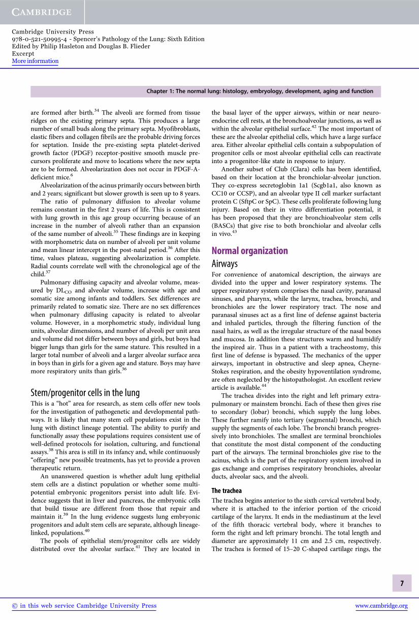
Within the lung, the primary bronchi divide to form sec-ondary (lobar) bronchi. One secondary bronchus goes to eachlobe, so the right lung has three secondary bronchi and the leftlung has two secondary bronchi. The right primary bronchusgives rise to the right upper lobe bronchus and continuesas the bronchus intermedius. It then divides into the rightmiddle lobe bronchus and right lower lobe bronchus. The leftprimary (main) bronchus is longer than the right and dividesinto the left upper lobe bronchus and left lower lobe bronchus.The secondary bronchi branch to form tertiary (segmental)bronchi (Figure 6). Each tertiary bronchus supplies air to asingle bronchopulmonary segment.
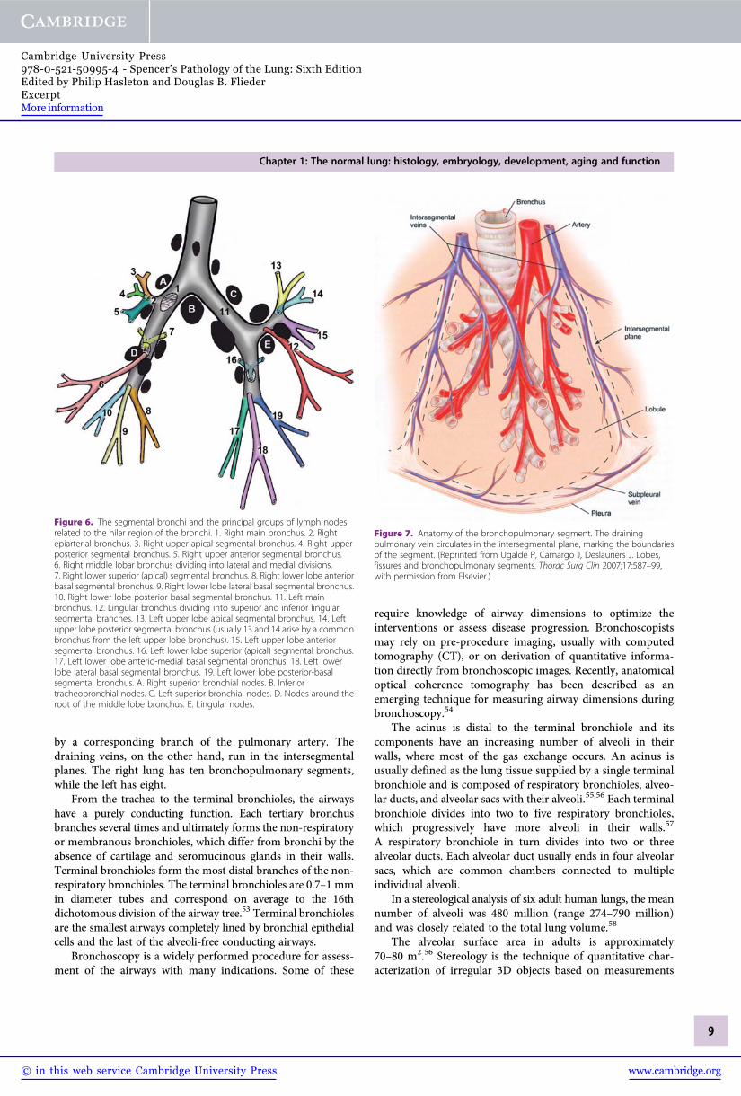
Bronchopulmonary segments are considered the anatomicunits of the lung, each possessing its own bronchus, andpulmonary arterial, venous and lymphatic systems. They areconstant in their topographic anatomy (Figures 7, 8 and 9).47
Each segment is surrounded by connective tissue septa, whichare continuous with the pleural surface. The segmental bron-chus traverses down the center of the segment, accompanied
Figure 5. Macroscopic image of the azygous lobe.
Chapter 1: The normal lung: histology, embryology, development, aging and function
8
www.cambridge.org© in this web service Cambridge University Press
Cambridge University Press978-0-521-50995-4 - Spencer’s Pathology of the Lung: Sixth EditionEdited by Philip Hasleton and Douglas B. FliederExcerptMore information
by a corresponding branch of the pulmonary artery. Thedraining veins, on the other hand, run in the intersegmentalplanes. The right lung has ten bronchopulmonary segments,while the left has eight.
From the trachea to the terminal bronchioles, the airwayshave a purely conducting function. Each tertiary bronchusbranches several times and ultimately forms the non-respiratoryor membranous bronchioles, which differ from bronchi by theabsence of cartilage and seromucinous glands in their walls.Terminal bronchioles form the most distal branches of the non-respiratory bronchioles. The terminal bronchioles are 0.7–1 mmin diameter tubes and correspond on average to the 16thdichotomous division of the airway tree.53 Terminal bronchiolesare the smallest airways completely lined by bronchial epithelialcells and the last of the alveoli-free conducting airways.
Bronchoscopy is a widely performed procedure for assess-ment of the airways with many indications. Some of these
require knowledge of airway dimensions to optimize theinterventions or assess disease progression. Bronchoscopistsmay rely on pre-procedure imaging, usually with computedtomography (CT), or on derivation of quantitative informa-tion directly from bronchoscopic images. Recently, anatomicaloptical coherence tomography has been described as anemerging technique for measuring airway dimensions duringbronchoscopy.54
The acinus is distal to the terminal bronchiole and itscomponents have an increasing number of alveoli in theirwalls, where most of the gas exchange occurs. An acinus isusually defined as the lung tissue supplied by a single terminalbronchiole and is composed of respiratory bronchioles, alveo-lar ducts, and alveolar sacs with their alveoli.55,56 Each terminalbronchiole divides into two to five respiratory bronchioles,which progressively have more alveoli in their walls.57
A respiratory bronchiole in turn divides into two or threealveolar ducts. Each alveolar duct usually ends in four alveolarsacs, which are common chambers connected to multipleindividual alveoli.
In a stereological analysis of six adult human lungs, the meannumber of alveoli was 480 million (range 274–790 million)and was closely related to the total lung volume.58
The alveolar surface area in adults is approximately70–80 m2.56 Stereology is the technique of quantitative char-acterization of irregular 3D objects based on measurements
Figure 6. The segmental bronchi and the principal groups of lymph nodesrelated to the hilar region of the bronchi. 1. Right main bronchus. 2. Rightepiarterial bronchus. 3. Right upper apical segmental bronchus. 4. Right upperposterior segmental bronchus. 5. Right upper anterior segmental bronchus.6. Right middle lobar bronchus dividing into lateral and medial divisions.7. Right lower superior (apical) segmental bronchus. 8. Right lower lobe anteriorbasal segmental bronchus. 9. Right lower lobe lateral basal segmental bronchus.10. Right lower lobe posterior basal segmental bronchus. 11. Left mainbronchus. 12. Lingular bronchus dividing into superior and inferior lingularsegmental branches. 13. Left upper lobe apical segmental bronchus. 14. Leftupper lobe posterior segmental bronchus (usually 13 and 14 arise by a commonbronchus from the left upper lobe bronchus). 15. Left upper lobe anteriorsegmental bronchus. 16. Left lower lobe superior (apical) segmental bronchus.17. Left lower lobe anterio-medial basal segmental bronchus. 18. Left lowerlobe lateral basal segmental bronchus. 19. Left lower lobe posterior-basalsegmental bronchus. A. Right superior bronchial nodes. B. Inferiortracheobronchial nodes. C. Left superior bronchial nodes. D. Nodes around theroot of the middle lobe bronchus. E. Lingular nodes.
Figure 7. Anatomy of the bronchopulmonary segment. The drainingpulmonary vein circulates in the intersegmental plane, marking the boundariesof the segment. (Reprinted from Ugalde P, Camargo J, Deslauriers J. Lobes,fissures and bronchopulmonary segments. Thorac Surg Clin 2007;17:587–99,with permission from Elsevier.)
Chapter 1: The normal lung: histology, embryology, development, aging and function
9
www.cambridge.org© in this web service Cambridge University Press
Cambridge University Press978-0-521-50995-4 - Spencer’s Pathology of the Lung: Sixth EditionEdited by Philip Hasleton and Douglas B. FliederExcerptMore information
made on 2D sections. This technique acts as a bridge betweenthe understanding of lung structure and function in variousstudies on the lung.59,60
The connective tissues from the lung hila extend progres-sively into the lung parenchyma in the form of fibrous parti-tions or trabeculae. The smallest unit separated by thetrabeculae is called the pulmonary lobule or the secondarylobule of Miller (Figure 10).61 Each pulmonary lobule consistsof three to five acini.62 The trabeculae of the subpleural lobules
(at the periphery of the lung) are continuous with the connect-ive tissue of the visceral pleura.
Blood supplyThe lungs have a dual circulation. The low-pressure, high-volume pulmonary system carries deoxygenated blood fromthe right side of the heart to the lungs for gas exchangewith the inspired air in the alveolar spaces. The high-pressure,
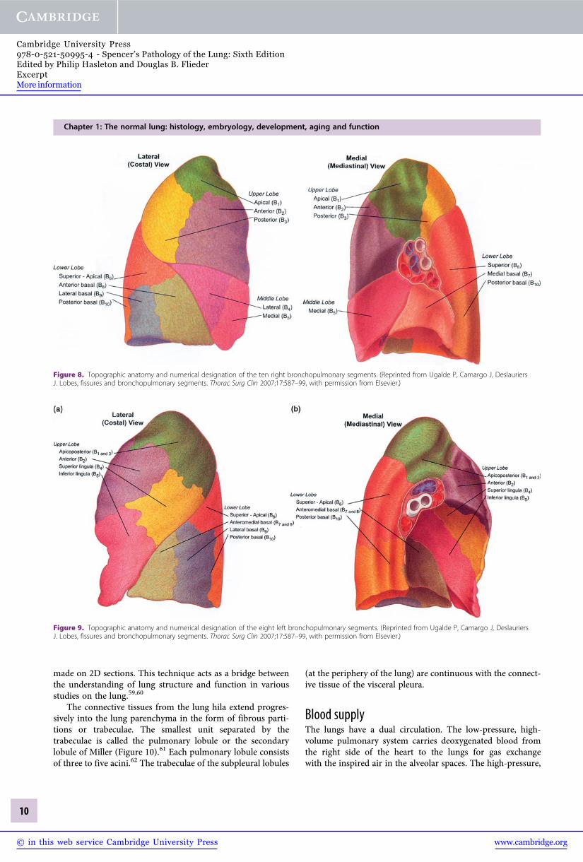
Figure 8. Topographic anatomy and numerical designation of the ten right bronchopulmonary segments. (Reprinted from Ugalde P, Camargo J, DeslauriersJ. Lobes, fissures and bronchopulmonary segments. Thorac Surg Clin 2007;17:587–99, with permission from Elsevier.)
Figure 9. Topographic anatomy and numerical designation of the eight left bronchopulmonary segments. (Reprinted from Ugalde P, Camargo J, DeslauriersJ. Lobes, fissures and bronchopulmonary segments. Thorac Surg Clin 2007;17:587–99, with permission from Elsevier.)
Chapter 1: The normal lung: histology, embryology, development, aging and function
10
www.cambridge.org© in this web service Cambridge University Press
Cambridge University Press978-0-521-50995-4 - Spencer’s Pathology of the Lung: Sixth EditionEdited by Philip Hasleton and Douglas B. FliederExcerptMore information