spermiogenesis particularities of a sperm storage species
TRANSCRIPT
Spermiogenesis particularities of a sperm storage species: Helicolenus dactylopterus (Teleostei:
Scorpaenidae)
SILVIA VILA, MARIA SÀBAT, MARTA MUÑOZ and MARGARIDA CASADEVALL
Departament de Ciències Ambientals, Universitat de Girona, Campus Montilivi, 17071 Girona, Spain. E-mail: [email protected]
SUMMARY: A study of the spermiogenesis and spermatozoa of Helicolenus dactylopterus was conducted. Females of this species have the capacity to store sperm within their ovaries, and male gametes have a considerable cytoplasmic mass sur-rounding their heads to survive the long period of intraovarian sperm storage. Our observations show that early spermatids are round-shaped cells and have a spherical nucleus with diffuse chromatin. The nuclear volume decreases as a result of progressive chromatin condensation during spermiogenesis, causing the nucleus to take on a U-shape. Flagellar insertion is not central to the nucleus but consistently occurs at an oblique angle towards one side of it. The flagellum is inserted into the nuclear fossa, without subsequent nuclear rotation. In mature spermatozoa, the flagellum is adjacent to the nucleus. A comparison of the spermatozoa in the testicular lobules and those in the intraovarian storage structures suggests that the in-crease in volume of the cytoplasmic mass may occur in the posterior region of the testis, in the testicular duct. Spermatozoa enter the ovary in groups that reach the ovarian lumen and are surrounded by the ovarian epithelium for storage in sperm storage crypts.
Keywords: Helicolenus dactylopterus, spermiogenesis, ultrastructure, sperm storage, spermatozoa.
RESUMEN: Particularidades en la espermiogénesis de una especie que almacena esperma: Helicolenus dac-tylopterus dactylopterus (Teleostei: Scorpaenidae). – Se estudian la espermatogénesis y el espermatozoide de la especie Helicolenus dactylopterus. Las hembras de esta especie tienen la capacidad de almacenar espermatozoides en el interior de sus ovarios y los gametos masculinos presentan una evidente masa citoplasmática rodeando sus cabezas para soportar el largo período de almacenaje intraovárico. Nuestras observaciones evidencian que las espermátidas tempranas son células redondas con un núcleo esférico que contiene cromatina difusa. El volumen nuclear disminuye debido a una progresiva condensación de la cromatina, provocando que el núcleo adopte forma de “U”. La inserción flagelar no es central respecto al núcleo sino que se realiza a un lado de éste, siempre de forma oblicua. El flagelo se inserta en la fosa nuclear y no existe rotación nuclear posterior. En los espermatozoides maduros, el flagelo es una estructura adyacente al núcleo. El aumento de volumen de las masas citoplasmáticas posiblemente ocurre en la región posterior del testículo, comparando los espermato-zoides del interior del lóbulo testicular con los que están dentro las estructuras de almacenaje intraováricas. Éstos entran en el ovario en grupos más o menos organizados y son envueltos por el epitelio ovárico de la zona lamelar para ser guardados en criptas de almacenaje de esperma.
Palabras clave: Helicolenus dactylopterus, espermiogenesis, ultra estructura, almacenaje esperma, espermatozoide.
Scientia Marina 74(4)December 2010, 687-704, Barcelona (Spain)
ISSN: 0214-8358doi: 10.3989/scimar.2010.74n4697
INTRODUCTION
The bluemouth, Helicolenus dactylopterus (Dela-Roche, 1809), is a benthic species which inhabits the seabed between 200 and 1000 m depth (Whitehead
et al., 1986). It is regularly caught in the Mediterra-nean Sea and Atlantic Ocean, the target of both semi-industrial and artisanal fishing. The information on bluemouth reproduction includes three studies from the Azores (Isidro, 1989; Estácio et al., 2001; Mendonça
698 • S. VILA et al.
SCI. MAR., 74(4), December 2010, 697-704. ISSN 0214-8358 doi: 10.3989/scimar.2010.74n4697
et al., 2006), several studies from the Atlantic (Kelly et al., 1999; Sequeira et al., 2003; White et al., 1998), and many studies from the Mediterranean Sea (Allain and Lorane, 2000; Muñoz et al., 1999; Muñoz et al., 2000; Muñoz et al., 2002a,b,c).
H. dactylopterus belongs to the family Scorpae-nidae, which is especially interesting from the repro-ductive point of view because scorpaenid modes of reproduction are extremely varied. The group contains families which lay individual pelagic eggs (Ano-plopomatidae, Congiopodidae, Hoplichthyidae and Triglidae) and others in which the spawn consists of sticky clusters of eggs (Agonidae, Cottidae, Cyclop-teridae and Hexagrammidae). As far as is known, most scorpaenids are basically oviparous species that produce pelagic egg masses enclosed in a gelatinous matrix. There are also viviparous genera such as Se-bastes and Sebastodes (Washington et al., 1984). In addition, there are some, such as Helicolenus, that have particularities in their patterns of reproduction which are considered typical of viviparous species. In the case of H. dactylopterus, sperm storage is intraovarian and can last for ten months, an extremely long period for a teleost (Muñoz et al., 1999). Spermatozoa are stored within cryptal structures situated at the base of the in-terlamellar spaces. Once oocytes are ripe, spermatozoa are released from the storage structure, where they are maintained in viable condition throughout the storage period, into the ovarian lumen, where fertilization oc-curs (Muñoz et al., 1999).
The species under study is zygoparous (Muñoz et al., 2002a), a term referring to an oviparous condition in which fertilized ova (i.e. zygotes) are retained within the female reproductive tract for short periods of time before being released into the marine environment (Wourms et al., 1988).
In previous studies conducted by our research team, we have analyzed this storage process ultrastructur-ally and histochemically (Muñoz et al., 2000; 2002b) and have observed that the bluemouth’s spermatozoa show a special particularity: while male germinal cells within testicular lobules show the typical morphology of internal fertilizing species (Muñoz et al., 2002a), when we observe them within the male spermatic ducts and within the ovaries, these spermatozoa have a cy-toplasmic mass surrounding their midpiece. This mass gradually decreases in volume as the spawning period approaches (Vila, 2004).
Given this peculiarity and the other special traits detected in relation to the reproduction of this species, in this study we aim to describe the transformation that male sexual cells undergo during spermiogenesis until spermatozoa are formed and released into the lumen of the testicular lobule. A further objective is the study of subsequent modifications in the testis related to the appearance of the cytoplasmic mass in the male gam-ete. This occurs in the spermatic ducts before sperm are introduced into the female’s body by the male’s urogenital papilla.
MATERIALS AND METHODS
Ten male specimens of Helicolenus dactylopterus were caught off the Costa Brava in the northwestern Mediterranean between March 2004 and February 2005, at a rate of approximately one per month, to study the fine structure of spermiogenesis. The sper-matic duct samples were obtained from seven speci-mens caught between December 2005 and February 2006. Several portions of recently killed specimens were fixed in a mixture of 2.5% glutaraldehyde and 2% paraformaldehyde in a 0.1 M cacodylate buffer. After fixation at 4ºC for 2 h, samples were washed several times with 0.1 M cacodylate buffer. Samples were then postfixed in 1% osmium tetroxide at 4ºC for 1 h in ca-codylate buffer. After washing, they were dehydrated through a graded alcohol series and finally embedded in Spurr’s mixture. Sections were examined in a Zeiss EM-910 transmission electron microscope, and images were analogically processed on Kodak 4489 electron microscopy film.
RESULTS
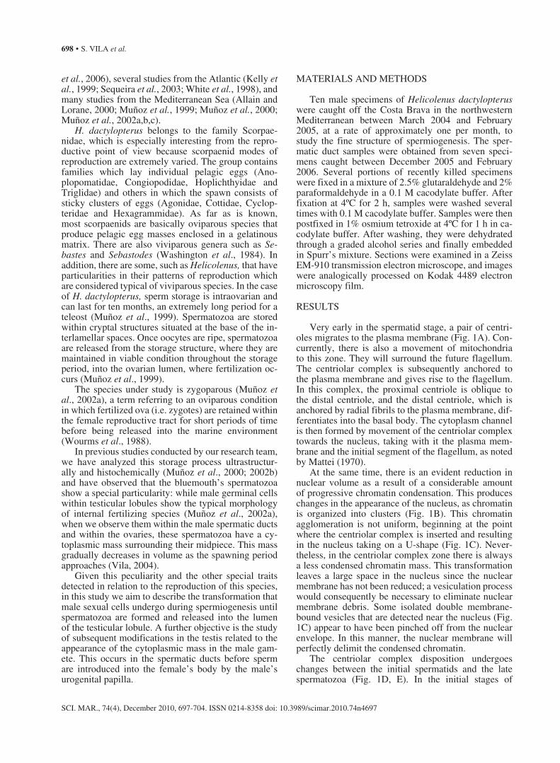
Very early in the spermatid stage, a pair of centri-oles migrates to the plasma membrane (Fig. 1A). Con-currently, there is also a movement of mitochondria to this zone. They will surround the future flagellum. The centriolar complex is subsequently anchored to the plasma membrane and gives rise to the flagellum. In this complex, the proximal centriole is oblique to the distal centriole, and the distal centriole, which is anchored by radial fibrils to the plasma membrane, dif-ferentiates into the basal body. The cytoplasm channel is then formed by movement of the centriolar complex towards the nucleus, taking with it the plasma mem-brane and the initial segment of the flagellum, as noted by Mattei (1970).
At the same time, there is an evident reduction in nuclear volume as a result of a considerable amount of progressive chromatin condensation. This produces changes in the appearance of the nucleus, as chromatin is organized into clusters (Fig. 1B). This chromatin agglomeration is not uniform, beginning at the point where the centriolar complex is inserted and resulting in the nucleus taking on a U-shape (Fig. 1C). Never-theless, in the centriolar complex zone there is always a less condensed chromatin mass. This transformation leaves a large space in the nucleus since the nuclear membrane has not been reduced; a vesiculation process would consequently be necessary to eliminate nuclear membrane debris. Some isolated double membrane-bound vesicles that are detected near the nucleus (Fig. 1C) appear to have been pinched off from the nuclear envelope. In this manner, the nuclear membrane will perfectly delimit the condensed chromatin.
The centriolar complex disposition undergoes changes between the initial spermatids and the late spermatozoa (Fig. 1D, E). In the initial stages of
SPERMIOGENESIS IN HELICOLENUS • 699
SCI. MAR., 74(4), December 2010, 697-704. ISSN 0214-8358 doi: 10.3989/scimar.2010.74n4697
Fig. 1. – A, very early spermatid, showing the centriolar complex in the spermatid plasma membrane ( cc= centriolar complex; cm= cytoplasm membrane; n= nucleus ); B, late spermatid with a nucleus that contains highly condensed, thin fibres of chromatin (m= mitochondria; n, nucleus; *, nuclear fossa; arrow, cytoplasmic channel); C, “U-shaped” nucleus chromatin in a late spermatid, showing different examples of condensed chromatin depending on the site of the nucleus (cc, cytoplasm channel; hcc, high condensed chromatin; lcc, low condensed chromatin; m, mitochondria; arrow, centriolar complex); D, distal and proximal centrioles within the nuclear fossa are not perfectly orthogonal (clu, clusters of chromatin; dc, distal centriole; n, nucleus; pc, proximal centriole); E, high magnification of distal and proximal centrioles of a mature spermatozoon (cc, cytoplasm channel; dc, distal centriole; n, nucleus; pc, proximal centriole); F, mature spermatozoa, showing the lateral nuclear fossa and the nucleus lobule that protects it (f, flagellum; h, head; l, nucleus lobule; m, mitochondria; nf, nuclear fossa). Scale
bars: A-C, F, 1 μm; D, 0.5 μm; E, 0.2 μm.
700 • S. VILA et al.
SCI. MAR., 74(4), December 2010, 697-704. ISSN 0214-8358 doi: 10.3989/scimar.2010.74n4697
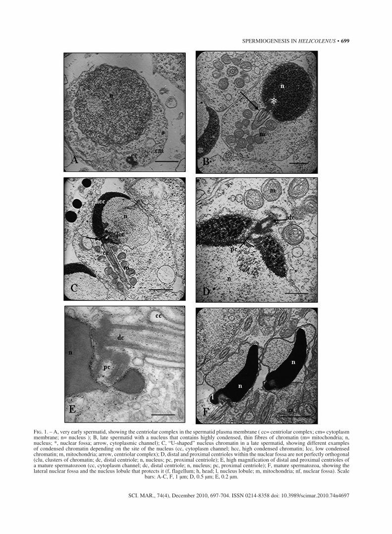
Fig. 2. – A, the flagellar axis is at an oblique angle to the spermatid nucleus (f, flagellar axis; ch, condensed chromatin; m, mitochondria; n, nucleus); B, spermatid midpiece, showing mitochondria with regularly organized cristae (f, flagellum; m, mitochondria); C, the spermatozoa inside testicular lobules do not exhibit cytoplasmic masses around their heads (f, flagella; h, heads); D, H. dactylopterus spermatozoa are of the complex type of anacrosomal introsperm, typical of internal fertilizing species (cm, cytoplasmic mass; h, spermatozoa’s head; f, flagellum; m, mitochondria; t, sperm tails). The arrows show the lateral projections of the flagellum; E, spermatozoon midpiece, in which we can clearly see a cytoplasmic channel (c, centriole; cc, cytoplasm channel; f, flagellum; m, mitochondria; v, vesicles); F, undulating surface of spermatozoon
flagellum (f, flagellum; pm, plasma membrane). Scale bars: A, 1 μm; B, 0.25 μm; C, 2.5 μm; D, 2 μm; E-F, 0.5 μm.
SPERMIOGENESIS IN HELICOLENUS • 701
SCI. MAR., 74(4), December 2010, 697-704. ISSN 0214-8358 doi: 10.3989/scimar.2010.74n4697
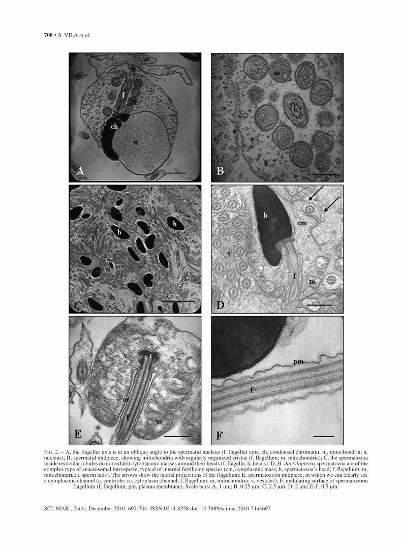
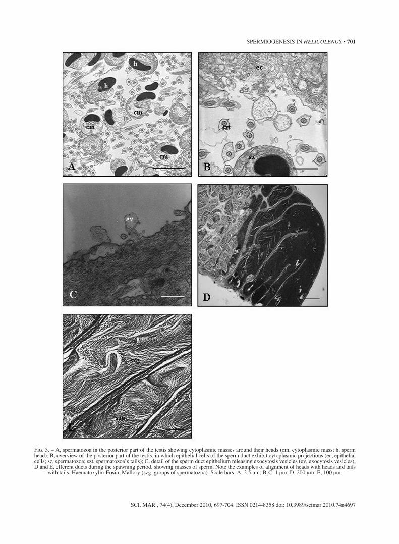
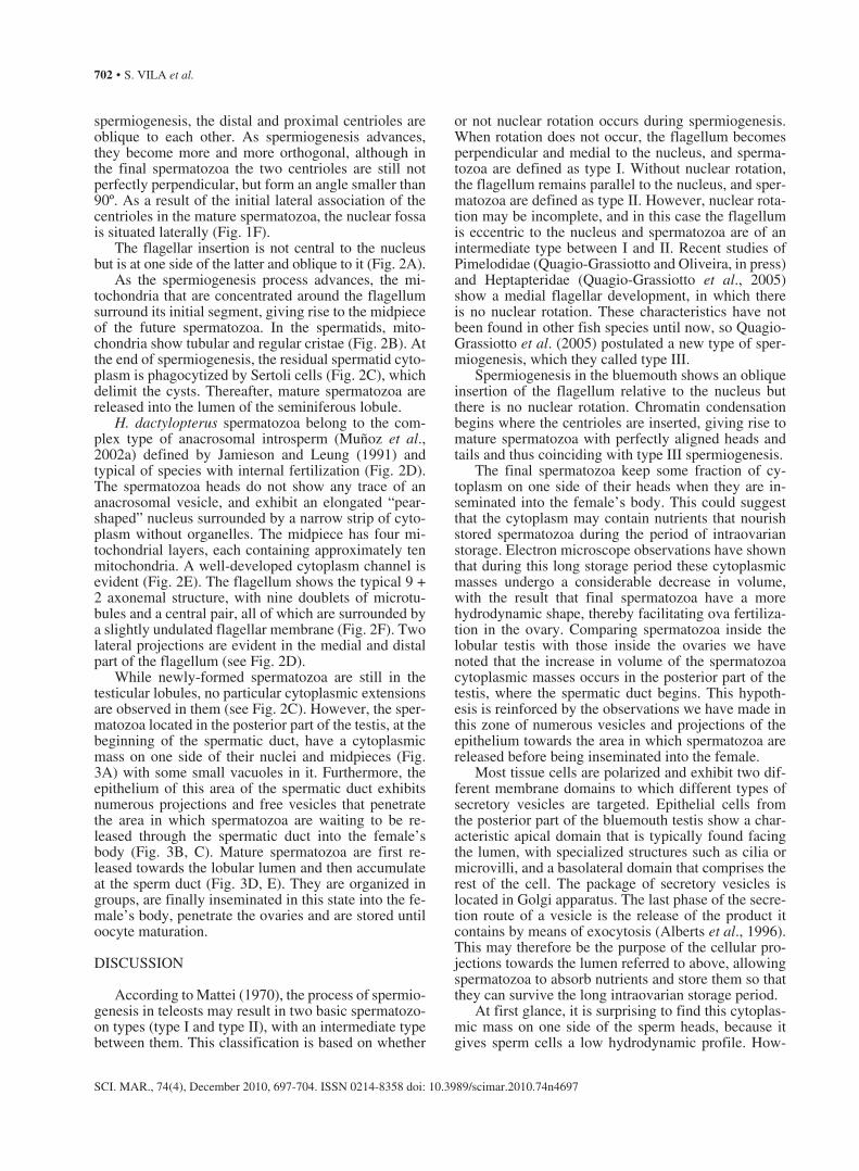
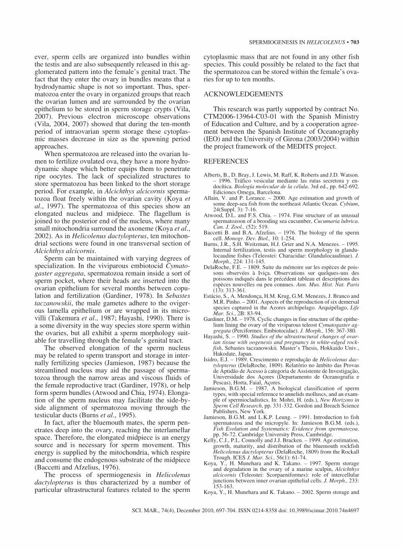
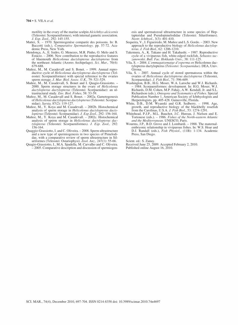
Fig. 3. – A, spermatozoa in the posterior part of the testis showing cytoplasmic masses around their heads (cm, cytoplasmic mass; h, sperm head); B, overview of the posterior part of the testis, in which epithelial cells of the sperm duct exhibit cytoplasmic projections (ec, epithelial cells; sz, spermatozoa; szt, spermatozoa’s tails); C, detail of the sperm duct epithelium releasing exocytosis vesicles (ev, exocytosis vesicles), D and E, efferent ducts during the spawning period, showing masses of sperm. Note the examples of alignment of heads with heads and tails
with tails. Haematoxylin-Eosin. Mallory (szg, groups of spermatozoa). Scale bars: A, 2.5 μm; B-C, 1 μm; D, 200 μm; E, 100 μm.
702 • S. VILA et al.
SCI. MAR., 74(4), December 2010, 697-704. ISSN 0214-8358 doi: 10.3989/scimar.2010.74n4697
spermiogenesis, the distal and proximal centrioles are oblique to each other. As spermiogenesis advances, they become more and more orthogonal, although in the final spermatozoa the two centrioles are still not perfectly perpendicular, but form an angle smaller than 90º. As a result of the initial lateral association of the centrioles in the mature spermatozoa, the nuclear fossa is situated laterally (Fig. 1F).
The flagellar insertion is not central to the nucleus but is at one side of the latter and oblique to it (Fig. 2A).
As the spermiogenesis process advances, the mi-tochondria that are concentrated around the flagellum surround its initial segment, giving rise to the midpiece of the future spermatozoa. In the spermatids, mito-chondria show tubular and regular cristae (Fig. 2B). At the end of spermiogenesis, the residual spermatid cyto-plasm is phagocytized by Sertoli cells (Fig. 2C), which delimit the cysts. Thereafter, mature spermatozoa are released into the lumen of the seminiferous lobule.
H. dactylopterus spermatozoa belong to the com-plex type of anacrosomal introsperm (Muñoz et al., 2002a) defined by Jamieson and Leung (1991) and typical of species with internal fertilization (Fig. 2D). The spermatozoa heads do not show any trace of an anacrosomal vesicle, and exhibit an elongated “pear-shaped” nucleus surrounded by a narrow strip of cyto-plasm without organelles. The midpiece has four mi-tochondrial layers, each containing approximately ten mitochondria. A well-developed cytoplasm channel is evident (Fig. 2E). The flagellum shows the typical 9 + 2 axonemal structure, with nine doublets of microtu-bules and a central pair, all of which are surrounded by a slightly undulated flagellar membrane (Fig. 2F). Two lateral projections are evident in the medial and distal part of the flagellum (see Fig. 2D).
While newly-formed spermatozoa are still in the testicular lobules, no particular cytoplasmic extensions are observed in them (see Fig. 2C). However, the sper-matozoa located in the posterior part of the testis, at the beginning of the spermatic duct, have a cytoplasmic mass on one side of their nuclei and midpieces (Fig. 3A) with some small vacuoles in it. Furthermore, the epithelium of this area of the spermatic duct exhibits numerous projections and free vesicles that penetrate the area in which spermatozoa are waiting to be re-leased through the spermatic duct into the female’s body (Fig. 3B, C). Mature spermatozoa are first re-leased towards the lobular lumen and then accumulate at the sperm duct (Fig. 3D, E). They are organized in groups, are finally inseminated in this state into the fe-male’s body, penetrate the ovaries and are stored until oocyte maturation.
DISCUSSION
According to Mattei (1970), the process of spermio-genesis in teleosts may result in two basic spermatozo-on types (type I and type II), with an intermediate type between them. This classification is based on whether
or not nuclear rotation occurs during spermiogenesis. When rotation does not occur, the flagellum becomes perpendicular and medial to the nucleus, and sperma-tozoa are defined as type I. Without nuclear rotation, the flagellum remains parallel to the nucleus, and sper-matozoa are defined as type II. However, nuclear rota-tion may be incomplete, and in this case the flagellum is eccentric to the nucleus and spermatozoa are of an intermediate type between I and II. Recent studies of Pimelodidae (Quagio-Grassiotto and Oliveira, in press) and Heptapteridae (Quagio-Grassiotto et al., 2005) show a medial flagellar development, in which there is no nuclear rotation. These characteristics have not been found in other fish species until now, so Quagio-Grassiotto et al. (2005) postulated a new type of sper-miogenesis, which they called type III.
Spermiogenesis in the bluemouth shows an oblique insertion of the flagellum relative to the nucleus but there is no nuclear rotation. Chromatin condensation begins where the centrioles are inserted, giving rise to mature spermatozoa with perfectly aligned heads and tails and thus coinciding with type III spermiogenesis.
The final spermatozoa keep some fraction of cy-toplasm on one side of their heads when they are in-seminated into the female’s body. This could suggest that the cytoplasm may contain nutrients that nourish stored spermatozoa during the period of intraovarian storage. Electron microscope observations have shown that during this long storage period these cytoplasmic masses undergo a considerable decrease in volume, with the result that final spermatozoa have a more hydrodynamic shape, thereby facilitating ova fertiliza-tion in the ovary. Comparing spermatozoa inside the lobular testis with those inside the ovaries we have noted that the increase in volume of the spermatozoa cytoplasmic masses occurs in the posterior part of the testis, where the spermatic duct begins. This hypoth-esis is reinforced by the observations we have made in this zone of numerous vesicles and projections of the epithelium towards the area in which spermatozoa are released before being inseminated into the female.
Most tissue cells are polarized and exhibit two dif-ferent membrane domains to which different types of secretory vesicles are targeted. Epithelial cells from the posterior part of the bluemouth testis show a char-acteristic apical domain that is typically found facing the lumen, with specialized structures such as cilia or microvilli, and a basolateral domain that comprises the rest of the cell. The package of secretory vesicles is located in Golgi apparatus. The last phase of the secre-tion route of a vesicle is the release of the product it contains by means of exocytosis (Alberts et al., 1996). This may therefore be the purpose of the cellular pro-jections towards the lumen referred to above, allowing spermatozoa to absorb nutrients and store them so that they can survive the long intraovarian storage period.
At first glance, it is surprising to find this cytoplas-mic mass on one side of the sperm heads, because it gives sperm cells a low hydrodynamic profile. How-
SPERMIOGENESIS IN HELICOLENUS • 703
SCI. MAR., 74(4), December 2010, 697-704. ISSN 0214-8358 doi: 10.3989/scimar.2010.74n4697
ever, sperm cells are organized into bundles within the testis and are also subsequently released in this ag-glomerated pattern into the female’s genital tract. The fact that they enter the ovary in bundles means that a hydrodynamic shape is not so important. Thus, sper-matozoa enter the ovary in organized groups that reach the ovarian lumen and are surrounded by the ovarian epithelium to be stored in sperm storage crypts (Vila, 2007). Previous electron microscope observations (Vila, 2004, 2007) showed that during the ten-month period of intraovarian sperm storage these cytoplas-mic masses decrease in size as the spawning period approaches.
When spermatozoa are released into the ovarian lu-men to fertilize ovulated ova, they have a more hydro-dynamic shape which better equips them to penetrate ripe oocytes. The lack of specialized structures to store spermatozoa has been linked to the short storage period. For example, in Alcichthys alcicornis sperma-tozoa float freely within the ovarian cavity (Koya et al., 1997). The spermatozoa of this species show an elongated nucleus and midpiece. The flagellum is joined to the posterior end of the nucleus, where many small mitochondria surround the axoneme (Koya et al., 2002). As in Helicolenus dactylopterus, ten mitochon-drial sections were found in one transversal section of Alcichthys alcicornis.
Sperm can be maintained with varying degrees of specialization. In the viviparous embiotocid Cymato-gaster aggregata, spermatozoa remain inside a sort of sperm pocket, where their heads are inserted into the ovarian epithelium for several months between copu-lation and fertilization (Gardiner, 1978). In Sebastes taczanowskii, the male gametes adhere to the oviger-ous lamella epithelium or are wrapped in its micro-villi (Takemura et al., 1987; Hayashi, 1990). There is a some diversity in the way species store sperm within the ovaries, but all exhibit a sperm morphology suit-able for travelling through the female’s genital tract.
The observed elongation of the sperm nucleus may be related to sperm transport and storage in inter-nally fertilizing species (Jamieson, 1987) because the streamlined nucleus may aid the passage of sperma-tozoa through the narrow areas and viscous fluids of the female reproductive tract (Gardiner, 1978), or help form sperm bundles (Atwood and Chia, 1974). Elonga-tion of the sperm nucleus may facilitate the side-by-side alignment of spermatozoa moving through the testicular ducts (Burns et al., 1995).
In fact, after the bluemouth mates, the sperm pen-etrates deep into the ovary, reaching the interlamellar space. Therefore, the elongated midpiece is an energy source and is necessary for sperm movement. This energy is supplied by the mitochondria, which respire and consume the endogenous substrate of the midpiece (Baccetti and Afzelius, 1976).
The process of spermiogenesis in Helicolenus dactylopterus is thus characterized by a number of particular ultrastructural features related to the sperm
cytoplasmic mass that are not found in any other fish species. This could possibly be related to the fact that the spermatozoa can be stored within the female’s ova-ries for up to ten months.
ACKNOWLEDGEMENTS
This research was partly supported by contract No. CTM2006-13964-C03-01 with the Spanish Ministry of Education and Culture, and by a cooperation agree-ment between the Spanish Institute of Oceanography (IEO) and the University of Girona (2003/2004) within the project framework of the MEDITS project.
REFERENCES
Alberts, B., D. Bray, J. Lewis, M. Raff, K. Roberts and J.D. Watson. – 1996. Tráfico vesicular mediante las rutas secretora y en-docítica. Biología molecular de la célula. 3rd ed., pp. 642-692. Ediciones Omega, Barcelona.
Allain, V. and P. Lorance. – 2000. Age estimation and growth of some deep-sea fish from the northeast Atlantic Ocean. Cybium, 24(Suppl. 3): 7-16.
Atwood, D.L. and F.S. Chia. – 1974. Fine structure of an unusual spermatozoon of a brooding sea cucumber, Cucumaria lubrica. Can. J. Zool., (52): 519.
Baccetti B. and B.A. Afzelius. – 1976. The biology of the sperm cell. Monogr. Dev. Biol., 10: 1-254.
Burns, J.R., S.H. Weitzman, H.J. Grier and N.A. Menezes. – 1995. Internal fertilization, testis and sperm morphology in glandu-locaudine fishes (Teleostei: Characidae: Glandulocaudinae). J. Morph., 224: 131-145.
DelaRoche, F.E. – 1809. Suite du mémoire sur les espèces de pois-sons observées à Iviça. Observations sur quelques-uns des poissons indiqués dans le précédent tableau et descriptions des espèces nouvelles ou peu connues. Ann. Mus. Hist. Nat. Paris (13): 313-361.
Estácio, S., A. Mendonça, H.M. Krug, G.M. Menezes, J. Branco and M.R. Pinho. – 2001. Aspects of the reproduction of six demersal species captured in the Azores archipelago. Arquipélago, Life Mar. Sci., 2B: 83-94.
Gardiner, D.M. – 1978. Cyclic changes in fine structure of the epithe-lium lining the ovary of the viviparous teleost Cymatogaster ag-gregata (Perciformes: Embiotocidae). J. Morph., 156: 367-380.
Hayashi, S. – 1990. Studies of the ultrastructural changes of ovar-ian tissue with oogenesis and pregnancy in white-edged rock-fish, Sebastes taczanowskii. Master’s Thesis, Hokkaido Univ., Hakodate, Japan.
Isidro, E.J. – 1989. Crescimento e reprodução de Helicolenus dac-tylopterus (DelaRoche, 1809). Relatório no âmbito das Provas de Aptidão de Acesso à categoria de Assistente de Investigação, Universidade dos Açores (Departamento de Oceanografia e Pescas), Horta, Faial, Açores.
Jamieson, B.G.M. – 1987. A biological classification of sperm types, with special reference to annelids molluscs, and an exam-ple of spermiocladistics. In: Mohri, H. (eds.), New Horizons in Sperm Cell Research, pp. 331-332. Gordon and Breach Science Publishers, New York.
Jamieson, B.G.M. and L.K.P. Leung. – 1991. Introduction to fish spermatozoa and the micropyle. In: Jamieson B.G.M. (eds.), Fish Evolution and Systematics: Evidence from spermatozoa. pp. 56-72. Cambridge University Press, Cambridge.
Kelly, C.J., P.L. Connolly and J.J. Bracken. – 1999. Age estimation, growth, maturity, and distribution of the bluemouth rockfish Helicolenus dactylopterus (DelaRoche, 1809) from the Rockall Trough. ICES J. Mar. Sci., 56(1): 61-74.
Koya, Y., H. Munehara and K. Takano. – 1997. Sperm storage and degradation in the ovary of a marine sculpin, Alcichthys alcicornis (Teleostei: Scorpaeniformes): role of intercellular junctions between inner ovarian epithelial cells. J. Morph., 233: 153-163.
Koya, Y., H. Munehara and K. Takano. – 2002. Sperm storage and
704 • S. VILA et al.
SCI. MAR., 74(4), December 2010, 697-704. ISSN 0214-8358 doi: 10.3989/scimar.2010.74n4697
motility in the ovary of the marine sculpin Alcichthys alcicornis (Teleostei: Scorpaeniformes), with internal gametic association. J. Exp. Zool., 292: 145-155.
Mattei, X. – 1970. Spermiogenèse comparée des poissons. In: B. Baccetti (eds.), Comparative Spermatology. pp. 57-72. Aca-demic Press, New York.
Mendonça, A., E. Isidro, G. Menezes, M.R. Pinho, O. Melo and S. Estácio. – 2006. New contribution to the reproductive features of bluemouth Helicolenus dactylopterus dactylopterus from the northeast Atlantic (Azores Archipelago). Sci. Mar., 70(4): 679-688.
Muñoz, M., M. Casadevall and S. Bonet. – 1999. Annual repro-ductive cycle of Helicolenus dactylopterus dactylopterus (Tel-eostei: Scorpaeniformes) with special reference to the ovaries sperm storage. J. Mar. Biol. Assoc. U.K., 79: 521-529.
Muñoz, M., M. Casadevall, S. Bonet and I. Quagio-Grassiotto. – 2000. Sperm storage structures in the ovary of Helicolenus dactylopterus dactylopterus (Teleostei: Scorpaenidae): an ul-trastructural study. Env. Biol. Fishes, 58: 53-59.
Muñoz, M., M. Casadevall and S. Bonet. – 2002a. Gametogenesis of Helicolenus dactylopterus dactylopterus (Teleostei: Scorpae-nidae). Sarsia, 87(2): 119-127.
Muñoz, M., Y. Koya and M. Casadevall. – 2002b. Histochemical analysis of sperm storage in Helicolenus dactylopterus dacty-lopterus (Teleostei: Scorpaenidae). J. Exp Zool., 292: 156-164.
Muñoz, M., Y. Koya and M. Casadevall. – 2002c. Histochemical analysis of sperm storage in Helicolenus dactylopterus dac-tylopterus (Teleostei: Scorpaeniformes). J. Exp. Zool., 292: 156-164.
Quaggio-Grassiotto, I. and C. Oliveira. – 2008. Sperm ultrastructure and a new type of spermiogenesis in two species of Pimelodi-dae, with a comparative review of sperm ultrastructure in Sil-uriformes (Teleostei: Ostariophysi). Zool. Anz., 247(1): 55-66.
Quagio-Grassiotto, I., M.A. Spadella, M. Carvalho and C. Oliveira. – 2005. Comparative description and discussion of spermiogen-
esis and spermatozoal ultrastructure in some species of Hep-tapteridae and Pseudopimelodidae (Teleostei: Siluriformes). Neotr. Ichthyol., 3(3): 401-410.
Sequeira, V., I. Figueiredo, M. Muñoz and L.S. Gordo. – 2003. New approach to the reproductive biology of Helicolenus dactylop-terus. J. Fish Biol., 62: 1206-1210.
Takemura, A., K. Takano and H. Takahashi. – 1987. Reproductive cycle of a viviparous fish, white-edged rockfish, Sebastes tac-zanowskii. Bull. Fac. Hokkaido Univ., 38: 111-125.
Vila, S. – 2004. L’emmagatzematge d’esperma en Helicolenus dac-tylopterus dactylopterus (Teleostei: Scorpaenidae). DEA, Univ. Girona.
Vila, S. – 2007. Annual cycle of stored spermatozoa within the ovaries of Helicolenus dactylopterus dactylopterus (Teleostei, Scorpaenidae). J. Fish Biol., 71: 596-609.
Washington, B.B., H.G. Moser, W.A. Laroche and W.J. Richards. – 1984. Scorpaeniformes: development. In: H.G. Moser, W.J. Richards, D.M. Cohen, M.P. Fahay, A.W. Kendall, Jr. and S.L. Richardson (eds.), Ontogeny and Systematics of Fishes. Special Publication Number 1, American Society of Ichthyologists and Herpetologists. pp. 405-428. Gainesville, Florida.
White, D.B., D.M. Wyanski and G.R. Sedberry. – 1998. Age, growth, and reproductive biology of the blackbelly rosefish from the Carolinas, U.S.A. J. Fish Biol., 53: 1274-1291.
Whitehead, P.J.P., M.L. Bauchot, J.C. Hureau, J. Nielsen and E. Tortonese (eds.). – 1986. Fishes of the North-eastern Atlantic and the Mediterranean. UNESCO, Paris.
Wourms, J.P., B.D. Grove and J. Lombardi. – 1988. The maternal-embryonic relationship in viviparous fishes. In: W.S. Hoar and D.J. Randall (eds.), Fish Physiol., (11B): 1-134. Academic Press, San Diego.
Scient. ed.: S. Zanuy.Received June 25, 2009. Accepted February 2, 2010.Published online August 16, 2010.