spezifische, quantitative und metabolomische ... · pdf filemake a basis for chemotaxonomy of...
TRANSCRIPT

« Forum für Lebensmittelsicherheit»
Waldbronn 30/31 Januar 2013
Spezifische, quantitative und
metabolomische Analysentecniken für
Marine Biotoxine mit dem
Agilent Q-ToF 6540
Philipp Hess1
Thomas Glauner2, Bernhard Wüst2,
Manoëlla Sibat1, Florence Mondeguer1, Zita Zendong1
1 Laboratoire Phycotoxines IFREMER – 44300 Nantes - France
2 Agilent Technologies R&D - 76337 Waldbronn - Germany
2
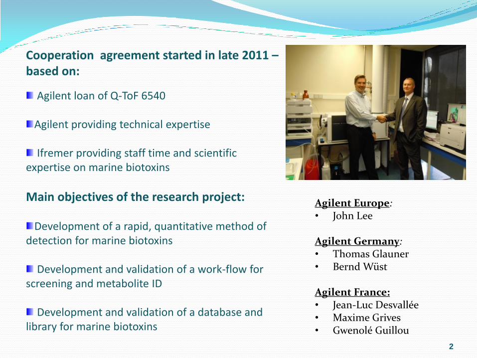
Cooperation agreement started in late 2011 –based on:
Agilent loan of Q-ToF 6540 Agilent providing technical expertise Ifremer providing staff time and scientific
expertise on marine biotoxins
Main objectives of the research project:
Development of a rapid, quantitative method of detection for marine biotoxins
Development and validation of a work-flow for screening and metabolite ID
Development and validation of a database and
library for marine biotoxins
Agilent Europe: • John Lee
Agilent Germany: • Thomas Glauner • Bernd Wüst Agilent France: • Jean-Luc Desvallée • Maxime Grives • Gwenolé Guillou

Content
Context
Development of a quantitative, rapid method for marine biotoxins
using full scan Q-ToF mass spectrometry Full scan techniques
Chromatographic developments
Development of a database and library for marine biotoxins Databases (collection of molecular formulae)
Libraries (database plus structures, spectra and metadata)
“Home-made” versus commercially available tools
Metabolomic techniques in chemotaxonomy and the elucidation of
unknowns (dereplication) “Metabolome of a sample”
Metabolomes of micro-algae (indivudual, comparative, chemotaxonomy)
Standardisation of work-flow
Linking dereplication work-flow to miniaturised bioassays
Conclusion
3

Biodiversity = Chemodiversity

Unknown toxins France 2003 – 2008
(as detected per mouse bioassay for lipophilic toxins)
27 % of unexplained mouse bioassays

European or global perspective:
Europe is a single market, hence all toxins
encountered in Europe must be monitored
Europe imports shellfish from a wide variety of third
countries, hence an even larger, virtually global
range of toxins must be taken into account for
imports (AU, NZ, JP, KO, Thailand, Vietnam,
Jamaica, CA, Chile, Peru, Uruguay, Greenland,
Morocco, Tunisia and Turkey)
Legislative change from mouse bioassay for
lipophilic toxins to targeted LC-MS/MS methods as
reference requires increased vigilance
6

7
1- Full scan analysis:
Okadaic acid
Example of full scan power MS Mass Hunter software to evaluate molecular and pseudo-molecular ion clusters.

Development of fast quantitative method for marine biotoxins
8
Fast LC-MS/MS method tested on:
Kinetex C18 100*2.1mm 2.6µm (Phenomenex)
Poroshell 120 EC-C18 100*2.1mm 2.7µm (Agilent)
Zorbax Extend-C18 50*2.1mm 1.8 µm (Agilent)
Zorbax SB C8 50*2.1mm 1.8µm (Agilent)

9

Calibration curves
10
Quantitative rapid method (full scan)
Certified standards
Six calibration levels
R2 > 0,98
Matrix effects remain to be evaluated…

Agilent suite of software
11
Database in construction (currently 275 compounds)
All major phycotoxins in Europe
Structures created in ChemDraw and then inserted into the PCDL library as .mol-files
PCDL Manager - module

12
458 ion is specific to
PnTX-G and does
not exist for SPXs
Using LC-HRMS
for definitive
confirmation of
identity:
Example of
isobaric PnTX-G
and SPX-B & 13-
dm SPX-D

Database development – literature search…
13
Toxin family Toxin group Abbreviation
Number
Compound Molecular structure
ASP Domoic acid DA 9 9
DSP
Okadaic acid &
dinophysistoxins OA+DTXs 65 10
Azaspiracids AZAs 30 /
Pectenotoxins PTXs 16 4
Yessotoxins YTXs 31 /
Cyanobacteria Oscillatoxins n/a 9 /
Cyclic imines
(FAT)
Gymnodimines GYMs 4 4
Spirolides SPXs 11 11
Pinnatoxins and Pteriatoxins PnTXs+PterTX
s 12 12
PSP Saxitoxins STXs 18 18
Tetrodotoxins TTXs 18 /
NSP
Palytoxins PLTXs 8 1
Brevetoxins PbTXs (BTX) 16 /
Pacific Ciguatoxins P-CTXs 27 /
Caribbean Ciguatoxins C-CTXs 2 /

LC-MS/MS spectra are reputed to be non-
reproducible between instruments of different
manufacturers
This can be overcome to some extent by acquiring
spectra at low, medium and high collision energies
We are working with Agilent to establish whether a
simple mass indicator can be used to set increasing
CEs as a standard approach
14
Database development – entering spectra…

Agilent work-flow for non-targeted analysis
15

Sample Prep
Fingerprints(LC/HRMS)
Data retreatment
Statistical analyses
16
1000’s ions Biomarkers ?
m/z
rt
intensités
133.07 133.08 133.09 133.10
m/z
0
10
20
30
40
50
60
70
80
90
100
110
Re
lative
Ab
un
da
nce
133.08020
133.07378
133.09665
133.08236
NL:9.69E2
C 4 H10 O2 N3: C 4 H10 O2 N3
p (gss, s /p:40) Chrg 1R: 20.0 PPM @FWHM
NL:1.11E6
281008023#93 RT: 2.46 AV: 1 SB: 1782 3.11-49.81 , 0.01-2.31 T: + c ESI Full ms [50.00-800.00]
Structure elucidation
MSn
Also:
Fingerprints
Footprints
Crosstalk
Metabolomics Approach:

Examples of algal metabolomes:
Alexandrium ostenfeldii
7,232 min 13,19 Didesmethyl-SPX-C (M+H)+ = 692,4520

Examples of algal metabolomes:
Karenia selliformis
6,48 min Gymnodimine A (M+H)+ = 508,3433

Examples of algal metabolomes:
Prorocentrum lima
11,38 min OA (M+H)+ = 805,4733 12,71 min DTX1 (M+H)+ = 819,4901

Examples of algal metabolomes:
Pseudo-nitzschia
5,49 min DA (M+H)+ 312,1445

Azadinium spinosum
Comparative metabolomes:
77 features only in A. obesum, 59 features only in A. spinosum, and 95 common features !
AZA1-methyl ester (M+H)+ 856.5201
AZA? (M+H)+ 716.4748
AZA2 (M+H)+ 856.5224 AZA1 (M+H)+ 842.5068
Azadinium obesum


From metabolomes to chemotaxonomy:

From metabolomes to chemotaxonomy:

Comparative metabolomes of algal fingerprints and algal footprints
25
SPATT A. spinosum vs A. obesum
147 features in SPATT vs. 196 features in A. spinosum
Acetone extract of A. spinosum vs A. obesum

Pilot-scale culture of V. rugosum
Batch 1 2 3 4 5 6 7 8 9 10 Total
Mass of pellet (g) 26 19 16 13 16 28 28 58 56 29 289g
Masse PnTX G (µg) 614 442 303 237 259 411 276 140 246 181 3mg
Culture conditions L1 L1 L1* L1 L1 L1* L1 L1 L1 L1
Duration of culture (d) 41 33 29 39 25 47 37** 63 56 43

Metabolome of Vulcanodinium rugosum
- bioguided fractionation
Fractions
Chemical analysis Evaluation of biological activity
Data analysis
(comparison to large natural product libraries2)
-Triple quad
-Q-ToF -Cytotoxicity KB cells
-Fly larvae
-bacteria
Vulcanodinium
rugosum Crude extract
Biological Screening
and
Metabolomics: Dereplication1,2
Fractioning
(1) Kristian F. Nielsen et al. J Nat. Prod. (2011) 74, p2338-2348
(2) http://www.chem.canterbury.ac.nz/marinlit/marinlit.shtml

Fractionation of sample for polar lipids
Scheme of extraction and purification of PnTX-G
CRUDE (A)
(2050 mg)
DCM fraction (B) (442 mg; 22% of A)
Aqueous phase
Hexane phase Aq. MeOH fraction (C)
(168 mg; 8% of A)
SiO2-F2 (73 mg; 43 % of C)
SiO2-F3 (39 mg; 23 % of C)
PnTX G
Crude extr.
DCM-fract.
Aq. MeOH

Evaluation of the activity of algal extracts
using cytotoxicity assay
0
20
40
60
80
100
F1bP3 F2bP3 F3bP3 F4bP3 F5bP3
Masse fractions (%)
Masse PnTX G (%)
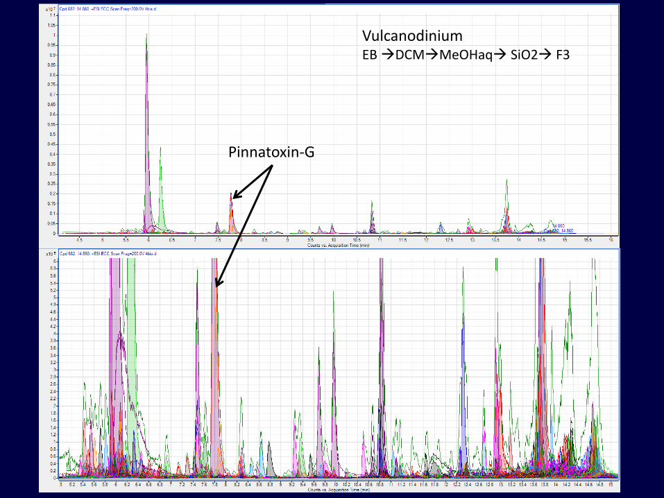
Dereplication: database screening results for SiO2 F3 Vulcanodinium EB DCMMeOHaq SiO2 F3
Pinnatoxin-G

Dereplication: SiO2 F2:
Screen against MarinLit
(database by Bunt & Munro
with > 30 000 cpds
(this process takes around
15 min per 10 min of full
scan data)
Name RT m/z Score (DB) Diff (DB, ppm) Nakijiquinone A 5.68 402.2278 99.4 -0.82

Interestingly…
Out of the 144 compounds present in these two
fractions, we were able to identify 45 compounds
applying a filter of 5 ppm, or 36 compounds when
applying a filter of 2 ppm, and 22 compounds at < 1
ppm
About 100 unknowns to follow up on…
Several present that had been initially identified from
sponges: nakijiquinone, petrosaspongiolide, plakinic
acid and sarcotin
We expect to be able to clarify the biological origin
and biogeography of many natural products…

Conclusions
Developed rapid & quantitative method for marine biotoxins
using full scan techniques
Linear over appropriate range
Allows for quantitation of all regulated (EU) lipophilic toxins in
< 9 min
Developed work-flow for assessing finger- and footprints of
microalgae
Exemplified how to use MP in comparative work
Make a basis for chemotaxonomy of microalgae
Used dereplication in conjunction with bioscreening
Developed a database and library for marine biotoxins
Ca. 275 compounds entered (ca. 90 structures added)
Demonstrated capability of rapid screening against large-scale
commercial databases

34
Jean-François de Troy « The oyster lunch » 1735 (originally decorating the dining room in Versailles), Musée Condé,
Chantilly, France
Thanks for your attention !!

ni PinnTx ni Nakijikinone ne sont des marqueurs discriminants de l’axenisation de la souche
Si avec le traitement anti bactérien 72>65 , 7 composés supplémentaires s’ajoutent donc pas utile d’axéniser