spherical herbert ulnar head prosthesis (uhp) - kebomed.dk · the safe solution for failed...
TRANSCRIPT

www.klsmartin.com
Spherical Herbert Ulnar HeadProsthesis (UHP)Management of failedSauvé-Kapandji Procedures
In the field of hand surgery we not only offer you solutions for standard restorations, but alsoproducts for unusual and difficult situations. We therefore regard ourselves as being a truehighly specialized partner in all matters relatingto hand surgery with our intelligent system solutions.
3
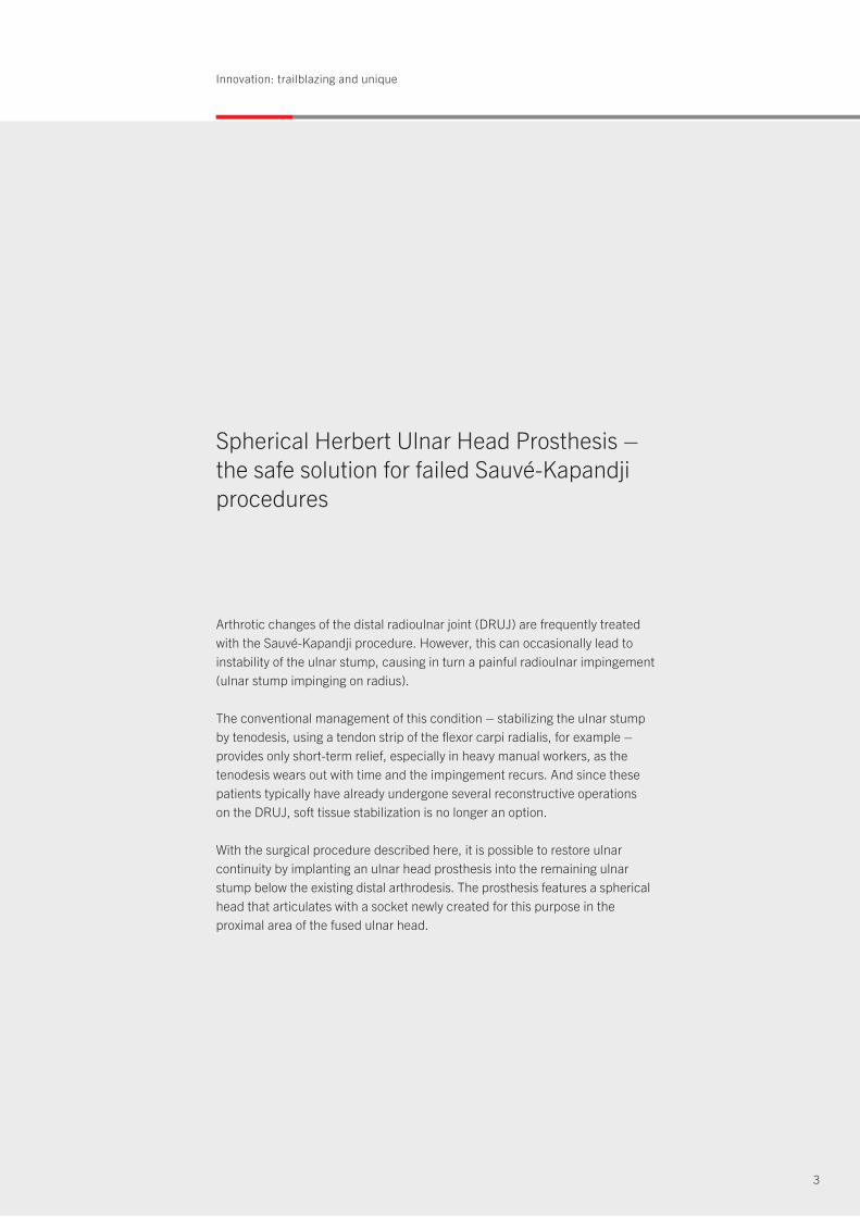
Spherical Herbert Ulnar Head Prosthesis –the safe solution for failed Sauvé-Kapandji procedures
Arthrotic changes of the distal radioulnar joint (DRUJ) are frequently treatedwith the Sauvé-Kapandji procedure. However, this can occasionally lead toinstability of the ulnar stump, causing in turn a painful radioulnar impingement (ulnar stump impinging on radius).
The conventional management of this condition – stabilizing the ulnar stump by tenodesis, using a tendon strip of the flexor carpi radialis, for example – provides only short-term relief, especially in heavy manual workers, as the tenodesis wears out with time and the impingement recurs. And since thesepatients typically have already undergone several reconstructive operations on the DRUJ, soft tissue stabilization is no longer an option.
With the surgical procedure described here, it is possible to restore ulnar continuity by implanting an ulnar head prosthesis into the remaining ulnarstump below the existing distal arthrodesis. The prosthesis features a sphericalhead that articulates with a socket newly created for this purpose in the proximal area of the fused ulnar head.
Innovation: trailblazing and unique
Design: durable and stable
4
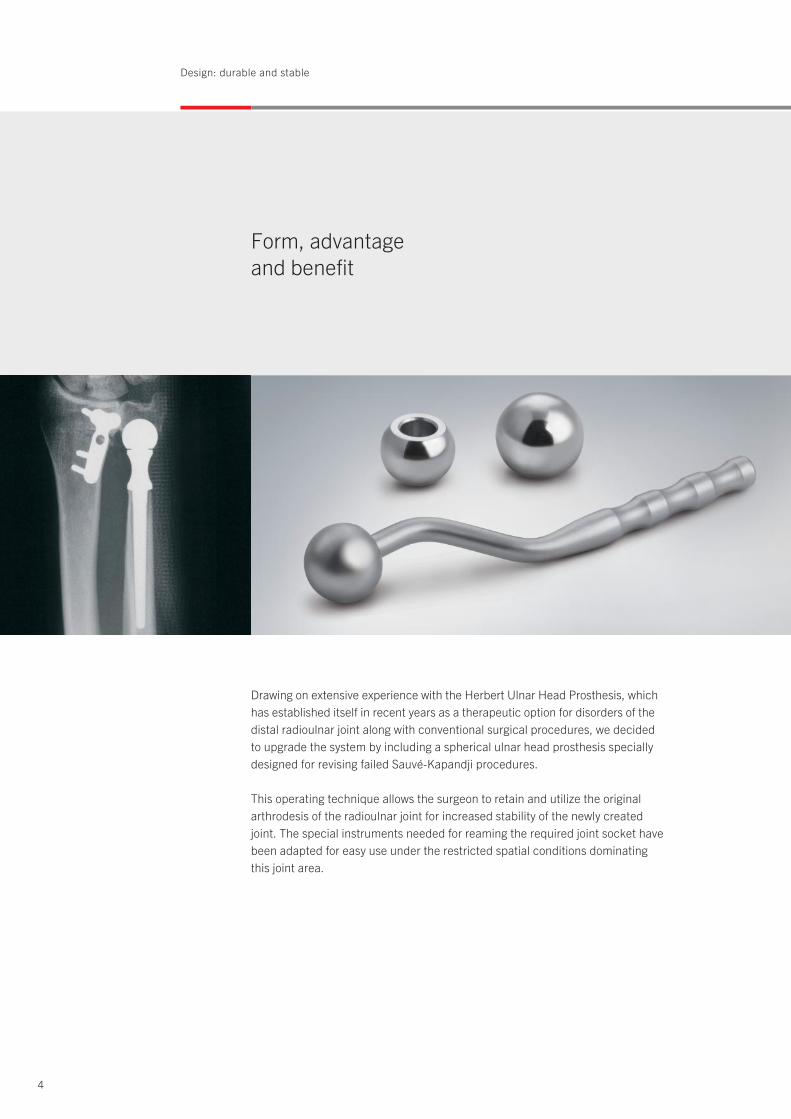
Form, advantage and benefit
Drawing on extensive experience with the Herbert Ulnar Head Prosthesis, whichhas established itself in recent years as a therapeutic option for disorders of thedistal radioulnar joint along with conventional surgical procedures, we decidedto upgrade the system by including a spherical ulnar head prosthesis speciallydesigned for revising failed Sauvé-Kapandji procedures.
This operating technique allows the surgeon to retain and utilize the originalarthrodesis of the radioulnar joint for increased stability of the newly createdjoint. The special instruments needed for reaming the required joint socket havebeen adapted for easy use under the restricted spatial conditions dominatingthis joint area.
5
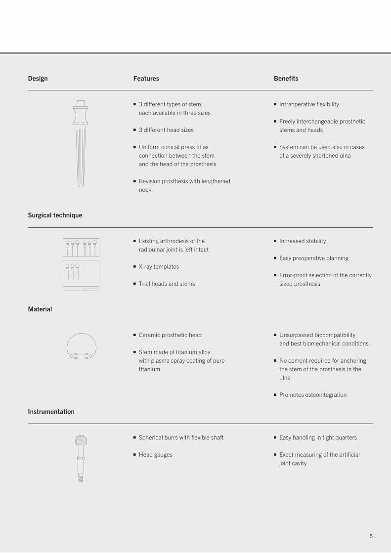
Design Features Benefits
■ 3 different types of stem, each available in three sizes
■ 3 different head sizes
■ Uniform conical press fit as connection between the stem and the head of the prosthesis
■ Revision prosthesis with lengthened neck
■ Intraoperative flexibility
■ Freely interchangeable prosthetic stems and heads
■ System can be used also in cases of a severely shortened ulna
Surgical technique
■ Existing arthrodesis of the radioulnar joint is left intact
■ X-ray templates
■ Trial heads and stems
■ Increased stability
■ Easy preoperative planning
■ Error-proof selection of the correctly sized prosthesis
Material
■ Ceramic prosthetic head
■ Stem made of titanium alloy with plasma spray coating of pure titanium
■ Unsurpassed biocompatibility and best biomechanical conditions
■ No cement required for anchoring the stem of the prosthesis in the ulna
■ Promotes osteointegration
Instrumentation
■ Spherical burrs with flexible shaft
■ Head gauges
■ Easy handling in tight quarters
■ Exact measuring of the artificial joint cavity
6
Surgical technique: simple and precise
Step by step to optimal fixation
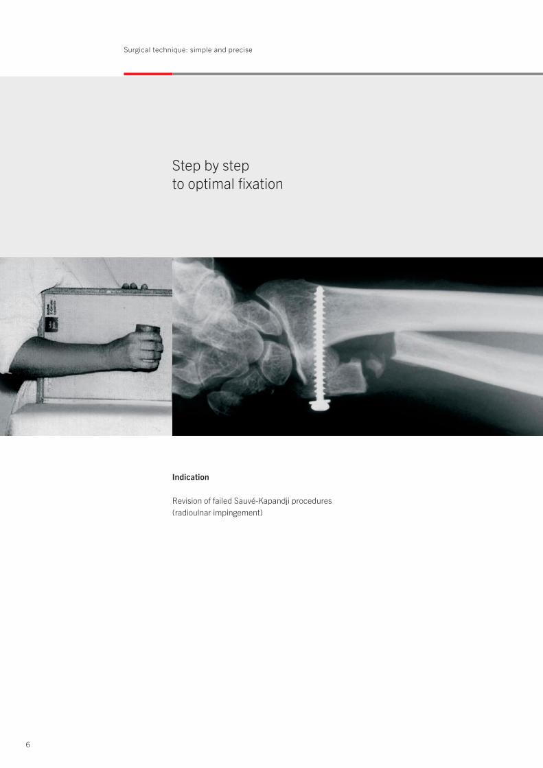
Indication
Revision of failed Sauvé-Kapandji procedures (radioulnar impingement)
7
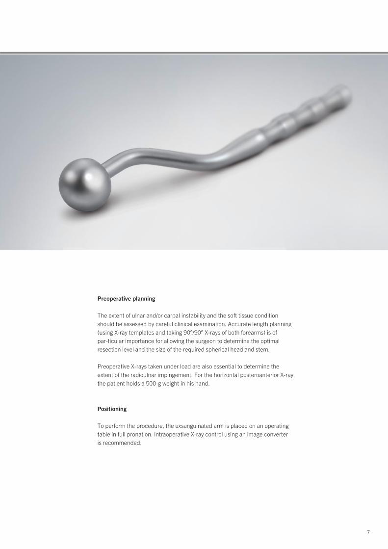
Preoperative planning
The extent of ulnar and/or carpal instability and the soft tissue condition should be assessed by careful clinical examination. Accurate length planning(using X-ray templates and taking 90°/90° X-rays of both forearms) is of par-ticular importance for allowing the surgeon to determine the optimal resection level and the size of the required spherical head and stem.
Preoperative X-rays taken under load are also essential to determine theextent of the radioulnar impingement. For the horizontal posteroanterior X-ray,the patient holds a 500-g weight in his hand.
Positioning
To perform the procedure, the exsanguinated arm is placed on an operatingtable in full pronation. Intraoperative X-ray control using an image converter is recommended.

8
Design: durable and stable
Fig. 1: Radioulnar impingement syndromewith severe instability of the proximalulnar stump after Sauvé-Kapandji procedure and several previous surgical operations on the distalradioulnar joint.
Fig. 3: Spherical burrs are now used to hollow out the proximal surface of thefused ulnar head to create the newsocket.
Fig. 2: Surgical exposure through the oldscar, which is usually slightly extendedin proximal direction. Care should beexercised to identify and preserve thedorsal sensory branch of the ulnar nerve during this step. This is followedby exposure of the fused ulnar headand the proximal ulnar stump betweenthe extensor carpi ulnaris and extensordigiti quinti muscles. Longitudinal opening of a relatively dense scar plate between these two structures is required as well.
Spherical burrFlexible shaft
Surgical documentation: Prof. Dr. Diego L. Fernandez, MD
9
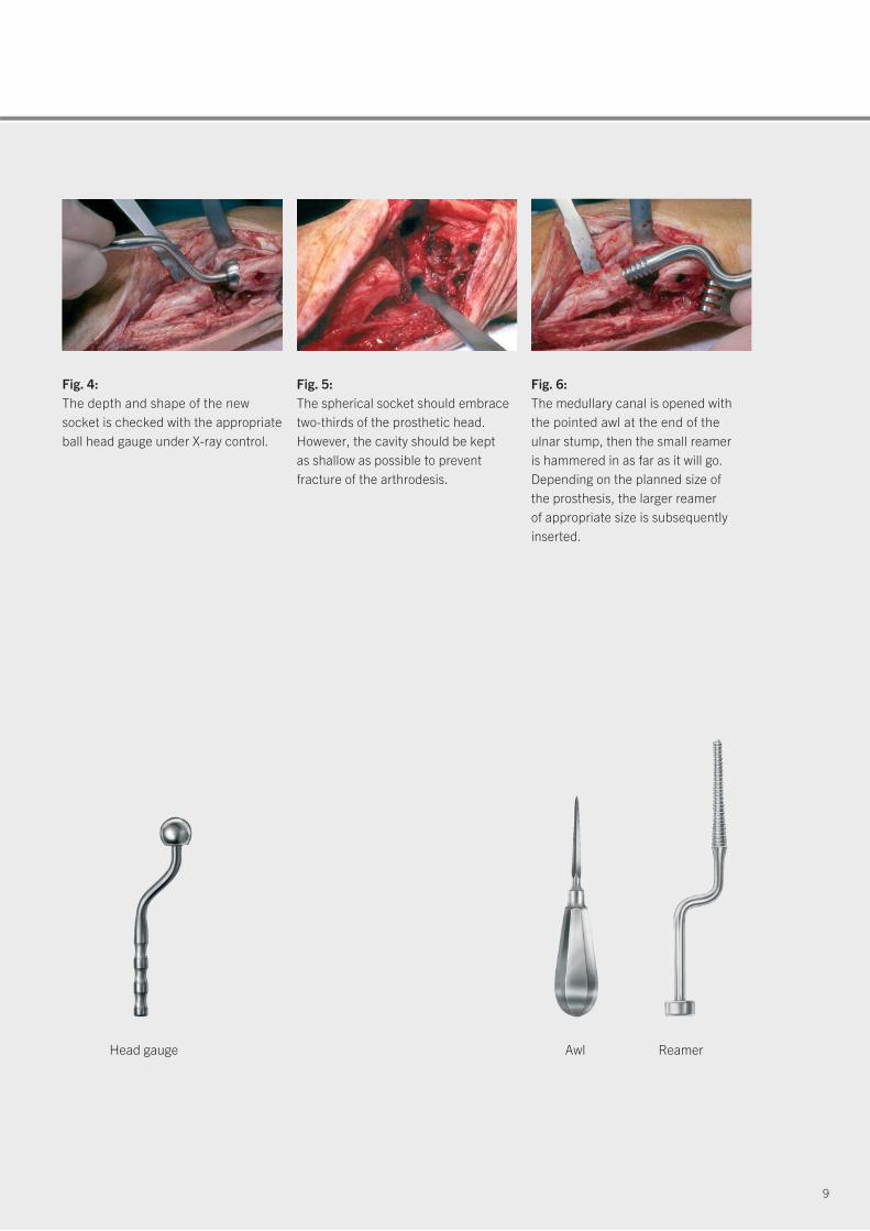
Fig. 5: The spherical socket should embracetwo-thirds of the prosthetic head.However, the cavity should be kept as shallow as possible to prevent fracture of the arthrodesis.
Fig. 6: The medullary canal is opened withthe pointed awl at the end of the ulnar stump, then the small reamer is hammered in as far as it will go.Depending on the planned size of the prosthesis, the larger reamer of appropriate size is subsequentlyinserted.
Head gauge Awl Reamer
Fig. 4: The depth and shape of the newsocket is checked with the appropriateball head gauge under X-ray control.
Surgical technique: simple and precise
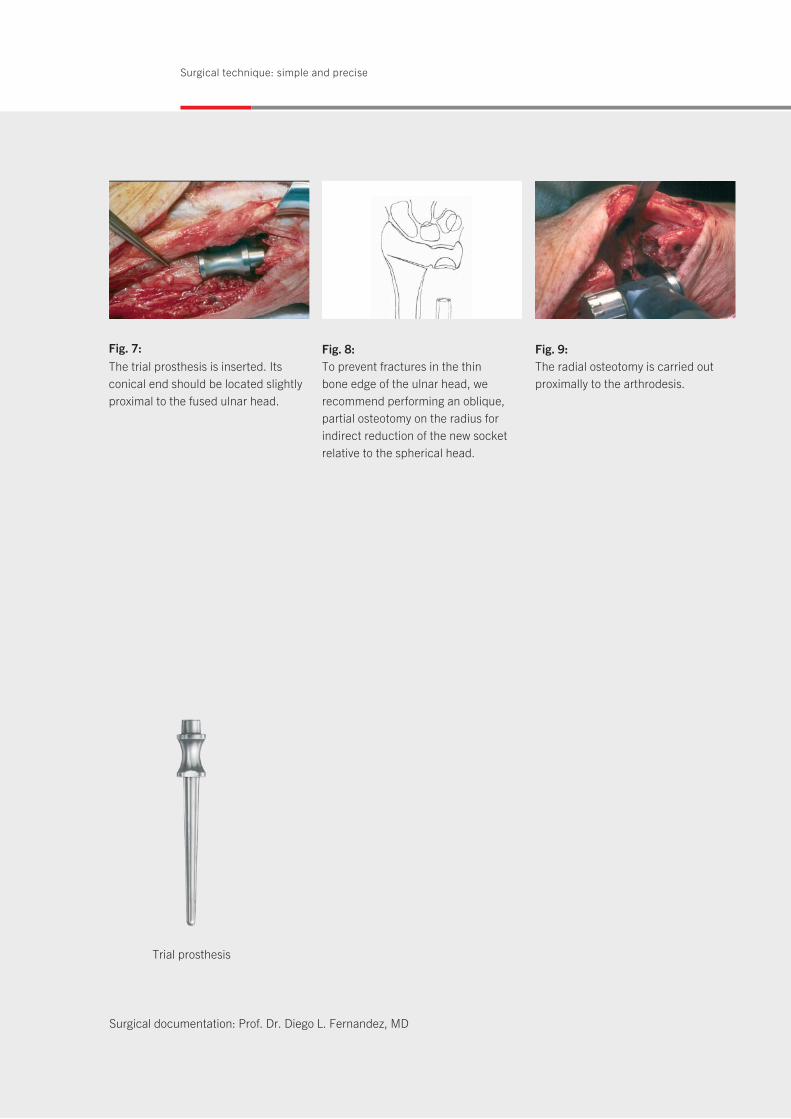
Fig. 7:The trial prosthesis is inserted. Itsconical end should be located slightlyproximal to the fused ulnar head.
Fig. 9: The radial osteotomy is carried outproximally to the arthrodesis.
Fig. 8: To prevent fractures in the thin bone edge of the ulnar head, werecommend performing an oblique,partial osteotomy on the radius forindirect reduction of the new socketrelative to the spherical head.
Trial prosthesis
Surgical documentation: Prof. Dr. Diego L. Fernandez, MD
11
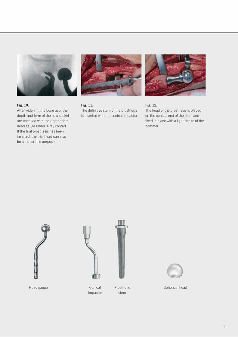
Fig. 11: The definitive stem of the prosthesis is inserted with the conical impactor.
Fig. 12: The head of the prosthesis is placedon the conical end of the stem andfixed in place with a light stroke of thehammer.
Fig. 10:After widening the bone gap, thedepth and form of the new socket are checked with the appropriate head gauge under X-ray control. If the trial prosthesis has been inserted, the trial head can also be used for this purpose.
Head gauge Conical impactor
Prosthetic stem
Spherical head
12
Surgical technique: simple and precise
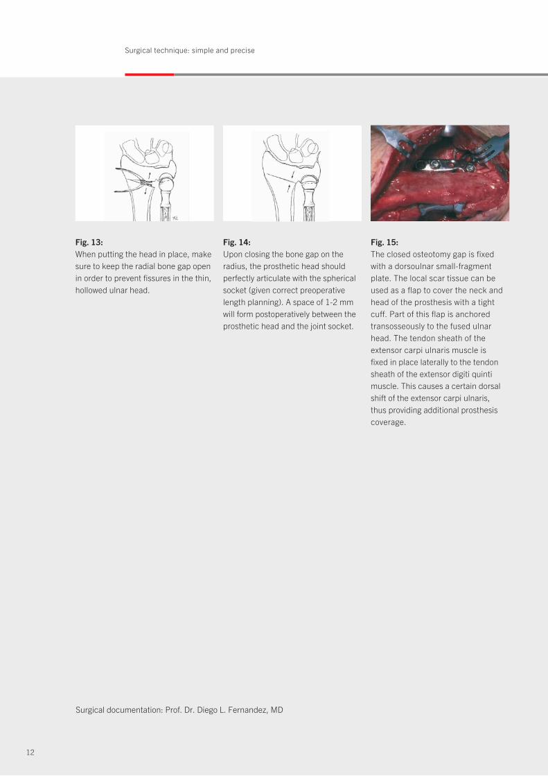
Fig. 15:The closed osteotomy gap is fixedwith a dorsoulnar small-fragment plate. The local scar tissue can beused as a flap to cover the neck andhead of the prosthesis with a tightcuff. Part of this flap is anchoredtransosseously to the fused ulnarhead. The tendon sheath of theextensor carpi ulnaris muscle is fixed in place laterally to the tendonsheath of the extensor digiti quintimuscle. This causes a certain dorsalshift of the extensor carpi ulnaris, thus providing additional prosthesiscoverage.
Fig. 13:When putting the head in place, makesure to keep the radial bone gap openin order to prevent fissures in the thin,hollowed ulnar head.
Fig. 14: Upon closing the bone gap on theradius, the prosthetic head shouldperfectly articulate with the sphericalsocket (given correct preoperativelength planning). A space of 1-2 mmwill form postoperatively between theprosthetic head and the joint socket.
Surgical documentation: Prof. Dr. Diego L. Fernandez, MD
13
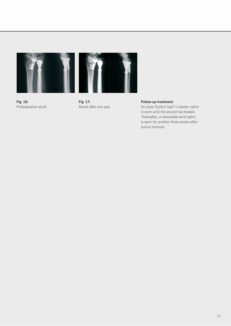
Fig. 17: Result after one year
Fig. 16: Postoperative result
Follow-up treatment:An ulnar Scotch Cast® U plaster splintis worn until the wound has healed.Thereafter, a removable wrist splint is worn for another three weeks aftersuture removal.
14
Clinical example: effective and successful
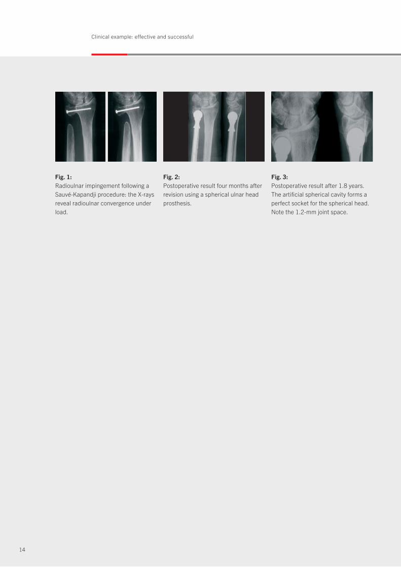
Fig. 1: Radioulnar impingement following aSauvé-Kapandji procedure: the X-raysreveal radioulnar convergence underload.
Fig. 3:Postoperative result after 1.8 years.The artificial spherical cavity forms aperfect socket for the spherical head.Note the 1.2-mm joint space.
Fig. 2: Postoperative result four months afterrevision using a spherical ulnar headprosthesis.
15
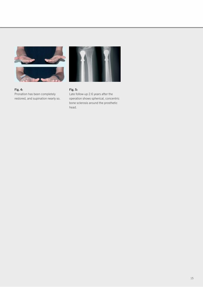
Fig. 5: Late follow-up 2.6 years after the operation shows spherical, concentricbone sclerosis around the prosthetichead.
Fig. 4: Pronation has been completely restored, and supination nearly so.
16
Product range: stems and heads
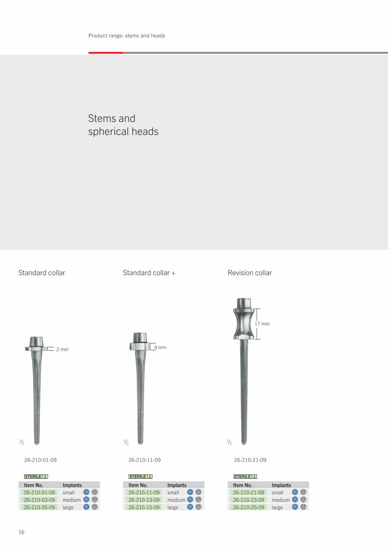
Stems and spherical heads
unit(s)1
unit(s)1
unit(s)1
26-210-01-09 26-210-21-0926-210-11-09
Item No. Implants 26-210-11-09 small26-210-13-09 medium26-210-15-09 large
Item No. Implants 26-210-21-09 small26-210-23-09 medium26-210-25-09 large
17 mm
2 mm 4 mm
Standard collar Standard collar + Revision collar
Item No. Implants 26-210-01-09 small26-210-03-09 medium26-210-05-09 large
1⁄21⁄21⁄2
unit(s)1
unit(s)1
unit(s)1
unit(s)1
unit(s)1
unit(s)1

17
Spherical ceramic heads
26-220-09-04 26-220-11-04 26-220-13-04
Spherical cobalt-chromium heads
26-230-09-04 26-230-11-04 26-230-13-04
Item No. Implants 26-220-09-04 small26-220-11-04 medium26-220-13-04 large
Item No. Implants 26-230-09-04 small26-230-11-04 medium26-230-13-04 large
unit(s)1
unit(s)1
unit(s)1
unit(s)1
unit(s)1
unit(s)1
13 mm 15 mm 18 mm13 mm 15 mm 18 mm
1⁄21⁄2
Icon explanations
unit(s)1
Titanium
Ceramics
Cobalt chromium
Items/pack
Sterile packed implants

18
Product range: storage module and set list
Storage module forinstruments
Item No. Instruments: Trial heads, spherical 26-231-09-05 small26-231-11-05 medium26-231-13-05 large Head gauges, spherical 26-241-19-07 small26-241-21-07 medium26-241-23-07 large Instruments 26-241-01-07 Handle26-241-03-07 Flexible shaft26-241-99-07 Wrench Spherical burrs 26-241-09-07 mini26-241-13-07 small26-241-15-07 medium26-241-18-07 large Storage module
55-910-20-04
Spherical Herbert Ulnar Head Prosthesis Set, complete26-230-00-04
consisting of:
unit(s)1
unit(s)1
unit(s)1
unit(s)1
unit(s)1
unit(s)1
unit(s)1
unit(s)1
unit(s)1
unit(s)1
unit(s)1
unit(s)1
unit(s)1
unit(s)1
19
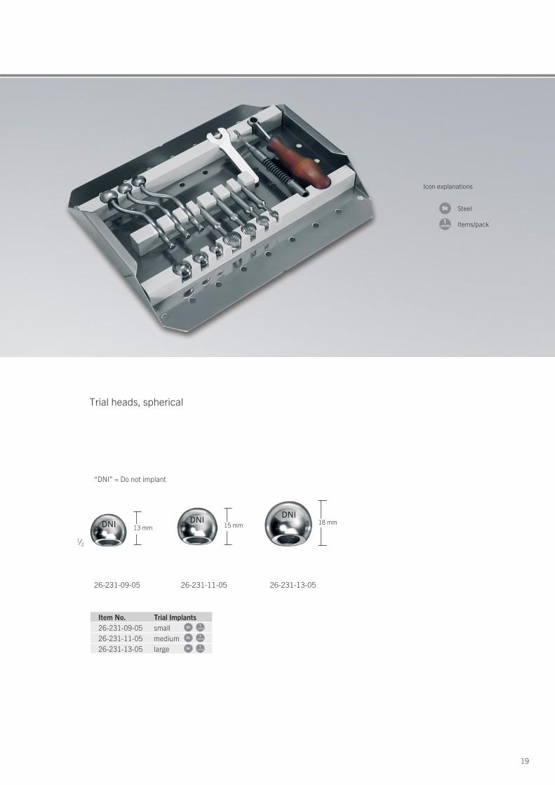
26-231-09-05 26-231-11-05 26-231-13-05
Item No. Trial Implants 26-231-09-05 small26-231-11-05 medium26-231-13-05 large
“DNI” = Do not implant
DNI
Trial heads, spherical
13 mm 15 mm 18 mmDNI
DNI
Icon explanations
unit(s)1
Steel
Items/pack
unit(s)1
unit(s)1
unit(s)1
1⁄2
20
Product range: trial implants and instruments
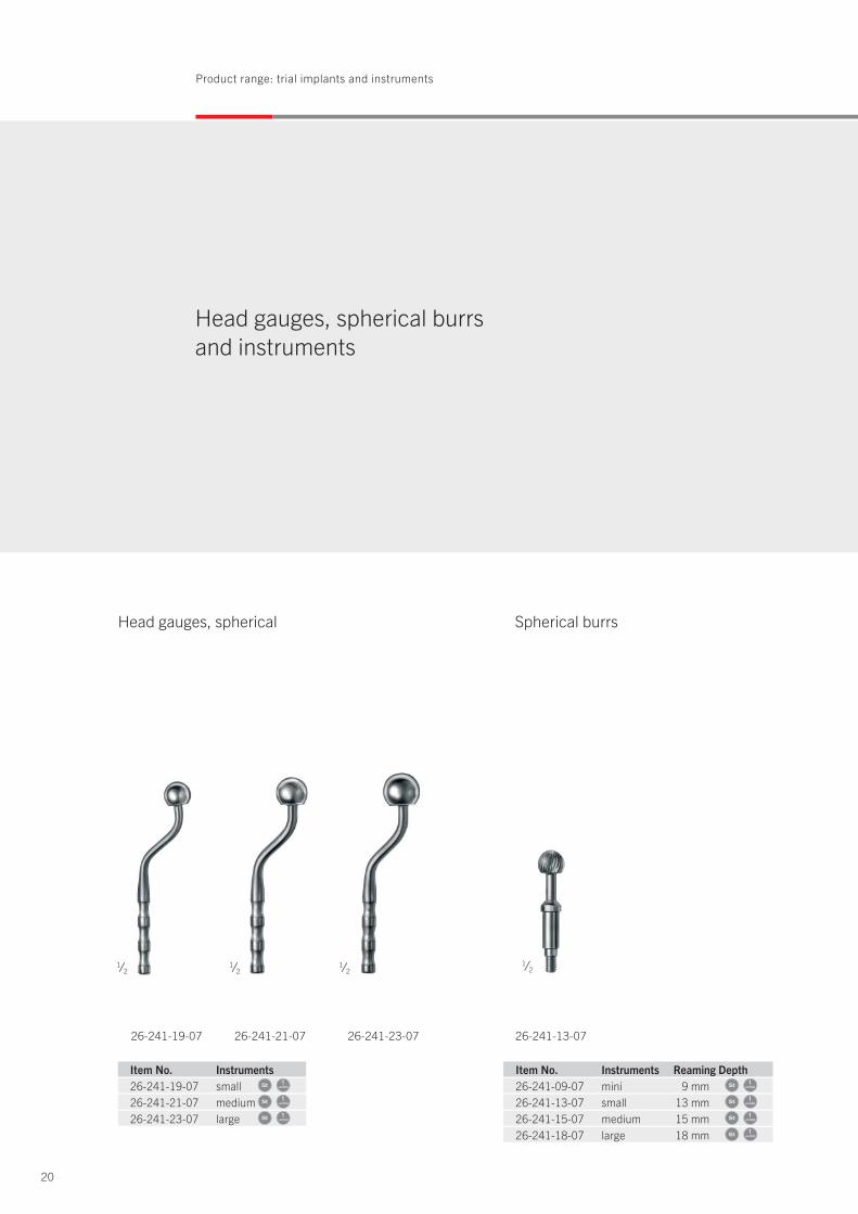
Head gauges, spherical burrs and instruments
1⁄2 1⁄2 1⁄2
26-241-19-07 26-241-21-07 26-241-23-07
1⁄2
Spherical burrs
26-241-13-07
Item No. Instruments 26-241-19-07 small26-241-21-07 medium26-241-23-07 large
Item No. Instruments Reaming Depth 26-241-09-07 mini 9 mm26-241-13-07 small 13 mm26-241-15-07 medium 15 mm26-241-18-07 large 18 mm
Head gauges, spherical
unit(s)1
unit(s)1
unit(s)1
unit(s)1
unit(s)1
unit(s)1
unit(s)1
21
1⁄21⁄2 1⁄2
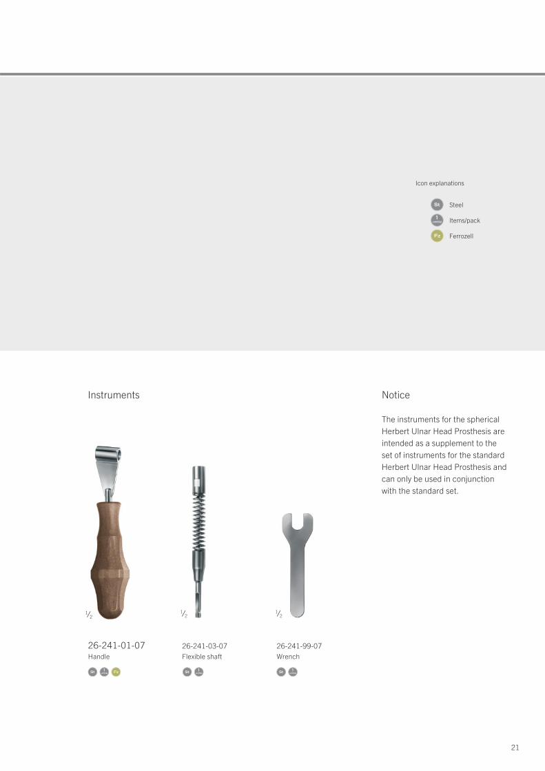
26-241-01-07Handle
26-241-03-07Flexible shaft
26-241-99-07Wrench
Instruments Notice
The instruments for the spherical Herbert Ulnar Head Prosthesis areintended as a supplement to the set of instruments for the standardHerbert Ulnar Head Prosthesis andcan only be used in conjunction with the standard set.
Icon explanations
unit(s)1
Steel
Items/pack
Ferrozell
unit(s)1
unit(s)1
unit(s)1
22
Service: information material and catalogs
Should any more questions remain … … just contact us!
Apart from our range of products specially tailored to the requirements posed by traumatological and reconstructive interventions in hand surgery, we also offer you a wide selection of different systems for use in classical traumatology. Please do not hesitate to order our Special Catalog for the Upper and Lower Extremities, which is available in printed and digital form (CD). To facilitate the ordering process for you, we have created a special Order Form that is available on request at any time.
Of course, you can reach us personally at your convenience, either by e-mail – [email protected] – or telephone (customer hotline): +49-7461-706-109.
23
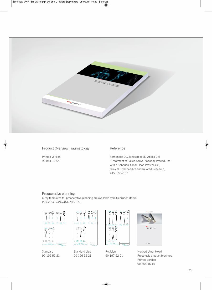
Product Overview Traumatology
Printed version90-851-16-04
Reference
Fernandez DL, Joneschild ES, Abella DM“Treatment of Failed Sauvé-Kapandji Procedures with a Spherical Ulnar Head Prosthesis”,Clinical Orthopaedics and Related Research,445, 100–107
Preoperative planning X-ray templates for preoperative planning are available from Gebrüder Martin.Please call +49-7461-706-109.
Standard90-195-52-21
Standard plus90-196-52-21
Revision90-197-52-21
Herbert Ulnar Head Prosthesis product brochurePrinted version90-665-16-10
Spherical UHP_En_2018.qxp_90-269-01 MicroStop dt.qxd 05.02.18 15:57 Seite 23

02.18 · 90-318-02-07 · Printed in Germany · Copyright by Gebrüder Martin GmbH & Co. KG · Alle Rechte vorbehalten · Technische Änderungen vorbehaltenWe reserve the right to make alterations · Cambios técnicos reservados · Sous réserve de modifications techniques · Ci riserviamo il diritto di modifiche tecniche
Gebrüder Martin GmbH & Co. KGA company of the KLS Martin GroupKLS Martin Platz 1 · 78532 Tuttlingen · Germany P.O. Box 60 · 78501 Tuttlingen · GermanyTel. +49 7461 706-0 · Fax +49 7461 [email protected] · www.klsmartin.com
KLS Martin Group
KLS Martin Australia Pty Ltd.Sydney · AustraliaTel. +61 2 9439 [email protected]
KLS Martin do Brasil Ltda.São Paulo · BrazilTel. +55 11 3554 [email protected]
KLS Martin Medical (Shanghai) International Trading Co., Ltd.Shanghai · ChinaTel. +86 21 5820 [email protected]
KLS Martin India Pvt Ltd.Chennai · India Tel. +91 44 66 442 [email protected]
Martin Italia S.r.l.Milan · ItalyTel. +39 039 605 67 [email protected]
Nippon Martin K.K.Tokyo · JapanTel. +81 3 3814 [email protected]
KLS Martin SE Asia Sdn. Bhd.Penang · Malaysia Tel. +604 505 [email protected]
Martin Nederland/Marned B.V.Huizen · The Netherlands Tel. +31 35 523 45 [email protected]
Gebrüder Martin GmbH & Co. KGMoscow · RussiaTel. +7 499 [email protected]
Gebrüder Martin GmbH & Co. KGDubai · United Arab Emirates Tel. +971 4 454 16 [email protected]
KLS Martin UK Ltd.London · United Kingdom Tel. +44 1189 000 [email protected]
KLS Martin LP Jacksonville · Florida, USA Tel. +1 904 641 77 [email protected]