spinal glia and proinflammatory cytokines mediate …spinal glia and proinflammatory cytokines...
TRANSCRIPT

Spinal Glia and Proinflammatory Cytokines MediateMirror-Image Neuropathic Pain in Rats
Erin D. Milligan,1 Carin Twining,1 Marucia Chacur,2 Joseph Biedenkapp,1 Kevin O’Connor,1 Stephen Poole,3
Kevin Tracey,4 David Martin,5 Steven F. Maier,1 and Linda R. Watkins1
1Department of Psychology and the Center for Neuroscience, University of Colorado at Boulder, Boulder, Colorado 80309-0345, 2Laboratory ofPathophysiology, Butantan Institute, 05503-900, San Paulo, SP, Brazil,3Division of Endocrinology, National Institute for Biological Standards and Control,South Mimms, Potters Bar, Herts EN6 3QG, United Kingdom, 4Laboratory of Biomedical Science, North Shore–LIJ Research Institute, Manhasset, NewYork 11030, and 5Department of Pharmacology, Amgen, Thousand Oaks, California 91320
Mirror-image allodynia is a mysterious phenomenon that occurs in association with many clinical pain syndromes. Allodynia refers topain in response to light touch/pressure stimuli, which normally are perceived as innocuous. Mirror-image allodynia arises from thehealthy body region contralateral to the actual site of trauma/inflammation. Virtually nothing is known about the mechanisms under-lying such pain. A recently developed animal model of inflammatory neuropathy reliably produces mirror-image allodynia, thus allowingthis pain phenomenon to be analyzed. In this sciatic inflammatory neuropathy (SIN) model, decreased response threshold to tactilestimuli (mechanical allodynia) develops in rats after microinjection of immune activators around one healthy sciatic nerve at mid-thighlevel. Low level immune activation produces unilateral allodynia ipsilateral to the site of sciatic inflammation; more intense immuneactivation produces bilateral (ipsilateral � mirror image) allodynia. The present studies demonstrate that both ipsilateral and mirror-image SIN-induced allodynias are (1) reversed by intrathecal (peri-spinal) delivery of fluorocitrate, a glial metabolic inhibitor; (2)prevented and reversed by intrathecal CNI-1493, an inhibitor of p38 mitogen-activated kinases implicated in proinflammatory cytokineproduction and signaling; and (3) prevented or reversed by intrathecal proinflammatory cytokine antagonists specific for interleukin-1,tumor necrosis factor, or interleukin-6. Reversal of ipsilateral and mirror-image allodynias was rapid and complete even when SIN wasmaintained constantly for 2 weeks before proinflammatory cytokine antagonist administration. These results provide the first evidencethat ipsilateral and mirror-image inflammatory neuropathy pain are created both acutely and chronically through glial and proinflam-matory cytokine actions.
Key words: microglia; astrocyte; interleukin-1; tumor necrosis factor; interleukin-6; allodynia; p38 MAP kinase
IntroductionTrauma and inflammation of peripheral nerves induce patholog-ical pain, referred to as neuropathic pain (Zimmermann, 2001).Although neuropathic pain is perceived to arise from the skininnervated by the damaged/inflamed nerve, pathological paincan also arise from sites contralateral to (the mirror image of) thesite of pathology (Watkins and Maier, 2002). Mirror-image painoccurs in chronic pain conditions, including reflex sympatheticdystrophy (Maleki et al., 2000), causalgia (Shir and Seltzer, 1991),atypical facial pain (Woda and Pionchon, 2000), idiopathic facialarthromyalgia (Woda and Pionchon, 2000), and stomatodynia(Woda and Pionchon, 2000). It is typically characterized by me-chanical allodynia (Moriwaki and Yuge, 1999; Baron, 2000). Thatis, mirror-image pain is perceived in response to light touch/pressure stimuli such as clothing and bed sheets (Slart et al.,1997).
How mirror-image pain is created is unknown. Althoughneuropathic pain from the area of nerve trauma can be accountedfor, in part, by ectopic action potentials and hyperexcitability(Woolf and Salter, 2000), no abnormal activity has been reportedin the healthy contralateral nerve. Hence abnormal contralateralperipheral nerve responsivity cannot account for mirror-imagepain. Rather, mirror-image pain likely arises from altered spinalprocessing of incoming sensory information (Koltzenburg et al.,1999; Watkins and Maier, 2002). Various neurocircuits havebeen proposed for how altered contralateral neural processing ofpain may occur (Koltzenburg et al., 1999; Ossipov et al., 2000),but whether such neurocircuits adequately account for mirror-image allodynia is unknown. Furthermore, virtually nothing isknown regarding the neurochemical bases of this painphenomenon.
The ability to study mirror-image allodynia has recently beenfacilitated by the development of the sciatic inflammatory neu-ropathy (SIN) model (Chacur et al., 2001; Gazda et al., 2001).This animal model creates neuropathic pain as a result of local-ized inflammation of one healthy sciatic nerve. Inflammation isinduced by perisciatic microinjection of an immune activator(yeast cell walls; zymosan). This procedure creates rapid, robust,low-threshold mechanical allodynia. Low levels of immune acti-vation create a unilateral mechanical allodynia ipsilateral to theinflamed sciatic nerve. Stronger immune activation creates bilat-
Received July 23, 2002; revised Nov. 1, 2002; accepted Nov. 12, 2002.This work was supported by National Institutes of Health Grants MH01558, MH00314, MH45045, and NS38020.
We thank Amgen (Thousand Oaks, CA) for their gift of IL1ra, TNFbp, and vehicles appropriate for each. Affinity-purified anti-rat IL6 was provided by Dr. Stephen Poole (National Institute for Biological Standards and Control, UK).This antibody was raised as part of the European Community-funded Concerted Action Program Biomed I “Cytokinesin the Brain” (PL931450).
Correspondence should be addressed to Linda R. Watkins, Department of Psychology, Campus Box 345, Univer-sity of Colorado at Boulder, Boulder, CO 80309-0345. E-mail: [email protected].
Copyright © 2003 Society for Neuroscience 0270-6474/03/231026-15$15.00/0
1026 • The Journal of Neuroscience, February 1, 2003 • 23(3):1026 –1040

eral allodynia; that is, allodynia is observed both ipsilateral andcontralateral (mirror-image) to the nerve inflammation (Chacuret al., 2001; Gazda et al., 2001). Mirror-image allodynia producedby SIN cannot be accounted for by systemic spread of the im-mune activator (Chacur et al., 2001). Rather, expression of con-tralateral allodynia is correlated with well defined immunologicaland anatomical changes in and around the inflamed sciatic nerve(Gazda et al., 2001).
The purpose of the present series of experiments is to providean initial investigation of spinal cord mechanisms underlyingmirror-image allodynia. It also provides the first investigation ofspinal mediators of inflammatory neuropathy pain. In contrast tothe wealth of studies focused on the spinal neurochemistry ofpain from traumatic neuropathy (Zimmermann, 2001), the spi-nal mediators of inflammatory neuropathy pain have not beenidentified. Specifically, these experiments examine whether spi-nal cord glia are involved in SIN-induced ipsilateral and con-tralateral pain changes and, if so, which glially derived substanc-e(s) is critically involved in creating or maintaining suchpathological pain.
Materials and MethodsSubjectsPathogen-free adult male Sprague Dawley rats (300 – 450 gm; HarlanLabs, Madison, WI) were used in all experiments. Rats were housed intemperature-controlled (23 � 3°C) and light-controlled (12 hr light/dark; lights on at 7:00 A.M.) rooms with standard rodent chow and wateravailable ad libitum. Behavioral testing was performed during the lightcycle. There were five to six rats per in every experiment. All procedureswere approved by the Institutional Animal Care and Use Committee ofthe University of Colorado at Boulder.
DrugsZymosan (yeast cell walls; Sigma, St. Louis, MO) was made fresh daily bysuspension in a vehicle of incomplete Freund’s adjuvant (Sigma) to finalconcentrations of 0.08 �g/�l and 3.2 �g/�l. The glial metabolic inhibitorfluorocitrate (Sigma) (Paulsen et al., 1987; Hassel et al., 1992) was dis-solved initially in 2 M HCl and then diluted in sterile, endotoxin-free 10mM PBS (Invitrogen, Gaithersburg, MD) to attain a final concentrationof 1 nmol fluorocitrate per microliter, pH 6.0. This solution was ali-quoted and stored at �70°C. The vehicle (0.3% 2 M HCl in PBS, pH 6.0)was aliquoted and stored at 4°C. The p38 mitogen-activated protein(MAP) kinase inhibitor CNI-1493 (Denham et al., 2000) was synthesizedat North Shore University Hospital as described previously (Bianchi etal., 1995). CNI-1493 was dissolved in endotoxin-free sterile distilled wa-ter (9 �g/�l), aliquoted, and stored at �70°C. Endotoxin-free solutionsof recombinant met-human interleukin-1 receptor antagonist (IL1ra;100 �g/�l; lot number 2010316L6; Amgen, Thousand Oaks, CA) andIL1ra vehicle (lot number 0210306L6; Amgen) were stored at 4°C. Ly-ophilized tumor necrosis factor binding protein (TNFbp; endotoxin-freepolyethylene glycol recombinant human soluble TNF receptor type I; lotnumber 36000D8; Amgen) was reconstituted at 30 �g/�l (its limit ofsolubility) in endotoxin-free sterile distilled water, aliquoted on ice, andstored at �75°C. TNFbp vehicle (lot number 1105208E8; Amgen) wasstored at 4°C. Lyophilized affinity-purified sheep anti-rat interleukin-6(IL6) IgG (National Institute for Biological Standards and Control, Pot-ters Bar, UK) was reconstituted at 1.3 �g/�l in endotoxin-free steriledistilled water, aliquoted, and stored at �70°C. At the time of testing, athawed aliquot of anti-rat IL6 was diluted in sterile 0.9% saline to a finalconcentration of 0.065 �g/�l. Affinity-purified normal sheep IgG (con-trol; lot number 31K9105; Sigma) was reconstituted to 1.3 �g/�l, ali-quoted, stored, and diluted at the time of test to 0.065 �g/�l in an iden-tical manner.
Behavioral measuresThe von Frey test (Chaplan et al., 1994) was performed within the sciaticinnervation area of the hindpaws as described previously in detail (Mil-ligan et al., 2000, 2001b; Chacur et al., 2001; Gazda et al., 2001). Briefly, a
logarithmic series of 10 calibrated Semmes-Weinstein monofilaments(von Frey hairs; Stoelting, Wood Dale, IL) was applied randomly to theleft and right hindpaws to determine the stimulus intensity thresholdstiffness required to elicit a paw withdrawal response. Log stiffness of thehairs is determined by log10 (milligrams � 10). The 10 stimuli had thefollowing log-stiffness values (value in grams is given in parentheses):3.61 (407 mg), 3.84 (692 mg), 4.08 (1202 mg), 4.17 (1479 mg), 4.31 (2041mg), 4.56 (3630 mg), 4.74 (5495 mg), 4.93 (8511 mg), 5.07 (11749 mg),and 5.18 (15136 mg). The range of monofilaments used in these experi-ments (0.407–15.136 gm) produces a logarithmically graded slope wheninterpolating a 50% response threshold of stimulus intensity (expressedas log10 (milligrams � 10)) (Chaplan et al., 1994). Assessments weremade before (baseline) and at specific times after perisciatic and intra-thecal drug administration, as detailed below for each experiment. Be-havioral testing was performed blind with respect to drug administra-tion. The behavioral responses were used to calculate the 50% pawwithdrawal threshold (absolute threshold), by fitting a Gaussian integralpsychometric function using a maximum-likelihood fitting method(Harvey, 1986; Treutwein and Strasburger, 1999), as described in detailpreviously (Milligan et al., 2000, 2001b). This fitting method allows para-metric statistical analyses (Milligan et al., 2000, 2001b).
Surgery and microinjectionsChronic intrathecal catheters. Lumbosacral intrathecal catheters wereconstructed and implanted by lumbar approach as described previouslyin detail (Milligan et al., 1999). The indwelling catheters were used tomicroinject drugs into the CSF space surrounding the lumbosacral spinalcord. Only one intrathecal injection was made per animal. All intrathecalmicroinjections were performed as detailed previously, using an 8 �lvoid volume to ensure complete drug delivery (Milligan et al., 1999). Allcatheter placements were verified after completion of behavioral testingby visual inspection. Data were only analyzed from animals with cathe-ters verified as having the catheter tip at the lumbosacral spinal level.
Chronic perisciatic catheters. Perisciatic catheters were constructed andimplanted at mid-thigh level of the left hindleg as described previously indetail (Chacur et al., 2001; Gazda et al., 2001; Milligan et al., 2003). Thismethod allowed multiday recovery of the animal from isofluorane anes-thesia before microinjection of an immune activator around the sciaticnerve. This avoids the deleterious effects of anesthetics on the function ofboth immune (Lockwood et al., 1993; Sato et al., 1995; Miller et al., 1996)and glial cells (Tas et al., 1987; Mantz et al., 1993; Miyazaki et al., 1997;Feinstein et al., 2001). In addition, this indwelling catheter method al-lowed perisciatic immune activation to be either acute (single injection ofan immune activator) or chronic (repeated injections across weeks)(Milligan et al., 2003). Both methods were used in the present experi-ments in awake unrestrained rats. These acute and chronic perisciaticmicroinjections over the left sciatic nerve were performed as detailedpreviously (Milligan et al., 1999; Chacur et al., 2001). For all experiments,catheters were verified by visual inspection at the time animals werekilled. Data were analyzed only from confirmed sites.
Data analysisAll statistical comparisons were computed using Statview 5.0.1 for theMacintosh. Data from the von Frey test were analyzed as the interpolated50% threshold (absolute threshold) in log base 10 of stimulus intensity(monofilament stiffness in milligrams � 10). Pre-drug baseline measureswere analyzed by one-way ANOVA. Post-drug time course measureswere analyzed by repeated measures ANOVAs followed by Fisher’s pro-tected least significant difference post hoc comparisons, whereappropriate.
Experiment 1: effect of intrathecal fluorocitrate on sciaticinflammatory neuropathy-induced allodynia: blockadeof allodyniaFluorocitrate selectively inhibits aconitase, an enzyme that is in theKrebs’ energy cycle of glia, but not neurons (Paulsen et al., 1987; Hassel etal., 1992). Although prolonged glial disruption can indirectly lead toaltered neuronal function (e.g., by altering glially regulated uptake ofexcitatory amino acids; for discussion, see Milligan et al. (2000)), effectsobserved at short post-drug times after 1 nmol fluorocitrate reflect glial
Milligan et al. • Spinal Cytokines Mediate SIN-Induced Pain States J. Neurosci., February 1, 2003 • 23(3):1026 –1040 • 1027

inactivation (Paulsen et al., 1987; Hassel et al., 1992). Blockade of theinitial development of exaggerated pain states by 1 nmol intrathecal fluo-rocitrate has provided supportive evidence that spinal cord glia are in-volved in enhanced pain induced by peripheral inflammation (Meller etal., 1994; Watkins et al., 1997) and direct spinal immune activation (Mil-ligan et al., 2000). Thus, intrathecal fluorocitrate was tested here to assesswhether spinal cord glia may participate in the development of ipsilateralor mirror-image SIN-induced low-threshold mechanical allodynias.
After baseline behavioral assessments, the glial metabolic inhibitorfluorocitrate (1 nmol) (Paulsen et al., 1987; Hassel et al., 1992) or equiv-olume vehicle (1 �l) was microinjected intrathecally. This intrathecaldose has previously been documented to block enhanced pain statesproduced by peripheral inflammation or spinal glial activation (Meller etal., 1994; Watkins et al., 1997; Milligan et al., 2000; Chacur et al., 2001).Thirty minutes after the intrathecal injection, rats received perisciaticmicroinjections of 0, 4, or 160 �g zymosan in 50 �l of vehicle. The 4 and160 �g zymosan doses were chosen for this and all subsequent experi-ments on the basis of their effectiveness in producing ipsilateral (relativeto the site of injection) and bilateral allodynia, respectively, in intrathecalcatheterized rats (Milligan et al., 2003). Behavior was reassessed 1, 2, and3 hr later. Behavioral testing was restricted to these early postinjectiontimes to avoid potential nonselective effects of fluorocitrate on neuronalfunction that may occur at later time points (for discussion see Milliganet al. (2000)).
Experiment 2: effect of intrathecal CNI-1493 on sciaticinflammatory neuropathy-induced allodynia: preventionof allodyniaCNI-1493 is a p38 MAP kinase inhibitor (Denham et al., 2000). p38 MAPkinase participates in one of the major intracellular signaling cascadesleading to the production and release of proinflammatory cytokines(TNF, IL1, IL6) in glia and immune cells (Lee et al., 2000). In addition,p38 MAP kinase is part of the intracellular signaling cascade activated byproinflammatory cytokines binding to their receptors (Lee et al., 2000).Thus, p38 MAP kinase inhibitors can disrupt both production of andsignaling by these proteins. As an initial screen for potential proinflam-matory cytokine mediation of SIN-induced allodynias, intrathecal CNI-1493 was administered before induction of SIN.
After baseline behavioral assessments, the p38 MAP kinase inhibitorCNI-1493 (9 �g) (Denham et al., 2000) or equivolume saline (1 �l) wasmicroinjected intrathecally. p38 MAP kinase is implicated in the intra-cellular signaling cascades (1) activated in response to proinflammatorycytokines binding their receptors (Raingeaud et al., 1995; Ridley et al.,1997) and (2) which lead to the production of proinflammatory cyto-kines (Lee et al., 1994, 2000). The CNI-1493 dose used here has previ-ously been documented to block enhanced pain states produced by directspinal glial activation (Watkins et al., 1997; Milligan et al., 2000). Thirtyminutes after intrathecal injection, each rat received a perisciatic micro-injection of 0, 4, or 160 �g zymosan, performed as above. Behavior wasreassessed 1, 1.5, 2, 3, and 24 hr later.
Experiment 3: effect of intrathecal CNI-1493 on sciaticinflammatory neuropathy-induced allodynia: reversal of allodyniaAfter baseline behavioral assessments, perisciatic microinjections of 0, 4,or 160 �g zymosan were performed as above. Behavior was reassessed 13hr later to confirm the effectiveness of the perisciatic injections beforeintrathecal drug delivery. At 14.5 hr after perisciatic injection, each ratreceived an intrathecal injection of either CNI-1493 (9 �g) or equivol-ume saline (1 �l). Behavior was then assessed 0.5, 2.5, and 4.5 hr later(that is, 15, 17, and 19 hr after the perisciatic injection).
Experiment 4: effect of intrathecal tumor necrosis factor bindingprotein on sciatic inflammatory neuropathy-induced allodynia:blockade of allodyniaIt should be noted here that CNI-1493 has recently been demonstrated tobe capable of crossing the blood– brain barrier into spinal cord aftersystemic administration (Milligan et al., 2001a). Whether it could crossthe blood– brain barrier from lumbar cerebrospinal fluid to reach thedorsal root ganglia is unknown. However, if this were possible, the CNI-1493 effects in experiments 2 and 3 might be accounted for by alterations
in dorsal root ganglia proinflammatory cytokine function. Because dor-sal root ganglia proinflammatory cytokines are upregulated in at leastsome pathological pain states (Watkins and Maier, 2002), this is an in-triguing possibility. Testing TNF, IL6, and IL1 antagonists in this andsubsequent experiments will clarify whether spinal proinflammatory cy-tokines are involved because these antagonists are all large proteins thatdo not diffuse across the blood– brain barrier; however, this does leavethe question of potential dorsal root ganglia proinflammatory cytokineinvolvement open for future studies.
After baseline behavioral assessments, and 60 min before perisciaticinjections, the TNF antagonist TNFbp (TNF soluble receptor; 300 �g) orequivolume vehicle (10 �l) was microinjected intrathecally. The TNFbpdose and timing of its administration were based on previous studies inwhich TNFbp blocked enhanced pain states produced by direct spinalglial activation (Milligan et al., 2001b). Behavior was reassessed 1, 3, and24 hr after perisciatic microinjection of 0, 4, or 160 �g zymosan.
Experiment 5: effect of intrathecal anti-rat interleukin-6 on sciaticinflammatory neuropathy-induced allodynia: reversal of allodynia1 d laterAfter baseline behavioral assessments, perisciatic microinjections of 0, 4,or 160 �g zymosan were performed as above. Behavior was reassessed 13hr later to confirm the effectiveness of the perisciatic injections beforeintrathecal drug delivery. At 14.5 hr after perisciatic injection, each ratreceived an intrathecal injection of either affinity-purified sheep anti-ratIL6 IgG (0.065 �g in 5 �l) or affinity-purified normal sheep IgG (0.065�g in 5 �l). Behavior was then assessed 0.5, 2.5, and 4.5 hr later (that is,15, 17, and 19 hr after the perisciatic injection).
Experiment 6: effect of intrathecal interleukin-1 receptorantagonist on sciatic inflammatory neuropathy-induced allodynia:reversal of allodynia 1 d laterAfter baseline behavioral assessments, perisciatic microinjections of 0, 4,or 160 �g zymosan were performed as above. Behavior was reassessed 13hr later to confirm the effectiveness of the perisciatic injections beforeintrathecal drug delivery. At 14.5 hr after perisciatic injection, each ratreceived an intrathecal injection of either IL1ra (100 �g) or equivolumevehicle (1 �l). Behavior was then assessed 0.5, 2.5, and 4.5 hr later (that is,15, 17, and 19 hr after the perisciatic injection).
Experiment 7: effect of intrathecal interleukin-1 receptorantagonist on sciatic inflammatory neuropathy-induced allodynia:reversal of allodynia 2 weeks laterBehavioral assessments were recorded before (baseline) and at 1, 4, 8, 10,12, and 14 d after baseline. In half of the animals, a perisciatic microin-jection of 160 �g zymosan was delivered immediately after baseline (day0) and 3, 5, 7, 9, 11, and 13 d later. This injection schedule was based onpilot studies aimed at maintaining robust allodynia across days. Theremaining animals were identically implanted with indwelling perisciaticcatheters and injected with equivolume saline on corresponding days.On day 14, after an initial behavioral assessment (day 14 baseline), allanimals received either IL1ra (100 �g) or equivolume (1 �l) vehicleintrathecally. Behavior was reassessed 0.5, 1, 1.5, 2 and 2.5 hr later.
ResultsExperiment 1: effect of intrathecal fluorocitrate on sciaticinflammatory neuropathy-induced allodynia: blockadeof allodyniaImmunohistochemical evidence of bilateral astrocyte and microglialactivation has been observed after traumatic neuropathies (Colburnet al., 1999; Winkelstein et al., 2001). Whether glial activation is theconsequence of nerve trauma or nerve inflammation in these modelsis unknown. However, intense sciatic nerve inflammation has re-cently been reported to also induce immunohistochemical evidenceof bilateral glial activation (Herzberg and Sagen, 2001). This suggeststhat glial activation may occur in response to SIN. Thus the purposeof the present experiment was to determine whether fluorocitrate, a
1028 • J. Neurosci., February 1, 2003 • 23(3):1026 –1040 Milligan et al. • Spinal Cytokines Mediate SIN-Induced Pain States

glial metabolic inhibitor, could block pathological pain induced byinflammatory neuropathy.
As in our previous studies (Chacur et al., 2001; Gazda et al.,2001), low-dose zymosan induced a unilateral allodynia (Fig.1A), whereas higher dose zymosan induced a bilateral allodynia(Fig. 1B), compared with vehicle controls (Fig. 1A). Pretreat-ment with intrathecal fluorocitrate prevented the development ofSIN-induced pain changes (Fig. 1A,B).
These observations were supported by statistical analyses.ANOVA revealed reliable main effects of zymosan dose (F(2,210) �214.125; p � 0.0001), intrathecal fluorocitrate (F(2,62) � 182.756;p � 0.0001), laterality (F(1,62 � 88.069; p � 0.0001), and time(F(2,124) � 33.601; p � 0.0001), and interactions between intrathecalfluorocitrate and zymosan dose (F(2,62) � 70.867; p � 0.0001),zymosan dose and laterality (F(2,62) � 31.775; p � 0.0001), intra-thecal fluorocitrate, zymosan dose, and laterality (F(2,62) �
12.070; p � 0.0001) and time, intrathecal fluorocitrate, zymosandose, and laterality (F(4,124) � 2.692; p � 0.05).
Post hoc means comparison revealed several important points.After 4 �g zymosan (Fig. 1A), mechanical allodynia was observedin the left (ipsilateral) hindpaw compared with the right (con-tralateral) hindpaw ( p � 0.0001). Mechanical responses of theright hindpaw after 4 �g zymosan did not differ from that afterperisciatic vehicle (Fig. 1A), supporting the conclusion that 4 �gzymosan induced only a unilateral allodynia ipsilateral to the siteof injection. Fluorocitrate greatly reduced the allodynic effects of4 �g zymosan ( p � 0.0001 comparing the ipsilateral paw of ratsreceiving 4 �g zymosan with versus without intrathecal fluoroci-trate) (Fig. 1A), because mild allodynia was observed only at 1 hrafter perisciatic zymosan ( p � 0.001). Intrathecal fluorocitrate,in the absence of perisciatic zymosan, had no effect on paw with-drawal thresholds, compared with intrathecal vehicle controls
Figure 1. Blockade of perisciatic SIN-induced mechanical allodynias by intrathecal fluorocitrate, a glial metabolic inhibitor. Rats were assessed for low-threshold mechanical sensitivity (von Freytest) both before (baseline) and 1, 2, and 3 hr after completion of intrathecal drug administration. Replicating our earlier studies (Chacur et al., 2001; Gazda et al., 2001), low-dose zymosan induceda unilateral allodynia ( A), whereas high-dose zymosan induced a bilateral allodynia ( B). Although fluorocitrate had no effect in the absence of perisciatic zymosan ( A), it greatly reduced bothunilateral ( A) and bilateral ( B) allodynias induced by perisciatic zymosan. The 10 stimuli tested had the following log-stiffness values (value in grams is given in parentheses): 3.61 (407 mg), 3.84(692 mg), 4.08 (1202 mg), 4.17 (1479 mg), 4.31 (2041 mg), 4.56 (3630 mg), 4.74 (5495 mg), 4.93 (8511 mg), 5.07 (11,749 mg), and 5.18 (15,136 mg). i.t., Intrathecal; Inj, injection; Veh, vehicle;perisci, perisciatic; Lo Zym, low-dose zymosan; Hi Zym, high-dose zymosan. Abbreviations apply to Figures 1–7.
Milligan et al. • Spinal Cytokines Mediate SIN-Induced Pain States J. Neurosci., February 1, 2003 • 23(3):1026 –1040 • 1029

( p � 0.5) (Fig. 1A). Post hoc means comparison also revealed thatbilateral mechanical allodynia occurred in response to 160 �gzymosan. That is, the thresholds of the left and right hindpawsdid not differ ( p � 0.05 comparing the ipsilateral and contralat-eral paws of rats receiving 160 �g zymosan but no fluorocitrate)(Fig. 1B), but the thresholds for both the left and right paws for allof these groups were reliably different from those of the vehiclecontrols ( p � 0.0001 and p � 0.0001, respectively) (Fig. 1A).Fluorocitrate greatly reduced the perisciatic allodynia in both theipsilateral ( p � 0.0001) and contralateral ( p � 0.0001) paws,compared with 160 �g zymosan-injected rats receiving vehicleintrathecally (Fig. 1B). Indeed, only mild, transient allodynia wasobserved in the fluorocitrate-treated animals at 1 hr after peris-ciatic zymosan ( p � 0.05).
Experiment 2: effect of intrathecal CNI-1493 on sciaticinflammatory neuropathy-induced allodynia: preventionof allodyniaExperiment 1 provided initial evidence that spinal cord glia maybe involved in the mediation of both ipsilateral and mirror-image
SIN-induced allodynias. This is the first evidence that pain in-duced by inflammation around healthy peripheral nerves likelyinvolves spinal cord glia. This suggests that SIN-induced painchanges would be mediated by pain-enhancing substancesknown to be released by activated glia. Although various sub-stances are released by activated glia, proinflammatory cytokineshave recently been implicated as mediators of diverse exaggeratedpain states (Watkins et al., 2001). Hence, CNI-1493, a globalinhibitor of proinflammatory cytokine function, was tested.
As in our previous studies (Chacur et al., 2001; Gazda et al.,2001), low-dose zymosan induced a unilateral allodynia (Fig.2A), whereas higher dose zymosan induced a bilateral allodynia(Fig. 2B) compared with vehicle controls (Fig. 2A). Pretreatmentwith intrathecal CNI-1493 abolished these SIN-induced painchanges through 3 hr after perisciatic injection (Fig. 2A,B). Allo-dynia recovered by 24 hr, in accordance with known pharmaco-kinetics for this compound (Cerami et al., 1996).
These observations were supported by statistical analyses.ANOVA revealed reliable main effects of zymosan dose (F(2,120) �30.742; p � 0.0001), intrathecal CNI-1493 (F(1,120) � 132.044; p �
Figure 2. Blockade of perisciatic SIN-induced mechanical allodynias by intrathecal CNI-1493, a p38 mitogen-activated kinase inhibitor. Rats were assessed for low-threshold mechanicalsensitivity (von Frey test) both before (baseline) and 1, 1.5, 2, 3, and 24 hr after completion of intrathecal drug administration. Replicating our earlier studies (Chacur et al., 2001; Gazda et al., 2001),low-dose zymosan induced a unilateral allodynia ( A), whereas high-dose zymosan induced a bilateral allodynia ( B). Although CNI-1493 had no effect in the absence of perisciatic zymosan ( A), itabolished unilateral allodynia ( A) and greatly reduced bilateral allodynia ( B) induced through 3 hr by perisciatic zymosan. Both unilateral and bilateral allodynias returned by 24 hr (A, B).
1030 • J. Neurosci., February 1, 2003 • 23(3):1026 –1040 Milligan et al. • Spinal Cytokines Mediate SIN-Induced Pain States

0.0001), and laterality (F(1,120) � 46.807; p � 0.0001), and interac-tions between intrathecal CNI-1493 and zymosan dose (F(2,60) �28.656; p � 0.0001), zymosan dose and laterality(F(2,60) � 7.481; p �0.01), and intrathecal CNI-1493, zymosan dose, and laterality (F(2,60)
� 11.843; p � 0.0001). Allodynia recovered by 24 hr. Both ipsilateralallodynia (Fig. 2A) and bilateral allodynia (Fig. 2B) were fullyrestored by this time. At 24 hr, ANOVA revealed reliable maineffects of zymosan dose (F(1,60) � 13.925; p � 0.0001) and later-ality (F(1,60) � 21.370; p � 0.0001), and interactions betweenzymosan dose and laterality (F(1,60) � 9.864; p � 0.001). As inexperiment 1, 4 �g zymosan (Fig. 2A) produced mechanicalallodynia in the left (ipsilateral) hindpaw compared with the right(contralateral) hindpaw ( p � 0.0001). Also as in experiment 1,mechanical responses of the right hindpaw after 4 �g zymosandid not differ from that after perisciatic vehicle ( p � 0.2) (Fig.2A), indicating that 4 �g zymosan induced only a unilateral al-lodynia ipsilateral to the site of injection. CNI-1493 abolished theallodynic effects of 4 �g zymosan through 3 hr ( p � 0.0001)comparing the ipsilateral paw of rats receiving 4 �g zymosan �intrathecal CNI-1493 with the ipsilateral paw of rats receivingno zymosan � intrathecal CNI-1493 (Fig. 2 A). IntrathecalCNI-1493, in the absence of perisciatic zymosan, had no effecton paw withdrawal thresholds, compared with intrathecalvehicle controls ( p � 0.5) (Fig. 2 A).
Post hoc means comparison also revealed that bilateral me-chanical allodynia occurred in response to 160 �g zymosan. Thatis, the thresholds of the left and right hindpaws did not differ,except at the 1 hr time point ( p � 0.001) (Fig. 2B). The thresh-olds for both the left and right paws for all of these groups werereliably different from those of the appropriate vehicle controls( p � 0.0001) (Fig. 2A). CNI-1493 greatly reduced the perisciaticallodynia in both the ipsilateral ( p � 0.0001) and contralateral( p � 0.0001) paws through 3 hr, compared with 160 �gzymosan-injected rats receiving vehicle intrathecally (Fig. 2A,B).
Experiment 3: effect of intrathecal CNI-1493 on sciaticinflammatory neuropathy-induced allodynia: reversalof allodyniaExperiment 2 provided initial evidence that p38 MAP kinasepathways are involved in the initiation of both ipsilateral andmirror-image SIN-induced allodynias. The present experimenttests whether p38 MAP kinases may be involved in the mainte-nance of these ipsilateral and mirror-image allodynias as well.This is a key issue for pathological pain states, because drug ma-nipulations can often prevent but not reverse pathological painonce it develops (Traub, 1996; Bianchi and Panerai, 1997). Thus,CNI-1493 was administered the day after the allodynias werefully developed to determine whether either allodynia would bereversed by this drug.
Compared with vehicle controls (Fig. 3A), low-dose perisci-atic zymosan induced a unilateral allodynia (Fig. 3A), whereashigher dose perisciatic zymosan induced a bilateral allodynia(Fig. 3B), measured 13 hr later. CNI-1493 injected intrathecally at14.5 hr abolished these SIN-induced pain changes within 2.5– 4.5hr (that is, 17–19 hr after perisciatic injection) (Fig. 3B).
These observations were supported by statistical analyses.ANOVA revealed reliable main effects of zymosan dose (F(2,66) �125.279; p � 0.0001) and laterality (F(1,66) � 80.804; p � 0.0001),and interactions between zymosan dose and laterality (F(2,66) �30.638; p � 0.0001). Post hoc means comparison revealed that 4�g zymosan (Fig. 3A) induced mechanical allodynia in the left(ipsilateral) hindpaw compared with the right (contralateral)hindpaw ( p � 0.0001). Mechanical responses of the right hind-
paw after 4 �g perisciatic zymosan did not differ from that afterperisciatic vehicle (Fig. 3A), showing that 4 �g zymosan inducedonly a unilateral allodynia ipsilateral to the site of injection. Inaddition, post hoc analyses showed that bilateral mechanical allo-dynia occurred in response to 160 �g perisciatic zymosan. Thatis, the thresholds of the left and right hindpaws did not differ (Fig.3B). The thresholds for both the left and right paws for all of thesegroups were reliably different from those of the vehicle controls( p � 0.0001) (Fig. 3A). At 14.5 hr, either CNI-1493 or vehicle wasinjected intrathecally. Behavior was recorded 0.5, 2.5, and 4.5 hrlater (that is, 15, 17, and 19 hr after perisciatic drug administra-tion). Although CNI-1493 had no effect in the absence of peris-ciatic zymosan (Fig. 3A), it reversed both unilateral (Fig. 3A) andbilateral (Fig. 3B) allodynias induced by perisciatic zymosan.ANOVA revealed reliable main effects of zymosan dose (F(2,60) �111.593; p � 0.0001), intrathecal CNI-1493 (F(1,60) � 162.991;p � 0.0001), and laterality (F(1,60) � 61.422; p � 0.0001), andinteractions between intrathecal CNI-1493 and zymosan dose(F(2,60) � 84.320; p � 0.0001), and intrathecal CNI-1493, zymo-san dose, and laterality (F(2,60) � 15.675; p � 0.0001).
Post hoc means comparisons showed that CNI-1493 reversed theallodynic effects of 4 �g zymosan by 17–19 hr ( p � 00001 compar-ing the ipsilateral paw of rats receiving 4 �g zymosan with vs withoutintrathecal CNI-1493) (Fig. 3A). Indeed, the response thresholds ofrats receiving 4 �g zymosan � CNI-1493 were not different fromvehicle controls at this time ( p � 0.3) (Fig. 3A). This drug reversedthe bilateral allodynic effects of 160 �g perisciatic zymosan by 17–19hr as well ( p � 0.0001 comparing the ipsilateral paw of rats receiving160 �g zymosan with vs without intrathecal CNI-1493; p � 0.0001comparing the contralateral paw of rats receiving 160 �g zymosanwith vs without intrathecal CNI-1493) (Fig. 3A,B). Again, the re-sponse thresholds of rats receiving 160 �g zymosan � CNI-1493were not different from vehicle controls at this time ( p � 0.1) (Fig.3A). Intrathecal CNI-1493, in the absence of perisciatic zymosan,had no effect on paw withdrawal thresholds, compared with intra-thecal vehicle controls (Fig. 3A).
Experiment 4: effect of intrathecal tumor necrosis factorbinding protein (soluble receptors) on sciatic inflammatoryneuropathy-induced allodynia: blockade of allodyniaExperiments 2 and 3 demonstrate that p38 MAP kinase is a keymediator in the intracellular signaling leading to SIN-inducedallodynias. Given that p38 MAP kinases are strongly associatedwith proinflammatory cytokine production and signaling (Lee etal., 2000), this suggests that disruption of SIN-induced allodyniasby CNI-1493 may result from disruption of proinflammatorycytokine function. There are three proinflammatory cytokinesknown to be affected by p38 MAP kinase inhibitors: TNF, IL1,and IL6 (Lee et al., 2000). All three have been implicated in thespinal mediation of pain arising from traumatic neuropathies(Arruda et al., 2000; Sweitzer et al., 2001; Winkelstein et al.,2001). Whether it is the trauma or associated inflammation of theperipheral nerve that recruits spinal proinflammatory cytokineinvolvement in these models is unknown. The present experi-ment tested whether disruption of TNF signaling with TNF sol-uble receptors (TNFbp) would mimic the effects of CNI-1493,that is, prevent the development of SIN-induced allodynias. Theexperiments that follow examine IL1 and IL6 in turn.
As before, low-dose zymosan again induced a unilateral allo-dynia (Fig. 4A), whereas higher dose zymosan induced a bilateralallodynia (Fig. 4B) through 24 hr, compared with vehicle con-trols (Fig. 4A). Pretreatment with intrathecal TNFbp preventedthese SIN-induced pain changes through 24 hr, in keeping with
Milligan et al. • Spinal Cytokines Mediate SIN-Induced Pain States J. Neurosci., February 1, 2003 • 23(3):1026 –1040 • 1031

the prolonged half-life of this compound relative to IL1ra (Ben-dele et al., 1998; Edwards, 1999) (Fig. 4A,B).
These observations were supported by statistical analyses.ANOVA revealed reliable main effects of intrathecal TNFbp(F(1,58) � 35.604; p � 0.0001) and laterality (F(1,58) � 23.061; p �0.0001) and time (F(2,116) � 6.141; p � 0.005), and interactionsbetween intrathecal TNFbp and zymosan dose (F(2,58) � 11.509;p � 0.0001), zymosan dose and laterality (F(2,58) � 6.736;p � 0.01), intrathecal TNFbp and zymosan dose and laterality(F(2,58) � 7.331; p � 0.005), time, and zymosan dose (F(4,116) �3.739; p � 0.01), and time, intrathecal TNFbp, zymosan dose,and laterality (F(4,116) � 3.681; p � 0.01). Post hoc means com-parison revealed that 4 �g zymosan induced a unilateral me-chanical allodynia observed in the left (ipsilateral) hindpawcompared with the right (contralateral) hindpaw ( p � 0.0001)(Fig. 4 A). Mechanical responses of the right hindpaw after 4�g zymosan did not differ from that after perisciatic vehicle( p � 0.05) (Fig. 4 A), showing that 4 �g zymosan induced onlya unilateral allodynia ipsilateral to the site of injection. TNFbpprevented the allodynic effects of 4 �g zymosan ( p � 0.0001comparing the ipsilateral paw of rats receiving 4 �g zymosan
with vs without intrathecal TNFbp; p � 0.25 comparing theipsilateral paw of rats receiving 4 �g zymosan � intrathecalTNFbp vs control groups receiving perisciatic vehicle) (Fig.4 A). Intrathecal TNFbp, in the absence of perisciatic zymosan,had no effect on paw withdrawal thresholds, compared withintrathecal vehicle controls ( p � 0.05) (Fig. 4 A).
Post hoc means comparison also revealed that bilateral me-chanical allodynia occurred in response to 160 �g zymosan.That is, the thresholds of the left and right hindpaws did notdiffer ( p � 0.05) (Fig. 4 B), but the thresholds for both the leftand right paws for all of these groups were reliably differentfrom those of the vehicle controls ( p � 0.0001 and p � 0.0001for ipsilateral and contralateral comparisons, respectively)(Fig. 4 A). TNFbp prevented the perisciatic 160 �g zymosan-induced allodynia in both the ipsilateral and contralateralpaws compared with 160 �g zymosan-injected rats receivingvehicle intrathecally ( p � 0.0001 and p � 0.0001 for ipsilateraland contralateral comparisons, respectively) and comparedwith control groups receiving perisciatic vehicle ( p � 0.05 andp � 0.05 for ipsilateral and contralateral comparisons, respec-tively) (Fig. 4 A, B).
Figure 3. Reversal of perisciatic SIN-induced mechanical allodynias by intrathecal CNI-1493, a p38 mitogen-activated kinase inhibitor. Rats were assessed for low-threshold mechanicalsensitivity (von Frey test) both before (baseline) and 13, 15, 17, and 19 hr after completion of perisciatic drug administration. Replicating and extending our earlier studies (Chacur et al., 2001; Gazdaet al., 2001), low-dose zymosan induced a unilateral allodynia ( A), whereas high-dose zymosan induced a bilateral allodynia ( B) at 13 hr after injection. CNI-1493 reversed both of these allodynias,although it had no effect on behavior in the absence of perisciatic zymosan (A).
1032 • J. Neurosci., February 1, 2003 • 23(3):1026 –1040 Milligan et al. • Spinal Cytokines Mediate SIN-Induced Pain States

Experiment 5: effect of intrathecal anti-rat interleukin-6 onsciatic inflammatory neuropathy-induced allodynia: reversalof allodynia 1 d laterExperiment 4 provided evidence that the proinflammatory cyto-kine TNF is a key mediator of SIN-induced allodynias. BecauseTNF can both induce the release of IL6 (Benveniste et al., 1990)and synergize with IL6 (Dinarello, 1997), involvement of TNF inSIN-induced effects does not exclude the possibility that IL6 mayalso be involved. Thus the present experiment sought to extendthe findings of experiment 4 by determining whether anti-IL6could reverse both ipsilateral and mirror-image pain states. Thus,this experiment with anti-IL6 paralleled the design of experiment3 (reversal of SIN-induced allodynias by CNI-1493).
As before, low-dose perisciatic zymosan induced a unilateralallodynia (Fig. 5A), whereas higher dose perisciatic zymosan in-duced a bilateral allodynia (Fig. 5B), measured 13 hr later. Anti-IL6 injected intrathecally at 14.5 hr reversed these SIN-inducedpain changes within 2.5– 4.5 hr (that is, 17–19 hr after perisciaticinjection) (Fig. 5A,B).
These observations were supported by statistical analyses. Be-
fore intrathecal anti-IL6 administration, ANOVA revealed reli-able main effects of zymosan dose (F(2,60) � 94.296; p � 0.0001)and laterality (F(1,60) � 23.977; p � 0.0001), and interactionsbetween zymosan dose and laterality (F(2,60) � 21.085; p �0.0001). Post hoc means comparison revealed that 4 �g zymosan(Fig. 5A) induced mechanical allodynia in the left (ipsilateral)hindpaw compared with the right (contralateral) hindpaw ( p �0.0001). Mechanical responses of the right hindpaw after 4 �gperisciatic zymosan did not differ from that after perisciatic ve-hicle ( p � 0.5) (Fig. 5A), showing that 4 �g zymosan inducedonly a unilateral allodynia ipsilateral to the site of injection. Inaddition, post hoc analyses showed that bilateral mechanical allo-dynia occurred in response to 160 �g perisciatic zymosan. Thatis, the thresholds of the left and right hindpaws did not differ( p � 0.5) (Fig. 5B). The thresholds for both the left and rightpaws for all of these groups were reliably different from those ofthe vehicle controls ( p � 0.0001) (Fig. 5A). At 14.5 hr, eitheraffinity-purified sheep anti-rat IL6 IgG or affinity-purified nor-mal sheep IgG (control) was injected intrathecally. Behavior wasrecorded 0.5, 2.5, and 4.5 hr later (that is, 15, 17, and 19 hr after
Figure 4. Blockade of perisciatic SIN-induced mechanical allodynias by intrathecal TNFbp (TNF-soluble receptors), a TNF antagonist. Rats were assessed for low-threshold mechanical sensitivity(von Frey test) both before (baseline) and 1, 3, and 24 hr after completion of intrathecal drug administration. Replicating our earlier studies (Chacur et al., 2001; Gazda et al., 2001), low-dose zymosaninduced a unilateral allodynia ( A), whereas high-dose zymosan induced a bilateral allodynia ( B). Although TNFbp had no effect in the absence of perisciatic zymosan ( A), it abolished both unilateral( A) and bilateral allodynia ( B) induced through 3 hr by perisciatic zymosan. There was no evident return of allodynia by 24 hr, in accord with its prolonged half-life (A, B).
Milligan et al. • Spinal Cytokines Mediate SIN-Induced Pain States J. Neurosci., February 1, 2003 • 23(3):1026 –1040 • 1033

perisciatic drug administration). Although anti-IL6 had no effectin the absence of perisciatic zymosan (Fig. 5A), it completelyreversed unilateral allodynia (Fig. 5A) and greatly reduced bilat-eral allodynia (Fig. 5B) induced by perisciatic zymosan. ANOVArevealed reliable main effects of zymosan dose (F(2,60) � 40.371;p � 0.0001), intrathecal anti-IL6 (F(1,60) � 29.745; p � 0.0001),and laterality (F(1,60) � 16.992; p � 0.0001), and interactionsbetween zymosan dose and intrathecal anti-IL6 (F(2,60) � 18.022;p � 0.0001), zymosan dose and laterality (F(2,60) � 5.186; p �0.01), and zymosan dose and intrathecal anti-IL6 and laterality(F(2,60) � 5.849; p � 0.005).
Post hoc means comparisons showed that anti-IL6 reversedthe allodynic effects of 4 �g zymosan by 17–19 hr ( p � 0.0001comparing the ipsilateral paw of rats receiving 4 �g zymosan withvs without intrathecal anti-IL6; p � 0.2 comparing the ipsilateralpaw of rats receiving 4 �g zymosan � intrathecal anti-IL6 versusperisciatic vehicle controls) (Fig. 5A). This antiserum reversedthe bilateral allodynic effects of 160 �g perisciatic zymosan by17–19 hr as well ( p � 0.001 comparing the ipsilateral paw of ratsreceiving 160 �g zymosan with vs without intrathecal anti-IL6;p � 0.001 comparing the contralateral paw of rats receiving 160�g zymosan with vs without intrathecal anti-IL6; p � 0.1 and p �
0.1 comparing the ipsilateral and contralateral paw of rats receiv-ing 160 �g zymosan � intrathecal anti-IL6 vs perisciatic vehiclecontrols) (Fig. 5A). Intrathecal anti-IL6, in the absence of peris-ciatic zymosan, had no effect on paw withdrawal thresholds,compared with intrathecal IgG controls ( p � 0.09) (Fig. 5A).
Experiment 6: effect of intrathecal interleukin-1 receptorantagonist on sciatic inflammatory neuropathy-inducedallodynia: reversal of allodynia 1 d laterExperiments 4 and 5 provided evidence that the proinflamma-tory cytokines TNF and IL6 are key mediators of SIN-inducedallodynias. Because TNF and IL6 can both (1) induce the releaseof IL1 (Watkins et al., 1999; Milligan et al., 2001b) and (2) syn-ergize with IL1 (Dinarello, 1997), involvement of TNF and IL6 inSIN-induced effects does not exclude the possibility that IL1 mayalso be involved. Thus the present experiment sought to extendthe findings of experiments 4 and 5 by examining whether IL1receptor antagonist could reverse SIN-induced pain states.
As before, low-dose perisciatic zymosan induced a unilateralallodynia (Fig. 6A), whereas higher dose perisciatic zymosan in-duced a bilateral allodynia (Fig. 6B), measured 13 hr later. IL1rainjected intrathecally at 14.5 hr greatly reduced these SIN-
Figure 5. Reversal of perisciatic SIN-induced mechanical allodynias by intrathecal anti-rat IL6, 1 d later. Rats were assessed for low-threshold mechanical sensitivity (von Frey test) both before(baseline) and 13, 15, 17, and 19 hr after completion of perisciatic drug administration. Replicating experiment 3 and extending our earlier studies (Chacur et al., 2001; Gazda et al., 2001), low-dosezymosan induced a unilateral allodynia ( A), whereas high-dose zymosan induced a bilateral allodynia ( B) at 13 hr after injection. Both were reversed by intrathecal anti-rat IL6 at this time, whereasanti-IL6 had no effect on behavior in the absence of perisciatic zymosan (A).
1034 • J. Neurosci., February 1, 2003 • 23(3):1026 –1040 Milligan et al. • Spinal Cytokines Mediate SIN-Induced Pain States
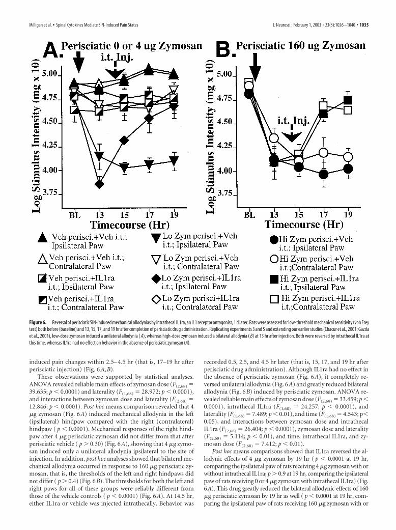
induced pain changes within 2.5– 4.5 hr (that is, 17–19 hr afterperisciatic injection) (Fig. 6A,B).
These observations were supported by statistical analyses.ANOVA revealed reliable main effects of zymosan dose (F(2,68) �39.635; p � 0.0001) and laterality (F(1,68) � 28.972; p � 0.0001),and interactions between zymosan dose and laterality (F(2,68) �12.846; p � 0.0001). Post hoc means comparison revealed that 4�g zymosan (Fig. 6A) induced mechanical allodynia in the left(ipsilateral) hindpaw compared with the right (contralateral)hindpaw ( p � 0.0001). Mechanical responses of the right hind-paw after 4 �g perisciatic zymosan did not differ from that afterperisciatic vehicle ( p � 0.30) (Fig. 6A), showing that 4 �g zymo-san induced only a unilateral allodynia ipsilateral to the site ofinjection. In addition, post hoc analyses showed that bilateral me-chanical allodynia occurred in response to 160 �g perisciatic zy-mosan, that is, the thresholds of the left and right hindpaws didnot differ ( p � 0.4) (Fig. 6B). The thresholds for both the left andright paws for all of these groups were reliably different fromthose of the vehicle controls ( p � 0.0001) (Fig. 6A). At 14.5 hr,either IL1ra or vehicle was injected intrathecally. Behavior was
recorded 0.5, 2.5, and 4.5 hr later (that is, 15, 17, and 19 hr afterperisciatic drug administration). Although IL1ra had no effect inthe absence of perisciatic zymosan (Fig. 6A), it completely re-versed unilateral allodynia (Fig. 6A) and greatly reduced bilateralallodynia (Fig. 6B) induced by perisciatic zymosan. ANOVA re-vealed reliable main effects of zymosan dose (F(2,68) � 33.459; p �0.0001), intrathecal IL1ra (F(1,68) � 24.257; p � 0.0001), andlaterality (F(1,68) � 7.489; p � 0.01), and time (F(1,68) � 4.543; p�0.05), and interactions between zymosan dose and intrathecalIL1ra (F(2,68) � 26.404; p � 0.0001), zymosan dose and laterality(F(2,68) � 5.114; p � 0.01), and time, intrathecal IL1ra, and zy-mosan dose (F(2,68) � 7.412; p � 0.01).
Post hoc means comparisons showed that IL1ra reversed the al-lodynic effects of 4 �g zymosan by 19 hr ( p � 0.0001 at 19 hr,comparing the ipsilateral paw of rats receiving 4 �g zymosan with orwithout intrathecal IL1ra; p � 0.9 at 19 hr, comparing the ipsilateralpaw of rats receiving 0 or 4 �g zymosan with intrathecal IL1ra) (Fig.6A). This drug greatly reduced the bilateral allodynic effects of 160�g perisciatic zymosan by 19 hr as well ( p � 0.0001 at 19 hr, com-paring the ipsilateral paw of rats receiving 160 �g zymosan with or
Figure 6. Reversal of perisciatic SIN-induced mechanical allodynias by intrathecal IL1ra, an IL1 receptor antagonist, 1 d later. Rats were assessed for low-threshold mechanical sensitivity (von Freytest) both before (baseline) and 13, 15, 17, and 19 hr after completion of perisciatic drug administration. Replicating experiments 3 and 5 and extending our earlier studies (Chacur et al., 2001; Gazdaet al., 2001), low-dose zymosan induced a unilateral allodynia ( A), whereas high-dose zymosan induced a bilateral allodynia ( B) at 13 hr after injection. Both were reversed by intrathecal IL1ra atthis time, whereas IL1ra had no effect on behavior in the absence of perisciatic zymosan (A).
Milligan et al. • Spinal Cytokines Mediate SIN-Induced Pain States J. Neurosci., February 1, 2003 • 23(3):1026 –1040 • 1035

without intrathecal IL1ra; p � 0.0001 at 19 hr, comparing the con-tralateral paw of rats receiving 160 �g zymosan with vs withoutintrathecal IL1ra) (Fig. 6B). Intrathecal IL1ra in the absence of peris-ciatic zymosan had no effect on paw withdrawal thresholds, com-pared with intrathecal vehicle controls ( p � 0.05) (Fig. 6A).
Experiment 7: effect of intrathecal interleukin-1 receptorantagonist on sciatic inflammatory neuropathy-inducedallodynia: reversal of allodynia 2 weeks laterExperiment 6 provided evidence that the proinflammatory cyto-kine IL1 is a key mediator of SIN-induced allodynias and is in-volved in the maintenance of allodynia. Because chronic pain canpersist for long periods of time, it was of interest to determinewhether IL1 would still be a key mediator of allodynia once painenhancement was maintained for weeks. If so, this would supportthe idea that IL1 in particular, and proinflammatory cytokines ingeneral, may be clinically relevant targets for pain control. Thus,this last experiment tested whether IL1ra would still be able toreverse long-standing (2 weeks) SIN-induced allodynias.
Figure 7, A and B (left panels), presents the behavioral results ofchronic perisciatic injections, with no intrathecal injections made atany time. That is, all rats in any given left panel are identical in termsof the drugs administered to them through the day 14 time point.Thus the small differences between groups within any given left panelreflect random variability. On day 14, the group designations withineach panel of Figure 7 become meaningful because it is on day 14 thatthe single intrathecal injection of IL1ra or vehicle was administered.The day 14 data shown in the left panels simultaneously serve as thebaseline measure for the intrathecal IL1ra versus vehicle test pre-sented in the right panels.
Low dose perisciatic zymosan induced a unilateral allodyniaby 1 d and was stably maintained across the 2 week observationperiod (Fig. 7A, left panel). There was a trend toward mild allo-dynia developing in the contralateral paw with chronic zymosan,but there was still clearly a marked allodynia in the ipsilateral pawcompared with the contralateral paw (Fig. 7A, left panel). Higherdose perisciatic zymosan also induced a bilateral allodynia by 1 dand was also stably maintained across the 2 week observationperiod (Fig. 7B, left panel). This is the first demonstration thatstable allodynias can be chronically produced by this SIN model.IL1ra injected intrathecally on day 14 reversed these SIN-inducedpain changes within 2.5 hr (Fig. 7A,B, right panels).
These observations were supported by statistical analyses.ANOVA revealed reliable main effects of zymosan dose (F(2,36) �46.844; p � 0.0001) and laterality (F(1,36) � 18.455; p � 0.0001), andinteractions between sciatic treatment and laterality (F(2,36) � 8.550;p � 0.0001). Post hoc means comparison revealed that 4 �g zymosan(Fig. 7A) induced mechanical allodynia in the left (ipsilateral) hind-paw compared with the right (contralateral) hindpaw ( p � 0.0001).Mechanical responses of the right hindpaw after 4 �g perisciaticzymosan did not differ from that after perisciatic vehicle, showingthat 4 �g zymosan induced only a unilateral allodynia ipsilateral tothe site of injection. In addition, post hoc analyses showed that bilat-eral mechanical allodynia occurred in response to 160 �g perisciaticzymosan (Fig. 7B). The thresholds for both the left and right paws forall of these groups were reliably different from those of the vehiclecontrols ( p � 0.0001). At 14 d, either IL1ra or vehicle was injectedintrathecally. Behavior was then recorded for 2.5 hr. Although IL1rahad no effect in the absence of perisciatic zymosan, it completelyreversed unilateral allodynia (Fig. 7A, right) and greatly reduced bi-lateral allodynia (Fig. 7B, right) induced by perisciatic zymosan.ANOVA revealed reliable main effects of intrathecal IL1ra (F(4,42) �91.950; p � 0.0001), and laterality (F(1,42) � 53.390; p � 0.0001), and
time (F(4,168) � 48.251; p � 0.0001), and interactions between intra-thecal IL1ra and laterality (F(4,42) � 22.964; p � 0.0001) and timeand intrathecal IL1ra (F(16,168) � 8.799; p � 0.0001).
Post hoc means comparisons showed that IL1ra abolished theallodynic effects of 4 �g zymosan by 2.5 hr on day 14 ( p � 0.0001comparing the ipsilateral paw of rats receiving 0 or 4 �g zymosanwith intrathecal IL1ra) (Fig. 7A, right). This drug greatly reducedthe bilateral allodynic effects of 160 �g perisciatic zymosan by 2.5hr on day 14 as well ( p � 0.0001 comparing the ipsilateral paw ofrats receiving 160 �g zymosan with or without intrathecal anti-IL6; p � 0.0001 comparing the contralateral paw of rats receiving160 �g zymosan with vs without intrathecal IL1ra) (Fig. 7B,right). Intrathecal IL1ra, in the absence of perisciatic zymosan,had no effect on paw withdrawal thresholds, compared with in-trathecal vehicle controls.
DiscussionThese experiments provide the first identification of spinal me-diators of mirror-image low-threshold mechanical allodynia.They also provide the first identification of neurochemical basesof inflammatory neuropathy-induced pain. These data supportthe conclusion that spinal cord glia and proinflammatory cyto-kines (TNF, IL1, IL6) are key mediators of these pathological painphenomena. These experiments thus support and extend the re-cent report that spinal proinflammatory cytokines (IL1) induceallodynia and hyperexcitability of pain transmission neurons(Reeve et al., 2000). As in previous studies of SIN, low-level sciaticinflammation created ipsilateral allodynia, whereas higher levelsciatic inflammation created both ipsilateral and contralateral(mirror-image) allodynias. All allodynias were (1) blocked byintrathecal fluorocitrate, a glial metabolic inhibitor (Paulsen etal., 1987; Hassel et al., 1992), (2) blocked by an inhibitor of thep38 MAP kinase pathway (intrathecal CNI-1493) implicated inboth proinflammatory cytokine production and intracellular sig-naling cascades (Lee et al., 2000), (3) reversed by this inhibitorafter the allodynias were fully developed, (4) blocked by an intra-thecal TNF antagonist (TNFbp), (5) reversed by an intrathecalIL6 antagonist after the allodynias were fully developed (anti-IL6), and (6) reversed by an intrathecal IL1ra after allodynia wasmaintained for either 1 d or 2 weeks. This last experiment alsoprovides the first demonstration that the SIN model can be usedto study pain chronically maintained by repeated immune acti-vation. The fact that IL1ra reversed ipsilateral and mirror-imagepain after 2 weeks of sciatic inflammation strongly indicates thatproinflammatory cytokines are not important simply for the cre-ation of pathological pain; rather, spinal proinflammatory cyto-kines are critical for maintenance of pathological pain as well.Last, given that nerve trauma always results in inflammation,these data have implications for all neuropathic pain regardless ofwhether pain arises from traumatic or nontraumatic etiologies.
SIN is not alone in creating mirror-image effects. As notedabove, numerous clinical pain syndromes are associated withmirror-image pain, primarily allodynic in nature. Mirror-imagethermal hyperalgesia and mechanical allodynia have also beenobserved in diverse animal models of pathological pain (Seltzer etal., 1990; Coderre and Melzack, 1991; Aloisi et al., 1993; Tal andBennett, 1994; Rees et al., 1996; Takahashi et al., 1996; Sinnott etal., 1999; Hunt et al., 2001). Few studies have examined the mech-anisms involved. Mirror-image thermal hyperalgesia is mediated,at least in part, by substance P, NMDA receptors, non-NMDAreceptors, and dynorphin (Coderre and Melzack, 1991; Chen etal., 2000; Malan et al., 2000). Mirror-image allodynia is distinct
1036 • J. Neurosci., February 1, 2003 • 23(3):1026 –1040 Milligan et al. • Spinal Cytokines Mediate SIN-Induced Pain States

Figure 7. Reversal of perisciatic SIN-induced mechanical allodynias by intrathecal IL1ra, an IL1 receptor antagonist, 2 weeks later. Rats were assessed for low-threshold mechanical sensitivity(von Frey test) both before (baseline) and across 2 weeks after completion of perisciatic drug administration. Extending our earlier studies (Chacur et al., 2001; Gazda et al., 2001), low-dose zymosaninduced a unilateral allodynia ( A), whereas high-dose zymosan induced a bilateral allodynia ( B) by 1 d after injection and stably maintained for 2 weeks. Both were reversed by intrathecal IL1ra atthis time, whereas IL1ra had no effect on behavior in the absence of perisciatic zymosan (A, B, right panels).
Milligan et al. • Spinal Cytokines Mediate SIN-Induced Pain States J. Neurosci., February 1, 2003 • 23(3):1026 –1040 • 1037

from mirror-image thermal hyperalgesia because it is not medi-ated by NMDA or dynorphin (Malan et al., 2000). Until now, nospinal mediators of mirror-image allodynia had been identified.
The involvement of glia in creating mirror-image effects is in-triguing. Glia are well suited for creating expansions of the bodyregion from which pain is perceived, for two reasons. First, proin-flammatory cytokines act in a paracrine manner to excite distantcells (Watkins et al., 1999). This would potentially allow the proin-flammatory cytokines to reach spinal terminations of neighboringnerves, causing hyperexcitability of the pain transmission neurons(Reeve et al., 2000). Second, glia are organized as widespread net-works via gap junctions and propagated calcium waves (Haydon,2001). Excitation of glia at one site can activate distant glia, causingthem to release pain-enhancing substances as well (Hassinger et al.,1995; Innocenti et al., 2000; Parri et al., 2001). If the spread of exci-tation were able to reach the contralateral dorsal horns, mirror-image pain might be anticipated to occur through release of proin-flammatory cytokines, glutamate, nitric oxide, or other productsreleased by the newly excited glia. Indeed SIN-induced mirror-image pain is abolished by inhibiting the function of astrocyte gapjunctions (Watkins et al., 2003).
Although the present data support a role for glial activation inmediating the allodynic effects of sciatic nerve inflammation, they donot indicate how nerve inflammation leads to glial activation. Thereare three obvious possibilities. The first is that glia are directly acti-vated by neurotransmitters released in the dorsal horn by the in-flamed sensory neurons. Indeed astrocytes and microglia may beactivated by “pain” neurotransmitters, including substance P, ATP,calcitonin gene-related peptide (CGRP), and glutamate. Spinal cordastrocytes are activated by substance P-binding neurokinin-1(NK-1) receptors (Palma et al., 1997). Microglia express nonclassicalNK-1 receptors as well (Martin et al., 1993). Substance P synergizeswith IL1 and TNF, enhancing release of IL6 and prostaglandin fromhuman spinal cord glia (Palma et al., 1997). It also synergizes withlipopolysaccharide, enhancing IL1 release (Martin et al., 1993). Fur-thermore, substance P releases IL6 and prostaglandins from astro-cytes (Marriott et al., 1991; Cadman et al., 1994; Gitter et al., 1994).Indeed, astrocytes in spinal cord, but not astrocytes isolated fromvarious brain regions, release prostaglandins in response to sub-stance P (Marriott et al., 1991), suggesting that spinal glia areuniquely responsive to neurotransmitters in dorsal horn. Extracel-lular ATP and ATP metabolites also stimulate astrocytes to releaseprostaglandins (Marriott et al., 1991) and microglia to release TNF(Hide et al., 2000), IL1 (Chakfe et al., 2002), and IL6 (Shigemoto-Mogami et al., 2001). CGRP and glutamate stimulate IL6 release aswell (Kiriyama et al., 1997; Wu et al., 1997). Whether such responsesoccur in spinal astrocytes and microglia is not known; however, it islikely that rat spinal cord glia will respond to at least ATP and gluta-mate because they express ATP, NMDA, AMPA, and kainate recep-tors (Agrawal and Fehlings, 1997; Aicher et al., 1997; Fam et al.,2000).
The second possibility is that sciatic inflammation activates spi-nal glia indirectly. Dorsal horn neurons are strongly activated inresponse to SIN, and strong neuronal activation can release fracta-lkine from the neuronal external surface (Chapman et al., 2000).Fractalkine is a member of the immune-related family of proteinscalled chemokines (Broxmeyer et al., 1999). These are proinflamma-tory, causing activation of immune and glial cells (Kuby, 1992). In-deed, fractalkine receptors are expressed by microglia in dorsal horn(Verge et al., 2002), and blocking these receptors abolishes ipsilateraland mirror-image SIN-induced pain (Milligan et al., 2002a). Fur-thermore, intrathecal fractalkine induces mechanical allodynia,which is blocked by intrathecal IL1ra (Milligan et al., 2002a). Thus,
SIN-driven dorsal horn neuronal activation appears to activate gliaand release proinflammatory cytokines, at least in part, by release offractalkine as a neuron-to-glia signal.
The third possibility is that signals arising from nerve inflamma-tion activate a spinal cord–brain–spinal cord loop (Watkins andMaier, 1997), which in turn activates glia. Certainly, exaggeratedpain arising from peripheral tissue inflammation activates such acircuit (Wiertelak et al., 1994). Notably, the resultant exaggeratedpain state is mediated by spinal cord glial activation and proinflam-matory cytokines (Watkins et al., 1997). Furthermore, pain fromtraumatic neuropathies are created by spinal cord–brain–spinalcord loops (Ossipov et al., 2000) and have also been linked to spinalcord glial activation (Colburn et al., 1999; Winkelstein et al., 2001)and proinflammatory cytokines (Arruda et al., 2000; Sweitzer et al.,2001; Winkelstein et al., 2001). Thus, although such a loop circuitmay indeed exist for SIN in general and for mirror-image pain inparticular, present evidence is that such a pathway would ultimatelylead to spinal cord glial activation, most likely via centrifugal axonalrelease of substance P and excitatory amino acids (Watkins et al.,1997; Watkins and Maier, 2000).
Regardless of how glia ultimately become activated, the impli-cations for neuropathic pain treatment are clear. Neuropathicpain, regardless of whether it arises from traumatic or inflamma-tory insults, is essentially uncontrolled by currently availabledrug therapies. Indeed, it is common for these pharmacologicaltreatments to provide no pain relief for 60 – 80% of patients andonly partial relief for the rest (McQuay et al., 1995, 1996; Sindrupand Jensen, 1999). The present data suggest that these therapiesmay fail because they target neurons rather than glia. As such, thissuggests that targeting spinal cord glial and proinflammatory cy-tokine function may provide a novel approach for pain control. Itis notable along these lines that CNI-1493, the p38 MAP kinaseinhibitor used here to block and reverse ipsilateral and mirror-image allodynias, has recently been shown to be effective system-ically for controlling pathological pain arising from spinal glialactivation (Milligan et al., 2001a). A novel and highly effectivegene therapy approach has also been developed recently to dis-rupt spinal proinflammatory cytokine function (Milligan et al.,2002b; Watkins, 2002; Watkins et al., 2003). Here, the intrathecalinjection of a viral vector that encodes for interleukin-10 (IL10)(Moore et al., 2001) blocked chronic pain. IL10 is an anti-inflammatory cytokine that suppresses all levels of proinflamma-tory cytokine expression from transcription through protein re-lease, downregulates proinflammatory cytokine production, andupregulates production of endogenous proinflammatory cyto-kine antagonists (Moore et al., 2001). To date, this new genetherapy approach has been shown to block and reverse patholog-ical pain arising from spinal cord inflammation, SIN, and partialnerve injury (Milligan et al., 2002b; Watkins, 2002). Thus, con-trolling pathological pain via targeting of spinal proinflammatorycytokines is an exciting possibility for clinical pain control.
ReferencesAgrawal SK, Fehlings MG (1997) Role of NMDA and non-NMDA iono-
tropic glutamate receptors in traumatic spinal cord axonal injury. J Neu-rosci 17:1055–1063.
Aicher SA, Sharma S, Cheng PY, Pickel VM (1997) The N-methyl-D-aspartate (NMDA) receptor is postsynaptic to substance P-containingaxon terminals in the rat superficial dorsal horn. Brain Res 772:71– 81.
Aloisi AM, Porro CA, Cavazzuti M, Baraldi P, Carli G (1993) “Mirror pain” inthe formalin test: behavioral and 2-deoxyglucose studies. Pain 55:267–273.
Arruda JL, Rutkowski MD, Sweitzer SM, DeLeo JA (2000) Antibody andIgG attenuates mechanical allodynia in a mononeuropathy model in therat: potential role of immune modulation in neuropathic pain. Brain Res879:216 –225.
1038 • J. Neurosci., February 1, 2003 • 23(3):1026 –1040 Milligan et al. • Spinal Cytokines Mediate SIN-Induced Pain States

Baron R (2000) Peripheral neuropathic pain: from mechanisms to symp-toms. Clin J Pain 16(Suppl 2):S12–20.
Bendele A, McAbee T, Woodward M, Scherrer J, Collins D, Frazier J, ChlipalaE, McCabe D (1998) Effects of interleukin-1 receptor antagonist in aslow-release hylan vehicle on rat type II collagen arthritis. Pharmacol Res15:1557–1561.
Benveniste EN, Sparacio SM, Norris JG, Grennett HE, Fuller GM (1990)Induction and regulation of interleukin-6 gene expression in rat astro-cytes. J Neuroimmunol 30:201–212.
Bianchi M, Panerai AE (1997) Formalin injection in the tail facilitates hind-paw withdrawal reflexes induced by thermal stimulation in the rat: effectof paracetamol. Neurosci Lett 237:89 –92.
Bianchi M, Ulrich P, Bloom O, Meistrell MR, Zimmerman GA, ZimmermanG, Schmidmayerova H, Bukrinsky M, Donnelley T, Bucala R, Sherry B,Manogue K, Tortolani A, Cerami A, Tracey K (1995) An inhibitor ofmacrophage arginine transport and nitric oxide production (CNI-1493)prevents acute inflammation and endotoxin lethality. Mol Med1:254 –266.
Broxmeyer HE, Kim CH, Cooper SH, Hangoc G, Hromas R, Pelus LM(1999) Effects of CC, CXC, C, and CX3C chemokines on proliferation ofmyeloid progenitor cells, and insights into SDF-1-induced chemotaxis ofprogenitors. Ann NY Acad Sci 872:142–162.
Cadman ED, Witrte DG, Lee CM (1994) Regulation of the release ofinterleukin-6 from human astrocytoma cells. J Neurochem 63:980 –987.
Cerami C, Zhang X, Ulrich P, Bianchi M, Tracey KJ, Berger BJ (1996) High-performance liquid chromatographic method for guanylhydrazone com-pounds. J Chromatogr B Biomed Appl 675:71–75.
Chacur M, Milligan ED, Gazda LS, Armstrong C, Wang H, Tracey KJ, MaierSF, Watkins LR (2001) A new model of sciatic inflammatory neuritis(SIN): induction of unilateral and bilateral mechanical allodynia follow-ing acute unilateral peri-sciatic immune activation in rats. Pain94:231–244.
Chakfe Y, Seguin R, Antel JP, Morissete C, Malo D, Henderson D, Seguela P(2002) ADP and AMP induce interleukin-1� release from microglialcells through activation of ATP-primed P2X7 receptor channel. J Neuro-sci 22:3061–3069.
Chaplan SR, Bach FW, Pogrel JW, Chung JM, Yaksh TL (1994) Quantitativeassessment of tactile allodynia in the rat paw. J Neurosci Methods53:55– 63.
Chapman GA, Moores K, Harrison D, Campbell CA, Steward BR, StrijbosPJLM (2000) Fractalkine cleavage from neuronal membranes repre-sents an acute event in the inflammatory response to excitotoxic braindamage. J Neurosci 20:1–5.
Chen HS, Chen J, Sun YY (2000) Contralateral heat hyperalgesia induced byunilaterally intraplantar bee venom injection is produced by centralchanges: a behavioral study in the conscious rat. Neurosci Lett 284:45– 48.
Coderre TJ, Melzack R (1991) Central neural mediators of secondary hyper-algesia following heat injury in rats: neuropeptides and excitatory aminoacids. Neurosci Lett 131:71–74.
Colburn RW, Rickman AJ, DeLeo JA (1999) The effect of site and type ofnerve injury on spinal glial activation and neuropathic pain behavior. ExpNeurol 157:289 –304.
Denham W, Yang J, Wang H, Botchkina G, Tracey KJ, Norman J (2000)Inhibition of p38 mitogen activate kinase attenuates the severity ofpancreatitis-induced adult respiratory distress syndrome. Crit Care Med28:2567–2572.
Dinarello CA (1997) Role of pro- and anti-inflammatory cytokines duringinflammation: experimental and clinical findings. J Biol Regul HomeostAgents 11:91–103.
Edwards CK, Edwards III CK (1999) PEGylated recombinant human solu-ble tumour necrosis factor receptor type I (r-Hu-sTNF-RI): novel highaffinity TNF receptor designed for chronic inflammatory diseases. AnnRheum Dis 58(Suppl 1):173–181.
Fam SR, Gallagher CJ, Salter MW (2000) P2Y(1) purinoceptor-mediatedCa( 2�) signaling and Ca( 2�) wave propagation in dorsal spinal cordastrocytes. J Neurosci 20:2800 –2808.
Feinstein DL, Murphy P, Sharp A, Galea E, Gavrilyuk V, Weinberg G (2001)Local anesthetics potentiate nitric oxide synthase type 2 expression in ratglial cells. J Neurosurg Anesthesiol 13:99 –105.
Gazda LS, Milligan ED, Hansen MK, Twining CM, Paulos N, Chacur M,O’Connor KA, Armstrong C, Maier SF (2001) Sciatic inflammatoryneuritis (SIN): behavioral allodynia is paralleled by peri-sciatic proin-
flammatory cytokine and superoxide production. J Peripheral Nerv Sys6:111–129.
Gitter BD, Regoli D, Howbert JJ, Glasebrook AL, Waters DC (1994)Interleukin-6 secretion from human astrocytoma cells induced by sub-stance P. J Neuroimmunol 51:101–108.
Harvey LOJ (1986) Efficient estimation of sensory thresholds. Behav ResMethods Instrum Comput 18:623– 632.
Hassel B, Paulsen RE, Johnson A, Fonnum F (1992) Selective inhibition ofglial cell metabolism by fluorocitrate. Brain Res 249:120 –124.
Hassinger TD, Atkinson PB, Strecker GJ, Whalen LR, Dudek FE, Kossel AH,Kater SB (1995) Evidence for glutamate-mediated activation of hip-pocampal neurons by glial calcium waves. J Neurobiol 28:159 –170.
Haydon PG (2001) Glia: listening and talking to the synapse. Nat Rev Neu-rosci 2:185–193.
Herzberg U, Sagen J (2001) Peripheral nerve exposure to HIV viral envelopeprotein gp120 induces neuropathic pain and spinal gliosis. J Neuroimmu-nol 116:29 –39.
Hide I, Tanaka M, Inoue A, Nakajima K, Kohsaka S, Inoue K, Nakata Y(2000) Extracellular ATP triggers tumor necrosis factor-alpha releasefrom rat microglia. J Neurochem 2000:965–972.
Hunt JL, Winkelstein BA, Rutkowski MD, Weinstein JN, DeLeo JA (2001)Repeated injury to the lumbar nerve roots produces enhanced mechanicalallodynia and persistent spinal neuroinflammation. Spine 26:2073–2079.
Innocenti B, Parpura V, Haydon PG (2000) Imaging extracellular waves ofglutamate during calcium signaling in cultured astrocytes. J Neurosci2000:1800 –1808.
Kiriyama Y, Murayama T, Tokumisu Y, Nomura Y (1997) Protein kinaseA-dependent IL6 production induced by calcitonin in human glioblas-toma A172 cells. J Neuroimmunol 76:139 –144.
Koltzenburg M, Wall PD, McMahon SB (1999) Does the right side knowwhat the left is doing? Trends Neurosci 22:122–127.
Kuby J (1992) Immunology. New York: W. H. Freeman.Lee JC, Laydon JT, McDonnell PC, Gallagher TF, Kumar S, Green D, Mc-
Nulty D, Blumenthal MJ, Heys JR, Landvatter SW (1994) A protein ki-nase involved in the regulation of inflammatory cytokine biosynthesis.Nature 372:739 –746.
Lee JC, Kumar S, Griwsold DE, Underwood DC, Votta BJ, Adams JL (2000)Inhibition of p38 MAP kinase as a therapeutic strategy. Immunopharma-cology 47:185–201.
Lockwood LL, Silbert LH, Laudenslager ML, Watkins LR, Maier SF (1993)Anesthesia-induced modulation of in vivo antibody levels: a study ofpentobarbital, chloral hydrate, methoxyflurane, halothane, and ket-amine/xylazine. Anesthes Analg 77:769 –774.
Malan TP, Ossipov MH, Gardell LR, Ibrahim M, Bian D, Lai J, Porreca F(2000) Extraterritorial neuropathic pain correlates with multisegmentalelevation of spinal dynorphin in nerve-injured rats. Pain 86:185–194.
Maleki J, LeBel AA, Bennett GJ, Schwartzman RJ (2000) Patterns of spreadin complex regional pain syndrome, type I (reflex sympathetic dystro-phy). Pain 88:259 –266.
Mantz J, Cordier J, Giaume C (1993) Effects of general anesthetics on inter-cellular communications mediated by gap junctions between astrocytes inprimary culture. Anesthesiology 78:892–901.
Marriott D, Wilkin GP, Coote PR, Wood JN (1991) Eicosanoid synthesis byspinal cord astrocytes is evoked by substance P; possible implications fornociception and pain. Adv Prostaglandin Thromboxane Leukot Res21B:739 –741.
Martin FC, Anton PA, Gornbein JA, Shanahan F, Merrill JE (1993) Produc-tion of interleukin-1 by microglia in response to substance P: role for anon-classical NK-1 receptor. J Neuroimmunol 42:53– 60.
McQuay H, Carroll D, Jadad AR, Wiffen P, Moore A (1995) Anticonvulsantdrugs for management of pain: a systematic review. Br Med J311:1047–1052.
McQuay HJ, Tramer M, Nye BA, Carroll D, Wiffen PJ, Moore RA (1996) Asystematic review of antidepressants in neuropathic pain. Pain68:217–227.
Meller S, Dykstra C, Grzbycki D, Murphy S, Gebhart G (1994) The possiblerole of glia in nociceptive processing and hyperalgesia in the spinal cord ofthe rat. Neuropharmacology 33:1471–1478.
Miller LS, Morita Y, Rangan U, Kondo S, Clemens MG, Bulkley GB (1996)Suppression of cytokine-induced neutrophil accumulation in rat mesen-teric venules in vivo by general anesthesia. Int J Microcirc Clin Exp16:147–154.
Milligan et al. • Spinal Cytokines Mediate SIN-Induced Pain States J. Neurosci., February 1, 2003 • 23(3):1026 –1040 • 1039

Milligan ED, Hinde JL, Mehmert KK, Maier SF, Watkins LR (1999) Amethod for increasing the viability of the external portion of the lumbarcatheters placed in the spinal subarachnoid space of rats. J Neurosci Meth-ods 90:81– 86.
Milligan ED, Mehmert KK, Hinde JL, Harvey LOJ, Martin D, Tracey KJ, MaierSF, Watkins LR (2000) Thermal hyperalgesia and mechanical allodyniaproduced by intrathecal administration of the human immunodeficiencyvirus-1 (HIV-1) envelope glycoprotein, gp120. Brain Res 861:105–116.
Milligan ED, O’Connor KA, Armstrong CB, Martin D, Tracey KJ, Maier SF,Watkins LR (2001a) Systemic administration of CNI-1493, a p38 MAPkinase inhibitor, blocks HIV-1 gp120-induced enhanced pain states inrats. J Pain 6:326 –333.
Milligan ED, O’Connor KA, Nguyen KT, Armstrong CB, Twining C, Gay-kema R, Holguin A, Martin D, Maier SF, Watkins LR (2001b) Intrathe-cal HIV-1 envelope glycoprotein gp120 enhanced pain states mediated byspinal cord proinflammatory cytokines. J Neurosci 21:2808 –2819.
Milligan ED, Verge G, Twining C, Chapman G, Maier SF, Naeve G, WatkinsL (2002a) The potential role of fractalkine, a neural chemokine, in cre-ating spinally mediated exaggerated pain states. J Pain (Suppl) 1:30.
Milligan ED, O’Connor KA, Hammack SE, Wiesler-Frank JL, Langer SJ, Lein-wand LA, Maier SF, Watkins LR (2002b) A novel gene therapy approachfor controlling pathological pain states: intrathecal (i.t.) delivery in rats ofviral vectors encoding the anti-inflammatory cytokine, interleukin-10, p124. 10th World Congress on Pain Abstracts. Seattle: IASP.
Milligan ED, Maier SF, Watkins LR (2003) Sciatic inflammatory neuropa-thy: a new model for studying neuropathic pain of inflammatory origin.In: Pain research methods and protocols: methods of molecular medicine(Luo D, ed). New York: Humana, in press.
Miyazaki H, Nakamura Y, Arai T, Kataoka K (1997) Increase of glutamateuptake in astrocytes: a possible mechanism of action of volatile anesthet-ics. Anesthesiology 86:1359 –1366.
Moore KW, de Waal-Malefyt R, Coffman RL, O’Garra A (2001) Interleukin-10and the interleukin-10 receptor. Annu Rev Immunol 19:683–765.
Moriwaki K, Yuge O (1999) Topographical features of cutaneous tactile hy-poesthetic and hyperesthetic abnormalities in chronic pain. Pain 81:1– 6.
Ossipov MH, Lai J, Malan TP, Porreca F (2000) Spinal and supraspinalmechanisms of neuropathic pain. Ann NY Acad Sci 909:12–24.
Palma C, Minghetti L, Astolfi M, Ambrosini E, Silberstein FC, Manzini S, LeviG, Aloisi F (1997) Functional characterization of substance P receptorson cultured human spinal cord astrocytes: synergism of substance P withcytokines in inducing interleukin-6 and prostaglandin E2 production.Glia 21:183–193.
Parri HR, Gould TM, Crunelli V (2001) Spontaneous astrocytic Ca 2� oscil-lations in situ drive NMDAR-mediated neuronal excitation. Nat Neurosci4:803– 812.
Paulsen RE, Contestabile A, Villani L, Fonnum F (1987) An in vivo modelfor studying function of brain tissue temporarily devoid of glial cell me-tabolism: the use of fluorocitrate. J Neurochem 48:1377–1385.
Raingeaud J, Gupta S, Rogers JS, Dickens M, Han J, Ulevitch RJ, Davis RJ(1995) Proinflammatory cytokines and environmental stress cause p38mitogen-activated protein kinase activation by dual phosphorylation ontyrosine and threonine. J Biol Chem 270:7420 –7426.
Rees H, Sluka KA, Lu Y, Westlund KN, Willis WD (1996) Dorsal root re-flexes in articular afferents occur bilaterally in a chronic model of arthritisin rats. J Neurophysiol 76:4190 – 4193.
Reeve AJ, Patel S, Fox A, Walker K, Urban L (2000) Intrathecally adminis-tered endotoxin or cytokines produce allodynia, hyperalgesia and changesin spinal cord neuronal responses to nociceptive stimuli in the rat. Eur JPain 4:247–257.
Ridley SH, Sarsfield SJ, Lee JC, Bigg HF, Cawston TE, Taylor DJ, DeWitt DL,Saklatvala J (1997) Actions of IL1 are selectively controlled by p38 mitogen-activated protein kinase: regulation of prostaglandin H synthase-2, metallo-proteinases, and IL6 at different levels. J Immunol 158:3165–3173.
Sato W, Enzan K, Masaki Y, Kayaba M, Suzuki M (1995) The effect of isoflu-rane on the secretion of TNF-alpha and IL1 beta from LPS-stimulatedhuman peripheral blood monocytes. Masui 44:971–975.
Seltzer Z, Dubner R, Shir Y (1990) A novel behavioral model of neuropathicpain disorders produced in rats by partial sciatic nerve injury. Pain43:205–218.
Shigemoto-Mogami Y, Koizumi S, Tsuda M, Ohsawa K, Kohsaka S, Inoue K(2001) Mechanisms underlying extracellular ATP-evoked interleukin-6release in mouse microglial cell line, MG-5. J Neurochem 78:1339 –1349.
Shir Y, Seltzer Z (1991) Effects of sympathectomy in a model of causalgi-form pain produced by partial sciatic nerve injury in rats. Pain45:309 –320.
Sindrup SH, Jensen TS (1999) Efficacy of pharmacological treatments ofneuropathic pain: an update and effect related to mechanism of drugaction. Pain 83:389 – 400.
Sinnott CJ, Garfield JM, Strichartz GR (1999) Differential efficacy of intra-venous lidocaine in alleviating ipsilateral versus contralateral neuropathicpain in the rat. Pain 80:521–531.
Slart R, Yu AL, Yaksh TL, Sorkin LS (1997) An animal model of pain pro-duced by systemic administration of an immunotherapeutic anti-ganglioside antibody. Pain 69:119 –125.
Sweitzer SM, Martin D, DeLeo JA (2001) Intrathecal interleukin-1 receptorantagonist in combination with soluble tumor necrosis factor receptorexhibits an anti-allodynic action in a rat model of neuropathic pain. Neu-roscience 103:529 –539.
Takahashi H, Suguro T, Okazima Y, Motegi M, Okada Y, Kakiuchi T (1996)Inflammatory cytokines in the herniated disc of the lumbar spine. Spine21:218 –224.
Tal M, Bennett GJ (1994) Extra-territorial pain in rats with a peripheralmononeuropathy: mechano-hyperalgesia and mechano-allodynia in theterritory of an uninjured nerve. Pain 57:375–382.
Tas PW, Kress HG, Koschel K (1987) General anesthetics can competitivelyinterfere with sensitive membrane proteins. Proc Natl Acad Sci USA84:5972–5975.
Traub RJ (1996) The spinal contribution of substance P to the generation andmaintenance of inflammatory hyperalgesia in the rat. Pain 67:151–161.
Treutwein B, Strasburger H (1999) Fitting the psychometric function. Per-cept Psychophys 61:87–106.
Verge G, Milligan ED, Twining C, Maier SF, Watkins LR, Foster AC, NaeveGS (2002) Mapping fractalkine and its receptor (CX3CR1) in a ratmodel of inflammatory neuropathy, p 124. 10th World Congress on PainAbstracts. Seattle: IASP.
Watkins LR (2002) Glial activation as a mediator of pathological pain: im-plications for clinical pain control, p 123. 10th World Congress on PainAbstracts. Seattle: IASP.
Watkins LR, Maier SF (1997) The case of the missing brain: arguments for arole of brain-to-spinal cord pathways in pain facilitation. Behav Brain Sci20:469 – 470.
Watkins LR, Maier SF (2000) The pain of being sick: implications ofimmune-to-brain communication for understanding pain. Annu RevPsychol 51:29 –57.
Watkins LR, Maier SF (2002) Beyond neurons: evidence that immune andglial cells contribute to pathological pain states. Physiol Rev 82:981–1011.
Watkins L, Wiertelak EP, Martin D, Ulrich P, Tracey K, Maier S (1997)Evidence for involvement of spinal cord glia in subcutaneous formalininduced hyperalgesia in the rat. Pain 71:225–235.
Watkins LR, Hansen MK, Nguyen KT, Lee JE, Maier SF (1999) Dynamicregulation of the proinflammatory cytokine, interleukin-1 beta: molecu-lar biology for non-molecular biologists. Life Sci 65:449 – 481.
Watkins LR, Milligan ED, Maier SF (2001) Glial activation: a driving forcefor pathological pain. Trends Neurosci 24:450 – 455.
Watkins LR, Milligan ED, Maier SF (2003) Immune and glial involvementin physiological and pathological exaggerated pain states. In: Advances inpain research and therapy (Dostrovsky JO, Carr DB, Koltzenburg M, eds).Seattle: IASP, in press.
Wiertelak EP, Furness LE, Horan R, Martinez J, Maier SF, Watkins LR (1994)Subcutaneous formalin produces centrifugal hyperalgesia at a non-injected site via the NMDA-nitric oxide cascade. Brain Res 649:19 –26.
Winkelstein BA, Rutkowski MD, Sweitzer SM, Pahl JL, DeLeo JA (2001)Nerve injury proximal or distal to the DRG induces similar spinal glialactivation and selective cytokine expression but differential behavioralresponses to pharmacologic treatment. J Comp Neurol 439:127–139.
Woda A, Pionchon P (2000) A unified concept of idiopathic orofacial pain:pathophysiologic features. J Orofacial Pain 14:196 –212.
Woolf CJ, Salter MW (2000) Neuronal plasticity: increasing the gain inpain. Science 288:1765–1769.
Wu L, Cao Z, Zhu X (1997) Effects of glutamate and ATP on interleukin 6production by cultured neonatal rat astrocyte. Hunan I Ko Ta HsuehHsueh Pao 22:116 –118.
Zimmermann M (2001) Pathobiology of neuropathic pain. Eur J Pharma-col 429:23–37.
1040 • J. Neurosci., February 1, 2003 • 23(3):1026 –1040 Milligan et al. • Spinal Cytokines Mediate SIN-Induced Pain States