spontaneous heterotopic pregnancy: dual case report and ... · casereport spontaneous heterotopic...
TRANSCRIPT
Case ReportSpontaneous Heterotopic Pregnancy:Dual Case Report and Review of Literature
Annika Chadee,1 Shadi Rezai,1 Catherine Kirby,2
Ekaterina Chadwick,1 Sri Gottimukkala,1 Abraham Hamaoui,1
Vasiliy Stankovich,1 Theodore Hale,1 Hamid Gilak,1 Mohammad Momtaz,1
Harvey Sasken,3 and Cassandra E. Henderson1
1Department of Obstetrics and Gynecology, Lincoln Medical and Mental Health Center, Bronx, NY 10451, USA2West Virginia School of Osteopathic Medicine (WVSOM), Lewisburg, WV 24901, USA3Department of Pathology, Lincoln Medical and Mental Health Center, Bronx, NY 10451, USA
Correspondence should be addressed to Annika Chadee; [email protected] Cassandra E. Henderson; [email protected]
Received 7 February 2016; Revised 22 May 2016; Accepted 1 June 2016
Academic Editor: Julio Rosa-e-Silva
Copyright © 2016 Annika Chadee et al.This is an open access article distributed under the Creative Commons Attribution License,which permits unrestricted use, distribution, and reproduction in any medium, provided the original work is properly cited.
Introduction. Heterotopic pregnancy is a rare complication usually seen in populations at risk for ectopic pregnancy or thoseundergoing fertility treatments. It is a potentially dangerous condition occurring in only 1 in 30,000 spontaneous pregnancies.Withthe advent of Assisted Reproduction Techniques (ART) and ovulation induction, the overall incidence of heterotopic pregnancyhas risen to approximately 1 in 3,900 pregnancies. Other risk factors include a history of pelvic inflammatory disease (PID),tubal damage, pelvic surgery, uterine Mullerian abnormalities, and prior tubal surgery. Heterotopic pregnancy is a potentiallyfatal condition, rarely occurring in natural conception cycles. Most commonly, heterotopic pregnancy is diagnosed at the timeof rupture when surgical management is required. Case. This paper represents two cases of heterotopic pregnancies as well as aliterature review. Conclusion. Heterotopic pregnancy should be suspected in patients with an adnexal mass, even in the absenceof risk factors. Clinicians must be alert to the fact that confirming an intrauterine pregnancy clinically or by ultrasound does notexclude the coexistence of an ectopic pregnancy. A high index of suspicion in women is needed for early and timely diagnosis, andmanagement with laparotomy or laparoscopy can result in a favorable and successful obstetrical outcome.
1. Introduction
Heterotopic pregnancy is the simultaneous coexistence ofan intrauterine and an extrauterine gestation [1–6]. It isa rare and potentially dangerous condition occurring inonly 1 in 30,000 spontaneous pregnancies [7–12]. With theadvent of Assisted Reproduction Techniques (ART) [12–14]and ovulation induction, the overall incidence of heterotopicpregnancy has risen to approximately 1 in 3,900 pregnan-cies [15]. However, obstetricians and emergency medicinephysicians are unlikely to consider this diagnosis as a part ofthe differential in cases presenting with abdominal pain andvaginal bleeding.
Transvaginal ultrasound is the key to diagnosing het-erotopic pregnancy [8, 16]. However, it continues to have
a low sensitivity because the diagnosis is often missed oroverlooked [17, 18]. Therefore the diagnosis is often delayedleading to serious consequences.
Surgical intervention plays a key role in the manage-ment of heterotopic pregnancy [19]. The goal is to removethe ectopic pregnancy without jeopardizing the intrauterinepregnancy [20]. Laparoscopic salpingectomy is the standardsurgical approach of heterotopic pregnancy. Other man-agement options mentioned in the literature include localinjection of potassium chloride, hyperosmolar glucose, ormethotrexate into the sac under ultrasound guidance fol-lowed by aspiration of the ectopic pregnancy [21]. This paperrepresents two cases of heterotopic pregnancies as well asreview of literature.
Hindawi Publishing CorporationCase Reports in Obstetrics and GynecologyVolume 2016, Article ID 2145937, 5 pageshttp://dx.doi.org/10.1155/2016/2145937
2 Case Reports in Obstetrics and Gynecology
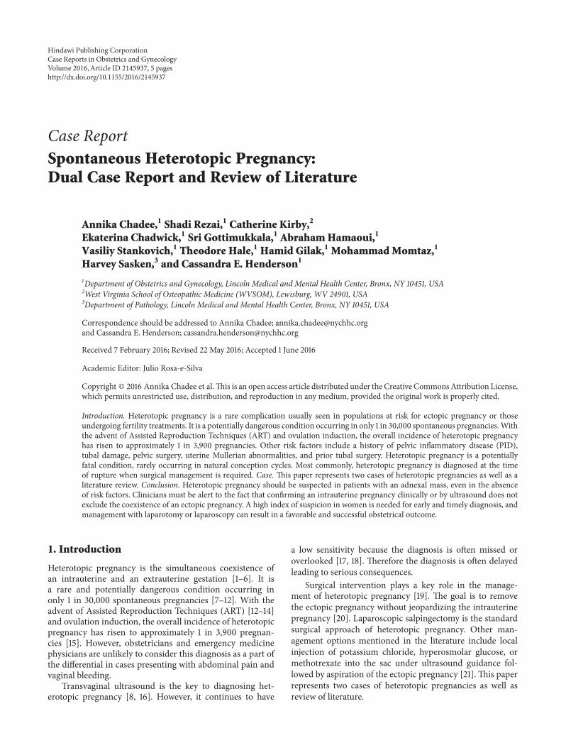
Figure 1: Patient number 1. Pelvic ultrasound showing the intrauterine and extrauterine/tubal ectopic pregnancies, both with fetal pole (FP),fetal heart rates (+FHM) present, and free fluid (FF) in the peritoneal cavity.
(a) (b)
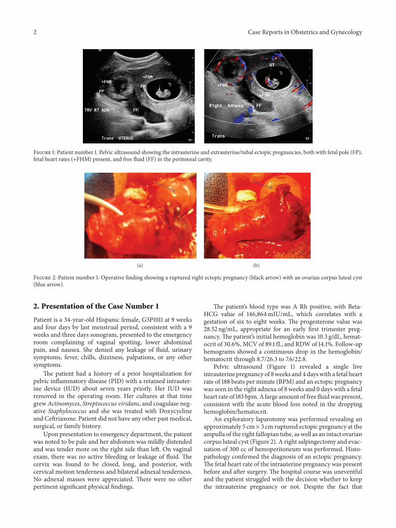
Figure 2: Patient number 1. Operative finding showing a ruptured right ectopic pregnancy (black arrow) with an ovarian corpus luteal cyst(blue arrow).
2. Presentation of the Case Number 1
Patient is a 34-year-old Hispanic female, G3P1011 at 9 weeksand four days by last menstrual period, consistent with a 9weeks and three days sonogram, presented to the emergencyroom complaining of vaginal spotting, lower abdominalpain, and nausea. She denied any leakage of fluid, urinarysymptoms, fever, chills, dizziness, palpations, or any othersymptoms.
The patient had a history of a prior hospitalization forpelvic inflammatory disease (PID) with a retained intrauter-ine device (IUD) about seven years priorly. Her IUD wasremoved in the operating room. Her cultures at that timegrew Actinomyces, Streptococcus viridans, and coagulase neg-ative Staphylococcus and she was treated with Doxycyclineand Ceftriaxone. Patient did not have any other past medical,surgical, or family history.
Upon presentation to emergency department, the patientwas noted to be pale and her abdomen was mildly distendedand was tender more on the right side than left. On vaginalexam, there was no active bleeding or leakage of fluid. Thecervix was found to be closed, long, and posterior, withcervical motion tenderness and bilateral adnexal tenderness.No adnexal masses were appreciated. There were no otherpertinent significant physical findings.
The patient’s blood type was A Rh positive, with Beta-HCG value of 146,864mIU/mL, which correlates with agestation of six to eight weeks. The progesterone value was28.52 ng/mL, appropriate for an early first trimester preg-nancy. The patient’s initial hemoglobin was 10.3 g/dL, hemat-ocrit of 30.6%,MCV of 89.1 fL, and RDWof 14.1%. Follow-uphemograms showed a continuous drop in the hemoglobin/hematocrit through 8.7/26.3 to 7.6/22.8.
Pelvic ultrasound (Figure 1) revealed a single liveintrauterine pregnancy of 8weeks and 4dayswith a fetal heartrate of 188 beats per minute (BPM) and an ectopic pregnancywas seen in the right adnexa of 8 weeks and 0 days with a fetalheart rate of 183 bpm.A large amount of free fluidwas present,consistent with the acute blood loss noted in the droppinghemoglobin/hematocrit.
An exploratory laparotomy was performed revealing anapproximately 5 cm × 3 cm ruptured ectopic pregnancy at theampulla of the right fallopian tube, as well as an intact ovariancorpus luteal cyst (Figure 2). A right salpingectomy and evac-uation of 300 cc of hemoperitoneum was performed. Histo-pathology confirmed the diagnosis of an ectopic pregnancy.The fetal heart rate of the intrauterine pregnancy was presentbefore and after surgery. The hospital course was uneventfuland the patient struggled with the decision whether to keepthe intrauterine pregnancy or not. Despite the fact that
Case Reports in Obstetrics and Gynecology 3
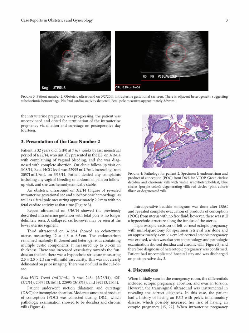
Figure 3: Patient number 2. Obstetric ultrasound on 3/2/2014: intrauterine gestational sac seen. There is adjacent heterogeneity suggestingsubchorionic hemorrhage. No fetal cardiac activity detected. Fetal pole measures approximately 2.9mm.
the intrauterine pregnancy was progressing, the patient wasunconvinced and opted for termination of the intrauterinepregnancy via dilation and curettage on postoperative dayfourteen.
3. Presentation of the Case Number 2
Patient is 32 years old, G1P0 at 7 6/7 weeks by last menstrualperiod of 1/22/14, who initially presented in the ED on 3/16/14with complaining of vaginal bleeding, and she was diag-nosed with complete abortion. On clinic follow-up visit on3/18/14, Beta-HCG level was 22995mIU/mL increasing from20571mIU/mL on 3/16/14. Patient denied any complaintsincluding any vaginal bleeding or abdominal pain on follow-up visit, and she was hemodynamically stable.
An obstetric ultrasound on 3/2/14 (Figure 3) revealedintrauterine gestational sac and subchorionic hemorrhage, aswell as a fetal pole measuring approximately 2.9mm with nofetal cardiac activity at that time (Figure 3).
Repeat ultrasound on 3/16/14 showed the previouslydescribed intrauterine gestation with fetal pole is no longerdefinitely seen. A collapsed sac however may be seen at thelower uterine segment.
Third ultrasound on 3/18/14 showed an echotextureuterus measuring 12 × 4.6 × 6.5 cm. The endometriumremained markedly thickened and heterogeneous containingmultiple cystic components. It measured up to 3.5 cm inthickness. There was increased vascularity towards the fun-dus; on the left, there was a hypoechoic structure measuring2.5 × 2.5 × 2.3 cm with mild vascularity. This was not clearlydelineated on prior imaging.There was no fluid in the cul-de-sac.
Beta-HCG Trend (mIU/mL). It was 2484 (2/26/14), 4211(3/2/14), 20571 (3/16/14), 22995 (3/18/15), and 3921 (3/21/14).
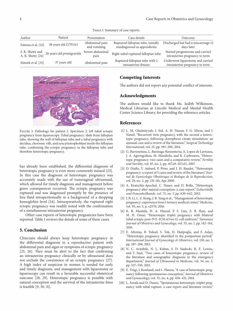
Patient underwent suction dilatation and curettage(D&C) for incomplete abortion.Moderate amount of productof conception (POC) was collected during D&C, whichpathologic examination showed to be decidua and chronicvilli (Figure 4).
Figure 4: Pathology for patient 2. Specimen 1: endometrium andproduct of conception (POC) from D&E for VTOP. Green circles:decidua and chorionic villi with viable syncytiotrophoblast; bluecircles (purple color): degenerating villi; red circles (pink color):fibrin or degenerated villi.
Intraoperative bedside sonogram was done after D&Cand revealed complete evacuation of products of conception(POC) from uterus with no free fluid; however, there was stilla hypoechoic structure along the fundus of the uterus.
Laparoscopic excision of left corneal ectopic pregnancywith mini-laparotomy for specimen retrieval was done andan approximately 4 cm × 4 cm left corneal ectopic pregnancywas excised, which was also sent to pathology, and pathologicexamination showed decidua and chronic villi (Figure 5) andtherefore diagnosis of heterotopic pregnancy was confirmed.Patient had uncomplicated hospital stay and was dischargedon postoperative day 3.
4. Discussions
When initially seen in the emergency room, the differentialsincluded ectopic pregnancy, abortion, and ovarian torsion.However, the transvaginal ultrasound was instrumental inrevealing the correct diagnosis. In this case, the patienthad a history of having an IUD with pelvic inflammatorydisease, which possibly increased her risk of having anectopic pregnancy [15, 22]. When intrauterine pregnancy
4 Case Reports in Obstetrics and Gynecology
Table 1: Summary of case reports.
Author Patient Presentation Case details Outcome
Fatema et al. [32] 38 years old G7P3A3 Abdominal painand vomiting
Ruptured fallopian tube, initiallymisdiagnosed as appendicitis
Discharged but had a miscarriage 12days later
S. K. Shetty andA. K. Shetty [24] 26 years old primigravida Severe abdominal
pain Right sided ruptured fallopian tube Started progesterone and carriedintrauterine pregnancy to term
Simsek et al. [33] 37 years old Abdominal pain Ruptured fallopian tube with 2intrauterine fetuses
Underwent laparotomy and carriedintrauterine pregnancy to term
Wall of fallopian tube
Tubal pregnancy
Figure 5: Pathology for patient 2. Specimen 2: left tubal ectopicpregnancy from laparoscopy. Tubal pregnancy: slide from fallopiantube, showing the wall of fallopian tube and a tubal pregnancy withdecidua, chorionic villi, and syncytiotrophoblast inside the fallopiantube, confirming the ectopic pregnancy in the fallopian tube andtherefore heterotopic pregnancy.
has already been established, the differential diagnosis ofheterotopic pregnancy is even more commonly missed [23].In this case the diagnosis of heterotopic pregnancy wasaccurately made with the use of transvaginal ultrasound,which allowed for timely diagnosis and management beforegrave consequences occurred. The ectopic pregnancy wasruptured and was diagnosed promptly by the presence offree fluid intraperitoneally in a background of a droppinghemoglobin level [24]. Intraoperatively, the ruptured rightectopic pregnancy was readily noted with the confirmationof a simultaneous intrauterine pregnancy.
Other case reports of heterotopic pregnancies have beenreported. Table 1 reviews the details of some of these cases.
5. Conclusion
Clinicians should always keep heterotopic pregnancy inthe differential diagnosis in a reproductive patient withabdominal pain and signs or symptoms of ectopic pregnancy[25, 26]. They must be alert to the fact that confirmingan intrauterine pregnancy clinically or by ultrasound doesnot exclude the coexistence of an ectopic pregnancy [27].A high index of suspicion in women is needed for earlyand timely diagnosis, and management with laparotomy orlaparoscopy can result in a favorable successful obstetricaloutcome [28, 29]. Heterotopic pregnancy is possible withnatural conception and the survival of the intrauterine fetusis feasible [9, 30, 31].
Competing Interests
The authors did not report any potential conflict of interests.
Acknowledgments
The authors would like to thank Ms. Judith Wilkinson,Medical Librarian at Lincoln Medical and Mental HealthCenter Science Library, for providing the reference articles.
References
[1] L. M. Ghulmiyyah, J. Eid, A. H. Nassar, F. G. Mirza, and J.Nassif, “Recurrent twin pregnancy, with the second a hetero-topic pregnancy, following clomiphene citrate stimulation: anunusual case and a review of the literature,” Surgical TechnologyInternational, vol. 25, pp. 195–200, 2014.
[2] G. Barrenetxea, L. Barinaga-Rementeria, A. Lopez de Larruzea,J. A. Agirregoikoa, M. Mandiola, and K. Carbonero, “Hetero-topic pregnancy: two cases and a comparative review,” Fertilityand Sterility, vol. 87, no. 2, pp. 417.e9–417.e15, 2007.
[3] D. Diallo, Y. Aubard, P. Piver, and J. H. Baudet, “Heterotopicpregnancy: a report of 5 cases and review of the literature,” Jour-nal de Gynecologie Obstetrique et Biologie de la Reproduction,vol. 29, no. 2, pp. 131–141, Apr 2000.
[4] A. Kratschla-Apochal, C. Nauer, and D. Bolla, “Heterotopicpregnancy after natural conception: a case report,” Geburtshilfeund Frauenheilkunde, vol. 72, no. 7, pp. 639–642, 2012.
[5] J. B. Li, L. Z. Kong, J. B. Yang et al., “Management of heterotopicpregnancy: experience from 1 tertiarymedical center,”Medicine,vol. 95, no. 5, p. e2570, 2016.
[6] K. B. Mustafa, H. A. Hamid, P. S. Lim, Z. R. Razi, andM. H. Omar, “Heterotopic triplet pregnancy with bilateraltubal ectopic post-IVF–ICSI of two 12-cell embryos,” TaiwaneseJournal of Obstetrics and Gynecology, vol. 55, no. 1, pp. 142–144,2016.
[7] E. Altıntas, B. Yuksel, S. Tok, H. Hatipoglu, and F. Aslan,“Heterotopic pregnancy identified in the postpartum period,”International Journal of Gynecology & Obstetrics, vol. 130, no. 3,pp. 287–288, 2015.
[8] N. C. Avitabile, N. L. Kaban, S. D. Siadecki, R. E. Lewiss,and T. Saul, “Two cases of heterotopic pregnancy: review ofthe literature and sonographic diagnosis in the emergencydepartment,” Journal of Ultrasound in Medicine, vol. 34, no. 3,pp. 527–530, 2015.
[9] E. Tingi, J. Rowland, and L. Hanna, “A case of heterotopic preg-nancy following spontaneous conception,” Journal of Obstetricsand Gynaecology, vol. 35, no. 4, pp. 430–431, 2014.
[10] L. Arsala and D. Danso, “Spontaneous heterotopic triplet preg-nancy with tubal rupture: a case report and literature review,”
Case Reports in Obstetrics and Gynecology 5
Journal of Investigative Medicine High Impact Case Reports, vol.2, no. 2, pp. 1–4, 2014.
[11] A. Nargund, S. Majumdar, and I. Stokes, “Heterotopic preg-nancy after spontaneous conception,” Journal of Obstetrics andGynaecology, vol. 33, no. 4, p. 425, 2013.
[12] F. Uysal, A. Uysal, D. C. Oztekin, and M. S. Avcı, “Heterotopicquadruplet pregnancy and successful twin outcome,” Archivesof Gynecology and Obstetrics, vol. 288, no. 3, pp. 715–717, 2013.
[13] M. Liu, X. Zhang, L. Geng et al., “Risk factors and earlypredictors for heterotopic pregnancy after in vitro fertilization,”PLoS ONE, vol. 10, no. 10, Article ID e0139146, 2015.
[14] J. K. Martin and R. B. Gala, “Adnexal mass in a spontaneouspregnancy diagnosed as heterotopic pregnancy at the time ofcesarean delivery,” Fall, vol. 15, no. 3, pp. 265–267, 2015.
[15] R. Kumar and M. Dey, “Spontaneous heterotopic pregnancywith tubal rupture and pregnancy progressing to term,”MedicalJournal Armed Forces India, vol. 71, supplement 1, pp. S73–S75,2015.
[16] D. I. P. Buca, D. Murgano, G. Impicciatore et al., “Earlydiagnosis of heterotopic triplet pregnancy with an intrauterineand bilateral tubal pregnancy after IVF: a case report,” Journalof Obstetrics and Gynaecology, vol. 35, no. 7, pp. 755–756, 2015.
[17] X. H. Li, Y. Ouyang, and G. X. Lu, “Value of transvaginalsonography in diagnosing heterotopic pregnancy after in-vitrofertilization with embryo transfer,” Ultrasound in Obstetrics &Gynecology, vol. 41, no. 5, pp. 563–569, 2013.
[18] L. Zhaoxia, Q. Honglang, and C. Danqing, “Ruptured hetero-topic pregnancy after assisted reproduction in a patient whounderwent bilateral salpingectomy,” Journal of Obstetrics andGynaecology, vol. 33, no. 2, pp. 209–210, 2013.
[19] J. T. Esterle and J. Schieda, “Hemorrhagic heterotopic pregnancyin a setting of prior tubal ligation and re-anastomosis,” Journalof Radiology Case Reports, vol. 9, no. 7, pp. 38–46, 2015.
[20] V. Vaishnav, “A very rare case of heterotopic pregnancy innatural conception with ectopic pregnancy as partial mole,”TheJournal of Obstetrics and Gynecology of India, vol. 64, no. 6, pp.433–435, 2014.
[21] Y. Yu, W. Xu, Z. Xie, Q. Huang, and S. Li, “Management andoutcome of 25 heterotopic pregnancies in Zhejiang, China,”European Journal of Obstetrics & Gynecology and ReproductiveBiology, vol. 180, pp. 157–161, 2014.
[22] T. Felekis, C. Akrivis, P. Tsirkas, and I. Korkontzelos, “Hetero-topic triplet pregnancy after in vitro fertilization with favorableoutcome of the intrauterine twin pregnancy subsequent tosurgical treatment of the tubal pregnancy,” Case Reports inObstetrics and Gynecology, vol. 2014, Article ID 356131, 4 pages,2014.
[23] H. S. Jeon, H. J. Shin, I. H. Kim, and D. Y. Chung, “A caseof spontaneous heterotopic pregnancy presenting with heartactivity of both embryos,” Korean Journal of Obstetrics &Gynecology, vol. 55, no. 5, pp. 339–342, 2012.
[24] S. K. Shetty and A. K. Shetty, “A case of heterotopic pregnancywith tubal rupture,” Journal of Clinical and Diagnostic Research,vol. 7, no. 12, pp. 3000–3001, 2013.
[25] V. Mihmanli, A. Kilickaya, N. Cetinkaya, G. Karahisar, andH. Uctas, “Spontaneous heterotopic pregnancy presenting withhemoperitoneum,” The Journal of Emergency Medicine, vol. 50,no. 1, pp. 44–46, 2016.
[26] D. L. Fylstra, “Ectopic pregnancy after hysterectomymay not beso uncommon: a case report and review of the literature,” CaseReports in Women’s Health, vol. 7, pp. 8–11, 2015.
[27] A. J. Headley and V. Adum, “Naturally occurring heterotopicpregnancy in a multiparous patient: a case report,” Journal ofReproductive Medicine, vol. 58, no. 11-12, pp. 541–544, 2013.
[28] B. Refaat, E. Dalton, and W. L. Ledger, “Ectopic pregnancysecondary to in vitro fertilisation-embryo transfer: pathogenicmechanisms and management strategies,” Reproductive Biologyand Endocrinology, vol. 13, no. 1, article 30, 2015.
[29] M. A. Bedaiwy, J. Volsky, N. Lazebnik, and J. Liu, “Laparo-scopic single-site linear salpingostomy for the management ofheterotopic pregnancy: a case report,” Journal of ReproductiveMedicine, vol. 59, no. 5, pp. 522–524, 2014.
[30] M. Varras, C. Akrivis, G. Hadjopoulos, and N. Antoniou, “Het-erotopic pregnancy in a natural conception cycle presentingwith tubal rupture: a case report and review of the literature,”European Journal of Obstetrics Gynecology and ReproductiveBiology, vol. 106, no. 1, pp. 79–82, 2003.
[31] F. Jan, G. M. Naikoo, M. H. Rather, T. A. Sheikh, and Y.H. Rather, “Ruptured heterotopic pregnancy: a rare cause forhemoperitoneum; report of three cases from Kashmir, India,”Indian Journal of Surgery, vol. 72, no. 5, pp. 404–406, 2010.
[32] N. Fatema, M. M. Al Badi, M. Rahman, and M. M. Elawdy,“Heterotopic pregnancy with natural conception; a rare eventthat is still being misdiagnosed: a case report,” Clinical CaseReports, vol. 4, no. 3, pp. 272–275, 2016.
[33] T. Simsek, A. Dogan, M. Simsek, and E. Pestereli, “Heterotopictriplet pregnancy (twin tubal) in a natural cycle with tubalrupture: case report and review of the literature,” Journal ofObstetrics and Gynaecology Research, vol. 34, no. 4, part 2, pp.759–762, 2008.