spontaneous leptospiral infection of the rat kidney. an ultrastructural study
TRANSCRIPT

EXPERIMENTAL AND MOLECULAR PATHOLOGY 10, 27-38 (1969)
Spontaneous Leptospiral Infection of the Rat Kidney. An
Ultrastructural Study’ CESARE DE MARTINO, CARMELO B. BRUNI, MARINELLA BELLOCCI,
AND PIER GIORGIO NATALI
Istituto di Medicinu Costituionule ed Endocrinologia; Istituto II Clinica Medica, UniversitiL di Roma, Rome, Italy
Received September 24,1%8
In the course of a study on the ultrastructural and biochemical changes in- duced by neptunium perchlorate in rats, a spontaneous leptospiral infection of the kidney was observed.
Spontaneous leptospiral infection in rata seems to be frequent, with large incidence of renal localization (Li and Davis, 1952). The relationship between the infection and toxic effects of neptunium is probably casual, since in the group of rata studied, only one out of seven was found affected.
The aim of the present report is to describe the morphologic changes asso- ciated with the localization of leptospirae in the cells of proximal tubules.
MATERIAL AND METHODS
Seven Sprague-Dawley male rats weighing 250 gm were treated with a single intraperitoneal injection of neptunium perchlorate in aqueous solution at a dose of 4 mg/kg of body weight. Twenty four hours after the injection the animals were killed by decapitation. Fragments of kidney were fixed in buffered 2% osmium tetroxide according to Millonig (1961)) dehydrated in graded acetone and embedded in Araldite (Durcupan ACM). Sections were obtained with glass knives. Thick (0.5-l cc) sections were stained with 1% toluidine blue in phos- phate buffer at pH 8. Thin sections were mounted on Formvar-coated grids and examined in a Siemens Elmiskop I electron microscope, after staining with lead hydroxide (Karnowsky, 1961).
RESULTS
Light microscopy
The lumens of some proximal tubules are almost completely obliterated by basophilic material (Fig. 1). The glomeruli, the distal and collecting tubules, and the loops of Henle seem to be normal. The interstitial tissue and the peritubular capillaries also have a normal appearance (Fig. 1).
1 This work was supported by Grant of Consiglio Nazionale delle Ricerche of Italy (C. N. R.) n”. 115/0815/O-1365.
27

28 CESARE DE MARTIN0 ET AL.
FIG. 1. Light microscopy. Proximal tubule. In the tubular lumen a finely granular material is
visible. The peritubular capillaries appear normal. Toluidine blue stained. X800. FIG. 2. Part of the same proximal tubule shown in Fig. 1. The lumen contains numerous
leptospirae some of which are visible among the microvilli of the brush border (circles). The apical
tubules (AT arrows), the apical vesicles (arrows), the cytosomes, and the mitochondria are normal in number and appearance. The basement membrane is well preserved. CL: Capillary lumen; BM:
basement membrane; BB: brush border. X 8000.

LEPTOSPIRAL INFECTION OF THE KIDNEY 29
FIG. 3. Distal tubule. Some leptospirae are visible within a cell (arrows). BM: Basement mem-
brane; TL: tubular lumen. X 15,400.
Electron microscopy
The basophilic material visible in the light microscope within the proximal tubules appears to be composed of clumps of microorganisms which occupy the lumen almost completely and which are in close contact with the brush border of the cells (Fig. 2). Only few microorganisms are found within the
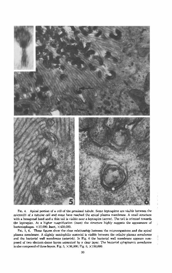
FIG. 4. Apical portion of a cell of the proximal tubule. Some leptospirae are visible between the microvilli of a tubular cell and some have reached the apical plasma membrane. A small structure
with a hexagonal head and a thin tail is visible near a leptospira (arrow). The tail is oriented towards the leptospira. At a higher magnification (inset) the structure highly suggests the appearance of bacteriophages. ~22,000. Inset, X430,000.
FIG. 5, 6. These figures show the close relationship between the microorganisms and the apical
plasma membrane. A slightly osmiophilic material is visible between the cellular plasma membrane and the bacterial wall membrane (asterisk). In Fig. 6 the bacterial wall membrane appears com- posed of two electron-dense layers separated by a clear layer. The bacterial cytoplasmic membrane is also composed of three layers. Fig. 5, X36,000; Fig. 6, X 150,000.
30

LEPTOSPIRAL INFECTION OF THE KIDNEY 31
FIG. 7. Apical portion of cells of a proximal tubule. Numerous Ieptospirae are visible in the lumen. Some cells have completely lost their brush border (right side). The apical vesicles and the
apical tubules are reduced in number. X 12,000. FIG. 8. Part of a tubular cell at a higher magnification. The brush border is almost completely
destroyed and the leptospirae are in contact with the plasma membrane. Few apical tubules are
visible (arrows). x 20,000.

32 CESARE DE MARTIN0 ET AL.
FIGS. 9, 10, 11. At the base of the microvilli the plasma membrane forms small invaginations. Some leptospirae, which have lost their normal aspect, appear as disorganized bacterial material. Fig. 9, x42,000; Fig. 10, x28,000; Fig. 11, ~21,000.
FIG. 12. The brush border of the tubular cell is partially damaged. Some leptospirae have destroyed the apical plasma membrane and are penetrating the cytoplasm (arrows). The number of apical tubules (AT arrows) and apical vesicles (V) is normal. x 16,000.

FIG. 13. In the cells of a proximal tubule the leptospirae are diffusely scattered in the cyto- plasm. Some of them are visible in the basal parts of the cells near the basement membrane. The mitochondria, scattered through the cytoplasm, are reduced in number and spherical in shape. Apical vesicles are no longer visible. The number of the apical tubules is greatly reduced. BM: Basement membrane; TL: tubular lumen; x 15,000.
FIG. 14. Part of two proximal tubular cells. The adjacent lateral plasma membranes are dis- placed and several leptospirae penetrate in the patent intercellular space. ICS: Intercellular space; BM: basement membrane. x 16,000.
33

34 CESARE DE MARTIN0 ET AL.
FIG. 15. A gap in the basement membrane of a proximal tubule (arrow), the lumen of which
contains several leptospirae. The tubular cells have completely lost the brush border and appear
flattened. Two altered mitochondria (m) of a tubular cell are visible outside the cell in the peritubu-
lar interstitial tissue. The leptospirae are visible in the ground substance, between the collagen
fibers and within the cytoplasm of a fibroblast. The nucleus of the fibroblast is pyknotic. A lepto-
spira is contained in an endothelial cell of a peritubular capillary (circle). TL: Tubular lumen; IN:
nucleus of a fibroblast; CL: capillary lumen; PT: proximal tubule; BM: basement membrane.
x 14,400.

LEPTOSPIRAL INFECTION OF THE KIDNEY 35
FIG. 16. A leptospira is visible in the lumen of a peritubular capillary (square), and another in the cytoplasm of an endothelial cell (circle). CL: Capillary lumen; E: endothelial cell; L: lympho- cyte; PT: proximal tubule. x7400; inset: the photograph shows at a higher magnification the leptospira present in the capillary lumen. The micrograph was obtained from the section following that of Fig. 16. X21,600.
FIG. 17. The figure shows the helicoidal shape of a leptospira, the wall membrane, and the cytoplasmic membrane. In the cytoplasm of the microorganisms some granules are visible. The axostyle is cut longitudinally (arrows). ~90,000.
FIGS. 18, 19; Some leptospirae exhibit a tubular structure in tangential, longitudinal, and cross sections (250 A in diameter) probably representing the axostyle (arrows). Fig. 18, ~40,000; Fig. 19, x80,000.

36 CESARE DE MARTIN0 ET AL.
cytoplasm of the distal tubules (Fig. 3). In the proximal tubules, some micro- organisms penetrate between the microvilli (Figs. 2, 4), displace them from their normal position, and reach the basal infoldings of the brush border (Fig. 4). The apical plasma membrane is separated from the cell wall of the micro- organisms by a slightly osmiophilic material (Figs. 5, 6), similar to that observed by Pease (1966) on the surface of the microvilli of the renal proximal tubules. In some epithelial cells, the brush border is destroyed and apical tubules and vesicles of the cytoplasm are reduced in number (Figs. 7, 8). At this phase, only a few microorganisms undergo degenerative changes and are phagocytized by the tubular cells in the little apical vesicles, similar to the “apical tubular in- vaginations” described by Ericsson (1964) (Figs. 9-11).
Some of the epithelial tubular cells show gaps in the apical plasma mem- brane (Fig. 12). Numerous microorganisms, which retain their normal appear- ance, are diffusely scattered in the cytoplasm (Fig. 13). The microorganisms are not contained within membrane-bounded bodies (Fig. 13). Other microorganisms are visible in the intercellular space (Fig. 14).
The infected cells do not show nuclear changes. The mitochondria have a spheric shape and are scattered within the cytoplasm. The number of mito- chondria and apical tubules is decreased. The apical vesicles are no longer visible (Fig. 13). In the interstitial tissue the microorganisms are found in the ground substance and in the cytoplasm of fibroblasts, some of which show degenerative changes. In these areas the basement membrane often appears interrupted. Altered mitochondria of tubular cells are visible in the interstitial space close to the gap of the basement membrane (Fig. 15).
A minor number of microorganisms are visible in the endothelium and in the lumen of peritubular capillaries (Fig. 16 and inset).
The microorganisms have a helicoidal shape, a diameter of 150 rnp and are surrounded by two envelopes, namely, the outer membrane and cytoplasmic membrane. Thep thickness of the former is 70-100 A and the thickness of the latter is 60-80 A (Fig. 5). The two protoplasmic envelopes are separated by a space of variable width and appear to have the structure of unit membrane. The cytoplasm of m@roorganisms is of variable electron density and contain; several granules 150 A in diameter (Figs. 5, 17) and an eccentric axostyle 250-A in diameter position (Figs. 18, 19).
In the lumen of some proximal tubules, structures composed of a hexagonal head, a tail, aqd three thin filaments are seen. The head, whose diameter is about 550-750 A, is connected to the tail, 400-600 A in length, which ends with three filaments (Fig. 4, inset).
DISCUSSION
The microorganisms observed in this study in the proximal and distal tubules of the rat kidney seem to belong to the genus Leptospira (Czekalowsky and Eaves, 1955; Simpson and White, 1961). This assumption is supported by their ultrastructural similarity with L. Icterohemorragiae (Pillot and Ryter, 1965),

LEPTOSPIRAL INFECTION OF THE KIDNEY 37
and with L. Pomona (Miller and Wilson, 1962); however, an exact classification is impossible on the sole morphologic observation.
The invasion of tubular cells by leptospirae seems to take place from the tubular lumen towards the tubular basement membrane. The degeneration of the brush border appears to be the first step in the penetration of the lepto- spirae in the cytoplasm of the tubular cell. In no instance did the brush border of parasitized cells appear intact. It is difficult to establish the sequential steps of the invasion into the interstitium. The hypothesis that leptospirae penetrate from the lumen into the tubular cells and then reach the interstitial spaces through gaps of the tubular basement membrane, was not confirmed by present observation. In fact, tubular cells overlying the gaps in no instances contained leptospirae, whereas the latter were numerous in the underlying interstitium. The presence of leptospirae in the lumen of peritubular capillaries may be due to the migration of the microorganisms from the interstitium into the capillaries, but could also be related to the presence of leptospirae in the blood stream.
The present observation seems to suggest that the integrity of the brush border of tubular cells is the only mechanism of defence against the bacterial infection. The tubular cells seems to have no capacity to phagocytize the micro- organims in toto, whereas they can engulf disorganized material within the small apical invaginations of the plasma membrane.
The structures with a hexagonal head, visible in the lumen of the proximal tubules and sometimes located close to the microorganisms in the brush border, could represent fragments of disorganized bacterial material. However, their shape, size, and presence of a tail-like structure strongly suggest that they might be bacteriophages.
SUMMARY
The morphologic features of the spontaneous leptospiral infection of renal proximal tubules of
rat kidney are described here. The main ultrastructural findings are: (1) Numerous microorganisms are visible in the tubular
lumens; (2) the brush border of the cells is severely damaged; (3) the leptospirae are present within the cytoplasm of the tubular cells which show degenerative changes; (4) the microorganisms are
also visible in the basilar cytoplasm, in the interstitial tissue, and sometimes in the endothelial cells and peritubular capillaries.
REFERENCES
CZEKALOWSKY, J. W., and EAVES, G. (1955). The structure of Leptospirae as revealed by electron
microscopy. J. Pothol. Bucteriol. 69, 129-132. ERICSSON, J. L. E. (1964). Absorption and decomposition of homologous hemoglobin in renal
proximal tubular cells. Acta P&d. Microbid. Stand., Suppl. 16&l-121. KARNOWSKY, M. J. (1961). Simple methods for “staining with lead” at high pH in electron micros-
copy. J. Biochem. Cytol. 11,729.
LI, H-Y, and DAVIS, D. E. (1952). The prevalence of carriers of Leptospira and Salmonella in Nor- way rats of Baltimore. Am. J. Hyg. 56,90-100.
MILLER, N. G., and WILSON, R. B. (1962). In uiuo and in uitro observation of Leptospim Pomona
by electron microscopy. J. Bacterial. 84,569-576.

38 CESARE DE MARTIN0 ET AL.
MILLONIG, G. (1961). The advantage of phosphate buffer for OSOJ solution in fixation. J. Appl. Physiol. 32, 1637.
PEASE, D. C. (1966). Polysaccharides associated with the exterior surface of epithelial cells: Kid- ney, Intestine, Brain. J. Ultrastruct. Res. U&555-588.
PILLOT, J., and RYTER, A. (1965). Structure des Spirochetes. I- Etude des genres Treponema, Borrelia et Leptospira au microscope electronique. Ann. Inst. Pasteur 108, 791-804.
SIMPSON, C. F., and WHITE, F. H. (1961). Electron microscope studies and staining reactions of leptospires. J. Inject. Diseases 109,243-250.