sport injuries: a review of outcomes - british medical bulletin
TRANSCRIPT
Sport injuries: a review of outcomes
Nicola Maffulli†*, Umile Giuseppe Longo‡, Nikolaos Gougoulias§,Dennis Caine}, and Vincenzo Denaro‡
†Centre for Sports and Exercise Medicine, Barts and The London School of Medicine and Dentistry,Mile End Hospital, 275 Bancroft Road, London E1 4DG, UK; ‡Department of Orthopaedic andTrauma Surgery, Campus Biomedico University, Via Alvaro del Portillo, 200, 00128 Trigoria,Rome, Italy; §Frimley Park Hospital, Portsmouth Road, Frimley, Surrey GU15 8UJ, UK, and}Department of Physical Education, Exercise Science and Wellness, University of North Dakota,Hyslop Sports Center, Room 114, 2751 2nd Avenue North Stop 8235, Grand Forks, ND 58202-8235, USA
Injuries can counter the beneficial aspects related to sports activities if an
athlete is unable to continue to participate because of residual effects of injury.
We provide an updated synthesis of existing clinical evidence of long-term
follow-up outcome of sports injuries. A systematic computerized literature
search was conducted on following databases were accessed: PubMed, Medline,
Cochrane, CINAHL and Embase databases. At a young age, injury to the physis
can result in limb deformities and leg-length discrepancy. Weight-bearing joints
including the hip, knee and ankle are at risk of developing osteoarthritis (OA) in
former athletes, after injury or in the presence of malalignment, especially in
association with high impact sport. Knee injury is a risk factor for OA. Ankle
ligament injuries in athletes result in incomplete recovery (up to 40% at 6
months), and OA in the long term (latency period more than 25 years). Spine
pathologies are associated more commonly with certain sports (e.g. wresting,
heavy-weight lifting, gymnastics, tennis, soccer). Evolution in arthroscopy allows
more accurate assessment of hip, ankle, shoulder, elbow and wrist intra-articular
post-traumatic pathologies, and possibly more successful management. Few
well-conducted studies are available to establish the long-term follow-up of
former athletes. To assess whether benefits from sports participation outweigh
the risks, future research should involve questionnaires regarding the health-
related quality of life in former athletes, to be compared with the general
population.
Keywords: sports/injury/athlete/knee/shoulder/ankle
Accepted: July 22, 2010
British Medical Bulletin 2011; 97: 47–80
DOI:10.1093/bmb/ldq026
& The Author 2010. Published by Oxford University Press. All rights reserved.
For permissions, please e-mail: [email protected]
*Correspondence address.
Centre for Sports and
Exercise Medicine, Barts
and The London School
of Medicine and
Dentistry, Mile End
Hospital, 275 Bancroft
Road, London E1 4DG,
UK. E-mail: n.maffulli@
qmul.ac.uk
Published Online August 14, 2010D
ownloaded from
https://academic.oup.com
/bmb/article/97/1/47/358051 by guest on 18 D
ecember 2021
Introduction
Participation in sports is widespread all over the world,1 with well-described physical, psychological and social consequences for involvedathletes.2–5 The benefits associated with physical activity in both youthand elderly are well documented.2,6–8 Regular participation in sports isassociated with a better quality of life and reduced risk of several dis-eases,1,9 allowing people involved to improve cardiovascular health.10,11
Both individual and team sports are associated with favourable physicaland physiological changes consisting of decreased percentage of bodyfat12 and increased muscular strength, endurance and power.13,14
Moreover, regular participation in high-volume impact-loading andrunning-based sports (such as basketball, gymnastics, tennis, soccer anddistance running) is associated with enhanced whole-body and regionalbone mineral content and density,14,15 whereas physical inactivity isassociated with obesity and coronary heart disease.16 Sports are associ-ated with several psychological and emotional benefits.7,17,18 First ofall, there is a strong relationship between the development of positiveself-esteem, due to testing of self in a context of sport competition,19
reduced stress, anxiety and depression.20 Physical activities also contrib-ute to social development of athletes, prosocial behaviour, fair play andsportspersonship21 and personal responsibility.22
Engaging in sports activities has numerous health benefits, but alsocarries the risk of injury.7,23,24 At every age, competitive and rec-reational athletes sustain a wide variety of soft tissue, bone, ligament,tendon and nerve injuries, caused by direct trauma or repetitivestress.25–35 Different sports are associated with different patterns andtypes of injuries, whereas age, gender and type of activity (e.g. competi-tive versus practice) influence the prevalence of injuries.7,36,37
Injuries in children and adolescents, who often tend to focus on highperformance in certain disciplines and sports,24 include susceptibilityto growth plate injury, nonlinearity of growth, limited thermoregula-tory capacity and maturity-associated variation.9 In the immature skel-eton, growth plate injury is possible38 and apophysitis is common. Themost common sites are at the knee (Osgood-Schlatter lesion), the heel(Sever’s lesion) and the elbow.39 Certain contact sports, such as rugby,for example, are associated with 5.2 injuries per 1000 total athleticexposures in high school children (usually boys). These were morecommon during competition compared with training and fracturesaccounted for 16% of these injuries, whereas concussions (15.8%) andligament sprains (15.7%) were almost as common.40
Sports trauma commonly affects joints of the extremities (knee,ankle, hip, shoulder, elbow, wrist) or the spine. Knee injuries are
N. Maffulli et al.
48 British Medical Bulletin 2011;97
Dow
nloaded from https://academ
ic.oup.com/bm
b/article/97/1/47/358051 by guest on 18 Decem
ber 2021
among the most common. Knee trauma can result in meniscal andchondral lesions, sometimes in combination with cruciate ligamentinjuries.37 Ankle injuries constitute 21% of all sports injuries.41 Ankleligament injuries are more commonly (83%) diagnosed as ligamentsprains (incomplete tears), and are common in sports such as basket-ball and volleyball. Ankle injuries occur usually during competitionand in the majority of cases, athletes can return to sports within aweek.42 Hip labral injuries have drawn attention in recent years withthe advent of hip arthroscopy.43,44 Upper extremity syndromes causedby a single stress or by repetitive microtrauma occur in a variety ofsports. Overhead throwing, long-distance swimming, bowling, golf,gymnastics, basketball, volleyball and field events can repetitively stressthe hand, wrist, elbow and shoulder. Shoulder and elbow problems arecommon in the overhead throwing athlete whereas elbow injuriesremain often unrecognized in certain sports.45 Hand and wrist traumaaccounts for 3–9% of all athletic injuries.46 Wrist trauma can affectthe triangular fibrocartilage complex47 or cause scaphoid fractures,48
whereas overuse problems (e.g. tenosynovitis) are not uncommon.49
Spinal problems can range from lumbar disc herniation,39–42 to fatiguefractures of the pars interarticularis,50 and ‘catastrophic’ cervical spineinjuries.51
Thus, in addition to the beneficial aspects related to sports activities,injuries can counter these if an athlete is unable to continue to partici-pate because of residual effects of injury. Do injuries in children, ado-lescents and young adults have long-term consequences? What are theoutcomes of the most commonly performed surgical procedures? Theaim of this review is to provide an updated synthesis of existing clinicalevidence of long-term follow-up outcome of sports injuries.
Literature search
An initial pilot Pubmed search using the keywords ‘sports’, ‘injury’,‘injuries’, ‘athletes’, ‘outcome’, ‘long term’, was performed. From 1467abstracts that were retrieved and scanned we identified the thematictopics (types of injury, management, area of the body involved) of thecurrent review, listed below:
(i) Physeal injuries and growth disturbance
(ii) Residual problems after injury in athletes(a) Osteoarthritis (OA) in former athletes
(b) Spine problems in former athletes
(c) Knee injury and OA
Sport injuries
British Medical Bulletin 2011;97 49
Dow
nloaded from https://academ
ic.oup.com/bm
b/article/97/1/47/358051 by guest on 18 Decem
ber 2021

(d) Ankle ligament injury and OA
(e) Residual upper limb symptoms in the ‘overhead’ athlete
(iii) Outcomes of operative management of common sports injuries
(a) Meniscectomy and OA
(b) Meniscal repair in athletes
(c) Anterior cruciate ligament (ACL) reconstruction and OA
(d) ACL reconstruction in children
(e) Ankle arthroscopy in athletes
(f ) Hip arthroscopy in athletes
(g) Operative management of shoulder injuries in athletes (focusing onsurgery for instability and labral tears)
(h) Operative management of wrist injuries in athletes (focusing ontriquetral fibrocartilage complex, TFCC, injuries and scaphoidfractures)
Then a more detailed search of PubMed, Medline, Cochrane, CINAHLand Embase databases followed. We used combinations of the key-words: ‘sport’, ‘sports’, ‘youth sports’, ‘young athletes’, ‘former ath-letes’, ‘children’, ‘skeletally immature’, ‘adolescent’, ‘paediatric’,‘pediatric’, ‘physeal’, ‘epiphysis’, ‘epiphyseal injuries’, ‘hip’, ‘knee’,‘ankle’, ‘spine’, ‘spinal’, ‘shoulder’, ‘elbow’, ‘wrist’, ‘football players’,‘football’, ‘soccer’, ‘tennis’, ‘swimmers’, ‘swimming’, ‘divers’, ‘wres-tlers’, ‘wrestling’, ‘cricket’, ‘gymnastics’, ‘skiers’, ‘baseball’, ‘basket-ball’, ‘osteoarthritis’, ‘former athletes’, ‘strain’, ‘contusion’, ‘distortion’,‘injury’, ‘injuries’, ‘trauma’, ‘drop out’, ‘dropping out’, ‘attrition’,‘young’, ‘ youth’, ‘sprain’, ‘ligament’, ‘ACL’, ‘cruciate ligament’,‘meniscus’, ‘meniscal’, ‘chondral’, ‘labrum’, ‘labral’, ‘reconstruction’,‘arthroscopy’, ‘throwing’, ‘overhead’, ‘rotator cuff’, ‘TFCC’, ‘scaphoid’,‘osteoarthritis’, ‘arthritis’, ‘long term’, ‘follow-up’ and ‘athlete’. Themost recent search was performed during the second week ofNovember 2009.
Given the different types of sports injuries in terms of location in thebody, several searches were carried out. The search was limited toarticles published in peer-reviewed journals.
From a total of 2596 abstracts that were scanned, 1247 studies wereirrelevant to the subject and were excluded. The remaining studieswere categorized in the topics identified earlier. We excluded fromour investigation case reports, letter to editors and articles not specifi-cally reporting outcomes, as well as ‘kin’ studies (studies reportingon the same patients’ population). The most recent study or thestudy with the longest follow-up was included. In some topics ofparticular importance, such as the effect of knee injuries (given their
N. Maffulli et al.
50 British Medical Bulletin 2011;97
Dow
nloaded from https://academ
ic.oup.com/bm
b/article/97/1/47/358051 by guest on 18 Decem
ber 2021
frequency), we included long-term studies reporting not only onathletes, but also on the general population (usually in these studies avery high proportion on sports injuries is included). Regarding kneeinjuries in adults, we included articles with follow-up more than10 years.
Given the linguistic capabilities of the research team, weconsidered publications in English, Italian, French, German, Spanishand Portuguese.
Physeal injuries and growth disturbance
A concern regarding children’s participation in sports is that the toler-ance limits of the physis may be exceeded by the mechanical stresses ofsports such as football and hockey or by the repetitive physical loadingrequired in sports such as baseball, gymnastics and distance running.52
Unfortunately, what is known about the frequency of acute sport-related physeal injuries is derived primarily from case reports and caseseries data. In a previous systematic review on the frequency andcharacteristics of sports-related growth plate injuries affecting childrenand youth, we found that 38.3% of 2157 acute cases were sportrelated and among these 14.9% were associated with growth disturb-ance.24 These injuries were incurred in a variety of sports, althoughfootball is the sport most often reported.53
There are accumulating reports of stress-related physeal injuries affect-ing young athletes in a variety of sports, including baseball, basketball,climbing, cricket, distance running, American football, soccer, gymnas-tics, rugby, swimming, tennis.24 Although most of these stress-relatedconditions resolved without growth complication during short-termfollow-up, there are several reports of stress-related premature partial orcomplete distal radius physeal closure of young gymnasts.25–29 Thesedata indicate that sport training, if of sufficient duration and intensity,may precipitate pathological changes of the growth plate and, inextreme cases, produce growth disturbance.24,32
Disturbed physeal growth as a result of injury can result in length dis-crepancy, angular deformity or altered joint mechanics and may causesignificant long-term disability.33 However, the incidence of long-termhealth outcome of physeal injuries in children’s and youth sports islargely unknown.
Based on the previously selection criteria, 20 studies54–73 wereretained for analysis (Table 1). Injury to the physis can result in limbdeformities and leg-length discrepancy, the latter being more commonafter motor vehicle accidents, rather than sports participation.
Sport injuries
British Medical Bulletin 2011;97 51
Dow
nloaded from https://academ
ic.oup.com/bm
b/article/97/1/47/358051 by guest on 18 Decem
ber 2021

Table 1 Evidence on acute physeal injury with subsequent adverse affects on growth.
Study Injury Patients Residual deformities
Stephens et al.58
(retrospective case
series)
Struck by car; automobile
accident; football; gymnastics;
baseball; fall;
20 Varus/valgus deformity of knee
(4/20); femoral shortening
(9/18); limitation knee motion
(4/20); ligament laxity (5/20)
Criswell et al.59
(retrospective case
series)
Football 15 Varus/valgus deformity of
distal femur (5/15); shortening
of injured leg (2/15)
Lombardo and
Harvey60 (retrospective
case series)
Motor-vehicle accident; fall;
football; bicycle accident
34 Limb-length discrepancy
(.1 cm) (13/28); varus/valgus
deformity of distal femur
(11/33); limitation of knee
motion (11/31); ligament laxity
(8/33); quadriceps atrophy
(5/30)
Goldberg and
Aadalen56
(retrospective case
series)
Football; basketball; skateboard;
skiing; gymnastics; ice skating
53 Ankle varus deformity (2/53);
shortening of injured leg
(12/53)
Burkhart and
Peterson61
(retrospective case
series)
Motor-vehicle accident; sledding;
bicycling; gymnastics football;
basketball; hurdling; high jump;
twist
26 Varus/valgus deformity of knee
(7/26); limb-length discrepancy
(4/26)
Cass and Peterson62
(retrospective case
series)
Automobile/motorcycle accident;
lawnmower accident; fall;
jumping; gymnastics; roller
skating; skiing; inversion
32 Varus/valgus deformity of knee
(5/18); limb-length discrepancy
(10/18)
Ogden63 (retrospective
case series)
Birth trauma; child abuse; fall;
vehicular accident
14 None
Landin et al.64
(retrospective case
series)
Sports injury; fall; traffic accident 65 Anterior angulation (5/65);
dorsal angulation (1/65);
valgus ankle deformity (1/65);
varus ankle deformity (1/65);
tibial shortening (1/65)
Hynes and O’Brien65
(retrospective case
series)
26 Medial physeal arrest of distal
tibia with varus deformity
(3/26); central physeal arrest of
distal tibia without deformity
(2/26)
Krueger-Franke et al.66
(retrospective case
series)
Soccer; skiing; track and field;
gymnastics; volleyball;
basketball; horseback riding;
skate boarding; field hockey;
ice hockey; judo; wrestling
85 Valgus deformity of knee
(2/49); leg-length discrepancy
(4/49); femoral rotational
deformity (1/49); varus ankle
deformity (1/49)
Berson et al.67
(retrospective case
series)
Sports injury; fall; vehicular
accident
24 Varus/valgus deformity (18/24);
leg-length discrepancy (5/24);
physeal bar without deformity
(6/24)
Continued
N. Maffulli et al.
52 British Medical Bulletin 2011;97
Dow
nloaded from https://academ
ic.oup.com/bm
b/article/97/1/47/358051 by guest on 18 Decem
ber 2021

Table 1 Continued
Study Injury Patients Residual deformities
Eid and Hafez68
(retrospective case
series)
Sport-related activities; road
traffic accidents; falls
151 Femoral shortening (58/151);
premature growth arrest (28/
151); varus deformity (21/151);
valgus deformity (14/151);
recurvatum (2/151); flexion
deformity (19/151); varus/
valgus with flexion deformity
(21/151); loss of knee motion
(43/151); ligamentous laxity
(21/151); thigh atrophy (42/151)
Cannata et al.57
(retrospective case
series)
163 Radial shortening (8/157);
ulnar shortening (5/157); radial
growth arrest/ulnar
overgrowth (2/157); radioulnar
length discrepancy (38/157);
ulnar styloid non-union (53/
157); atrophy of forearm
muscles (10/157)
Barmada et al.69
(retrospective case
series)
Fall; skateboard accidents; motor
vehicle accidents; football;
soccer; biking; baseball
92 Premature physeal closure of
distal tibia with shortening
and/or angular deformity
(25/92)
Nietosvaara et al.70
(retrospective case
series)
Fall; ballgames or playground
equipment; motor-vehicle
accidents
109 Growth arrest (2/20); persistent
symptomatic apex volar
angulation exceeded 108 (2/20)
Lalonde and Letts71
(retrospective case
series)
Motor-vehicle accident; fall;
sports activities
12 Leg-length discrepancy
(.1 cm) (3/12); varus deformity
(.58) (4/12); physeal bar
without deformity (6/12)
Nenopoulos et al.55
(retrospective case
series)
Falling down stairs; tripping over
a step, or slipping or falling
while roller skating or
skateboarding; sports injury;
traffic accident; direct violence
83 Varus deformity of ankle
(7/83); overgrowth of medial
malleolus (2/83); external
rotation (3/83); angulation of
distal fibula (1/83); growth
disturbance (3/83)
Kawamoto et al.72
(retrospective case
series)
Sports injury; fall; traffic accident 297 Leg-length discrepancy (1/297);
varus deformity (1/297); toe
angulation (1/297); toe
shortening (1/297); finger
dorsal angulation (2/297);
extention lag (1/297);
metacarpal dorsal angulation
(1/297)
Ilharreborde et al.54
(retrospective case
series)
Struck by cars; sports-related
accidents (ski, soccer, judo); fall
20 Leg-length discrepancy
(.1 cm) (5/20); varus/valgus
deformity of knee (13/20);
motion restriction (5/20)
Arkader et al.73
(retrospective case
series)
Motor vehicle accidents
(including pedestrian versus
motor vehicle) and sports-related
injuries (most predominately
football)
83 Physeal bar without deformity
(7/73); leg-length discrepancy
(9/73); angular deformity
(8/73); loss of reduction (3/73);
loss of range of motion (3/73);
malunion (1/73)
Sport injuries
British Medical Bulletin 2011;97 53
Dow
nloaded from https://academ
ic.oup.com/bm
b/article/97/1/47/358051 by guest on 18 Decem
ber 2021

Residual problems after injury in athletes
OA in former athletes
Two studies investigated former top-level female gymnasts for residualsymptoms (back pain) and radiographical changes.74,75 Both studiesreported no significant differences in back pain between gymnast andcontrol groups; however, the prevalence of radiographical abnormal-ities was greater in gymnasts than controls in one study.74
Lower limb weight-bearing joints such as the hip and the knee are atrisk of developing OA after injury or in the presence of malalignment,especially in association with high impact sport.76 Varus alignmentwas present in 65 knees (81%) in 81 former professional footballers(age 44–70 years), whereas radiographic OA in 45 (56%).77 Othersshowed that prevalence of knee OA in soccer players and weight lifterswas 26% (eight athletes) and 31% (nine athletes), respectively,whereas it was only 14% in runners (four athletes).78 By stepwiselogistic regression analysis, the increased risk is explained by knee inju-ries in soccer players and by high body mass in weight lifters. A surveyin English former professional soccer players revealed that 47% retiredbecause of an injury. The knee was most commonly involved (46%),followed by the ankle (21%). Of all respondents, 32% had OA in atleast one lower limb joint and 80% reported joint pain.79 Anotherstudy examined the incidence of knee and ankle arthritis in injured anduninjured elite football players. The mean time from injury was 25years.80 Arthritis was present in 63% of the injured knees and in 33%of the injured ankles, whereas the incidence of arthritis in uninjuredplayers was 26% in the knee and 18% in the ankle. Obviously, itshould be kept in mind that radiographic studies can only ascertain thepresence of degenerative joint disease, which is just one of the featuresof OA. Clinical examination is always necessary to clarify the diagno-sis, and formulate a management plan.
Ex-footballers also had high prevalence of hip OA (odds ratio:10.2),81 whereas in another study the incidence of hip arthritis was5.6% among former soccer players (mean age: 55 years) comparedwith 2.8% in an age-matched control group. In 71 elite players it washigher (14%). Female ex-elite athletes (runners, tennis players) werecompared with an age-matched population of women, and were foundto have higher rates (2–3 fold increase) of radiographic OA (particu-larly the presence of osteophytes) of the hip and knee.82 The risk wassimilar in ex-elite athletes and in a subgroup from the general popu-lation who reported long-term sports activity, suggesting that durationrather than frequency of training is important. An older study83 isrunners associated degenerative changes with genu varum and history
N. Maffulli et al.
54 British Medical Bulletin 2011;97
Dow
nloaded from https://academ
ic.oup.com/bm
b/article/97/1/47/358051 by guest on 18 Decem
ber 2021

of injury. A cohort of 27 Swiss long-distance runners was at increasedrisk of developing ankle arthritis compared with a control group.84
Similarly elite tennis players were at risk of developing glenohumeralOA,85 whereas handball players of developing premature hip OA,86
and former elite volleyball players had marginally increased risk forankle OA.87 Interestingly a study that investigated the health-relatedquality of life (HRQL) in 284 former professional players in the UKfound that medical treatment for football-related injuries was acommon feature, as was arthritis, with the knee being most commonlyaffected. Respondents with arthritis reported poorer outcomes in allaspects of HRQL.88
In summary, OA is more common among former athletes, comparedwith the general population. The lower limb joints are commonlyaffected, in association with high impact and injury.
Evidence from follow-up studies on spine of former athletes
Heavy physical work and activity lead to degenerative changes in thespine. Studies on different athletic disciplines and heavy workers havegiven variable degenerative changes and abnormalities in the lumbarspine. Even though sporting activity is regarded as an important predis-posing factor in the development of spinal pathologies,89–99 there arefew studies on the late spinal sequelae of competitive youth sport. Anycomparison in terms of back pain between top athletes and the generalpopulation is difficult. Experience of pain may be influenced by factorssuch as susceptibility, motivation and physical activity. Minor painmay be provoked by vigorous body movements that hamper athleticperformance, thereby ascribing the pain a greater impact than in thegeneral population. On the other hand, a well-motivated athlete mayignore even severe pain to maintain or improve his/her athletic per-formance. Also, varying rate/prevalence of osteophytosis has beenreported in players associated with various disciplines of sports.
Efforts should be made to understand the aetiology of injuries to theintervertebral discs during athletic performance and thereby preventthem.74
Based on the previously selection criteria, seven studies74,89,98,100–103
were retained for analysis (Table 2). In summary, spine pathologies areassociated more commonly with certain sports (e.g. wresting, heavy-weight lifting, gymnastics, tennis, soccer). Degenerative changes in theathlete’s spine can occur, but they are not necessarily associated withclinically relevant symptoms of OA. Therefore, it cannot be determinedwhether it threatens the athlete’s career, or whether it has a worseimpact on athletes compared with the general population.
Sport injuries
British Medical Bulletin 2011;97 55
Dow
nloaded from https://academ
ic.oup.com/bm
b/article/97/1/47/358051 by guest on 18 Decem
ber 2021

Table 2 Evidence from follow-up studies on spine of former athletes.
Study Sport Joint(s) Patients Spine alterations
McCarroll et al.98
(retrospective case series)
Football Lumbar spine 145 Spondylolysis (3/126)
Granhed and Morelli101
(retrospective case series)
Wrestling;
heavyweight
liftering
– 45 (wrestlers, 32; heavyweight
lifters, 13)
Disk height reduced (9/32 of wrestlers; 8/13 of lifters);
spondylolysis (4/32 of wrestlers; 2/13 of lifters)
Burnett et al.89
(retrospective case series)
Cricket Thoraco-lumbar
spine
19 (fast bowlers) Disc degeneration (11 of 19)
Lundin et al.74
(retrospective case series)
Wrestling;
gymnastics; soccer;
tennis
Thoraco-lumbar
spine
134 (wrestlers, 28; gymnasts,
48); soccer players, 30; tennis
players, 28)
Spondylolysis, disc height reduction, apophyseal abnormalities,
abnormal configuration of the vertebral bodies and
osteophytes
Schmitt et al.102
(retrospective case series)
Jawelin throwing Lumbar spine 21 Spondylolisthesis (10/21); spondylolysis without
spondylolisthesis (3/21); early ankylosis (1/21)
Baranto et al.100
(retrospective case series)
Divers Thoraco-lumbar
spine
18 Reduced disc height (12/17); disc bulging (8/17); injury to the
ring apophyses (1/17); Schmorl’s nodes (7/17); abnormal
configuration of vertebral body (3/17)
Ozturk et al.103
(retrospective case series)
Football Lumbar spine 70 Disc height reduction; osteophytosis
N.
Maffu
lliet
al.
56
British
Med
ical
Bu
lletin
2011;9
7
Dow
nloaded from https://academ
ic.oup.com/bm
b/article/97/1/47/358051 by guest on 18 Decem
ber 2021

Knee injury and OA in athletes
A population-based case-control study investigated the risk of knee OAwith respect to sports activity and previous knee injuries of 825 athletescompeting in different sports. They were matched with 825 controls.After confounding factors were adjusted, the sports-related increase riskof OA was explained by knee injuries.104 Another study leads to the sameconclusion: 23 American football high-school players were comparedwith 11 age-matched controls, 20 years after high-school competition.No significant increase in OA could be demonstrated clinically or radio-graphically. However, a significant increase in knee joint OA was foundin the subgroup of football players who had sustained a knee injury.105
A cohort of 286 former soccer players (71 elite, 215 non-elite) with amean age of 55 years was compared with 572 age-matched controls,regarding the prevalence of radiographic features of knee arthritis.Arthritis in elite players, non-elite players and controls was 15%, 4.2%and 1.6%, respectively. In non-elite players, absence of history of kneeinjury was associated with arthritis prevalence similar to the controls.106
An interesting study involved a cohort of 19 high-level athletes of theOlympic program of former East Germany. They sustained an ACLtear between 1963 and 1965. None were reconstructed, and all wereable to return to sports within 14 weeks. Subsequent meniscectomieswere necessary in 15/19 (79%) athletes at 10 years and 18/19 (95%) at20 years, when in 18 of the 19 knees, arthroscopy was performed, 13patients (68%) had a grade four chondral lesion. By year 2000 (morethan 35 years after ACL rupture), 10/19 knees required a jointreplacement.107
The incidence of radiographic advanced degeneration (Kellgren–Lawrence grade 2 or higher) was 41% in a cohort of 122 Swedish malesoccer players (from a total of 154) who consented to radiographicfollow-up, 14 years after an ACL rupture. No difference was foundbetween players treated with or without surgery for their ACL rupture.The prevalence of Kellgren–Lawrence grade 2 or higher knee OA was4% in the uninjured knees.108
Similar results were evident among Swedish female soccer playerswho were injured before the age of 20. The prevalence of radiographicOA was 51%, compared with 8% only in the uninjured knee, 12 yearslater. The presence of symptoms was documented in 63 of 84 (75%)athletes who answered the questionnaire, and was similar (P ¼ 0.2) inthe two management groups (operative versus non-operative). The pres-ence of symptoms did not necessarily correlate with radiographic OA(P ¼ 0.4).109
In summary, knee injury is a recognized risk factor for OA. Injuredathletes develop OA more commonly than the general population in
Sport injuries
British Medical Bulletin 2011;97 57
Dow
nloaded from https://academ
ic.oup.com/bm
b/article/97/1/47/358051 by guest on 18 Decem
ber 2021

the long term. Approximately half of the injured knees could haveradiographic changes 10–15 years later. It is not clear whether radio-graphic changes correspond to presence of symptoms.
Ankle ligament injuries and OA in athletes
Ankle sprains are common sporting injuries generally believed to bebenign and self-limiting. However, some studies report a significantproportion of patients with ankle sprains having persistent symptomsfor months or even years. Nineteen patients with a mean age of 20years (range: 13–28), who were referred to a sports medicine clinicafter an ankle inversion injury, were followed for 29 months (average),and compared with matched controls. Only five (26%) injured patientshad recovered fully, whereas 74% had symptoms 1.5–4 years after theinjury. Assessments of quality of life using the short form-36 question-naires revealed a difference in the general health subscale between thetwo groups, favouring the controls (P , 0.05).110
Similar conclusions were drawn from another study, regarding ankleinjuries in a young (age range: 17–24 years) athletic population.111
There were 104 ankle injuries (96 sprains, 7 fractures and 1 contusion),accounting for 23% of all injuries seen. Of the 96 sprains, 4 were pre-dominately medial injuries, 76 lateral and 16 syndesmosis sprains.Although 95% had returned to sports at 6 weeks, 55% reported painor loss of function. At 6 months, 40% had not fully recovered, report-ing residual symptoms. Syndesmosis injuries were associated with pro-longed recovery.
The association between ligamentous ankle injuries has been high-lighted in a study that, retrospectively, reviewed data from 30 patients(mean age: 59 years, 33 ankles) with ankle osteoarthritis.112 Theyfound that 55% had a history of sports injuries (33% from soccer),and 85% had a lateral ankle ligament injury. The mean latency timebetween injury and OA was 34.3 years. The latency period for acutesevere injuries was significantly lower (25.7 years), compared withchronic instability (38 years). Varus malalignment and persistentinstability were present in 52% of those patients.
In summary, ankle ligamentous injuries in athletes can result in con-siderable morbidity, residual symptoms and arthritis 25–30 years later.
Residual upper limb symptoms in the ‘overhead’ athlete
Shoulder injuries account for 7% of sports injuries and often limit theathlete in his or her ability to continue with their chosen sport.113
Repetitive overhead throwing imparts high valgus and extension loads
N. Maffulli et al.
58 British Medical Bulletin 2011;97
Dow
nloaded from https://academ
ic.oup.com/bm
b/article/97/1/47/358051 by guest on 18 Decem
ber 2021
to the athlete’s shoulder and elbow, often leading to either acute orchronic injury or progressive structural change and long-term problemsin the overhead athlete.45
Schmitt et al.102 examined 21 elite javelin throwing athletes at anaverage of 19 years after the end of their high-performance phase(mean age at follow-up was 50 years). Five athletes (24%) complainedabout transient shoulder pain and three (16%) about elbow pain intheir throwing arm affecting activities of daily living. All dominantelbows had advanced degeneration (osteophytes).
Elbow intra-articular lesions are recognized as consequences ofrepetitive stress and overuse. Shanmugam and Maffulli9 reportedfollow-up (mean 3.6 years) of lesions of the articular surface of theelbow joint in a group of 12 gymnasts (six females and six males).This group showed a high frequency of osteochondritic lesions,intra-articular loose bodies and precocious signs of joint ageing.Residual mild pain in the elbow at full extension occurring afteractivity was present in 10 patients and all patients showed marked lossof elbow extension compared with their first visit.
Glenoid labral tears require repair, and shoulder instability is cur-rently approached operatively more often. A review article found thatconservative management of traumatic shoulder dislocations in adoles-cents was associated with high rates of recurrent instability (up to100%). Therefore, surgical shoulder stabilization is recommended. Theoutcomes of surgical management are presented in the next section.
A distinct clinical entity is the ‘little league shoulder’, which ischaracterized by progressive upper arm pain with throwing and is morecommonly seen in male baseball pitchers between ages 11 and 14years. It is thought to be Salter-Harris type I stress fracture. Activitymodification, education to improve throwing mechanics and coremuscle training are recommended. It is not known how this conditionbehaves in the long term, regarding structural damage and developmentof degenerative changes.
Overhead athletes are plagued by shoulder and elbow injuries oroveruse syndromes that can affect their performance and causedegeneration and pain in the long term.
Outcomes of operative management of commonsports injuries
Meniscectomy and OA
The association between knee OA and meniscectomy has been welldocumented. In former athletes114–116 it is associated with OA
Sport injuries
British Medical Bulletin 2011;97 59
Dow
nloaded from https://academ
ic.oup.com/bm
b/article/97/1/47/358051 by guest on 18 Decem
ber 2021

(Table 3). Meniscectomy in children and adolescents117–123 has beenassociated with unfavourable results and radiographic arthritic changesin the long term (Table 4). However, radiographic criteria were notalways clearly defined. To assess the long-term outcomes of meniscect-omy, we also evaluated studies with a minimum follow-up of 10 yearsin the adult general population106,124–129 (Table 5). Many of the‘older’ studies providing the long-term outcomes represent results ofopen total meniscectomies. The overall message is that radiographicdegeneration is common in meniscectomized knees, and patients are atrisk of developing OA. The condition of the articular cartilage is aprognostic factor. However, clinical and radiographic findings do notalways correlate. Resection should be limited to the torn part of themeniscus.
Meniscal repair in athletes
Given the long-term problems associated with meniscectomies, preser-vation of the substance of the meniscus after injury is currently advo-cated. Based on this concept, arthroscopic meniscal repair techniqueshave been developed.125 In the general population, encouraging clinicalresults with failure rates of 27–30% at 6–7 years follow-up have beenreported.130–132 One study133 evaluated 45 meniscal repairs in 42 eliteathletes followed for an average of 8.5 years. In 83% of them an ACL
Table 3 Menicectomy and osteoarthritis in athletes.
Study Patients Follow-up Operation Outcome
Muckle114
(retrospective case
series)
91 soccer
players (50
professional)
7–12 years Meniscectomy All had arthritic
changes
Jørgensen et al.115
(prospective case series)
147 athletes At median
of 4.5 years;
14.5 years
Meniscectomy Residual symptoms,
53% at 4.5 years; 67%
at 14.5 years;
radiographic arthritic
changes, 40% at 4.5
years; 89% at 14.5
years; 46% had given
up or reduced their
sporting activity; 6.5%
had changed their
occupation
Bonneux and
Vandekerckhove116
(prospective case series)
31 athletes 8 years
(mean)
Partial
arthroscopic
lateral
meniscectomy
Tegner score dropped
from 7.2 to 5.7;
Lysholm score: 65%
good/excellent;
radiographic changes:
93%
N. Maffulli et al.
60 British Medical Bulletin 2011;97
Dow
nloaded from https://academ
ic.oup.com/bm
b/article/97/1/47/358051 by guest on 18 Decem
ber 2021

reconstruction was performed as well. Return to their sport was poss-ible in 81% at an average of 10 months after surgery. They identified11 failures (24%), seven of which were associated with a new injury.The medial meniscus re-ruptured more frequently compared with thelateral (36.4 versus 5.6%, respectively).
Table 4 Menicectomy in children and adolescents.
Study Patients Follow-up Operation Outcome
Medlar et al.119
(prospective case
series)
26 skeletally
immature
8.3 years
(mean)
Total meniscectomy Radiographic arthritis: 22/
26 (75%)
Zaman and
Leonard121
(prospective case
series)
59 children 7.5 years
(mean)
Total meniscectomy Radiographic early
arthritic changes in 11/59
(19%)
Manzione et al.122
(prospective case
series)
20 children 5.5 years
(mean)
Total meniscectomy Radiographic
degeneration: 16/20
(75%)
Wroble et al.120
(retrospective case
series)
39 patients
,16 years
21 years
(mean)
Total meniscectomy Asymptomatic: 10/39
(27%); pain: 27/39 (71%);
limitations in sports: 24/39
(62%); limitations at
work: 4/39 (10%);
radiographic
degeneration: 35/39
(90%)
Dai et al.123
(prospective case
series)
24 children
(7–16 years)
16.1 years
(mean)
Total meniscectomy Good/excellent results: 15/
24 (63%); radiographic
degeneration: 21/24
(87%)
McNicholas et al.117
(retrospective case
series)
Cohort of 100
adolescents
(10–18 years);
63 were
reviewed at
last follow-up
30 years
(mean)
Total meniscectomy Patients’ satisfaction: 45/
63 (71%); radiographic
findings (53 of 63
patients) in the operated
versus contralateral knee:
Osteophytes: 41/53
(79%) versus 13/53
(25%)
Joint space narrowing:
19/53 (36%) versus 6/
53 (11%)
One patient underwent
knee arthroplasty at age
42; compared with
patients follow-up at 17
years,118 satisfaction rate
had increased, ROM had
decreased and joint
narrowing had increased
at 30 years
Sport injuries
British Medical Bulletin 2011;97 61
Dow
nloaded from https://academ
ic.oup.com/bm
b/article/97/1/47/358051 by guest on 18 Decem
ber 2021

Table 5 Meniscectomy in adults / general popaltion—long-term outcomes.
Study Patients Follow-up Operation Outcome
Neyret et al.182
(retrospective case
series)
195 knees
(93 ACL
ruptures)
20–35
years
‘Rim preserving’
meniscectomy
Radiographic OA; ACL
deficient: 61% at 20–24
years and 86% if .30
years of follow-up; ACL
intact: respective values
were 40 and 50%
Rockborn and
Gillquist124
(retrospective case
series)
33
patients,
43 knees
12–15
years
Total meniscectomy Radiographic early OA:
62%; joint space
narrowing: 42%; active in
sports: 70%, compared
with 90% preoperatively
Maletius and
Messner126 (prospective
case series)
40 knees 12–15
years
Partial
meniscectomy
Good/excellent results:
If articular cartilage
damaged: 50%
If articular cartilage
intact: 85%
Radiographic joint space
narrowing:
If articular cartilage
damaged: 80%
If articular cartilage
intact: 30%
Activity levels decreased
equally in the two groups
Roos et al.106
(prospective case
series)
107 knees 21 years
(mean)
Total meniscectomy Mild radiographic
changes: 71%; OA
changes Kellgren–
Lawrence grade .2: 48%;
relative risk of 14.0 for
developing OA, compared
with age-matched controls
Schimmer et al.127
(prospective case series)
119
patients
12 years
(mean)
Arthroscopic partial
meniscectomy
Good/excellent results:
At 4 years: 92%
At 12 years: 78%
At 12 years if articular
cartilage damaged: 62%
At 12 years if articular
cartilage intact: 95%
Rockborn and
Messner125
(comparative study,
non-randomized)
60 patients 13 years
(mean)
Arthroscopic partial
meniscectomy
(n ¼ 30) versus
repair (n ¼ 30)
No difference between in
radiographic findings,
knee function, subjective
complaints, or
examination findings;
re-operation was needed
in 20% of meniscectomies
versus 23% of repairs
Continued
N. Maffulli et al.
62 British Medical Bulletin 2011;97
Dow
nloaded from https://academ
ic.oup.com/bm
b/article/97/1/47/358051 by guest on 18 Decem
ber 2021

Mintzer et al.134 retrospectively reviewed the outcome of meniscalrepair in 26 young athletes involved in several sports at an averagefollow-up of 5 years (range: 2–13.5). No failures were reported, with85% of patients performing high level of sports activities.
In general, the results of meniscal repairs in the general population,as well as in athletes, are encouraging.
ACL reconstruction and OA
Knee injuries can result in ligament ruptures and/or meniscal tears andare recognized as a risk factor of OA. A systematic review on studiespublished until 2006135 reported on the prognosis of conservativelymanaged ACL injuries showed that there was an average reduction of21% at the level of activities (Tegner score evaluation). ACL recon-struction is therefore a procedure frequently performed in athletic indi-viduals, as they desire to maintain a high level of activities. However,does ACL reconstruction affect the incidence of knee degeneration andsymptoms in the long term? We identified three studies108,109,136 com-paring operative versus non-operative management of ACL rupturesspecifically in athletes, in regard to OA.
Two studies from Sweden investigating the prevalence of OA afterACL rupture in male108 and female109 soccer players were discussedearlier. Both found no difference in the incidence of radiographic
Table 5 Continued
Study Patients Follow-up Operation Outcome
Anderson-Molina
et al.128 (comparative
study,
non-randomized)
36 patients 14 years
(mean)
Total (n ¼ 18) versus
partial (n ¼ 18)
meniscectomy
Radiographic
degeneration rate higher
after total meniscectomy
(72 versus 33%); little
influence on activity and
knee function; Lysholm
score .94 (normal) in
70%
Englund et al.129
(prospective case
series)
155
patients
16 years
(mean)
‘Limited’
meniscectomy
OA changes Kellgren–
Lawrence grade .2: 43%;
only 59% of knees with
radiographic OA were
symptomatic; in total 50%
of knees were
symptomatic; the relative
risk for combined
radiographic and
symptomatic OA after
post-traumatic meniscal
tear was 7.0
Sport injuries
British Medical Bulletin 2011;97 63
Dow
nloaded from https://academ
ic.oup.com/bm
b/article/97/1/47/358051 by guest on 18 Decem
ber 2021

arthritis between surgically and conservatively treated players, morethan 10 years after their injury.
A comparative study136 on high-level athletes with ACL injuryshowed no statistical difference between the patients treated conserva-tively or operatively (patella tendon graft) with respect to OA or menis-cal lesions of the knee, as well as activity level, objective and subjectivefunctional outcome. The patients who were treated operatively had asignificantly better stability of the knee at examination.
Several studies present outcomes of ACL injuries in the general popu-lation. A recent systematic review included 31 studies (seven were pro-spective) reporting radiographic outcomes regarding OA, with morethan 10 years follow-up after ACL injury.137 The prevalence of OA inthe injured knee varied from 1 to 100%, whereas in the contralateralknee it was 0–38%. Isolated ACL tears were associated with low OAincidence between 0 and 13%, whereas in the presence of additionalmeniscal injury, it was 21–48%. Meniscal injury and meniscectomywere the most frequently reported risk factors for OA. The authorsscored the quality of the studies and found that studies scoring highreported low incidence of OA. Data extraction indicated that ACLreconstruction as a single factor did not prevent the development ofknee OA.137
There is lack of evidence to support a protective role of reconstructivesurgery of the ACL against OA, both in athletes as well as in thegeneral population.
ACL reconstruction in children
ACL reconstruction in skeletally immature patients is a relatively newtrend.138 The concern is intra-operative epiphysis damage and growthdisturbance, a complication which has been avoided in severalstudies.139–143
The earliest published study144 compared non-operative versus oper-ative management of ACL ruptures in 42 skeletally immature athletes(age range: 4–17 years) followed for a mean of 5.3 years. They used acomposite knee score based on clinical examination and a patient ques-tionnaire and found superior results in the operatively treated patients.Age and growth plate maturity did not influence results. They rec-ommended ACL reconstruction for active athletic children.
One of the early reports showed that there were no growth disturbancesat a mean of 3.3 years after surgery in 9 children, however, with twore-ruptures. Those children could not return to athletic activities.139
In a series of 57 ACL reconstructions, 15 patients had reached com-pletion of growth when examined at follow-up, none had signs of
N. Maffulli et al.
64 British Medical Bulletin 2011;97
Dow
nloaded from https://academ
ic.oup.com/bm
b/article/97/1/47/358051 by guest on 18 Decem
ber 2021
growth disturbance, whereas clinical scoring was good or excellent inall patients.142
Another study compared the outcomes of two management strategiesin 56 children with ACL ruptures, namely ligament reconstruction inthe presence of open physis, or delayed reconstruction after skeletalmaturity. The ‘early’ reconstruction group had evidence of less medialmeniscal tears (16 versus 41%), and no evidence of growth disturb-ances, at 27 months mean follow-up.140
After 1.5–7.5 years follow-up of 19 ACL reconstructions in 20 ath-letic teenagers (age range: 11.8–15.6 years), all but one had returnedto sports, none had tibiofemoral malalignment or a leg-length discre-pancy of more than 1 cm, and the modified Lysholm score was 93 outof 95.143
Finally, 55 children (ages 8 to 16 years, mean 13 years) were fol-lowed for a mean of 3.2 years (range: 1–7.5 years) after ACL recon-struction, with no evidence of growth disturbances. Clinical scoresshowed normal or almost normal values (higher than 90 out of 100possible points) and 88% of the patients went back to normal oralmost normal sports according to the Tegner score.141
Overall, the clinical results are encouraging and iatrogenic epiphysisdamage does not seem to be a problem, possibly because physealsparing procedures were used. The study designs, however, areinadequate to answer the question of whether early or delayed ACLreconstruction results in the best possible outcome in skeletally imma-ture patients.
Ankle arthroscopy in athletes
Anterior impingement syndrome is a generally accepted diagnosis for acondition characterized by anterior ankle pain with limited and painfuldorsiflexion. The cause can be either soft tissue or bony obstruction.Arthroscopic debridement is currently considered a routine procedure,and chondral lesions are now more frequently identified as causes ofankle pain. Few reports specifically in athletes are available145–149
(Table 6). Short-term outcomes only are available. It is not knownwhether arthritis is a long-term consequence.
Hip arthroscopy in athletes
Only recently has the hip received attention as a recognized site ofsports injuries, possibly as a result of the evolution of hip arthroscopywhich allowed recognition of intra-articular pathology.150 Acetabular
Sport injuries
British Medical Bulletin 2011;97 65
Dow
nloaded from https://academ
ic.oup.com/bm
b/article/97/1/47/358051 by guest on 18 Decem
ber 2021

Table 6 Ankle arthroscopy in athletes.
Study Patients Follow-up Problem Operation Outcome
Saxena and Eakin145
(comparative study,
non-randomized)
46 athletes 2–8 years Cartilage lesions of talar
dome
Arthroscopy and
microfractures (n ¼ 26) or
arthrotomy and bone
grafting (n ¼ 20)
Return to sports: 100%; excellent/good
AOFAS score: 96%; no difference
between the two methods
Rolf et al.146
(prospective case series)
61 athletes (26 professional,
35 semi-professional), soccer,
49%, rugby, 14%
2 years
(mean) for
51/61
patients
Cartilage lesions Arthroscopic debridement Returned to sports at 16 weeks (range
3–32); pre-injury level: 73% (37/51);
reduced level: 24% (12/51); ended
career: 4% (2/51); residual symptoms:
43% (22/51)
Baums et al.147
(prospective case series)
26 athletes 2–4 years
(mean 2.6
years)
Anterior ankle pain and
limited dorsiflexion (soft
tissue n ¼ 12, bony
n ¼ 14)
Arthroscopic debridement Athletes’ satisfaction: 25/26 (96%); return
to competitive sport: 100%; Tegner score
improved from 3 to 8 (average); Karlsson
ankle score improved from 66 to 92
(average)
DeBerardino et al.148
(prospective case series)
61 athletes 0.5–6 years
(mean 2.3
years)
Anterolateral soft tissue
impingement
Arthroscopic debridement Excellent/good clinical results: 95%
(58/61)
Jerosch et al.149
(prospective case series)
35 athletes 2.7 years
(mean)
Anterior synovitis Arthroscopic debridement Not significant change in clinical scoring;
same athletic activity: 26% (9/35);
reduced athletic activity: 54% (19/35);
stopped athletic activity: 20% (7/35);
iatrogenic nerve damage: 17% (6/35)
N.
Maffu
lliet
al.
66
British
Med
ical
Bu
lletin
2011;9
7
Dow
nloaded from https://academ
ic.oup.com/bm
b/article/97/1/47/358051 by guest on 18 Decem
ber 2021

labrum and chondral lesions can be addressed arthroscopically, andpatients’ satisfaction rates up to 75% have been reported.44 One studyevaluated the outcome of hip arthroscopy in 15 athletes (mean age: 32years, range: 14–70) followed for 10 years. Nine were recreational ath-letes, four high school and two intercollegiate athletes. Diagnosesincluded cartilage lesion (8), labral tear (7), arthritis (5), avascularnecrosis (1), loose body (1) and synovitis (1). The median improvementin the modified Harris hip score was 45 points (from 51 preoperativelyto 96, on the 100-point scale), with 13 patients (87%) returning totheir sport. All five athletes with arthritis eventually underwent totalhip arthroplasty at an average of 6 years.43 Long-term outcomesregarding progression of joint degeneration after traumatic chondral orlabral damage are not available.
Operative management of shoulder injuries in athletes
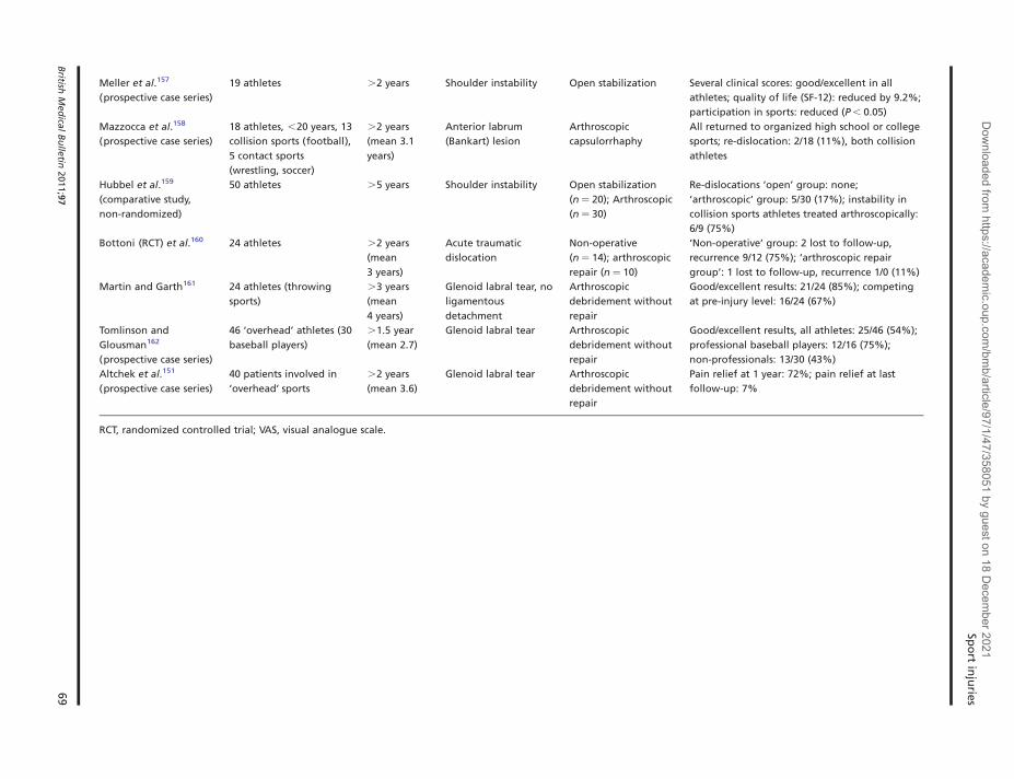
Labral tears require repair, whereas shoulder instability is currentlyapproached operatively more often. Conservative management of trau-matic shoulder dislocations in adolescents is associated with high ratesof recurrent instability (up to 100%), whereas recurrent dislocationswere reported in up to 12%, at an average of 3 years after arthroscopicstabilization. Shoulder dislocations are particularly common in rugby,the characteristic mechanism of injury being tackling, whereas labraltears are common in the ‘overhead’ athlete’. Published results in ath-letes151–162 (Table 7) show that operative stabilization of the shoulderis initially successful, but instability and pain can recur in the longterm. Results of arthroscopic techniques in the management ofintra-articular pathologies are promising, but long-term outcomes areunknown (Table 7).
Operative management of elbow injuries in athletes
Elbow ulnar collateral ligament (UCL) insufficiency is one of the fre-quently recognized injuries in the overhead athlete, as a result of exces-sive valgus stress. It constitutes a potentially career threatening injuryand requires surgical repair.163 The use of a muscle-splitting approach,avoiding handling of the ulnar nerve, and the use of the docking tech-nique for stabilization is recommended164,165 (Table 8). Recent advan-tages in arthroscopic surgical techniques and ligament reconstructionin the elbow have improved the prognosis for return to competition forhighly motivated athletes. The results of arthroscopic debride-ment150,166 (Table 7) need to be evaluated in the long term.
Sport injuries
British Medical Bulletin 2011;97 67
Dow
nloaded from https://academ
ic.oup.com/bm
b/article/97/1/47/358051 by guest on 18 Decem
ber 2021

Table 7 Operative management of shoulder injuries in athletes
Study Patients Follow-up Problem Operation Outcome
Owens et al.153
(prospective case series)
39 athletes (40 shoulders) 9–14 years
(mean 11.7
years)
First-time traumatic
anterior shoulder
dislocations
Acute arthroscopic
Bankart repair
Re-dislocations: 14% (6/40); subluxation: 21% (9/
40); revision stabilization surgery: 14% (6/40);
SF-36 (mean): 94.4 of 100; Tegner score (mean):
6.5 (3–10); patients’ rating of shoulder function
compared with pre-injury: 93%; would they
recommend the surgery? VAS¼9.1 of 10 (only
three patients ,7)
Baker et al.152
(prospective case series)
40 athletes (43 shoulders) .2 years
(mean 2.8)
Multidirectional
instability
Arthroscopic
capsulorrhaphy
Clinical scores: mean .91 points out of 100;
strength: 98% normal or slightly decreased;
range of motion: 91% full or satisfactory; return
to sport: 86%
Kartus et al.154
(prospective case series)
71 patients (73% involved
in ‘overhead’ sports)
Median 9
years
Anterior labrum
(Bankart) lesion
Arthroscopic
capsulorrhaphy
Shoulder instability: 37/71 (38%); re-dislocation:
16/71 (23%); Overhead sports participation: 45%
(compared to 73% before the injury)
Radkowski et al.155
(prospective case series)
98 athletes (107
shoulders)
Mean 2.3
years
Unidirectional
(posterior) instability
Arthroscopic
capsulorrhaphy
Good/excellent clinical score in 89% of
‘throwers’ and 93% of ‘non-throwers’; return to
pre-injury level: ‘throwers’ 55%; ‘non-throwers’
71%
Bonnevialle et al.156
(prospective case series)
31 Rugby players .5 years Shoulder instability Open stabilization Return to rugby: 97%; recurrence after trauma:
17%; patients’ satisfaction: 88%; radiographic
arthritis: 32%
N.
Maffu
lliet
al.
68
British
Med
ical
Bu
lletin
2011;9
7
Dow
nloaded from https://academ
ic.oup.com/bm
b/article/97/1/47/358051 by guest on 18 Decem
ber 2021
Meller et al.157
(prospective case series)
19 athletes .2 years Shoulder instability Open stabilization Several clinical scores: good/excellent in all
athletes; quality of life (SF-12): reduced by 9.2%;
participation in sports: reduced (P , 0.05)
Mazzocca et al.158
(prospective case series)
18 athletes, ,20 years, 13
collision sports (football),
5 contact sports
(wrestling, soccer)
.2 years
(mean 3.1
years)
Anterior labrum
(Bankart) lesion
Arthroscopic
capsulorrhaphy
All returned to organized high school or college
sports; re-dislocation: 2/18 (11%), both collision
athletes
Hubbel et al.159
(comparative study,
non-randomized)
50 athletes .5 years Shoulder instability Open stabilization
(n ¼ 20); Arthroscopic
(n ¼ 30)
Re-dislocations ‘open’ group: none;
‘arthroscopic’ group: 5/30 (17%); instability in
collision sports athletes treated arthroscopically:
6/9 (75%)
Bottoni (RCT) et al.160 24 athletes .2 years
(mean
3 years)
Acute traumatic
dislocation
Non-operative
(n ¼ 14); arthroscopic
repair (n ¼ 10)
‘Non-operative’ group: 2 lost to follow-up,
recurrence 9/12 (75%); ‘arthroscopic repair
group’: 1 lost to follow-up, recurrence 1/0 (11%)
Martin and Garth161 24 athletes (throwing
sports)
.3 years
(mean
4 years)
Glenoid labral tear, no
ligamentous
detachment
Arthroscopic
debridement without
repair
Good/excellent results: 21/24 (85%); competing
at pre-injury level: 16/24 (67%)
Tomlinson and
Glousman162
(prospective case series)
46 ‘overhead’ athletes (30
baseball players)
.1.5 year
(mean 2.7)
Glenoid labral tear Arthroscopic
debridement without
repair
Good/excellent results, all athletes: 25/46 (54%);
professional baseball players: 12/16 (75%);
non-professionals: 13/30 (43%)
Altchek et al.151
(prospective case series)
40 patients involved in
‘overhead’ sports
.2 years
(mean 3.6)
Glenoid labral tear Arthroscopic
debridement without
repair
Pain relief at 1 year: 72%; pain relief at last
follow-up: 7%
RCT, randomized controlled trial; VAS, visual analogue scale.
Spo
rtin
jurie
s
British
Med
ical
Bu
lletin
2011;9
769
Dow
nloaded from https://academ
ic.oup.com/bm
b/article/97/1/47/358051 by guest on 18 Decem
ber 2021

Table 8 Operative management of elbow injuries in athletes.
Study Patients Follow-up Problem Operation(s) Outcome
Vitale and Ahmad164
(systematic review of 8
retrospective studies)
‘Overhead’
athletes
.1 year UCL injury UCL repair
Muscle-splitting approach
versus Detachment of
flexor-pronator mass
Unlar nerve transposition
versus no transposition
Docking versus figure of
eight technique
Overall: good/excellent results: 83%; complication
rate: 10%; ulnar neuropathy: 6%; muscle, splitting
approach: better results and less complications;
ulnar nerve transposition: less favourable results,
higher neuropathy rate (9% versus 4%); docking
technique: better outcomes
Savoie et al.165
(prospective case series)
60 high school,
college athletes
Mean 5
years
UCL injury Direct repair
(suture placation with repair
to bone)
Good/excellent results: 93%; return to sports
(pre-injury level) within 6 months: 97%; transient
ulnar neuropathy: 5%; failures:
Rahusen et al.166
(prospective case series)
16 athletes .2.5 years
(mean 3.2)
Posterior elbow
impingement
Arthroscopic debridement Extension deficit: reduced from 88 to 28; VAS in rest:
reduced from 3 to 0; VAS during sports: reduced
from 7 to 2 (all differences were significant,
P , 0.05)
Byrd and Jones150
(prospective case series)
10 baseball
players
Mean 4
years
Osteochondritis
dissecans of the
capitellum
Arthroscopic debridement Excellent clinical results; radiographs:
Primary lesion evident in 2/10 athletes
Degenerative changes in 2/10 athletes
Return to baseball: 4/10 athletes
UCL, ulnar collateral ligament.
N.
Maffu
lliet
al.
70
British
Med
ical
Bu
lletin
2011;9
7
Dow
nloaded from https://academ
ic.oup.com/bm
b/article/97/1/47/358051 by guest on 18 Decem
ber 2021

Operative management of wrist injuries in athletes
A review of the literature shows that 3–9% of all athletic injuriesoccur in the hand or wrist, and are more common in adolescent ath-letes than adults.46 In this article, we focused on TFCC injuries andacute scaphoid fractures in athletes.
TFCC injuries are an increasingly recognized cause of ulnar-sidedwrist pain, and can be particularly disabling in the competitive athlete.Advances in wrist arthroscopy made endoscopic debridement andrepair of the TFCC possible. McAdams et al.47 treated arthroscopicallyTFCC tears in 16 competitive athletes (mean age: 23.4 years). Repairof unstable tears was performed in 11 (69%) and debridement only in5 (31%). Return to play averaged 3.3 months (range: 3–7 months).The mean duration of follow-up was 2.8 years (range: 2–4.2 years).Clinical scores (mini-DASH and mini-DASH sports module) improvedsignificantly. No long-term outcomes are available.
Operative management of scaphoid fractures in athletes, even ifundisplaced, is recommended if early return to sports is desired. Onestudy followed 12 athletes treated operatively for a scaphoid fracture.They were able to return to sports at 6 weeks. At an average follow-upof 2.9 years, 9 of 12 athletes had range of motion equal to the unin-jured side, and grip strength was equal to the unaffected side in 10 of12 athletes.49
Discussion
Participation in sports offers potential benefits for individuals of allages, such as combating obesity and enhancing cardiovascular fitness.1
On the other hand, negative consequences of musculoskeletal injuriessustained during sports may compromise function in later life, limitingthe ability to experience pain-free mobility and engage in fitness-enhan-cing activity.167 Increasingly, successful management of sports-relatedinjuries has allowed more athletes to return to participation. The kneeis the joint most commonly associated with sports injuries, and there-fore is most at risk of developing degenerative changes. It is not clearwhether radiographic OA always correlates with symptoms andreduced quality of life. Furthermore, even effective management ofmeniscal or ACL injury does not reduce the risk of developing sub-sequent OA.137,168 OA in an injured joint is caused by intra-articularpathogenic processes initiated at the time of injury, combined withlong-term changes in dynamic joint loading. Variation in outcomesinvolves not only the exact type of injury (e.g. ACL rupture with orwithout meniscal damage),137 but also additional variables associated
Sport injuries
British Medical Bulletin 2011;97 71
Dow
nloaded from https://academ
ic.oup.com/bm
b/article/97/1/47/358051 by guest on 18 Decem
ber 2021
with the individual such as age, sex, genetics, obesity, muscle strength,activity and reinjury. A better understanding of these variables mayimprove future prevention and treatment strategies.169
In many of the long-term studies (the majority being retrospectivecase series), several methodological flaws have to be highlighted. Arecent systematic review on OA after ACL injuries137 suggested thatsome studies may overestimate the prevalence of long-term OA. Theauthors in several studies mention that a proportion of the index groupof injured athletes were available for follow-up or consented for radio-graphic examination. One can argue that these patients were the oneswith symptoms, therefore the prevalence of OA (after ACL rupture forexample) may appear higher than it really is. Presentation of outcomeswas not always based on robust criteria. Different clinical scores andradiographic classifications have been used, and therefore resultsbetween studies are not directly comparable. In the majority of thestudies, it was not clarified whether radiographic appearance correlatedwith symptoms, and how important these were for the quality of life ofthe patients. Disabling arthritis requiring intervention may actually bedelayed for more than 20–30 years.107,112 Furthermore, long-termstudies present outcomes of older techniques, not used any more inclinical practice (e.g. primary ACL repair or total meniscectomy).Evolution in surgical or rehabilitation techniques might have improvedoutcomes of certain injuries. Therefore, currently known ‘long-termoutcomes’ may only reflect the results of techniques used in the pastand not what we should expect in the future. Increasing awareness ofathletes and trainers, new diagnostic and musculoskeletal imagingmodalities, improved surgical and rehabilitation methods, but alsoanalysis of injury patterns in different sports and development of injuryprevention strategies might be beneficial to minimize the effects ofsports injuries in the years to come.
What is the true incidence of arthritis in the long term? Will it be adisabling condition for the former athlete, in the coming decades?Currently, joint preserving procedures (e.g. microfractures,145 mosaic-platy,170 autologous chondrocyte implantation,171,172 realignmentosteotomies173 and implant arthroplasties174) have evolved and allowmiddle aged or older patients to live without pain and maintain anactive life style. Meniscal transplantation shows encouraging results.175
Should therefore an increased risk for developing musculoskeletal pro-blems prevent children and adults from being active in sports?176 Dothe benefits of participating in sports outweigh the risks?
A survey in Sweden showed that 80% of former track and field ath-letes with an age range of 50–80 years felt they were in good health,compared with 61% of the referents, despite higher prevalence of hiparthritis in former athletes. Low back disorders were similar in the two
N. Maffulli et al.
72 British Medical Bulletin 2011;97
Dow
nloaded from https://academ
ic.oup.com/bm
b/article/97/1/47/358051 by guest on 18 Decem
ber 2021

groups, shoulder and neck problems were lower in former athletes, andknee arthritis was similar in the two groups.177
No definite answer can be given to the previously addressed ques-tions, based on available evidence. Future research should involve ques-tionnaires assessing the HRQL in former athletes, to be compared withthe general population.27,178–181
Conclusions
Physical injury is an inherent risk in sports participation and, to acertain extent, must be considered an inevitable cost of athletic trainingand competition. Injury may lead to incomplete recovery and residualsymptoms, drop out from sports, and can cause joint degeneration inthe long term. Few well-conducted studies are available on the long-term follow-up of former athletes, and, in general, we lack studiesreporting on the HRQL to be compared with the general population.Advances in arthroscopic techniques allow operative management ofmost intra-articular post-traumatic pathologies in the lower and upperlimb joints, but long-term outcomes are not available yet. It is impor-tant to balance the negative effects of sports injuries with the manysocial, psychological and health benefits that a serious commitment tosport brings.9
References
1 Maffulli N. The growing child in sport. Br Med Bull 1992;48:561–8.
2 Baxter-Jones AD, Helms P, Maffulli N et al. Growth and development of male gymnasts,swimmers, soccer and tennis players: a longitudinal study. Ann Hum Biol 1995;22:381–94.
3 Baxter-Jones AD, Maffulli N. Intensive training in elite young female athletes. Effects ofintensive training on growth and maturation are not established. Br J Sports Med2002;36:13–5.
4 Baxter-Jones AD, Maffulli N. Parental influence on sport participation in elite young ath-letes. J Sports Med Phys Fitness 2003;43:250–5.
5 Baxter-Jones AD, Maffulli N. Endurance in young athletes: it can be trained. Br J SportsMed 2003;37:96–7.
6 Caine D, Cochrane B, Caine C et al. An epidemiologic investigation of injuries affecting
young competitive female gymnasts. Am J Sports Med 1989;17:811–20.7 Caine DJ, Maffulli N. Epidemiology of children’s individual sports injuries. An important
area of medicine and sport science research. Med Sport Sci 2005;48:1–7.8 Caine DJ, Nassar L. Gymnastics injuries. Med Sport Sci 2005;48:18–58.9 Shanmugam C, Maffulli N. Sports injuries in children. Br Med Bull 2008;86:33–57.
10 De Mozzi P, Longo UG, Galanti G et al. Bicuspid aortic valve: a literature review and itsimpact on sport activity. Br Med Bull 2008;85:63–85.
11 van Mechelen W, Hlobil H, Kemper HC. Incidence, severity, aetiology and prevention of
sports injuries. A review of concepts. Sports Med 1992;14:82–99.12 Brites F, Verona J, De Geitere C et al. Enhanced cholesterol efflux promotion in well-trained
soccer players. Metabolism 2004;53:1262–7.
Sport injuries
British Medical Bulletin 2011;97 73
Dow
nloaded from https://academ
ic.oup.com/bm
b/article/97/1/47/358051 by guest on 18 Decem
ber 2021

13 Hansen L, Bangsbo J, Twisk J et al. Development of muscle strength in relation to training
level and testosterone in young male soccer players. J Appl Physiol 1999;87:1141–7.14 Tsunawake N, Tahara Y, Moji K et al. Body composition and physical fitness of female vol-
leyball and basketball players of the Japan inter-high school championship teams. J PhysiolAnthropol Appl Human Sci 2003;22:195–201.
15 Ginty F, Rennie KL, Mills L et al. Positive, site-specific associations between bone mineral
status, fitness, and time spent at high-impact activities in 16- to 18-year-old boys. Bone2005;36:101–10.
16 Freedman DS, Khan LK, Dietz WH et al. Relationship of childhood obesity to coronaryheart disease risk factors in adulthood: the Bogalusa Heart Study. Pediatrics 2001;108:712–8.
17 Caine D, Lewis R, O’Connor P et al. Does gymnastics training inhibit growth of females?Clin J Sport Med 2001;11:260–70.
18 Caine D, Maffulli N, Caine C. Epidemiology of injury in child and adolescent sports: injuryrates, risk factors, and prevention. Clin Sports Med 2008;27:19–50. vii.
19 Fox KR, Riddoch C. Charting the physical activity patterns of contemporary children and
adolescents. Proc Nutr Soc 2000;59:497–504.20 Hassmen P, Koivula N, Uutela A. Physical exercise and psychological well-being: a popu-
lation study in Finland. Prev Med 2000;30:17–25.
21 Gibbons SL, Ebbeck V, Weiss MR. Fair Play for Kids: effects on the moral development ofchildren in physical education. Res Q Exerc Sport 1995;66:247–55.
22 Maffulli N, Baxter-Jones AD, Grieve A. Long term sport involvement and sport injury ratein elite young athletes. Arch Dis Child 2005;90:525–7.
23 Erlandson MC, Sherar LB, Mirwald RL et al. Growth and maturation of adolescent female
gymnasts, swimmers, and tennis players. Med Sci Sports Exerc 2008;40:34–42.24 Maffulli N, Longo UG, Spiezia F et al. Sports injuries in young athletes: long-term outcome
and prevention strategies. Phys Sportsmed 2010;38:29–34.25 Longo UG, Fazio V, Poeta ML et al. Bilateral consecutive rupture of the quadriceps tendon
in a man with BstUI polymorphism of the COL5A1 gene. Knee Surg Sports TraumatolArthrosc 2010;18:514–8.
26 Longo UG, Forriol F, Maffulli N et al. Evaluation of histological scoring systems for
tissue-engineered, repaired and osteoarthritic cartilage. Osteoarthritis Cartilage 2010;18:1001.Author reply 1002.
27 Longo UG, Lamberti A, Maffulli N et al. Tendon augmentation grafts: a systematic review.
Br Med Bull 2010;94:165–88.28 Maffulli N, Longo UG, Loppini M et al. Current treatment options for tendinopathy.
Expert Opin Pharmacother 2010; June 23. [Epub ahead of print].
29 Maffulli N, Longo UG, Maffulli GD et al. Achilles tendon ruptures in diabetic patients.Arch Orthop Trauma Surg 2010; April 26. [Epub ahead of print].
30 Maffulli N, Longo UG, Maffulli GD et al. Marked pathological changes proximal and distalto the site of rupture in acute Achilles tendon ruptures. Knee Surg Sports TraumatolArthrosc 2010; June 19. [Epub ahead of print].
31 Becher C, Driessen A, Hess T et al. Microfracture for chondral defects of the talus: mainten-ance of early results at midterm follow-up. Knee Surg Sports Traumatol Arthrosc2010;18:656–63.
32 Denaro V, Ruzzini L, Longo UG et al. Effect of dihydrotestosterone on cultured humantenocytes from intact supraspinatus tendon. Knee Surg Sports Traumatol Arthrosc2010;18:971–6.
33 Longo UG, Garau G, Denaro V et al. Surgical management of tendinopathy of biceps
femoris tendon in athletes. Disabil Rehabil 2008;30:1602–7.34 Longo UG, Oliva F, Denaro V et al. Oxygen species and overuse tendinopathy in athletes.
Disabil Rehabil 2008;30:1563–71.
35 Longo UG, Ramamurthy C, Denaro V et al. Minimally invasive stripping for chronicAchilles tendinopathy. Disabil Rehabil 2008;30:1709–13.
36 Beynnon BD, Vacek PM, Murphy D et al. First-time inversion ankle ligament trauma: theeffects of sex, level of competition, and sport on the incidence of injury. Am J Sports Med2005;33:1485–91.
N. Maffulli et al.
74 British Medical Bulletin 2011;97
Dow
nloaded from https://academ
ic.oup.com/bm
b/article/97/1/47/358051 by guest on 18 Decem
ber 2021

37 DeHaven KE, Lintner DM. Athletic injuries: comparison by age, sport, and gender. Am JSports Med 1986;14:218–24.
38 Caine D, Roy S, Singer KM et al. Stress changes of the distal radial growth plate. A radio-
graphic survey and review of the literature. Am J Sports Med 1992;20:290–8.39 Adirim TA, Cheng TL. Overview of injuries in the young athlete. Sports Med
2003;33:75–81.
40 Collins CL, Micheli LJ, Yard EE et al. Injuries sustained by high school rugby players in theUnited States, 2005–2006. Arch Pediatr Adolesc Med 2008;162:49–54.
41 Malliaropoulos N, Ntessalen M, Papacostas E et al. Reinjury after acute lateral anklesprains in elite track and field athletes. Am J Sports Med 2009;37:1755–61.
42 Nelson AJ, Collins CL, Yard EE et al. Ankle injuries among United States high school
sports athletes, 2005–2006. J Athl Train 2007;42:381–7.43 Byrd JW, Jones KS. Hip arthroscopy in athletes: 10-year follow-up. Am J Sports Med
2009;37:2140–3.44 Shetty VD, Villar RN. Hip arthroscopy: current concepts and review of literature. Br J
Sports Med 2007;41:64–8. Discussion 68.
45 Cain EL Jr, Dugas JR, Wolf RS et al. Elbow injuries in throwing athletes: a current conceptsreview. Am J Sports Med 2003;31:621–35.
46 Geissler WB. Carpal fractures in athletes. Clin Sports Med 2001;20:167–88.
47 McAdams TR, Swan J, Yao J. Arthroscopic treatment of triangular fibrocartilage wrist inju-ries in the athlete. Am J Sports Med 2009;37:291–7.
48 Rizzo M, Shin AY. Treatment of acute scaphoid fractures in the athlete. Curr Sports MedRep 2006;5:242–8.
49 Rettig AC. Athletic injuries of the wrist and hand: Part II: overuse injuries of the wrist and
traumatic injuries to the hand. Am J Sports Med 2004;32:262–73.50 Sys J, Michielsen J, Bracke P et al. Nonoperative treatment of active spondylolysis in elite
athletes with normal X-ray findings: literature review and results of conservative treatment.Eur Spine J 2001;10:498–504.
51 Boden BP, Tacchetti RL, Cantu RC et al. Catastrophic cervical spine injuries in high school
and college football players. Am J Sports Med 2006;34:1223–32.52 Caine DJ. Are kids having a rough time of it in sports? Br J Sports Med 2010;44:1–3.
53 Maffulli N, Longo UG, Gougoulias N et al. Long-term health outcomes of youth sportsinjuries. Br J Sports Med 2010;44:21–5.
54 Ilharreborde B, Raquillet C, Morel E et al. Long-term prognosis of Salter-Harris type 2 inju-
ries of the distal femoral physis. J Pediatr Orthop B 2006;15:433–8.55 Nenopoulos SP, Papavasiliou VA, Papavasiliou AV. Outcome of physeal and epiphyseal inju-
ries of the distal tibia with intra-articular involvement. J Pediatr Orthop 2005;25:518–22.
56 Goldberg VM, Aadalen R. Distal tibial epiphyseal injuries: the role of athletics in 53 cases.Am J Sports Med 1978;6:263–8.
57 Cannata G, De Maio F, Mancini F et al. Physeal fractures of the distal radius and ulna: long-term prognosis. J Orthop Trauma 2003;17:172–9. Discussion 179–80.
58 Stephens DC, Louis E, Louis DS. Traumatic separation of the distal femoral epiphyseal car-
tilage plate. J Bone Joint Surg Am 1974;56:1383–90.59 Criswell AR, Hand WL, Butler JE. Abduction injuries of the distal femoral epiphysis. Clin
Orthop Relat Res 1976;115:189–94.60 Lombardo SJ, Harvey JP Jr. Fractures of the distal femoral epiphyses. Factors influencing
prognosis: a review of thirty-four cases. J Bone Joint Surg Am 1977;59:742–51.
61 Burkhart SS, Peterson HA. Fractures of the proximal tibial epiphysis. J Bone Joint Surg Am1979;61:996–1002.
62 Cass JR, Peterson HA. Salter-Harris Type-IV injuries of the distal tibial epiphyseal growthplate, with emphasis on those involving the medial malleolus. J Bone Joint Surg Am1983;65:1059–70.
63 Ogden JA. Distal clavicular physeal injury. Clin Orthop Relat Res 1984;118:68–73.64 Landin LA, Danielsson LG, Jonsson K et al. Late results in 65 physeal ankle fractures. Acta
Orthop Scand 1986;57:530–4.65 Hynes D, O’Brien T. Growth disturbance lines after injury of the distal tibial physis. Their
significance in prognosis. J Bone Joint Surg Br 1988;70:231–3.
Sport injuries
British Medical Bulletin 2011;97 75
Dow
nloaded from https://academ
ic.oup.com/bm
b/article/97/1/47/358051 by guest on 18 Decem
ber 2021

66 Krueger-Franke M, Siebert CH, Pfoerringer W. Sports-related epiphyseal injuries of the
lower extremity. An epidemiologic study. J Sports Med Phys Fitness 1992;32:106–11.67 Berson L, Davidson RS, Dormans JP et al. Growth disturbances after distal tibial physeal
fractures. Foot Ankle Int 2000;21:54–8.68 Eid AM, Hafez MA. Traumatic injuries of the distal femoral physis. Retrospective study on
151 cases. Injury 2002;33:251–5.
69 Barmada A, Gaynor T, Mubarak SJ. Premature physeal closure following distal tibia physealfractures: a new radiographic predictor. J Pediatr Orthop 2003;23:733–9.
70 Nietosvaara Y, Hasler C, Helenius I et al. Marked initial displacement predicts compli-cations in physeal fractures of the distal radius: an analysis of fracture characteristics,primary treatment and complications in 109 patients. Acta Orthop 2005;76:873–7.
71 Lalonde KA, Letts M. Traumatic growth arrest of the distal tibia: a clinical and radiographicreview. Can J Surg 2005;48:143–7.
72 Kawamoto K, Kim WC, Tsuchida Y et al. Incidence of physeal injuries in Japanese children.J Pediatr Orthop B 2006;15:126–30.
73 Arkader A, Warner WC Jr, Horn BD et al. Predicting the outcome of physeal fractures of
the distal femur. J Pediatr Orthop 2007;27:703–8.74 Lundin O, Hellstrom M, Nilsson I et al. Back pain and radiological changes in the thoraco-
lumbar spine of athletes. A long-term follow-up. Scand J Med Sci Sports 2001;11:103–9.
75 Tsai L, Wredmark T. Spinal posture, sagittal mobility, and subjective rating of back pro-blems in former female elite gymnasts. Spine (Phila Pa 1976) 1993;18:872–5.
76 Conaghan PG. Update on osteoarthritis part 1: current concepts and the relation to exercise.Br J Sports Med 2002;36:330–3.
77 Chantraine A. Knee joint in soccer players: osteoarthritis and axis deviation. Med Sci SportsExerc 1985;17:434–9.
78 Kujala UM, Kettunen J, Paananen H et al. Knee osteoarthritis in former runners, soccer
players, weight lifters, and shooters. Arthritis Rheum 1995;38:539–46.79 Drawer S, Fuller CW. Propensity for osteoarthritis and lower limb joint pain in retired pro-
fessional soccer players. Br J Sports Med 2001;35:402–8.
80 Larsen E, Jensen PK, Jensen PR. Long-term outcome of knee and ankle injuries in elite foot-ball. Scand J Med Sci Sports 1999;9:285–9.
81 Shepard GJ, Banks AJ, Ryan WG. Ex-professional association footballers have an increasedprevalence of osteoarthritis of the hip compared with age matched controls despite nothaving sustained notable hip injuries. Br J Sports Med 2003;37:80–1.
82 Spector TD, Harris PA, Hart DJ et al. Risk of osteoarthritis associated with long-termweight-bearing sports: a radiologic survey of the hips and knees in female ex-athletes andpopulation controls. Arthritis Rheum 1996;39:988–95.
83 McDermott M, Freyne P. Osteoarthrosis in runners with knee pain. Br J Sports Med1983;17:84–7.
84 Knobloch M, Marti B, Biedert R et al. [Risk of arthrosis of the upper ankle joint in long dis-tance runners: controlled follow-up of former elite athletes]. Sportverletz Sportschaden1990;4:175–9.
85 Maquirriain J, Ghisi JP, Amato S. Is tennis a predisposing factor for degenerative shoulderdisease? A controlled study in former elite players. Br J Sports Med 2006;40:447–50.
86 L’Hermette M, Polle G, Tourny-Chollet C et al. Hip passive range of motion and frequencyof radiographic hip osteoarthritis in former elite handball players. Br J Sports Med2006;40:45–9. Discussion 45–9.
87 Gross P, Marti B. Risk of degenerative ankle joint disease in volleyball players: study offormer elite athletes. Int J Sports Med 1999;20:58–63.
88 Turner AP, Barlow JH, Heathcote-Elliott C. Long term health impact of playing professionalfootball in the United Kingdom. Br J Sports Med 2000;34:332–6.
89 Burnett AF, Khangure MS, Elliott BC et al. Thoracolumbar disc degeneration in young fast
bowlers in cricket: a follow-up study. Clin Biomech (Bristol, Avon) 1996;11:305–10.90 Rossi F, Dragoni S. Lumbar spondylolysis: occurrence in competitive athletes. Updated
achievements in a series of 390 cases. J Sports Med Phys Fitness 1990;30:450–2.91 Morita T, Ikata T, Katoh S et al. Lumbar spondylolysis in children and adolescents. J Bone
Joint Surg Br 1995;77:620–5.
N. Maffulli et al.
76 British Medical Bulletin 2011;97
Dow
nloaded from https://academ
ic.oup.com/bm
b/article/97/1/47/358051 by guest on 18 Decem
ber 2021
92 Foster D, John D, Elliott B et al. Back injuries to fast bowlers in cricket: a prospective study.
Br J Sports Med 1989;23:150–4.93 Annear PT, Chakera TM, Foster DH et al. Pars interarticularis stress and disc degeneration
in cricket’s potent strike force: the fast bowler. Aust N Z J Surg 1992;62:768–73.94 Hardcastle P, Annear P, Foster DH et al. Spinal abnormalities in young fast bowlers. J Bone
Joint Surg Br 1992;74:421–5.
95 Dixon M, Fricker P. Injuries to elite gymnasts over 10 yr. Med Sci Sports Exerc1993;25:1322–9.
96 Jackson DW, Wiltse LL, Cirincoine RJ. Spondylolysis in the female gymnast. Clin OrthopRelat Res 1976;117:68–73.
97 Ferguson RJ, McMaster JH, Stanitski CL. Low back pain in college football linemen. JSports Med 1974;2:63–9.
98 McCarroll JR, Miller JM, Ritter MA. Lumbar spondylolysis and spondylolisthesis in college
football players. A prospective study. Am J Sports Med 1986;14:404–6.99 Semon RL, Spengler D. Significance of lumbar spondylolysis in college football players.
Spine (Phila Pa 1976) 1981;6:172–4.
100 Baranto A, Hellstrom M, Nyman R et al. Back pain and degenerative abnormalities in thespine of young elite divers: a 5-year follow-up magnetic resonance imaging study. Knee SurgSports Traumatol Arthrosc 2006;14:907–14.
101 Granhed H, Morelli B. Low back pain among retired wrestlers and heavyweight lifters. AmJ Sports Med 1988;16:530–3.
102 Schmitt H, Brocai DR, Carstens C. Long-term review of the lumbar spine in javelinthrowers. J Bone Joint Surg Br 2001;83:324–7.
103 Ozturk A, Ozkan Y, Ozdemir RM et al. Radiographic changes in the lumbar spine in
former professional football players: a comparative and matched controlled study. Eur SpineJ 2008;17:136–41.
104 Thelin N, Holmberg S, Thelin A. Knee injuries account for the sports-related increased riskof knee osteoarthritis. Scand J Med Sci Sports 2006;16:329–33.
105 Moretz JA III, Harlan SD, Goodrich J et al. Long-term followup of knee injuries in high
school football players. Am J Sports Med 1984;12:298–300.106 Roos H, Lauren M, Adalberth T et al. Knee osteoarthritis after meniscectomy: prevalence of
radiographic changes after twenty-one years, compared with matched controls. ArthritisRheum 1998;41:687–93.
107 Nebelung W, Wuschech H. Thirty-five years of follow-up of anterior cruciate ligament-
deficient knees in high-level athletes. Arthroscopy 2005;21:696–702.108 von Porat A, Roos EM, Roos H. High prevalence of osteoarthritis 14 years after an anterior
cruciate ligament tear in male soccer players: a study of radiographic and patient relevant
outcomes. Ann Rheum Dis 2004;63:269–73.109 Lohmander LS, Ostenberg A, Englund M et al. High prevalence of knee osteoarthritis, pain,
and functional limitations in female soccer players twelve years after anterior cruciate liga-ment injury. Arthritis Rheum 2004;50:3145–52.
110 Anandacoomarasamy A, Barnsley L. Long term outcomes of inversion ankle injuries. Br JSports Med 2005;39:e14. Discussion e14.
111 Gerber JP, Williams GN, Scoville CR et al. Persistent disability associated with ankle
sprains: a prospective examination of an athletic population. Foot Ankle Int1998;19:653–60.
112 Valderrabano V, Hintermann B, Horisberger M et al. Ligamentous posttraumatic ankle
osteoarthritis. Am J Sports Med 2006;34:612–20.113 Wallace WA. Sporting injuries to the shoulder. J R Coll Surg Edinb 1990;35:S21–S26.
114 Muckle DS. Factors affecting the prognosis of meniscectomy in soccer players. Br J SportsMed 1983;17:88–90.
115 Jorgensen U, Sonne-Holm S, Lauridsen F et al. Long-term follow-up of meniscectomy in
athletes. A prospective longitudinal study. J Bone Joint Surg Br 1987;69:80–3.116 Bonneux I, Vandekerckhove B. Arthroscopic partial lateral meniscectomy long-term results
in athletes. Acta Orthop Belg 2002;68:356–61.117 McNicholas MJ, Rowley DI, McGurty D et al. Total meniscectomy in adolescence. A thirty-
year follow-up. J Bone Joint Surg Br 2000;82:217–21.
Sport injuries
British Medical Bulletin 2011;97 77
Dow
nloaded from https://academ
ic.oup.com/bm
b/article/97/1/47/358051 by guest on 18 Decem
ber 2021

118 Abdon P, Turner MS, Pettersson H et al. A long-term follow-up study of total meniscectomy
in children. Clin Orthop Relat Res 1990;257:166–70.119 Medlar RC, Mandiberg JJ, Lyne ED. Meniscectomies in children. Report of long-term
results (mean, 8.3 years) of 26 children. Am J Sports Med 1980;8:87–92.120 Wroble RR, Henderson RC, Campion ER et al. Meniscectomy in children and adolescents.
A long-term follow-up study. Clin Orthop Relat Res 1992;279:180–9.
121 Zaman M, Leonard MA. Meniscectomy in children: results in 59 knees. Injury1981;12:425–8.
122 Manzione M, Pizzutillo PD, Peoples AB et al. Meniscectomy in children: a long-termfollow-up study. Am J Sports Med 1983;11:111–5.
123 Dai L, Zhang W, Xu Y. Meniscal injury in children: long-term results after meniscectomy.
Knee Surg Sports Traumatol Arthrosc 1997;5:77–9.124 Rockborn P, Gillquist J. Outcome of arthroscopic meniscectomy. A 13-year physical and
radiographic follow-up of 43 patients under 23 years of age. Acta Orthop Scand1995;66:113–7.
125 Rockborn P, Messner K. Long-term results of meniscus repair and meniscectomy: a 13-year
functional and radiographic follow-up study. Knee Surg Sports Traumatol Arthrosc2000;8:2–10.
126 Maletius W, Messner K. Chondral damage and age depress the long-term prognosis after
partial meniscectomy. A 12- to 15-year follow-up study. Knee Surg Sports TraumatolArthrosc 1996;3:211–4.
127 Schimmer RC, Brulhart KB, Duff C et al. Arthroscopic partial meniscectomy: a 12-yearfollow-up and two-step evaluation of the long-term course. Arthroscopy 1998;14:136–42.
128 Andersson-Molina H, Karlsson H, Rockborn P. Arthroscopic partial and total meniscect-
omy: a long-term follow-up study with matched controls. Arthroscopy 2002;18:183–9.129 Englund M, Roos EM, Lohmander LS. Impact of type of meniscal tear on radiographic and
symptomatic knee osteoarthritis: a sixteen-year followup of meniscectomy with matchedcontrols. Arthritis Rheum 2003;48:2178–87.
130 Eggli S, Wegmuller H, Kosina J et al. Long-term results of arthroscopic meniscal repair. An
analysis of isolated tears. Am J Sports Med 1995;23:715–20.131 Lee GP, Diduch DR. Deteriorating outcomes after meniscal repair using the Meniscus
Arrow in knees undergoing concurrent anterior cruciate ligament reconstruction: increasedfailure rate with long-term follow-up. Am J Sports Med 2005;33:1138–41.
132 Siebold R, Dehler C, Boes L et al. Arthroscopic all-inside repair using the Meniscus Arrow:
long-term clinical follow-up of 113 patients. Arthroscopy 2007;23:394–9.133 Logan M, Watts M, Owen J et al. Meniscal repair in the elite athlete: results of 45 repairs
with a minimum 5-year follow-up. Am J Sports Med 2009;37:1131–4.
134 Mintzer CM, Richmond JC, Taylor J. Meniscal repair in the young athlete. Am J SportsMed 1998;26:630–3.
135 Muaidi QI, Nicholson LL, Refshauge KM et al. Prognosis of conservatively managedanterior cruciate ligament injury: a systematic review. Sports Med 2007;37:703–16.
136 Meuffels DE, Favejee MM, Vissers MM et al. Ten year follow-up study comparing conser-
vative versus operative treatment of anterior cruciate ligament ruptures. A matched-pairanalysis of high level athletes. Br J Sports Med 2009;43:347–51.
137 Oiestad BE, Engebretsen L, Storheim K et al. Knee osteoarthritis after anterior cruciate liga-ment injury: a systematic review. Am J Sports Med 2009;37:1434–43.
138 Bales CP, Guettler JH, Moorman CT III. Anterior cruciate ligament injuries in children with
open physes: evolving strategies of treatment. Am J Sports Med 2004;32:1978–85.139 Bisson LJ, Wickiewicz T, Levinson M et al. ACL reconstruction in children with open
physes. Orthopedics 1998;21:659–63.140 Henry J, Chotel F, Chouteau J et al. Rupture of the anterior cruciate ligament in children:
early reconstruction with open physes or delayed reconstruction to skeletal maturity? KneeSurg Sports Traumatol Arthrosc 2009;17:748–55.
141 Marx A, Siebold R, Sobau C et al. ACL reconstruction in skeletally immature patients.
Sportverletz Sportschaden 2009;23:47–51.142 Schneider FJ, Kraus T, Linhart WE. Anterior cruciate ligament reconstruction with semiten-
dinosus tendon in children. Oper Orthop Traumatol 2008;20:409–22.
N. Maffulli et al.
78 British Medical Bulletin 2011;97
Dow
nloaded from https://academ
ic.oup.com/bm
b/article/97/1/47/358051 by guest on 18 Decem
ber 2021
143 Edwards PH, Grana WA. Anterior cruciate ligament reconstruction in the immature
athlete: long-term results of intra-articular reconstruction. Am J Knee Surg 2001;14:232–7.
144 Pressman AE, Letts RM, Jarvis JG. Anterior cruciate ligament tears in children: an analysisof operative versus nonoperative treatment. J Pediatr Orthop 1997;17:505–11.
145 Saxena A, Eakin C. Articular talar injuries in athletes: results of microfracture and autoge-
nous bone graft. Am J Sports Med 2007;35:1680–7.146 Rolf CG, Barclay C, Riyami M et al. The importance of early arthroscopy in athletes with
painful cartilage lesions of the ankle: a prospective study of 61 consecutive cases. J OrthopSurg Res 2006;1:4.
147 Baums MH, Kahl E, Schultz W et al. Clinical outcome of the arthroscopic management of
sports-related ‘anterior ankle pain’: a prospective study. Knee Surg Sports TraumatolArthrosc 2006;14:482–6.
148 DeBerardino TM, Arciero RA, Taylor DC. Arthroscopic treatment of soft-tissue impinge-ment of the ankle in athletes. Arthroscopy 1997;13:492–8.
149 Jerosch J, Steinbeck J, Schneider T et al. Arthroscopic treatment of anterior synovitis of the
upper ankle joint in the athlete. Sportverletz Sportschaden 1994;8:67–72.150 Byrd JW, Jones KS. Arthroscopic surgery for isolated capitellar osteochondritis dissecans in
adolescent baseball players: minimum three-year follow-up. Am J Sports Med 2002;
30:474–8.151 Altchek DW, Warren RF, Wickiewicz TL et al. Arthroscopic labral debridement. A three-
year follow-up study. Am J Sports Med 1992;20:702–6.152 Baker CL III, Mascarenhas R, Kline AJ et al. Arthroscopic treatment of multidirectional
shoulder instability in athletes: a retrospective analysis of 2- to 5-year clinical outcomes. AmJ Sports Med 2009;37:1712–20.
153 Owens BD, DeBerardino TM, Nelson BJ et al. Long-term follow-up of acute arthroscopic
Bankart repair for initial anterior shoulder dislocations in young athletes. Am J Sports Med2009;37:669–73.
154 Kartus C, Kartus J, Matis N et al. Long-term independent evaluation after arthroscopic
extra-articular Bankart repair with absorbable tacks. Surgical technique. J Bone Joint SurgAm 2008;90(Suppl. 2 Pt 2):262–74.
155 Radkowski CA, Chhabra A, Baker CL III et al. Arthroscopic capsulolabral repair for pos-terior shoulder instability in throwing athletes compared with nonthrowing athletes. Am JSports Med 2008;36:693–9.
156 Bonnevialle N, Mansat P, Bellumore Y et al. Surgical treatment of anterior shoulder instabil-ity in rugby players: clinical and radiographic results with minimum five-year follow-up.Rev Chir Orthop Reparatrice Appar Mot 2008;94:635–42.
157 Meller R, Krettek C, Gosling T et al. Recurrent shoulder instability among athletes: changesin quality of life, sports activity, and muscle function following open repair. Knee SurgSports Traumatol Arthrosc 2007;15:295–304.
158 Mazzocca AD, Brown FM Jr, Carreira DS et al. Arthroscopic anterior shoulder stabilizationof collision and contact athletes. Am J Sports Med 2005;33:52–60.
159 Hubbell JD, Ahmad S, Bezenoff LS et al. Comparison of shoulder stabilization using arthro-scopic transglenoid sutures versus open capsulolabral repairs: a 5-year minimum follow-up.
Am J Sports Med 2004;32:650–4.160 Bottoni CR, Wilckens JH, DeBerardino TM et al. A prospective, randomized evaluation of
arthroscopic stabilization versus nonoperative treatment in patients with acute, traumatic,
first-time shoulder dislocations. Am J Sports Med 2002;30:576–80.161 Martin DR, Garth WP Jr. Results of arthroscopic debridement of glenoid labral tears. Am J
Sports Med 1995;23:447–51.162 Tomlinson RJ Jr, Glousman RE. Arthroscopic debridement of glenoid labral tears in ath-
letes. Arthroscopy 1995;11:42–51.
163 Langer P, Fadale P, Hulstyn M. Evolution of the treatment options of ulnar collateral liga-ment injuries of the elbow. Br J Sports Med 2006;40:499–506.
164 Vitale MA, Ahmad CS. The outcome of elbow ulnar collateral ligament reconstruction inoverhead athletes: a systematic review. Am J Sports Med 2008;36:1193–205.
Sport injuries
British Medical Bulletin 2011;97 79
Dow
nloaded from https://academ
ic.oup.com/bm
b/article/97/1/47/358051 by guest on 18 Decem
ber 2021
165 Savoie FH III, Trenhaile SW, Roberts J et al. Primary repair of ulnar collateral ligament inju-
ries of the elbow in young athletes: a case series of injuries to the proximal and distal endsof the ligament. Am J Sports Med 2008;36:1066–72.
166 Rahusen FT, Brinkman JM, Eygendaal D. Arthroscopic treatment of posterior impingementof the elbow in athletes: a medium-term follow-up in sixteen cases. J Shoulder Elbow Surg2009;18:279–82.
167 Garrick JG, Requa RK. Sports and fitness activities: the negative consequences. J Am AcadOrthop Surg 2003;11:439–43.
168 Feller J. Anterior cruciate ligament rupture: is osteoarthritis inevitable? Br J Sports Med2004;38:383–4.
169 Lohmander LS, Englund PM, Dahl LL et al. The long-term consequence of anterior cruciate
ligament and meniscus injuries: osteoarthritis. Am J Sports Med 2007;35:1756–69.170 Hangody L, Vasarhelyi G, Hangody LR et al. Autologous osteochondral grafting—tech-
nique and long-term results. Injury 2008;39(Suppl. 1):S32–S39.171 Franceschi F, Longo UG, Ruzzini L et al. Simultaneous arthroscopic implantation of autolo-
gous chondrocytes and high tibial osteotomy for tibial chondral defects in the varus knee.
Knee 2008;15:309–13.172 Gikas PD, Bayliss L, Bentley G et al. An overview of autologous chondrocyte implantation.
J Bone Joint Surg Br 2009;91:997–1006.
173 Gougoulias N, Khanna A, Maffulli N. Sports activities after lower limb osteotomy. Br MedBull 2009;91:111–21.
174 Ethgen O, Bruyere O, Richy F et al. Health-related quality of life in total hip and total kneearthroplasty. A qualitative and systematic review of the literature. J Bone Joint Surg Am2004;86-A:963–74.
175 Rijk PC. Meniscal allograft transplantation—Part I: background, results, graft selection andpreservation, and surgical considerations. Arthroscopy 2004;20:728–43.
176 Maffulli N, Helms P. Controversies about intensive training in young athletes. Arch DisChild 1988;63:1405–7.
177 Vingard E, Sandmark H, Alfredsson L. Musculoskeletal disorders in former athletes. A
cohort study in 114 track and field champions. Acta Orthop Scand 1995;66:289–91.178 Longo UG, Franceschi F, Loppini M et al. Rating systems for evaluation of the elbow. Br
Med Bull 2008;87:131–61.179 Lippi G, Longo UG, Maffulli N. Genetics and sports. Br Med Bull 2010;93:27–47.180 Longo UG, Franceschetti E, Maffulli N et al. Hip arthroscopy: state of the art. Br Med Bull
2010; July 6. [Epub ahead of print].181 Longo UG, Loppini M, Denaro L et al. Rating scales for low back pain. Br Med Bull
2010;94:81–144.
182 Neyret P, Donell ST, Dejour H. Results of partial meniscectomy related to the state of theanterior cruciate ligament. Review at 20 to 35 years. J Bone Joint Surg Br 1993;75:36–40.
N. Maffulli et al.
80 British Medical Bulletin 2011;97
Dow
nloaded from https://academ
ic.oup.com/bm
b/article/97/1/47/358051 by guest on 18 Decem
ber 2021