sports medicine - rhode island medical society · sports medicine guest editors ... the warren...
TRANSCRIPT
M E D I C A L J O U R N A LR H O D E I S LA N D
V O L U M E 97 • N U M B E R 1 1N O V E M B E R 2 0 1 4 I S S N 2 3 2 7 - 2 2 2 8
SPORTS MEDICINEGUEST EDITORS
ROY K. AARON, MD; JENNIFER R. RACINE, BA; ROBERT M. SHALVOY, MD

M E D I C A L J O U R N A LR H O D E I S LA N D
18 The Female Athlete TriadELIZABETH HORN, MD; NICOLE GERGEN, MD;
KELLY A. MCGARRY, MD, FACP
22 Exercise-Induced RhabdomyolysisGEORGE LEE, MD
25 Post-Traumatic Osteoarthritis after ACL InjuryJENNIFER R. RACINE, BA; ROY K. AARON, MD
29 Predicting Success in ACL ReconstructionROBERT M. SHALVOY, MD
17 Sports MedicineGUEST EDITORS: ROY K. AARON, MD;
JENNIFER R. RACINE, BA; ROBERT M. SHALVOY, MD
G. Lee, PhD
K.A. McGarry, MD
R.M. Shalvoy, MD
J.R. Racine, BA
R.K. Aaron, MD
Your patients have back pain and/or leg pain problems?
Those are related to a herniated disc?
Your patients have and/or leg pain problems
Those are related to a herniated disc
joimax® Inc.14 Goodyear, Suite 145Irvine, CA 92618-3759, USANet www.joimaxusa.com
joimax® GmbHAmalienbadstrasse 41 · RaumFabrik 6176227 Karlsruhe · GermanyNet www.joimax.com © Copyright 2014 joimax® GmbH · All rights reserved · Patents pending
We have the expert solution!Transforaminal endoscopic spinal surgery with TESSYS® . A very gentle spinal surgery method and technology to make your patients healthy and active the quickest way possible!
Target area of the TESSYS® techniqueSafe access through the Kambin Triangle
The TESSYS® method: Minimally invasive endoscopic surgery by the natural „keyhole“ – the intervertebral foramen – where the nerve root exits.
17
17
EN
SPORTS MEDICINE
W W W. R I M E D . O R G | R I M J A R C H I V E S | N O V E M B E R W E B P A G E N O V E M B E R 2 0 1 4 R H O D E I S L A N D M E D I C A L J O U R N A L 17
Sports MedicineROY K. AARON, MD; JENNIFER R. RACINE, BA; ROBERT M. SHALVOY, MD
GUEST EDITORS
Athletic training and sports-related injuries can involve multiple organ systems. Examples include exercise-induced asthma, idiopathic hypertrophic subaortic stenosis (hyper-trophic cardiomyopathy), and fluid and electrolyte imbal-ance. In this issue of the Rhode Island Medical Journal, we present examples of sports-related pathology reflecting multiple organ involvement. “The Female Athlete Triad” describes the effects of exercise on both the endocrine sys-tem and bone density. Female athletes are susceptible to menstrual dysfunction and loss of bone density associated with negative energy balance. Originally thought to occur in women with eating disorders, it is now recognized to occur with normal caloric intake in the face of excessive caloric demands of exercise.
“Exercise Induced Rhabdomyolysis” describes the syn-drome of skeletal muscle breakdown associated with strenuous exercise. The clinical presentation consists of dis-proportionate muscle pain after exercise associated with ele-vated creatine phosphokinase. The mechanism seems to be related to cell membrane damage with intracellular influx of calcium and efflux of cellular breakdown products. In extreme cases, myoglobinuria and acute renal failure result.
Osteoarthritis (OA) following joint injuries in athletics (post-traumatic OA) is now recognized as a whole joint dis-ease involving multiple tissues – cartilage, bone, ligament, capsule, and possibly having contributions from bone vas-culature and inflammatory pathways. Anterior cruciate lig-ament tears are common causes of post-traumatic OA but
most certainly involves trauma to other tissues in the joint that may be unrecognized at the time of injury.
“Post-traumatic Osteoarthritis after ACL Injury” discusses the contributions of bone and cartilage to joint damage and describes the mechanics of injury in pre-clinical models. Epidemiological and clinical research initiatives that may result in treatment programs are described.
Contemporary knee ligament reconstructions are assisted by intraoperative computer guidance for optimal graft placement and tension. “Predicting Success in ACL Reconstruction” describes the challenges of individualizing reconstructions and optimizing knee stability. The role of state-of-the-art computer navigation for the assessment and correction of the ACL injury is described along with the func-tional outcomes and return to sports of ACL-injured athletes.
Guest editorsRoy K. Aaron, MD, is Professor of Orthopedic Surgery, Department
of Orthopaedics, The Warren Alpert Medical School of Brown University and Director of the Orthopedic Cell Biology Laboratory and the Orthopedic Program in Clinical/Translational Research.
Jennifer Racine, BA, is an MBA candidate at the University of Rhode Island, and Academic Coordinator and Research Associate, Department of Orthopaedics, The Warren Alpert Medical School of Brown University.
Robert M. Shalvoy, MD, is Assistant Professor of Orthopedic Surgery, The Warren Alpert Medical School of Brown University.
SPORTS MEDICINE
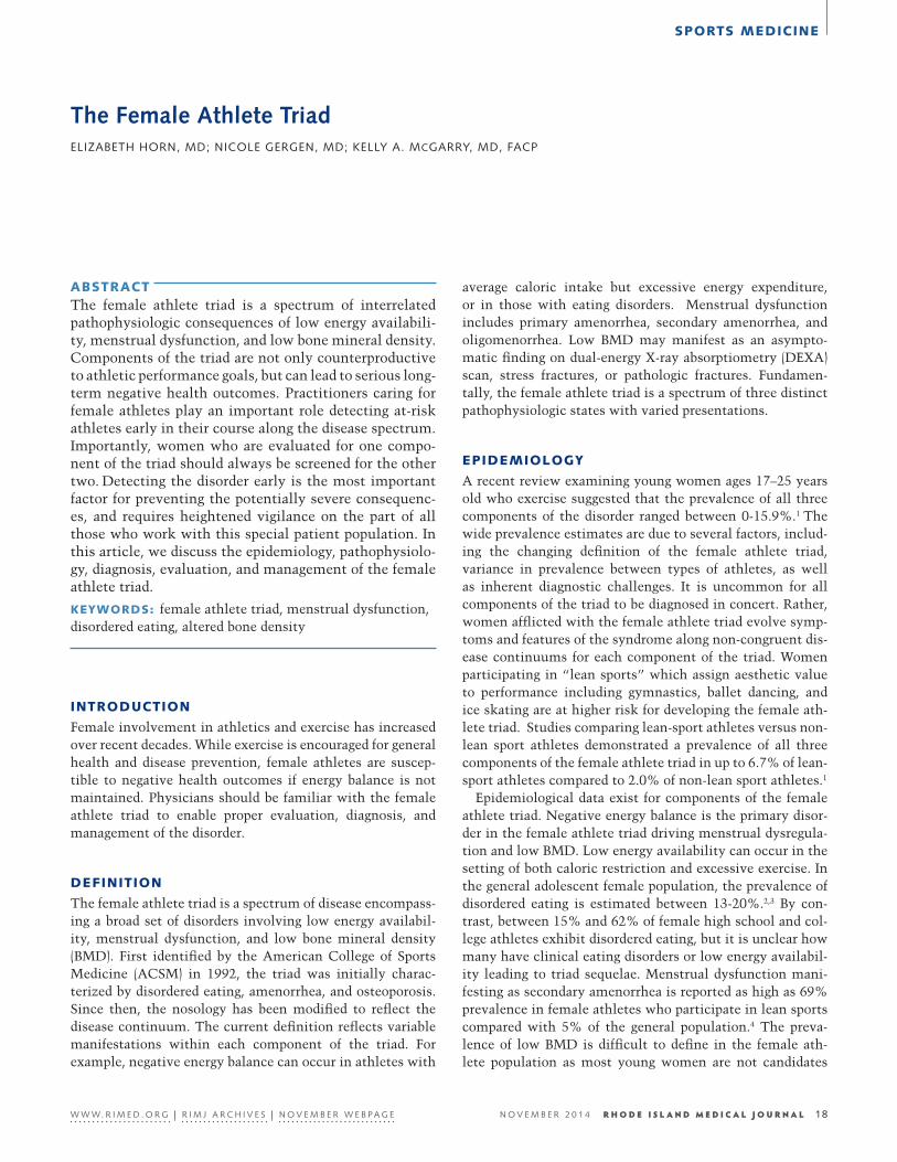
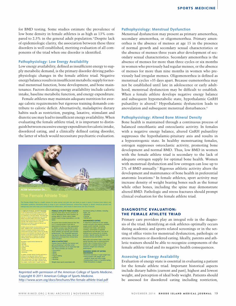
The Female Athlete TriadELIZABETH HORN, MD; NICOLE GERGEN, MD; KELLY A. MCGARRY, MD, FACP
ABSTRACT The female athlete triad is a spectrum of interrelated pathophysiologic consequences of low energy availabili-ty, menstrual dysfunction, and low bone mineral density. Components of the triad are not only counterproductive to athletic performance goals, but can lead to serious long-term negative health outcomes. Practitioners caring for female athletes play an important role detecting at-risk athletes early in their course along the disease spectrum. Importantly, women who are evaluated for one compo-nent of the triad should always be screened for the other two. Detecting the disorder early is the most important factor for preventing the potentially severe consequenc-es, and requires heightened vigilance on the part of all those who work with this special patient population. In this article, we discuss the epidemiology, pathophysiolo-gy, diagnosis, evaluation, and management of the female athlete triad.
KEYWORDS: female athlete triad, menstrual dysfunction, disordered eating, altered bone density
INTRODUCTION
Female involvement in athletics and exercise has increased over recent decades. While exercise is encouraged for general health and disease prevention, female athletes are suscep-tible to negative health outcomes if energy balance is not maintained. Physicians should be familiar with the female athlete triad to enable proper evaluation, diagnosis, and management of the disorder.
DEFINITION
The female athlete triad is a spectrum of disease encompass-ing a broad set of disorders involving low energy availabil-ity, menstrual dysfunction, and low bone mineral density (BMD). First identified by the American College of Sports Medicine (ACSM) in 1992, the triad was initially charac-terized by disordered eating, amenorrhea, and osteoporosis. Since then, the nosology has been modified to reflect the disease continuum. The current definition reflects variable manifestations within each component of the triad. For example, negative energy balance can occur in athletes with
average caloric intake but excessive energy expenditure, or in those with eating disorders. Menstrual dysfunction includes primary amenorrhea, secondary amenorrhea, and oligomenorrhea. Low BMD may manifest as an asympto-matic finding on dual-energy X-ray absorptiometry (DEXA) scan, stress fractures, or pathologic fractures. Fundamen-tally, the female athlete triad is a spectrum of three distinct pathophysiologic states with varied presentations.
EPIDEMIOLOGY
A recent review examining young women ages 17–25 years old who exercise suggested that the prevalence of all three components of the disorder ranged between 0-15.9%.1 The wide prevalence estimates are due to several factors, includ-ing the changing definition of the female athlete triad, variance in prevalence between types of athletes, as well as inherent diagnostic challenges. It is uncommon for all components of the triad to be diagnosed in concert. Rather, women afflicted with the female athlete triad evolve symp-toms and features of the syndrome along non-congruent dis-ease continuums for each component of the triad. Women participating in “lean sports” which assign aesthetic value to performance including gymnastics, ballet dancing, and ice skating are at higher risk for developing the female ath-lete triad. Studies comparing lean-sport athletes versus non-lean sport athletes demonstrated a prevalence of all three components of the female athlete triad in up to 6.7% of lean-sport athletes compared to 2.0% of non-lean sport athletes.1
Epidemiological data exist for components of the female athlete triad. Negative energy balance is the primary disor-der in the female athlete triad driving menstrual dysregula-tion and low BMD. Low energy availability can occur in the setting of both caloric restriction and excessive exercise. In the general adolescent female population, the prevalence of disordered eating is estimated between 13-20%.2,3 By con-trast, between 15% and 62% of female high school and col-lege athletes exhibit disordered eating, but it is unclear how many have clinical eating disorders or low energy availabil-ity leading to triad sequelae. Menstrual dysfunction mani-festing as secondary amenorrhea is reported as high as 69% prevalence in female athletes who participate in lean sports compared with 5% of the general population.4 The preva-lence of low BMD is difficult to define in the female ath-lete population as most young women are not candidates
18
21
EN
W W W. R I M E D . O R G | R I M J A R C H I V E S | N O V E M B E R W E B P A G E N O V E M B E R 2 0 1 4 R H O D E I S L A N D M E D I C A L J O U R N A L 18
SPORTS MEDICINE
for BMD testing. Some studies estimate the prevalence of low bone density in female athletes is as high as 13% com-pared to 2.3% in the general adult population.4 Despite lack of epidemiologic clarity, the association between these three disorders is well established, meriting evaluation of all com-ponents of the triad when one disorder is identified.
Pathophysiology: Low Energy AvailabilityLow energy availability, defined as insufficient energy to sup-ply metabolic demand, is the primary disorder driving patho-physiologic changes in the female athlete triad. Negative energy balance results in insufficient metabolic supply for nor-mal menstrual function, bone development, and bone main-tenance. Factors dictating energy availability include caloric intake, baseline metabolic function, and energy expenditure.
Female athletes may maintain adequate nutrition for aver-age caloric requirements but rigorous training demands con-tribute to caloric deficit. Alternatively, maladaptive dietary habits such as restriction, purging, laxative, stimulant and diuretic use may lead to insufficient energy availability. When evaluating the female athlete triad, it is important to distin-guish between excessive energy expenditure for caloric intake, disordered eating, and a clinically defined eating disorder, the latter of which would necessitate psychiatric evaluation.
Pathophysiology: Menstrual DysfunctionMenstrual dysfunction may present as primary amenorrhea, secondary amenorrhea, or oligomenorrhea. Primary amen-orrhea is the absence of menses at age 15 in the presence of normal growth and secondary sexual characteristics or the absence of menses three years after development of sec-ondary sexual characteristics. Secondary amenorrhea is the absence of menses for more than three cycles or six months in women who previously had regular menses, or the absence of menses for more than nine months in women who pre-viously had irregular menses. Oligomenorrhea is defined as menstrual cycles >35 days apart. Because eumenorrhea may not be established until late in adolescence or early adult-hood, menstrual dysfunction may be difficult to establish. When a female athlete develops negative energy balance and subsequent hypometabolic state, hypothalamic GnRH pulsatility is altered.5 Hypothalamic dysfunction leads to anovulation and subsequent menstrual disturbances.6
Pathophysiology: Altered Bone Mineral DensityBone health is maintained through a continuous process of balanced osteoblastic and osteoclastic activity. In females with a negative energy balance, altered GnRH pulsatility suppresses the hypothalamic-pituitary axis and results in a hypoestrogenic state. In healthy menstruating females, estrogen suppresses osteoclastic activity, promoting bone development and normal BMD. Thus, low BMD in women with the female athlete triad is secondary to the lack of adequate estrogen supply for optimal bone health. Women with menstrual dysfunction and low estrogen can lose up to 2% of BMD annually.7 Rigorous athletic activity alters the development and maintenance of bone health in preferential anatomic locations.8 In female athletes, sport activity may increase density of weight bearing bones such as the femur while other bones, including the spine may demonstrate altered BMD. Pathologic and stress fractures should prompt clinical evaluation for the female athlete triad.
DIAGNOSTIC EVALUATION: THE FEMALE ATHLETE TRIAD
Primary care providers play an integral role in the diagno-sis of the triad. Identifying at-risk athletes optimally occurs during academic and sports related screenings or in the set-ting of office visits for menstrual dysfunction, pathologic or stress fractures or disordered eating. Ideally, parents and ath-letic trainers should be able to recognize components of the female athlete triad and its negative health consequences.
Assessing Low Energy AvailabilityEvaluation of energy state is essential in evaluating a patient for the female athlete triad. Important historical aspects include dietary habits (current and past), highest and lowest weight, and perception of ideal body weight. Patients should be assessed for disordered eating including restriction,
W W W. R I M E D . O R G | R I M J A R C H I V E S | N O V E M B E R W E B P A G E N O V E M B E R 2 0 1 4 R H O D E I S L A N D M E D I C A L J O U R N A L 19
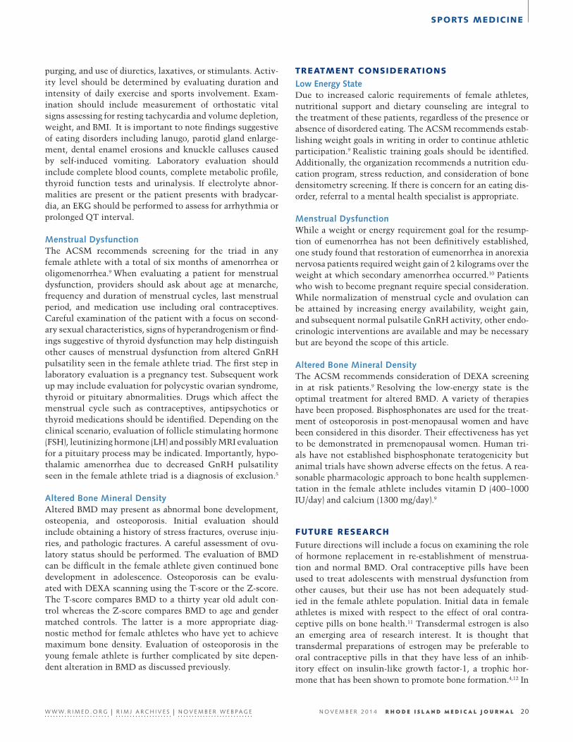
The Female Athlete TriadThe Female Athlete Triad is a health concern for active women and girls who are driven to excel in sports. It involves three distinct andinterrelated conditions: disordered eating (a range of poor nutritional behaviors), amenorrhea (irregular or absent menstrual periods) andosteoporosis (low bone mass and microarchitectural deterioration, which leads to weak bones and risk of fracture).
FEMALE ATHLETE TRIAD CAUSESExercise alone does not put someone atrisk for developing the Triad; however, anenergy deficit, in which caloric intakedoesn’t match energy expenditure, is a riskfactor.
All women face societal pressure that “thinis in.” A young woman or girl who isdetermined to achieve a lean appearanceor athletic success may attempt to excelthrough compulsive dieting and exercise.(Such athletes are typically goal-orientedperfectionists.) This misguided approachmay lead to disordered eating, menstrualdysfunction and lower-than-normal bonemass formation.
WHO IS AFFECTED?Anyone may be affected, but women andgirls participating in activities whichemphasize leanness are at especially highrisk. These activities can include:• Gymnastics• Ballet• Diving• Figure skating• Aerobics• Running
Weight class sports associated with
disordered eating in athletes, includingmales, are:• Wrestling• Rowing• Martial arts
DISORDERED EATING In response to pressure to lose weight,women and girls may practice unhealthyweight-control methods, includingrestricted food intake, self-inducedvomiting, consumption of appetitesuppressants and diet pills, and use oflaxatives and compounds to increaseurination. Specific eating disorders areanorexia nervosa and bulimia.
Many girls and women hide or deny theireating disorders due to embarrassment,shame, fear of losing control of theirdieting and a mistaken believe thatexcessive weight loss enhancesperformance.
WARNING SIGNS OF EATINGDISORDERS • Excessive leanness or rapid weight loss;• Preoccupation with weight, food,
mealtime rituals and body image;• Avoiding team meals, or secretive eating;• Wide fluctuations in weight;
A COMPLETE PHYSICAL ACTIVITY PROGRAM A well-rounded physical activity program includesaerobic exercise and strength training exercise, butnot necessarily in the same session. This blend helpsmaintain or improve cardiorespiratory and muscularfitness and overall health and function. Regularphysical activity will provide more health benefits thansporadic, high intensity workouts, so chooseexercises you are likely to enjoy and that you canincorporate into your schedule.
ACSM’s physical activity recommendations for healthyadults, updated in 2011, recommend at least 30minutes of moderate-intensity physical activity(working hard enough to break a sweat, but still ableto carry on a conversation) five days per week, or 20minutes of more vigorous activity three days perweek. Combinations of moderate- and vigorous-intensity activity can be performed to meet thisrecommendation.
Examples of typical aerobic exercises are: • Walking• Running• Stair climbing• Cycling• Rowing• Cross country skiing• Swimming.
In addition, strength training should be performed aminimum of two days each week, with 8-12repetitions of 8-10 different exercises that target allmajor muscle groups. This type of training can beaccomplished using body weight, resistance bands,free weights, medicine balls or weight machines.
ACSM Information On…
Reprinted with permission of the American College of Sports Medicine. Copyright © 2011 American College of Sports Medicine. http://www.acsm.org/docs/brochures/the-female-athlete-triad.pdf
SPORTS MEDICINE
purging, and use of diuretics, laxatives, or stimulants. Activ-ity level should be determined by evaluating duration and intensity of daily exercise and sports involvement. Exam-ination should include measurement of orthostatic vital signs assessing for resting tachycardia and volume depletion, weight, and BMI. It is important to note findings suggestive of eating disorders including lanugo, parotid gland enlarge-ment, dental enamel erosions and knuckle calluses caused by self-induced vomiting. Laboratory evaluation should include complete blood counts, complete metabolic profile, thyroid function tests and urinalysis. If electrolyte abnor-malities are present or the patient presents with bradycar-dia, an EKG should be performed to assess for arrhythmia or prolonged QT interval.
Menstrual DysfunctionThe ACSM recommends screening for the triad in any female athlete with a total of six months of amenorrhea or oligomenorrhea.9 When evaluating a patient for menstrual dysfunction, providers should ask about age at menarche, frequency and duration of menstrual cycles, last menstrual period, and medication use including oral contraceptives. Careful examination of the patient with a focus on second-ary sexual characteristics, signs of hyperandrogenism or find-ings suggestive of thyroid dysfunction may help distinguish other causes of menstrual dysfunction from altered GnRH pulsatility seen in the female athlete triad. The first step in laboratory evaluation is a pregnancy test. Subsequent work up may include evaluation for polycystic ovarian syndrome, thyroid or pituitary abnormalities. Drugs which affect the menstrual cycle such as contraceptives, antipsychotics or thyroid medications should be identified. Depending on the clinical scenario, evaluation of follicle stimulating hormone (FSH), leutinizing hormone (LH) and possibly MRI evaluation for a pituitary process may be indicated. Importantly, hypo-thalamic amenorrhea due to decreased GnRH pulsatility seen in the female athlete triad is a diagnosis of exclusion.5
Altered Bone Mineral DensityAltered BMD may present as abnormal bone development, osteopenia, and osteoporosis. Initial evaluation should include obtaining a history of stress fractures, overuse inju-ries, and pathologic fractures. A careful assessment of ovu-latory status should be performed. The evaluation of BMD can be difficult in the female athlete given continued bone development in adolescence. Osteoporosis can be evalu-ated with DEXA scanning using the T-score or the Z-score. The T-score compares BMD to a thirty year old adult con-trol whereas the Z-score compares BMD to age and gender matched controls. The latter is a more appropriate diag-nostic method for female athletes who have yet to achieve maximum bone density. Evaluation of osteoporosis in the young female athlete is further complicated by site depen-dent alteration in BMD as discussed previously.
TREATMENT CONSIDERATIONS
Low Energy StateDue to increased caloric requirements of female athletes, nutritional support and dietary counseling are integral to the treatment of these patients, regardless of the presence or absence of disordered eating. The ACSM recommends estab-lishing weight goals in writing in order to continue athletic participation.9 Realistic training goals should be identified. Additionally, the organization recommends a nutrition edu-cation program, stress reduction, and consideration of bone densitometry screening. If there is concern for an eating dis-order, referral to a mental health specialist is appropriate.
Menstrual DysfunctionWhile a weight or energy requirement goal for the resump-tion of eumenorrhea has not been definitively established, one study found that restoration of eumenorrhea in anorexia nervosa patients required weight gain of 2 kilograms over the weight at which secondary amenorrhea occurred.10 Patients who wish to become pregnant require special consideration. While normalization of menstrual cycle and ovulation can be attained by increasing energy availability, weight gain, and subsequent normal pulsatile GnRH activity, other endo-crinologic interventions are available and may be necessary but are beyond the scope of this article.
Altered Bone Mineral DensityThe ACSM recommends consideration of DEXA screening in at risk patients.9 Resolving the low-energy state is the optimal treatment for altered BMD. A variety of therapies have been proposed. Bisphosphonates are used for the treat-ment of osteoporosis in post-menopausal women and have been considered in this disorder. Their effectiveness has yet to be demonstrated in premenopausal women. Human tri-als have not established bisphosphonate teratogenicity but animal trials have shown adverse effects on the fetus. A rea-sonable pharmacologic approach to bone health supplemen-tation in the female athlete includes vitamin D (400–1000 IU/day) and calcium (1300 mg/day).9
FUTURE RESEARCH
Future directions will include a focus on examining the role of hormone replacement in re-establishment of menstrua-tion and normal BMD. Oral contraceptive pills have been used to treat adolescents with menstrual dysfunction from other causes, but their use has not been adequately stud-ied in the female athlete population. Initial data in female athletes is mixed with respect to the effect of oral contra-ceptive pills on bone health.11 Transdermal estrogen is also an emerging area of research interest. It is thought that transdermal preparations of estrogen may be preferable to oral contraceptive pills in that they have less of an inhib-itory effect on insulin-like growth factor-1, a trophic hor-mone that has been shown to promote bone formation.4,12 In
W W W. R I M E D . O R G | R I M J A R C H I V E S | N O V E M B E R W E B P A G E N O V E M B E R 2 0 1 4 R H O D E I S L A N D M E D I C A L J O U R N A L 20
SPORTS MEDICINE
postmenopausal women, transdermal estrogen has reduced fracture risk, but its potential benefits and harms have not been well studied in the young female athlete population.
References1. Gibbs JC, Williams NL, De Souza MJ. Prevalence of individual
and combined components of the female athlete triad. Med Sci Sports Exerc. 2012; epub ahead of print.
2. Johnson C, Powers PS, Dick R. Athletes and eating disorders: the National Collegiate Athletic Association study. Int J Eating Disord. 1999; 26:179-188.
3. Beals KA, Manore II. Disorders of the female athlete triad among collegiate athletes. Int J Sports Nutr Exerc Metab. 2002;12:281-293.
4. Nazem TG, Ackerman KE. The Female Athlete Triad. Sports Health. 2012;4:302.
5. Warren MP, Perlroth NE. The effects of intense exercise on the female reproductive system. Journal of Endocrinology. 2001;170:3-11.
6. Williams NI, Helmreich DL, Parfitt DB, et al. Evidence for a causal role of low energy availability in the induction of men-strual cycle disturbances during strenuous exercise training. J Clin Endocrinol Metab. 2001;86:5184-5193.
7. Deimel JF, Dunlap BJ. The female athlete triad. Clin Sports Med. 2012;31:247-254.
8. Cobb KL, Bachrach LK, Greendale G, et al. Disordered eating, menstrual irregularity, and bone mineral density in female run-ners. Med Sci Sports Exerc. 2002;711-719.
9. Nattiv A, Loucks AB, Manore MM, Sanborn CF, Sundgot-Bor-gen J, Warren MP. American College of Sports Medicine po-sition stand: the female athlete triad. Med Sci Sports Exerc. 2007;30:1867-1882.
10. Golden NH, Jacobson MS, Sterling WM, Hertz S. Treatment goal weight in adolescents with anorexia nervosa: use of BMI percentiles. Int J Eat Disord. 2008;41:301-306.
11. Liu SL, Lebrun CM. Effect of oral contraceptives and hormone replacement therapy on BMD in premenopausal and perimeno-pausal women: a systematic review. Br J Sports Med. 2006;40:11-24.
12. Grinspoon S, Baum H, Lee K, Anderson E, Herzog D, Klibans-ki A. Effects of a short-term recombinant human insulin-like growth factor I administration on bone turnover in osteopenic women with anorexia nervosa. Journal of Clinical Endocrinolo-gy and Metabolism. 1996;31:3864-3870.
13. Joy EA, Van Hala S, Cooper L. Health-related concerns of the fe-male athlete: a lifespan approach. American Family Physician. 2007;79(6):489-495.
AuthorsElizabeth Horn, MD, is a Housestaff Officer, Internal Medicine,
The Alpert Medical School of Brown University.
Nicole Gergen, MD, is a Housestaff Officer, Internal Medicine-Pediatrics, The Alpert Medical School of Brown University.
Kelly A. McGarry, MD, FACP, is Associate Professor of Medicine, The Alpert Medical School of Brown University and Program Director, General Internal Medicine Residency, Rhode Island Hospital, Providence, RI.
Correspondence Kelly A. McGarry, MDRhode Island Hospital593 Eddy StreetProvidence, RI 02903401-444-5953Fax [email protected]
W W W. R I M E D . O R G | R I M J A R C H I V E S | N O V E M B E R W E B P A G E N O V E M B E R 2 0 1 4 R H O D E I S L A N D M E D I C A L J O U R N A L 21
SPORTS MEDICINE
Exercise-Induced RhabdomyolysisGEORGE LEE, MD
ABSTRACT Exercise-induced rhabdomyolysis, or exertional rhabdo-myolysis (ER), is a clinical entity typically considered when someone presents with muscle stiffness, swelling, and pain out of proportion to the expected fatigue post exercise. The diagnosis is confirmed by myoglobinuria, and an elevated serum Creatinine Phosphokinase (CPK) level, usually 10 times the normal range. However, an elevation in CPK is seen in most forms of strenuous ex-ercise, up to 20 times the upper normal range. Therefore, there is no definitive pathologic CPK cut-off. Fortunate-ly the dreaded complication of acute renal failure is rare compared to other forms rhabdomyolysis. We review the risks, diagnosis, clinical course and treatment for exercise- induced rhabdomyolysis.
KEYWORDS: exertional rhabdomyolysis, CPK, myoglobinuria, acute renal failure
INTRODUCTION
Rhabdomyolysis (RM) is a condition of striated muscle dam- age, usually in conjunction with an elevation in creatine phosphokinase (CPK). The mechanism involves either trauma or intracellular depletion of ATP leading to intracel-lular influx of calcium. This in turn results in the disruption of the cell membrane, and subsequent release of intracellu-lar contents into the plasma and extracellular space. It is this translocation of intracellular debris that can potentially lead to serious complications, most notably acute renal failure (ARF). The incidence of acute renal failure complicating RM ranges from 15-50%. The most common causes of RM with resultant acute renal failure include ischemia, drugs, alcohol and trauma. Melli1 reported 475 hospitalized patients with RM at John Hopkins. Exogenous toxins, including illicit drugs, alcohol and medications were the most common cause, with an incidence of acute renal failure being 46%.
The incidence of exercise-induced or exertional rhabdo-myolysis (ER) in the general public is difficult to define, as many patients probably do not seek medical attention. How-ever, data have been accrued from military recruits undergo-ing basic training. Wildly ranging rates have been described, due to the varying definitions utilized. The largest data set was by Hill,2 who reported, in a retrospective review of
574,688 U.S. Army soldiers, 1203 cases of ER or 0.2%. This translates to a yearly rate of 7-8 cases/10,000. Rates were higher men vs. women. Olerud et al,3 using serum myo-globin as a screening test, diagnosed ER in 40% of military recruits within the first 6 days of basic training.
DIAGNOSIS
The varying rate of ER is due to the nebulous diagnostic cri-teria. Clinically it can manifest itself with prolonged mus-cle swelling and tenderness, lasting several days longer than expected. Ensuing dark urine may develop, signifying myo-globinuria. Elevation of CPK is one of the main serologic criteria to define the entity. However, there is no defining set point in the height of the CPK rise to identify clinically relevant EH. A confounding factor is that CPK elevation after strenuous activity is quite common, with the range being quite variable. Thus, no normal post-exercise CPK value has been established. Studies in male marathoners4 and triathletes have demonstrated, 24 hours after race com-pletion, CPK elevation in the several thousand range, 10-20 times the upper limit of normal. Of the exercises associated with ER, downhill running and those that induce eccentric (muscle lengthening) contractions tend to be more com-monly identified. Examples would include squat thrusts, pushups, and biceps curls. Clarkson5 measured CPK in 203 healthy, but relatively physically inactive college students after 2 sets of elbow curls with weights for 25 repetitions. Mean CPK rose to a peak of 7713 at day 4, with a range of 50-80,550. The enzyme elevation lasted until day 10 post exercise. No participant developed any medical complica-tions. ER has also been reported in a host of other physical activities including spinning, rock climbing, ice skating, and swimming. A common thread seen in ER is continued exer-tion beyond the point of fatigue. This is typically seen in a group setting, where peer pressure plays a role, or under the supervision of a demanding personal trainer.
RISK FACTORS
Asides from the type and duration of exercise, several other risks factors are associated with ER. Studies have shown that at baseline and post exercise, elevations in CPK are greater in men vs. women. Also CPK increments are greater in blacks vs. Caucasians.2,11 Increased muscle mass is thought to
22
24
EN
W W W. R I M E D . O R G | R I M J A R C H I V E S | N O V E M B E R W E B P A G E N O V E M B E R 2 0 1 4 R H O D E I S L A N D M E D I C A L J O U R N A L 22
SPORTS MEDICINE
explain the gender difference.4 The ethnic difference expla-nation is more elusive. One entertained mechanism is the prevalence of sickle cell trait, which may lead to an exag-gerated raise in post exercise CPK.13 Another potential risk is any factor that may hamper bodily heat release. Drugs, particularly amphetamines, are implicated, as they cause peripheral vasoconstriction. Concordant with this hypothe-sis, is the fact that history of heat stroke may also be another predisposing factor. This was noted in a retrospective review by Hill in the military recruits. As a result, rubber suits, used by wrestlers to lose water weight have been banned. If ER is recurrent in an otherwise healthy, young patient, inherited muscle enzyme defects should be considered. The most common include carnitine palmitoyl transferase defi-ciency, myophosphorylase deficiency (McArdle’s disease) and adenosine monophosphate deaminase deficiency.
COMPLICATIONS
The serious complications of RM include ARF, hyperkale-mia, DIC and compartment syndrome. Fortunately, these are all rare with ER. This is most likely due to the fact many of these patients are relatively young and healthy. If acute renal failure develops from ER, full renal recovery is nearly universal. Sinert6 reported 35 ED admissions for ER with a mean CPK of 40,471. No patient developed acute kidney injury. Hill’s2 (data reported an incidence of 8% in the 1203 cases of EH, all of whom recovered renal function.
One question frequently asked is, what level of CPK is associated with kidney injury? Although those who develop renal damage tend to have a higher CPK levels, the correla-tion between peak of CPK rise and acute renal failure is poor. Some studies have suggested renal injury is associated with CPK in excess of 20,000. However, there are also case reports of it occurring at 5,000.8 Complicating the issue is that there are frequently other contributing factors to renal damage in those studies. Meijer7 reported the clinical course of 26 ICU admissions with severe RM, defined as CPK >10,000. The most common causes were ischemic and trauma, none due to exercise. Those who developed acute renal failure had a mean peak CPK of 55,366 vs. 28,643. However, there was substantial overlap between the 2 groups, and no defining level could be ascertained. Therefore, no CPK level has been established in the literature to predict ARF.
ARF from RM is the most serious complication that phy-sicians are attuned to. It was first described in the medical literature in the 1940s by Beall and Bywater.12 They reported uremic deaths several days following crush injuries due to bombing raids in London. The mechanism of kidney injury is several-fold. Myoglobin, the heme-based oxygen carrying component in muscle is released into the circulation. It is believed to be toxic to the renal tubules. Secondly, there is a period of renal vasoconstriction hampering perfusion.
Lastly, there can be severe third spacing with fluid being sequestered into damaged muscle, leading to an effectively pre-renal condition. Due to the last mechanism, vigorous isotonic intravenous fluids have been the hallmark of pre-ventive therapy. Volumes suggested range from 6-10 liters over the first 24 hours to maintain a urine output of 200-300ml/hour. The earlier the fluid administration, the better, a conclusion Ori Better9 reported in crush victims from a collapsed building. However, therapy needs to be individual-ized, with close attention paid to the patient’s volume status. IVF administration to the point of overt fluid overload has been associated with increased mortality in ICU patients.
TREATMENT
The issue of type of intravenous fluid is still debated. Although urinary alkalinization can increase the solubil-ity of myoglobin, the superiority of bicarbonate containing solutions over saline has not been confirmed. The same holds true for mannitol, another agent frequently employed to prevent and treat RM-associated ARF. In a retrospective review of 74 cases of kidney injury due to trauma-induced RM, Brown8 found no benefit with mannitol or bicarbonate solution. In addition, there is a risk of osmotic induced tubu-lar injury with mannitol adminstration. A serum osmolar gap >50 can predispose to this untoward complication.
Hypothetically, extracorporeal removal of myoglobin can be beneficial. Due to the size of the heme protein, it is not removed with conventional hemodialysis. However, plas-mapharesis10 can effectively extract the compound from the vascular space. High flux continuous hemofiltration also can remove it as well. Regardless, there are no randomized stud-ies that establish either modality as a preventative measure or treatment for ARF. Thus neither can be recommended.
SUMMARY
A post-exercise CPK rise is a common phenomenon. The defining line from a normal physiologic response to a disease state is a blurry one. When complications are initially appar-ent, then the distinction is obvious, but frequently, they are not present. Avoidance of alcohol, amphetamine-based drugs and the gradual increments in exercise intensity are recommended to attenuate ER. One issue is when to admit patients. Acute renal failure is quite rare and when it does occur, it almost always resolves completely. Therefore, in the absence of serious complications, the decision to admit is generally intuition based. CPK tends to peak at day 4, but can remain elevated for 1-2 weeks. If a patient presents with-out evidence of ARF, it would be unlikely to develop after generous IV isotonic fluid administration. Rate and volume of fluid needs to be individualized and clinically-based.
W W W. R I M E D . O R G | R I M J A R C H I V E S | N O V E M B E R W E B P A G E N O V E M B E R 2 0 1 4 R H O D E I S L A N D M E D I C A L J O U R N A L 23
SPORTS MEDICINE
References 1. Melli G, Chaudhry V, Cornblath DR. Rhabdomyolysis: An eval-
uation of 475 hospitalized patients. Medicine. 2005;84:377-85.2. Hill O, et al. Medicine and Science in Sports and Exercise.
2012;44:442-49.3. Olerud JE, Homer LD, Carroll HW. Incidence of acute exertional
rhabdomyolysis. Arch Int Med. 1976;136:692-97.4. Rogers MA, Stull GA, Apple FS. Creatine kinase isoenzyme ac-
tivities in men and women following a marathon race. Med Sci Sports Exerc. 1985;17:679-82.
5. Clarkson et al. Serum creatine kinase levels and renal function measures in exertional muscle damage. Med Sci Sports Exerc. 2006;38:623-627.
6. Sinert et al. Exercise induced rhabdomyolysis. Annals of Emerg Med. June 1994;23:1301-06.
7. Meijer et al. Serum creatine kinase as predictor of clinical course in rhabdomyolysis: a 5 year intensive care survey. Inten-sive Care Medicine. 2003;29:1121-25.
8. Brown et al. Preventing renal failure in patients with rhabdo-myolysis: do bicarbonate and mannitol make a difference. Jn. of Trauma. 2004;56:1191-96.
9. Better O, Stein J. Early management of shock and prophylax-is of acute renal failure in traumatic rhabdomyolysis. NEJM. 1990;322:825-29.
10. Paaske et al. Plasma exchange after revascularization compart-ment syndrome with acute toxic nephropathy caused by rhabdo-myolysis. Jn of Vasc Surg.1988;7:757-8.
11. Noakes TD. Effect of exercise on serum enzyme activities in humans. Sports Med. 1987;4:245-267.
12. Bywaters EGL, Beall D. Crush injuries with impairment in renal function. Br. Med J. 1941;427-32.
13. Makaryus JN, Catanzaro JN, Katona KC. Exertional rhabdomy-olysis and renal failure in patients with sickle cell trait. Hema-tology. 2007;12(4):349-52.
AuthorGeorge Lee, MD, is Clinical Assistant Professor of Medicine at the
Alpert Medical School of Brown University.
CorrespondenceGeorge Lee, MDNephrology Associates318 Waterman Ave.401-438-5950Fax [email protected]
W W W. R I M E D . O R G | R I M J A R C H I V E S | N O V E M B E R W E B P A G E N O V E M B E R 2 0 1 4 R H O D E I S L A N D M E D I C A L J O U R N A L 24
SPORTS MEDICINE
Post-Traumatic Osteoarthritis after ACL InjuryJENNIFER R. RACINE, BA; ROY K. AARON, MD
ABSTRACT Post-traumatic osteoarthritis (PTOA) occurs as a conse-quence of joint trauma or occupations or sports that sub-ject joints to excessive loading stresses. Ligament injuries to the knee, particularly tears of the anterior cruciate lig-ament (ACL), often result in PTOA. Approximately half of the individuals with an ACL injury develop PTOA re-gardless of the reconstruction of the torn ligament. This observation has raised the possibility that other injuries occur to the knee in association with ACL tears that may involve ligamentous capsular structures, articular carti-lage, or subchondral bone. Many ACL injuries occur in noncontact sports and are the result of biomechanical ab-normalities. Female athletes are more likely than their male counterparts to suffer ACL injuries. This review outlines the epidemiology of ACL tears, its pathology in cartilage and bone, some of the demographic, biome-chanical, and neuromuscular factors involved in ACL tears, and PTOA and important information gained from preclinical injury models.
KEYWORDS: Osteoarthritis, Ligament Injury, Arthroscopy
INTRODUCTION
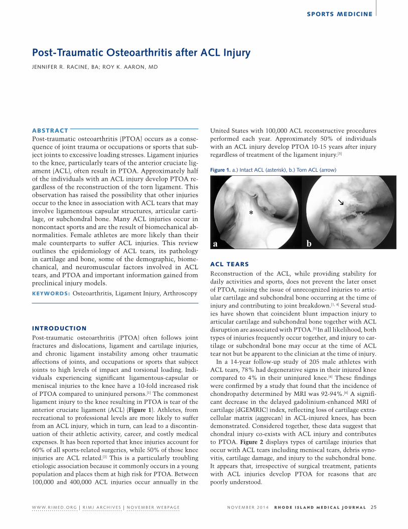
Post-traumatic osteoarthritis (PTOA) often follows joint fractures and dislocations, ligament and cartilage injuries, and chronic ligament instability among other traumatic affections of joints, and occupations or sports that subject joints to high levels of impact and torsional loading. Indi-viduals experiencing significant ligamentous-capsular or meniscal injuries to the knee have a 10-fold increased risk of PTOA compared to uninjured persons.[1] The commonest ligament injury to the knee resulting in PTOA is tear of the anterior cruciate ligament (ACL) (Figure 1). Athletes, from recreational to professional levels are more likely to suffer from an ACL injury, which in turn, can lead to a discontin-uation of their athletic activity, career, and costly medical expenses. It has been reported that knee injuries account for 60% of all sports-related surgeries, while 50% of those knee injuries are ACL related.[2] This is a particularly troubling etiologic association because it commonly occurs in a young population and places them at high risk for PTOA. Between 100,000 and 400,000 ACL injuries occur annually in the
United States with 100,000 ACL reconstructive procedures performed each year. Approximately 50% of individuals with an ACL injury develop PTOA 10-15 years after injury regardless of treatment of the ligament injury.[3]
25
28
EN
Figure 1. a.) Intact ACL (asterisk), b.) Torn ACL (arrow)
ACL TEARS
Reconstruction of the ACL, while providing stability for daily activities and sports, does not prevent the later onset of PTOA, raising the issue of unrecognized injuries to artic-ular cartilage and subchondral bone occurring at the time of injury and contributing to joint breakdown.[1, 4] Several stud-ies have shown that coincident blunt impaction injury to articular cartilage and subchondral bone together with ACL disruption are associated with PTOA.[5] In all likelihood, both types of injuries frequently occur together, and injury to car-tilage or subchondral bone may occur at the time of ACL tear not but be apparent to the clinician at the time of injury.
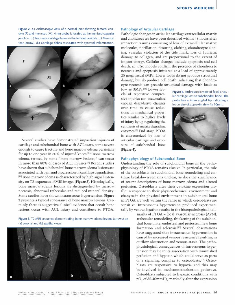
In a 14-year follow-up study of 205 male athletes with ACL tears, 78% had degenerative signs in their injured knee compared to 4% in their uninjured knee.[4] These findings were confirmed by a study that found that the incidence of chondropathy determined by MRI was 92-94%.[6] A signifi-cant decrease in the delayed gadolinium-enhanced MRI of cartilage (dGEMRIC) index, reflecting loss of cartilage extra-cellular matrix (aggrecan) in ACL-injured knees, has been demonstrated. Considered together, these data suggest that chondral injury co-exists with ACL injury and contributes to PTOA. Figure 2 displays types of cartilage injuries that occur with ACL tears including meniscal tears, debris syno-vitis, cartilage damage, and injury to the subchondral bone. It appears that, irrespective of surgical treatment, patients with ACL injuries develop PTOA for reasons that are poorly understood.
W W W. R I M E D . O R G | R I M J A R C H I V E S | N O V E M B E R W E B P A G E N O V E M B E R 2 0 1 4 R H O D E I S L A N D M E D I C A L J O U R N A L 25
SPORTS MEDICINE
Several studies have demonstrated impaction injuries of cartilage and subchondral bone with ACL tears, some severe enough to cause fracture and bone marrow edema persisting for up to one year in 60% of injured knees.[7, 8] Bone marrow edema, termed by some “bone marrow lesions,” can occur in more than 80% of cases of ACL injuries.[9] Recent studies have shown that subchondral bone marrow edema lesions are associated with pain and progression of cartilage degradation.[10] Bone marrow edema is characterized by high signal inten-sity on T2 sequences of MRI images (Figure 3). Histologically, bone marrow edema lesions are distinguished by marrow necrosis, abnormal trabeculae and reduced mineral density. Some studies have shown intraosseous hypertension. Figure 2 presents a typical appearance of bone marrow lesions. Cer-tainly there is suggestive clinical evidence that occult bone lesions occur with ACL injury and contribute to PTOA.
Pathology of Articular CartilagePathologic changes in articular cartilage extracellular matrix and chondrocytes have been described within 48 hours after impaction trauma consisting of loss of extracellular matrix molecules, fibrillation, fissuring, clefting, chondrocyte clon-ing, vascular violation of the tide mark, loss of lubricin, damage to collagen, and are proportional to the extent of impact energy. Cellular changes include apoptosis and cell death. In vitro models confirm the presence of chondrocyte necrosis and apoptosis initiated at a load of approximately 25 megapascal (MPa) Lower loads do not produce structural damage, but do produce cell death indicating that chondro-cyte necrosis can precede structural damage with loads as low as 3MPa.[11] Lower lev-els of repetitive compres-sive injuries can accumulate enough degradative changes over time to cause reduc-tions in mechanical proper-ties similar to higher levels of injury by up-regulating the synthesis of matrix degrading enzymes.[5] End stage PTOA is characterized by loss of articular cartilage and expo-sure of subchondral bone (Figure 4).
Pathophysiology of Subchondral BoneUnderstanding the role of subchondral bone in the patho-physiology of PTOA remains elusive. In particular, the role of the osteoblasts in subchondral bone remodeling and car-tilage breakdown remains unclear, as does the significance of recent descriptions of bone marrow edema and altered perfusion. Osteoblasts alter their cytokine expression pro-file in response to their physicochemical environment and changes in the physical environment in subchondral bone in PTOA are well within the range in which osteoblasts are sensitive. Intraosseous hypertension produced experimen-tally by venous ligation results in the histopathological hall-
marks of PTOA – focal avascular necrosis (AVN), trabecular remodeling, thickening of the subchon-dral bone plate, endosteal and periosteal new bone formation and sclerosis.[12] Several observations have suggested that intraosseous hypertension is caused by increased venous resistance resulting in outflow obstruction and venous stasis. The patho-physiological consequences of intraosseous hyper-tension may lie in its association with diminished perfusion and hypoxia which could serve as parts of a signaling complex to osteoblasts.[13] Osteo-blasts are responsive to hypoxia and this may be involved in mechanotransduction pathways. Osteoblasts subjected to hypoxic conditions with pO2 of 35-40mmHg, markedly alter the expression
Figure 2. a.) Arthroscopic view of a normal joint showing femoral con-
dyle (F) and meniscus (M); 4mm probe is located at the menisco-capsular
junction. b.) Traumatic cartilage lesion in the femoral condyle. c.) Meniscal
tear (arrow). d.) Cartilage debris associated with synovial inflammation.
Figure 3. T2 MRI sequence demonstrating bone marrow edema lesions (arrows) on
(a) coronal and (b) sagittal views.
Figure 4. Arthroscopic view of focal articu-lar cartilage loss to subchondral bone. The probe has a 4mm angled tip indicating a lesion size of approximately 4x 10mm.
W W W. R I M E D . O R G | R I M J A R C H I V E S | N O V E M B E R W E B P A G E N O V E M B E R 2 0 1 4 R H O D E I S L A N D M E D I C A L J O U R N A L 26
SPORTS MEDICINE
profile of growth factors associated with the pathologic findings of OA, increased bone remodeling and cartilage degradation. Osteoblasts derived from OA bone also express high levels of alkaline phosphatase, osteocalcin, and IGF-1 which are related to bone remodeling.
BIOMECHANICAL AND NEUROMUSCULAR FACTORS IN ACL TEARS
Sports medicine researchers and clinicians have focused on biomechanical risk factors for injuries and preventative measures. Studies have examined extrinsic risk factors including the level of sport (training versus competition), shoe type, and playing surface, and intrinsic risk factors such as age, gender, and even hormonal status. The most frequent mechanisms of ACL injuries in sports, almost 70%, are non-contact.[14] Non-contact ACL injuries include stop-ping mid-stride to respond to an opponent’s change in direc-tion, especially in soccer, and an increase in loading from jumping, such as in basketball. Studies of neuromuscular and biomechanical factors have shown that many ACL inju-ries are not the results of contact and that distinct biome-chanical patterns such as excessive coronal plane motion, less knee flexion, and landing flatfooted are associated with ACL injury.[15] These observations suggest that ACL injuries, especially in females, are primarily neuromuscular and bio-mechanical in nature and subject to modification.
Female athletes in particular, are more likely to incur ACL injuries than are male athletes. Over the past two decades there has been a marked increase in ACL injuries in young female athletes in sports that involve cutting, jumping, and pivoting.[15] One study looked at the effect of gender on injury rates among military recruits during basic training (n=861) and found that women had twice as many injuries as men (relative risk = 2.1, 95% CI =1.78 to 2.5).[16] Another study looked at ACL injuries among male and female recruits playing intercollegiate sports, coed intramural sports, and military training, and found a combined relative risk of 2.44 for women compared with men.[17] This has been ascribed to increased valgus movements during landing, hormone levels, narrower intercondylar notch width, and smaller AC ligaments.[14,18] Neuromuscular training programs suggest that enhancing body control may decrease ACL injuries in women.[14] It has been found that male athletes attenuate knee ligament stresses during jumping by knee flexion and absorption of strain by the quadriceps. By contrast, female athletes have been found to land with the knee relatively straighter with more stress taken up by the ACL. These relatively higher stresses are believed to contribute to ACL injuries. Programs have been devised to train female ath-letes to attenuate more stress with their quadriceps, thus reducing load, and hopefully injury, to the ACL. Prevention programs that involve proprioception, plyometrics, strength training, and improved jumping, stopping, and turning tech-niques are showing promising results.[18] With this type
of training, female athletes have been shown to reduce cor-onal plane motion, exert more muscular control, and reduce ACL stresses during movements such as jumping, landing, pivoting, and deceleration.
PRECLINICAL INJURY MODELS
Preclinical injury models are useful for several reasons; 1.) Human tissue is unavailable for study except in end-stage PTOA, 2.) Dosimetry of impact can be determined and related to pathological changes in cartilage and bone, 3.) Sequential histopathological examinations of joints can reveal the time course and magnitude of progressive devel-opment of PTOA. The fracture threshold for the femoral condyle in rabbits has been reported to be about 120MPa and most direct impaction injury studies have used magnitudes between 20-50MPa to produce chondrocyte death and loss of extracellular matrix integrity Models of impaction injury to cartilage and bone have been established in rabbits, dogs, and sheep in vivo, as well as in vitro explants and changes typical of PTOA have been demonstrated.
In vitro studies have reported that cell death and concom-itant extracellular matrix damage are initiated at stress mag-nitudes of 15 to 25 MPa.[19] A stress magnitude of >40 MPa has caused complete cell death under the impacted region. One key in vitro study determined the effects of stress magnitude on cartilage extracellular matrix damage and cell viability in the rabbit knee. Femoral condyles were impacted with stress magnitudes of 15-50MPa at a stress rate of 420MPa/s. The stress rate was based on predictions of joint impact that may occur in contact sports, joint injuries, and ACL tears. All specimens impacted with peak stresses >35MPa showed visible surface damage in the impacted region. Superficial matrix damage was observed in 2 of 4 specimens impacted with peak stresses between 30 to 35MPa. Below 30MPa there was no visible matrix damage. A limitation of in vitro models is that it is not possible to investigate the cartilage mechanobiologic response to injury over time.[20]
Using a novel in vivo animal test system that is capable of independently applying quantifiable, precise stress mag-nitudes and rates to the femoral condyle of the rabbit knee, PTOA has been induced as a result of a single impact trauma to the articular cartilage and subchondral bone.[20] The extent of cell death depends on the magnitude of the impact at the time of injury. Initial cartilage injuries progress to almost complete cartilage matrix and chondrocyte loss throughout the depth of the impacted region by 3 weeks after impact. The post impact morphological biochemical observations are similar to early-to-late stage pathologic observations typ-ically seen in human PTOA. In an important series of in vivo studies, the femoral condyle was impacted with a peak stress of 35MPa at a stress rate of 420MPa/s. Zero-time rabbits had histologic evidence of matrix damage patterns consisting of surface roughening and distinct cracks that propagated to 20% of the depth. Histologic sections of rabbits sacrificed
W W W. R I M E D . O R G | R I M J A R C H I V E S | N O V E M B E R W E B P A G E N O V E M B E R 2 0 1 4 R H O D E I S L A N D M E D I C A L J O U R N A L 27
SPORTS MEDICINE
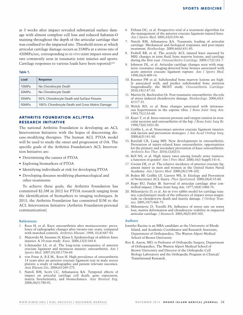
at 3 weeks after impact revealed substantial surface dam-age with almost complete cell loss and reduced Safranin-O staining throughout the depth of the articular cartilage that was confined to the impacted site. Threshold stress at which articular cartilage damage occurs as 25MPa at a stress rate of 420MPa/sec, corresponding to in vivo joint impact stress and rate commonly seen in traumatic joint injuries and sports. Cartilage responses to various loads have been reported:[21]
6. Fithian DC, et al. Prospective trial of a treatment algorithm for the management of the anterior cruciate ligament-injured knee. Am J Sports Med. 2005;33(3):335-46.
7. Natoli RM, Athanasiou KA. Traumatic loading of articular cartilage: Mechanical and biological responses and post-injury treatment. Biorheology. 2009;46(6):451-85.
8. Frobell RB, et al. The acutely ACL injured knee assessed by MRI: changes in joint fluid, bone marrow lesions, and cartilage during the first year. Osteoarthritis Cartilage. 2009;17(2):161-7.
9. Johnson DL, et al. Articular cartilage changes seen with mag-netic resonance imaging-detected bone bruises associated with acute anterior cruciate ligament rupture. Am J Sports Med. 1998;26(3):409-14.
10. Roemer FW et al. Subchondral bone marrow lesions are high-ly associated with, and predict subchondral bone attrition longitudinally: the MOST study. Osteoarthritis Cartilage. 2010;18(1):47-53.
11. Martin JA, Buckwalter JA. Post-traumatic osteoarthritis: the role of stress induced chondrocyte damage. Biorheology. 2006;43(3-4):517-21.
12. Welch RD, et al. Bone changes associated with intraosse-ous hypertension in the caprine tibia. J Bone Joint Surg Am. 1993;75(1):53-60.
13. Kiaer T, et al. Intra-osseous pressure and oxygen tension in avas-cular necrosis and osteoarthritis of the hip. J Bone Joint Surg Br. 1990;72(6):1023-30.
14. Griffin L, et al. Noncontact anterior cruciate ligament injuries: risk factors and prevention strategies. J Am Acad Orthop Surg. 2000;8(3):141-50.
15. Ratzlaff CR, Liang MH. New developments in osteoarthritis. Prevention of injury-related knee osteoarthritis: opportunities for the primary and secondary prevention of knee osteoarthritis. Arthritis Res Ther. 2010;12(4)215.
16. Bell NS, et al. High injury rates among female army trainees: a function of gender? Am J Prev Med. 2000;18(3 Suppl):141-6.
17. Gwinn DE, et al. The relative incidence of anterior cruciate lig-ament injury in men and women at the United States Naval Academy. Am J Sports Med. 2000;28(1):98-102.
18. Boden BP, Griffin LY, Garrett WE, Jr. Etiology and Prevention of Noncontact ACL Injury. Phys Sportsmed. 2000;28(4):53-60.
19. Repo RU, Finlay JB. Survival of articular cartilage after con-trolled impact. J Bone Joint Surg Am. 1977;59(8):1068-76.
20. Milentijevic D, et al. An in vivo rabbit model for cartilage trau-ma: a preliminary study of the influence of impact stress magni-tude on chondrocyte death and matrix damage. J Orthop Trau-ma. 2005;19(7):466-73.
21. Milentijevic D, Torzilli PA. Influence of stress rate on water loss, matrix deformation and chondrocyte viability in impacted articular cartilage. J Biomech. 2005;38(3):493-502.
AuthorsJennifer Racine is an MBA candidate at the University of Rhode
Island, and Academic Coordinator and Research Associate, Department of Orthopaedics, The Warren Alpert Medical School of Brown University.
Roy K. Aaron, MD, is Professor of Orthopedic Surgery, Department of Orthopaedics, The Warren Alpert Medical School of Brown University and Director of the Orthopedic Cell Biology Laboratory and the Orthopedic Program in Clinical/Translational Research.
Table 1.
ARTHRITIS FOUNDATION ACL/PTOA RESEARCH INITIATIVE
The national Arthritis Foundation is developing an ACL Intervention Initiative with the hopes of discovering dis-ease-modifying therapies. The human ACL PTOA model will be used to study the onset and progression of OA. The specific goals of the Arthritis Foundation’s ACL Interven-tion Initiative are:
• Determining the causes of PTOA
• Exploring biomarkers of PTOA
• Identifying individuals at risk for developing PTOA
• Developing diseases modifying pharmacological and other treatments
To achieve these goals, the Arthritis Foundation has committed $2.3M in 2012 for PTOA research ranging from the identification of biomarkers to imaging techniques. In 2013, the Arthritis Foundation has committed $1M to the ACL Intervention Initiative (Arthritis Foundation-personal communication).
References1. Roos H, et al. Knee osteoarthritis after meniscectomy: preva-
lence of radiographic changes after twenty-one years, compared with matched controls. Arthritis Rheum. 1998; 41(4):687-93.
2. Majewski M, Susanne H, Klaus S. Epidemiology of athletic knee injuries: A 10-year study. Knee. 2006;13(3):184-8.
3. Lohmander LS, et al. The long-term consequence of anterior cruciate ligament and meniscus injuries: osteoarthritis. Am J Sports Med. 2007;35(10):1756-69.
4. von Porat A, R.E.M., Roos H. High prevalence of osteoarthritis 14 years after an anterior cruciate ligament tear in male soccer players: a study of radiographic and patient relevant oucomes. Ann Rheum Dis. 2004;63:269-273.
5. Natoli RM, Scott CC, Athanasiou KA. Temporal effects of impact on articular cartilage cell death, gene expression, matrix biochemistry, and biomechanics. Ann Biomed Eng. 2008;36(5):780-92.
W W W. R I M E D . O R G | R I M J A R C H I V E S | N O V E M B E R W E B P A G E N O V E M B E R 2 0 1 4 R H O D E I S L A N D M E D I C A L J O U R N A L 28
Load Response
10MPa No Chondrocyte Death
20MPa No Chondrocyte Death
35MPa 50% Chondrocyte Death and Surface Fissures
50MPa 100% Chondrocyte Death and Gross Matrix Damage
SPORTS MEDICINE
29
31
EN
Predicting Success in ACL ReconstructionROBERT M. SHALVOY, MD
ABSTRACT Anterior Cruciate Ligament (ACL) injury and ACL reconstruction is common in the United States. Howev-er, when compared to the standards of other orthopedics procedures today, ACL reconstruction is NOT predictably successful in restoring patients to their pre-injury state. Only 60–70% of reconstructed patients resume their previous level of activity and many patients experience some degree of osteoarthritis. The reasons for such limitations of success are many. A recent renewal of interest in the many variables affecting ACL reconstruction and the understanding of the varying needs of patients with ACL injury holds promise for im-proving success even today as well as ultimately provid-ing a normal knee for patients after ACL reconstruction.
KEYWORDS: anterior cruciate ligament reconstruction, knee kinematics, computer-assisted surgery
INTRODUCTION
ACL reconstruction is performed on 150,000 to 200,000 patients in the US yearly, a number that has been steadily growing for the past 25 years.1 Despite this popularity, the long-term outcome has been surprisingly disappointing with regards to restoring the anatomy, returning patients to their previous level of activity and maintaining a healthy joint, free from the symptoms of osteoarthritis.2 This is in part the result of a perception that ACL reconstruction is a routine surgical procedure for the general orthopedic com-munity and supported by the fact that the majority of these reconstructions are performed by orthopedic surgeons per-forming less than 10 such procedures in a year. Additionally, after surgery the parameters of healing and rehabilitation needed to successfully return patients to pre-injury levels of function and performance remain poorly defined.3
The problem lies in approaching ACL reconstruction as a routine, generic or “one size fits all” procedure. In this paradigm, graft failures have been reported as high as 25% in athletes under the age of 25, even in the best of hands.4 Likewise, the ability to return to previous levels of func-tion among the most dedicated and elite athletes has been a disappointing 60%.8 The solution is likely to lie in a better understanding of the injury and an individualization of the
surgery to meet each patient’s needs. With the appropriate focus on the subtleties and variations of the ACL-injured knee, ACL reconstruction, rehabilitation and functional assessment, we can move towards recreating normalcy in the knee while at the same time indentify the limitations or short-comings of each reconstructed knee and reasonably predict success in terms of function and joint health. As we move in this direction, it is important that patients have a realistic expectation of ACL reconstruction and receive the appropriate counseling for making their best decision.
Knee Function and ACL Injury The knee is one of the more complex joints in the body requiring great mobility and stability to function properly. This is accomplished in part by the various ligaments, both intra-articular and extrarticular. The ACL predominately controls against excessive anterior movement or translation of the tibia along with internal rotation of the tibia with respect to the femur and the rest of the body above it. The ACL is a major stabilizer of the knee in pivoting activities and in positions of knee flexion ranging from 15–30 degrees.5 This range comprises the majority of athletic functions as well as many activities of work and daily living. Given the ACL’s limited blood supply and the effects of the synovial fluid environment that surrounds it, torn ACLs have no abil-ity to heal after injury leading to altered mechanical func-tion in the knee. It is important to note that the knee joint has a variable if not unique balance of mobility that suits one’s own neuromuscular system of control resulting in a likewise unique functional starting point. The ACL injury alters knee function at this balance point creating a new “pathologic” balance point. The effect on function can vary from minimal effect to greatly disabling. To further com-plicate this picture, the ACL is rarely injured in a vacuum, meaning that even without frank tearing of other ligaments or the menisci, the surrounding soft tissue structures can be strained or stretched in a variety of patterns corresponding to the forces of injury that further alter the stability in both subtle and overt ways that leads to what is arguably a unique instability from ACL injury.
Recent work using computer navigation technology to measure knee kinematics during ACL reconstruction has confirmed variable patterns and magnitudes of instability resulting from what has been considered an “isolated ACL tear.”6 Clearly, a mindset of treating a variable pattern of
W W W. R I M E D . O R G | R I M J A R C H I V E S | N O V E M B E R W E B P A G E N O V E M B E R 2 0 1 4 R H O D E I S L A N D M E D I C A L J O U R N A L 29
SPORTS MEDICINE
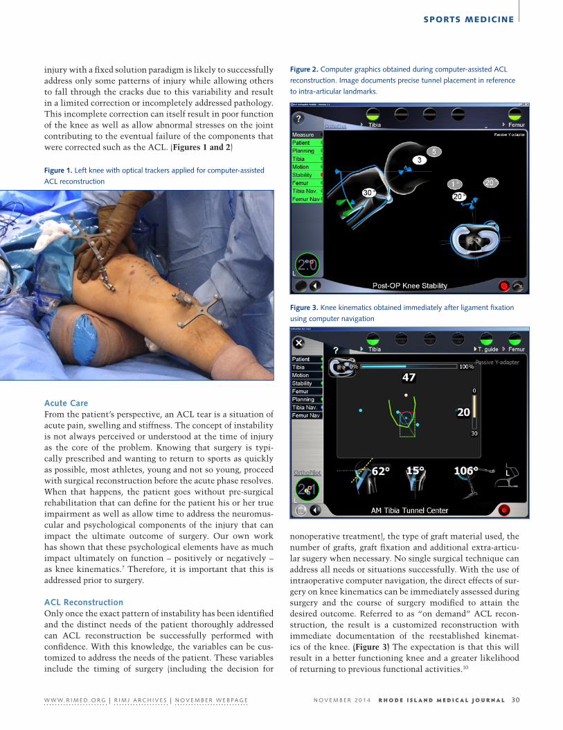
injury with a fixed solution paradigm is likely to successfully address only some patterns of injury while allowing others to fall through the cracks due to this variability and result in a limited correction or incompletely addressed pathology. This incomplete correction can itself result in poor function of the knee as well as allow abnormal stresses on the joint contributing to the eventual failure of the components that were corrected such as the ACL. (Figures 1 and 2)
Acute Care From the patient’s perspective, an ACL tear is a situation of acute pain, swelling and stiffness. The concept of instability is not always perceived or understood at the time of injury as the core of the problem. Knowing that surgery is typi-cally prescribed and wanting to return to sports as quickly as possible, most athletes, young and not so young, proceed with surgical reconstruction before the acute phase resolves. When that happens, the patient goes without pre-surgical rehabilitation that can define for the patient his or her true impairment as well as allow time to address the neuromus-cular and psychological components of the injury that can impact the ultimate outcome of surgery. Our own work has shown that these psychological elements have as much impact ultimately on function – positively or negatively – as knee kinematics.7 Therefore, it is important that this is addressed prior to surgery.
ACL ReconstructionOnly once the exact pattern of instability has been identified and the distinct needs of the patient thoroughly addressed can ACL reconstruction be successfully performed with confidence. With this knowledge, the variables can be cus-tomized to address the needs of the patient. These variables include the timing of surgery (including the decision for
Figure 1. Left knee with optical trackers applied for computer-assisted
ACL reconstruction
Figure 2. Computer graphics obtained during computer-assisted ACL
reconstruction. Image documents precise tunnel placement in reference
to intra-articular landmarks.
Figure 3. Knee kinematics obtained immediately after ligament fixation
using computer navigation
nonoperative treatment), the type of graft material used, the number of grafts, graft fixation and additional extra-articu-lar sugery when necessary. No single surgical technique can address all needs or situations successfully. With the use of intraoperative computer navigation, the direct effects of sur-gery on knee kinematics can be immediately assessed during surgery and the course of surgery modified to attain the desired outcome. Referred to as “on demand” ACL recon-struction, the result is a customized reconstruction with immediate documentation of the reestablished kinemat-ics of the knee. (Figure 3) The expectation is that this will result in a better functioning knee and a greater likelihood of returning to previous functional activities.10
W W W. R I M E D . O R G | R I M J A R C H I V E S | N O V E M B E R W E B P A G E N O V E M B E R 2 0 1 4 R H O D E I S L A N D M E D I C A L J O U R N A L 30
SPORTS MEDICINE
Post-op Healing and Rehabilitation After surgery, a healing process is required for the grafted tendon material to remodel into a viable, dynamic ligament. While the acute phase requires 12 weeks, healing, complete or otherwise, is not guaranteed. Furthermore, no known graft tissue has the unique structure and mechanical function of the native ACL.9 The goal of surgery therefore is to anatomi-cally restore the ligament tissue in the best possible way and through remodeling attain the best functional equivalent. While the nuances of surgery as previously described can greatly affect this, establishing a nurturing intra-articular environment post operatively is equally important. Physical therapy is an important adjuvant that can help create such an environment.
Physical therapy is used to restore range of motion, reduce swelling and restore neuromuscular function. When done appropriately, this stimulates articular cartilage, helps nor-malize the synovial fluid environment and provides a posi-tive stimulus for the healing ACL graft. Under stimulating or over stimulating the knee is thought to adversely affect graft healing and therefore knee function.9 ACL rehabilitation is a subspecialty of physical therapy requiring close super-vision and on-going feedback between patient, therapist and surgeon.
Function While biological healing largely occurs during the first 12 weeks following surgery, no good determination of restored function exists.12-14 While validated research instruments exist, these have been patient-reported outcomes and not true functional assessments. Single-leg hop and triple-hop testing through physical therapy have provided simple estimates of function but the need for better tools and methods of assessment is reflected in the alarmingly high incidence of reinjury in select groups of athletes returning to sport 6 or 7 months after surgery.4 Similarly, the high inci-dence of injury to the contralateral ACL upon returning to sports implies an incomplete restoration of function in the recovering ACL patient.11
CONCLUSIONACL injuries are common. They frequently have a devas-tating impact on knee function and can ultimately lead to joint degeneration. The traditional assumption has been that surgically restoring ligament anatomy will result in restored joint kinematics and thus joint function. Short- and long-term outcomes’ research has failed to identify the lim-itations of current practice. As Lord Kelvin stated over a cen-tury ago, “if it cannot be measured, it cannot be improved.” Currently available technologies, such as computer naviga-tion, can be part of an increased effort to better assess and therefore better correct the pathology of ACL injury and ultimately measure the effect of surgical reconstruction on functional outcome. Patient selection, detailed, individual-ized surgical planning, in-depth patient education, precise intra-operative joint assessment and ligament reconstruc-tion are all necessary now and are more likely to predict suc-cess in ACL reconstruction.
References 1. Miller RH III, Azar FM. Anterior Cruciate Ligament Injuries.
In: Canale St, Beaty JH, eds. Campbell’s Operative Orthopedics. 11th ed. Philadelphia, PA: Mosby, 2008: 2496-2500.
2. Leys T, Salmon L, Waller A, Linklater J, Pinczewski L. Clin-ical Results and Risk Factors for Reinjury 15 Years After An-terior Cruciate Ligament Reconstruction: A Prospective Study of Hamstring and Patellar Tendon Grafts. Am J Sports Med. 2012;40:595-605.
3. Myer G, Martin L, Ford K, Paterno M, Schmidt L, Heidt R, Co-losimo A, Hewett T. No Association of Time from Surgery with Functional Deficits in Athletes After Anterior Cruciate Liga-ment Reconstruction: Evidence for Objective Return to Sport Criteria. Am J Sports Med. 2012;40:2256.
4. Barrett AM, Craft JA, Replogle WH, Hydrick JM, Barrett GR. Anterior Cruciate Ligament Graft Failure: A Comparison of Graft Type Based on Age and Tegner Activity Level. Am J Sports Med. 2011;39:2194-2198.
5. Sakane M, Fox RJ, Woo SL-Y, et al. In Situ Forces in the Anterior Cruciate Ligament and Its Bundles in Response to Anterior Tib-ial Loads. J Ortho Res. 1997;15:285-293.
6. Zaffagnini S, Klos T, Bignozzi S. Computer-Assisted Anterior Cruciate Ligament Reconstruction: An Evidence Based Ap-proach of the First 15 Years. Arthros. 2010; 26:546-554.
7. Christino M, Fleming B, Machan J, Shalvoy R. Psychological Factors Associated with ACL Reconstruction Recovery. Medi-cine and Science in Sports and Exercise. 2013;45:S85-S86.
8. Shah V, Andrews J, Fleisig G, McMichael C, Lemak L. Return to Play After Anterior Cruciate Ligament Reconstruction in Na-tional Football League Athletes. Am J Sports Med. 2010;38:2233-2239.
9. Menetrey J, Duthon VB, Laumonier T, Fritschy D. “Biological Failure” of the Anterior Cruciate Ligament Graft. Knee Surg Sports Traumatol Arthros. 2008;16:224-231.
10. Pearle A, Kendoff D, Musahl V. Perspectives on Computer-As-sisted Orthopaedic Surgery: Movement Toward Quantitative Orthopedic Surgery. J Bone Joint Surg Am. 2009;91(supplement 1):7-12.
11. Brophy R, Schmitz L, Wright R, Dunn W, Parker R, Andrish J, McCarty E, Spindler K. Return to Play and Future ACL Inju-ry Risk After ACL Reconstruction in Soccer Athletes from the Multicenter Orthopaedic Outcomes Network (Moon) Group. Am J Sports Med. 2012;40:2517-2522.
12. Di Stasi S, Logerstedt D, Gardinier E, Snyder-Mackler L. Gait Patterns Differ Between ACL-Reconstructed Athletes Who Pass Return-to-Sport Criteria and Those Who Fail. Am J Sports Med. 2013;41:1310-1318.
13. Stearn K, Pollard C. Abnormal Frontal Plane Knee Mechanics During Sidestep Cutting in Female Soccer Athletes After Ante-rior Cruciate Ligament Reconstruction and Return to Sport. Am J Sports Med. 2013;41:918-923.
14. Paterno M, Schmitt L, Ford K, Rauh M, Myer G, Huang B, Hewett T. Biomechanical Measures During Landing and Postur-al Stability Predict Second Anterior Cruciate Ligament Injury after Anterior Cruciate Ligament Reconstruction and Return to Sport. Am J Sports Med. 2010;38:1968-1978.
AuthorRobert M. Shalvoy, MD, is Assistant Professor of Orthopedic
Surgery, The Warren Alpert Medical School of Brown University.
CorrespondenceRobert M. Shalvoy, MDUniversity Orthopedics, Inc.100 Butler DriveProvidence, RI 02906401-330-1433Fax [email protected]
W W W. R I M E D . O R G | R I M J A R C H I V E S | N O V E M B E R W E B P A G E N O V E M B E R 2 0 1 4 R H O D E I S L A N D M E D I C A L J O U R N A L 31

WE’ll guide you EVERY STEP OF THE WAY.
Founded in 1982, The Center For Orthopaedics provides quality orthopaedic and sports medicine care to patients throughout Rhode Island, Massachusetts and Connecticut.
Our facility and equipment are state-of-the-art, providing excellence in orthopaedic care. We offer our patients a full spectrum of orthopaedic care in an attentive, convenient and coordinated manner.
1524 Atwood Avenue, Suite 140 - Johnston, RI 02919
401.351.6200 | centerfororthopaedics.com
Orthopaedic SurgeryReconstructive SurgerySpine SurgeryJoint Replacement
OUR SERVICES
Sports MedicinePhysical TherapyTherapeutic exerciseManual Therapy
Electrical stimulationCervical/lumbar tractionBalance activitiesGait training