sql-1, homologue of the golgi protein gmap210, modulates...
TRANSCRIPT
1
SQL-1, homologue of the Golgi protein GMAP210, modulates Intraflagellar
Transport in C. elegans
Joost R. Broekhuis1, Suzanne Rademakers1, Jan Burghoorn1, and Gert Jansen1, *
1Department of Cell Biology, Erasmus MC, PO Box 2040, 3000 CA, Rotterdam, the
Netherlands
* Author for correspondence ([email protected])
© 2012. Published by The Company of Biologists Ltd.Jo
urna
l of C
ell S
cien
ceA
ccep
ted
man
uscr
ipt
JCS Advance Online Article. Posted on 26 February 2013

2
Summary
Primary cilia are microtubule-based organelles, which have important sensory functions. For
their function cilia rely on the delivery of specific proteins, both by intracellular trafficking
and intraflagellar transport (IFT). In C. elegans’ cilia, anterograde IFT is mediated by kinesin-
II and OSM-3. Previously, we have shown that expression of a dominant active G protein α
subunit (GPA-3QL) in amphid channel neurons affects the coordination of kinesin-II and
OSM-3 and cilia length, suggesting that environmental signals can modulate these processes.
Here, we show that loss-of-function of sql-1 (suppressor of gpa-3QL #1), which encodes the
homologue of the mammalian Golgi protein GMAP210, suppresses the gpa-3QL cilia length
phenotype. SQL-1 localizes to the Golgi apparatus, where it contributes to maintaining Golgi
organization. Loss of sql-1 by itself does not affect cilia length, while overexpression of sql-1
results in longer cilia. Using live imaging of fluorescently tagged IFT proteins, we show that
in sql-1 mutants OSM-3 moves faster, kinesin-II moves slower, and that some complex A and
B proteins move at an intermediate velocity, while others move at the same velocity as OSM-
3. This indicates that mutation of sql-1 destabilizes the IFT complex. Finally, we show that
simultaneous inactivation of sql-1 and activation of gpa-3QL affects the velocity of OSM-3.
In summary, we show that in C. elegans the Golgin protein SQL-1 plays an important role in
maintaining the stability of the IFT complex.
Jour
nal o
f Cel
l Sci
ence
Acc
epte
d m
anus
crip
t
3
Introduction
Primary cilia are microtubule-based protrusions that can be found on the surface of almost all
vertebrate cells, and have important sensory functions. Cilia dysfunction has been associated
with a number of genetic diseases, collectively called ciliopathies (Hildebrandt et al., 2011).
Many ciliopathies affect various tissues and are caused by defects in proteins that play a role
in transport to the cilium or within the cilium. Often these mutations do not completely block
ciliogenesis, but rather result in changes in cilia morphology and/or localization of signaling
proteins in cilia.
A large diversity in cilia lengths and morphologies exists, probably reflecting the specialized
functions cilia can have in different tissues and/or organisms. However, little is known about
how this diversity is achieved. Different types of signaling molecules, including second
messengers (Mukhopadhyay et al., 2008; Ou et al., 2009; Besschetnova et al., 2010; Abdul-
Majeed et al., 2012), kinases (Berman et al., 2003; Wang et al., 2006; Burghoorn et al., 2007;
Tam et al., 2007; Miyoshi et al., 2009; Omori et al., 2010), phosphatases (Kim et al., 2010;
Clement et al., 2011; Abdul-Majeed et al., 2012) and G proteins (Burghoorn et al., 2010) have
been shown to regulate cilia length and cilia morphology. Since these signaling pathways also
regulate transport, both in and toward the cilium, it is likely that these transport processes play
a role in regulating cilium length and morphology (Silverman and Leroux, 2009).
Ciliary proteins often originate from the Golgi apparatus (Pazour and Bloodgood, 2008;
Emmer et al., 2010). A number of small GTPases, including ARF4, ARL6 and RAB8
(Nachury et al., 2007; Mazelova et al., 2009; Jin et al., 2010; Wiens et al., 2010), are involved
in budding of vesicles from the Golgi apparatus and fusion at the ciliary base. In the absence
of components of the BBSome complex, composed of 7 Bardet-Biedl Syndrome (BBS)
proteins (Nachury et al., 2007), the GPCRs (G protein coupled receptors) rhodopsin, SSTR3,
and MCHR1 (Nishimura et al., 2004; Abd-El-Barr et al., 2007; Berbari et al., 2008), fail to
reach the cilium, indicating that the BBSome functions in trafficking of (G-protein coupled)
receptors to the cilium. However, whether the BBSome functions in vesicular transport, cargo
selection, or vesicle docking and fusion is not completely clear.
Once inside the cilium, signaling proteins and structural proteins are transported by
intraflagellar transport (IFT). IFT particles carry cargo along the microtubule axis of the
cilium, towards the distal tip and back. Movement of IFT particles is powered by kinesin-2
motors (anterograde transport) and cytosolic dynein 2 (retrograde transport) (Rosenbaum and
Jour
nal o
f Cel
l Sci
ence
Acc
epte
d m
anus
crip
t
4
Witman, 2002). IFT particles are organized in two distinct subcomplexes, complex A and
complex B (Cole and Snell, 2009). Complex A has been implicated in retrograde transport
and complex B in anterograde transport. In addition, proteins that are part of the BBSome
have been observed moving in the cilium, with similar velocities as components of the IFT
complex (Blacque et al., 2004; Nachury et al., 2007), suggesting that the BBSome associates
with or is part of the IFT complex.
GMAP210 was recently found to play a role in vesicular transport from the Golgi to the
cilium (Follit et al., 2008). It belongs to the Golgin family, whose members are characterized
by a large number of coiled-coil domains, and function in maintaining the organization and
position of the Golgi apparatus (Short et al., 2005). GMAP210 localizes to the cis-Golgi
network (Rios et al., 1994), and is important for Golgi integrity (Rios et al., 2004).
Interestingly, GMAP210 anchors the complex B protein IFT20 to the Golgi apparatus. IFT20
probably functions as an adaptor protein for targeting vesicles to the cilium. In line with its
novel role in vesicular trafficking to the cilium, absence of GMAP210 results in reduced
levels of ciliary polycystin-2 (Follit et al., 2008). GMAP210 knockout mice die at birth, most
likely due to heart and lung defects (Follit et al., 2008). Both heart and lung are among the
organs affected in ciliopathy patients (Cardenas-Rodriguez and Badano, 2009).
The sensory cilia of C. elegans’ amphid neurons provide an excellent model to study the
regulation of cilia morphology. They can be divided in a middle segment, which has nine
microtubule doublets, and a distal segment, which has nine microtubule singlets (Ward et al.,
1975; Perkins et al., 1986). In the middle segment, the IFT complex is transported by two
members of the kinesin-2 family, kinesin-II and OSM-3. At the end of the middle segment
kinesin-II dissociates from the complex, which leaves OSM-3 to transport the IFT complex in
the distal segment (Snow et al., 2004). We have previously shown that in gpa-3QL animals,
which carry a dominant active mutant version of the sensory Gα subunit, gpa-3, the
coordination of kinesin-II and OSM-3 is affected, and the sensory cilia are shorter (Burghoorn
et al., 2010). Interestingly, exposure of animals to dauer pheromone (a continuously secreted
compound that at high concentrations induces an alternative larval stage, the dauer larva
(Golden and Riddle, 1982), affects the coordination of kinesin-II and OSM-3 very similarly as
gpa-3QL (Burghoorn et al., 2010). IFT measurements on gpa-3 mutant animals exposed to
dauer pheromone suggest that gpa-3 functions in the pathway by which the pheromone affects
IFT. Together these results suggest that environmental cues transduced via GPA-3 can
modulate IFT.
Jour
nal o
f Cel
l Sci
ence
Acc
epte
d m
anus
crip
t
5
To find out how GPA-3 regulates IFT and cilia length we performed a screen for suppressors
of the gpa-3QL cilia defect. We identified and characterized one of the mutants, sql-1, and
found that it encodes the C. elegans’ homologue of the mammalian Golgin protein
GMAP210. We show that SQL-1 is ubiquitously expressed and localizes to the Golgi
apparatus. In sql-1 mutants the Golgi structure seems disorganized, in line with the function
of Golgin proteins. In addition, sql-1 affects the stability of the IFT machinery, resulting in a
partial separation of kinesin-II and OSM-3, and transport of the IFT complex predominantly
by OSM-3.
Jour
nal o
f Cel
l Sci
ence
Acc
epte
d m
anus
crip
t
6
Results
sql-1 encodes the C. elegans homologue of mammalian GMAP210
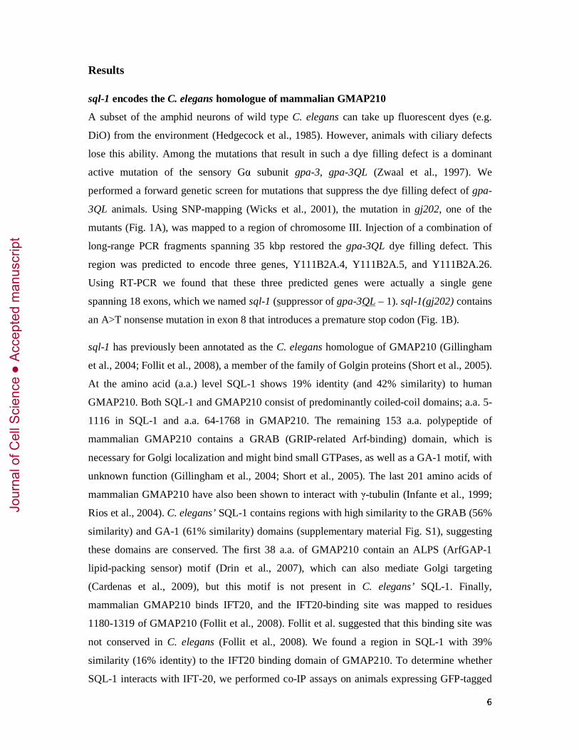
A subset of the amphid neurons of wild type C. elegans can take up fluorescent dyes (e.g.
DiO) from the environment (Hedgecock et al., 1985). However, animals with ciliary defects
lose this ability. Among the mutations that result in such a dye filling defect is a dominant
active mutation of the sensory Gα subunit gpa-3, gpa-3QL (Zwaal et al., 1997). We
performed a forward genetic screen for mutations that suppress the dye filling defect of gpa-
3QL animals. Using SNP-mapping (Wicks et al., 2001), the mutation in gj202, one of the
mutants (Fig. 1A), was mapped to a region of chromosome III. Injection of a combination of
long-range PCR fragments spanning 35 kbp restored the gpa-3QL dye filling defect. This
region was predicted to encode three genes, Y111B2A.4, Y111B2A.5, and Y111B2A.26.
Using RT-PCR we found that these three predicted genes were actually a single gene
spanning 18 exons, which we named sql-1 (suppressor of gpa-3QL – 1). sql-1(gj202) contains
an A>T nonsense mutation in exon 8 that introduces a premature stop codon (Fig. 1B).
sql-1 has previously been annotated as the C. elegans homologue of GMAP210 (Gillingham
et al., 2004; Follit et al., 2008), a member of the family of Golgin proteins (Short et al., 2005).
At the amino acid (a.a.) level SQL-1 shows 19% identity (and 42% similarity) to human
GMAP210. Both SQL-1 and GMAP210 consist of predominantly coiled-coil domains; a.a. 5-
1116 in SQL-1 and a.a. 64-1768 in GMAP210. The remaining 153 a.a. polypeptide of
mammalian GMAP210 contains a GRAB (GRIP-related Arf-binding) domain, which is
necessary for Golgi localization and might bind small GTPases, as well as a GA-1 motif, with
unknown function (Gillingham et al., 2004; Short et al., 2005). The last 201 amino acids of
mammalian GMAP210 have also been shown to interact with γ-tubulin (Infante et al., 1999;
Rios et al., 2004). C. elegans’ SQL-1 contains regions with high similarity to the GRAB (56%
similarity) and GA-1 (61% similarity) domains (supplementary material Fig. S1), suggesting
these domains are conserved. The first 38 a.a. of GMAP210 contain an ALPS (ArfGAP-1
lipid-packing sensor) motif (Drin et al., 2007), which can also mediate Golgi targeting
(Cardenas et al., 2009), but this motif is not present in C. elegans’ SQL-1. Finally,
mammalian GMAP210 binds IFT20, and the IFT20-binding site was mapped to residues
1180-1319 of GMAP210 (Follit et al., 2008). Follit et al. suggested that this binding site was
not conserved in C. elegans (Follit et al., 2008). We found a region in SQL-1 with 39%
similarity (16% identity) to the IFT20 binding domain of GMAP210. To determine whether
SQL-1 interacts with IFT-20, we performed co-IP assays on animals expressing GFP-tagged
Jour
nal o
f Cel
l Sci
ence
Acc
epte
d m
anus
crip
t
7
IFT-20. However, anti-SQL-1 antibodies did not bring down IFT-20::GFP, and anti-GFP
antibodies failed to bring down endogenous SQL-1 (supplementary material Fig. S2A,B). In
addition, we determined the subcellular localization of IFT-20 using a full length ift-20::gfp -
fusion construct. In the ciliated neurons, we observed IFT-20::GFP in the cilia and at their
base in the basal body remnants, and a weak, diffuse IFT-20::GFP signal in the dendrites and
cell bodies, but no distinct Golgi localization (supplementary material Fig. S2C, compare to
Fig. 2B). The localization pattern of IFT-20::GFP in sql-1 mutant animals was identical to
that of wild type animals. These data suggest that in C. elegans SQL-1 and IFT-20 do not
physically interact, and that IFT-20 does not localize to the Golgi apparatus. Possibly, IFT-
20’s function in the Golgi apparatus is not conserved in C. elegans. However, we cannot
exclude the possibility that IFT-20 is transported to and from the Golgi apparatus but that the
steady state level at the Golgi apparatus is low.
Initially, two sql-1 deletion mutants were obtained from the NBP-Japan, sql-1(tm2409), and
sql-1(tm2440). In the sql-1(tm2440) strain 192 bp of exon 4 was deleted, resulting in the loss
of 64 a.a. of SQL-1 (Fig. 1B). Western blot analysis, using antibodies raised against the N-
and C-terminal parts of the SQL-1 protein, showed that the sql-1(tm2440) strain indeed
expressed a smaller SQL-1 protein (Fig. 1C and supplementary material Fig. S3A). sql-
1(tm2440); gpa-3QL animals were dye filling defective, thus the deletion in sql-1(tm2440)
did not suppress the dye filling defect of gpa-3QL animals (data not shown). The sql-
1(tm2409) strain carries a 175 bp deletion, starting in intron 14 and ending in exon 15. RT-
PCR showed that this resulted in the loss of exon 15, a frameshift in exon 16, and an early
stop. We could not detect the truncated SQL-1 protein of sql-1(tm2409) animals on a Western
blot (expected molecular weight 137 kDa), or in immunofluorescence (Fig. 1C and
supplementary material Fig. S3A,B), demonstrating that sql-1(tm2409) is a null allele. In sql-
1(tm2409); gpa-3QL mutants the dye filling defect caused by gpa-3QL was suppressed.
Recently, we obtained an additional null mutant, tm4995, which carries a 632 bp deletion that
removes exon 1. Also the sql-1(tm4995) mutation suppressed the dye filling defect of gpa-
3QL (data not shown). In addition to the loss-of-function mutants, we generated animals that
contain extra copies of the sql-1 gene, sql-1XS. Western blot analysis showed that sql-1XS
animals (gj2077) expressed approximately three times more SQL-1 than wild type animals
(supplementary material Fig. S3C). Overexpression of sql-1 did not affect dye filling. All
loss-of-function and overexpression animals were healthy and showed no apparent phenotype.
Jour
nal o
f Cel
l Sci
ence
Acc
epte
d m
anus
crip
t
8
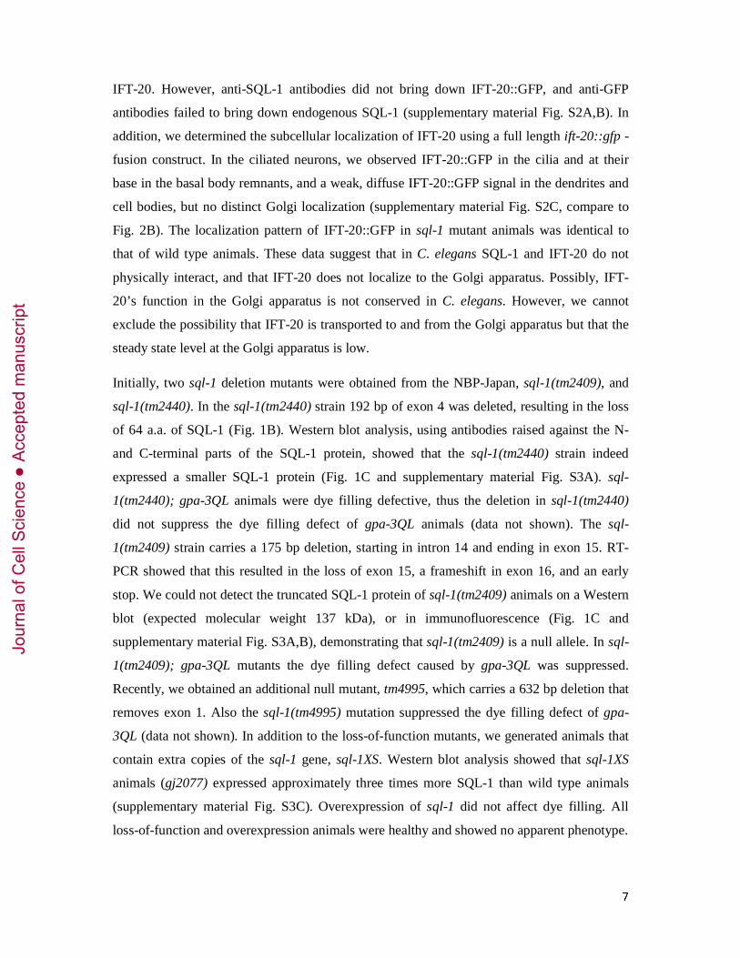
SQL-1 is ubiquitously expressed, and localizes to the Golgi apparatus
To determine the expression pattern and subcellular localization of SQL-1, two different
antibodies (Abs) were raised against SQL-1, one against an N-terminal part (a.a. 106-737) of
SQL-1, and one against a C-terminal part (a.a. 519-1328). Immunofluorescence on wild type
animals showed a spotted pattern throughout the whole body (supplementary material Fig.
S3B), suggesting SQL-1 is ubiquitously expressed. A similar pattern was seen in sql-
1(tm2440) animals, but no immunoreactivity could be detected in sql-1(tm2409)
(supplementary material Fig. S3B), confirming the specificity of the antibodies.
To confirm the localization pattern determined in the immunofluorescence experiments, we
generated animals that expressed full length SQL-1 fused to GFP, psql-1::sql-1::GFP animals.
In these animals we observed spots throughout their bodies (Fig. 2B), similar to the pattern
observed with SQL-1 Abs. Since the homologue of SQL-1, GMAP210, localizes to the Golgi
apparatus (Rios et al., 1994), we stained psql-1::sql-1::gfp animals with an antibody against the
glucuronyl transferase SQV-8, which stains the Golgi apparatus (Hadwiger et al., 2010). This
showed co-localization of SQL-1 and SQV-8 (Fig. 2B). Both the immunofluorescence and
GFP fusion data confirm that SQL-1 localizes to the Golgi apparatus. Interestingly, also an N-
terminal (a.a. 1-302) SQL-1::GFP fusion localized to spots throughout the animal, suggesting
that perhaps SQL-1 contains an N-terminal region that mediates Golgi localization. However,
this GFP-fusion was more abundant in the cytosol than the full length SQL-1 GFP-fusion
(supplementary material Fig. S3D).
Mutation of sql-1 suppresses the dye-filling defect of gpa-3QL animals. Therefore we
analyzed the expression and subcellular localization of SQL-1 in neurons. We generated
animals that expressed the Golgi marker AMAN-2, an alpha-mannosidase, tagged with YFP
only in one pair of ciliated sensory neurons, the ASI neurons (pgpa-4::aman-2::YFP), or in a
subset of inter- and motor neurons (pglr-1::aman-2::YFP). Staining of these animals with anti-
SQL-1 Ab indeed showed co-localization of AMAN-2::YFP and SQL-1 in the cell bodies of
glr-1 expressing neurons and the ASI neurons (Fig. 2C). In pgpa-4::sql-1::GFP animals, we
observed SQL-1::GFP in the Golgi apparatus in the cell bodies, and sometimes SQL-1 spots
in the dendrites (Fig. 2D). These SQL-1 spots were immobile, and were never observed in
close proximity of the cilium. Together these data show that SQL-1 localizes to the Golgi
apparatus in C. elegans’ ciliated sensory neurons.
Jour
nal o
f Cel
l Sci
ence
Acc
epte
d m
anus
crip
t
9
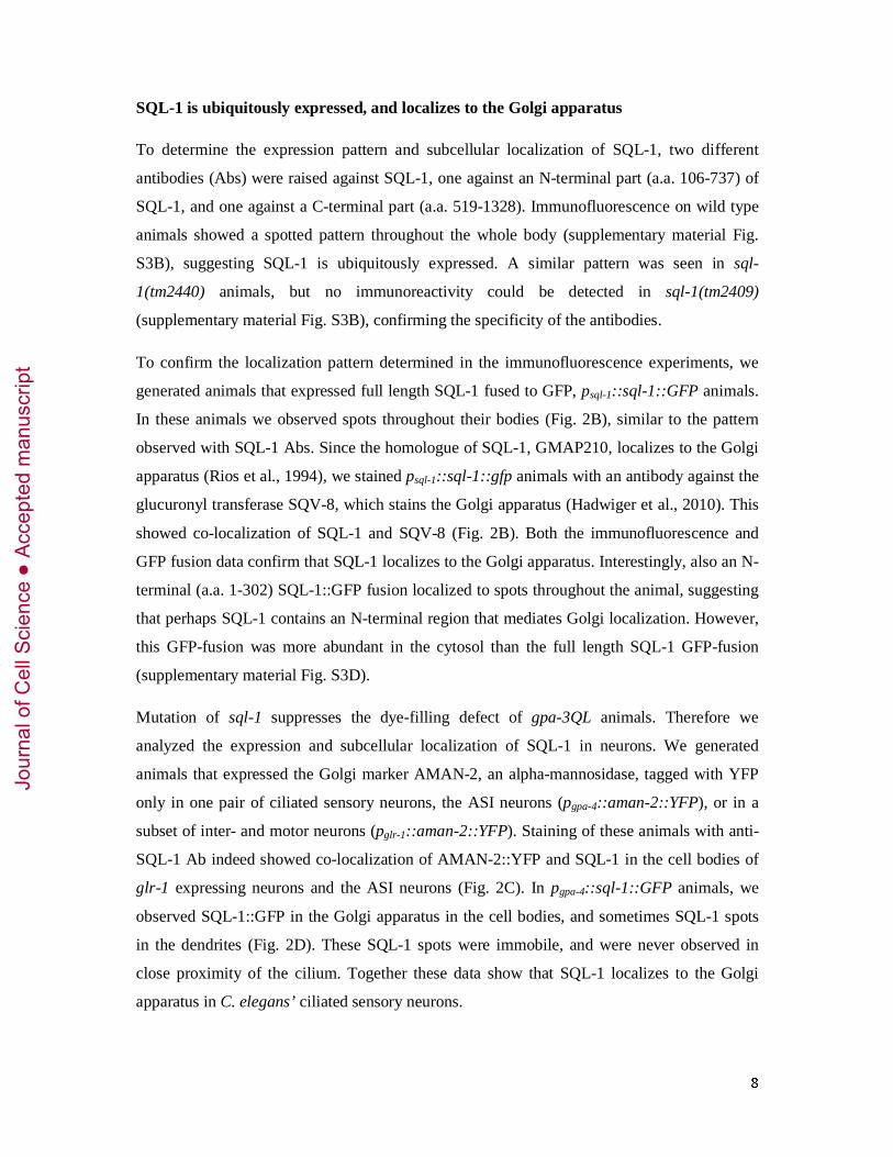
sql-1 is involved in maintaining Golgi apparatus structure, but is not required for
normal GPA-3 localization
Knock-down of GMAP210 has been shown to result in the dispersion of the Golgi
membranes (Rios et al., 2004; Yadav et al., 2009). However, no defects were observed in the
Golgi apparatus of GMAP210 mutant mice (Follit et al., 2008). We wondered whether the
absence of SQL-1 affects the integrity of the Golgi apparatus in C. elegans.
To investigate this we visualized the Golgi apparatus specifically in the ASI neurons using
AMAN-2::GFP in wild type and sql-1(tm2409) animals. In wild type animals we observed
distinct AMAN-2::GFP structures in the ASI cells (Fig. 3A,B). However, in the majority of
the sql-1(tm2409) animals the AMAN-2::GFP signal was more fragmented or even diffuse
(Fig. 3A,B). Very similar effects were seen in sql-1(tm2409); gpa-3QL animals (Fig. 3A,B).
In gpa-3QL animals we observed distinct AMAN-2 structures similar to those observed in
wild type animals (Fig 3A,B). These results suggest that loss of function of sql-1 affects the
organization of the Golgi apparatus, although we cannot exclude the possibility that the effect
of sql-1 mutation is AMAN-2 specific.
One way to explain the suppression of the dye filling defect in sql-1(tm2409); gpa-3QL
animals is a trafficking defect of dominant active GPA-3QL protein, caused by the absence of
SQL-1. Therefore we used immunofluorescence to visualize the localization of GPA-3. In
both wild type and sql-1(tm2409) animals GPA-3 can be observed at the ciliary membrane at
comparable levels (Fig. 3C), showing that sql-1 is not required for ciliary localization of
GPA-3. gpa-3QL and sql-1; gpa-3QL animals showed very strong anti-GPA-3 staining in the
cilia, dendrites, and cell bodies. Especially high levels of GPA-3 were observed at the base of
the cilia, possibly in the periciliary membrane compartment (Kaplan et al., 2012), making it
difficult to visualize GPA-3 levels in the cilia of these animals. Nonetheless, we observed no
obvious differences in the levels of GPA-3QL staining in the cilia of gpa-3QL and sql-1; gpa-
3QL animals (supplementary material Fig. S4A). Also, a Western blot showed no differences
in GPA-3 levels between the two strains (supplementary material Fig. S4B). Together these
data suggest that suppression of the Dyf phenotype in sql-1(tm2409); gpa-3QL animals is not
caused an effect on the amount or the localization of GPA-3.
Mutation of sql-1 regulates cilia length, and acts cell-autonomously
We have previously shown that the cilia of the ADF, ASH, ASI, ASK, and ADL neurons of
adult gpa-3QL animals are shorter than those of wild-type adult animals (Burghoorn et al.,
Jour
nal o
f Cel
l Sci
ence
Acc
epte
d m
anus
crip
t
10
2010), explaining the dye filling defect. To determine if sql-1 mutation affects cilia length of
gpa-3QL animals, we visualized cilia using an ASI neuron specific gpa-4::gfp construct, and
a gpa-15::gfp construct expressed in ASH, ASK, and ADL neurons (Jansen et al., 1999).
Cilium length of gpa-3QL animals was restored to approximately wild type length in sql-
1(tm2409); gpa-3QL animals (Fig. 4A,B).
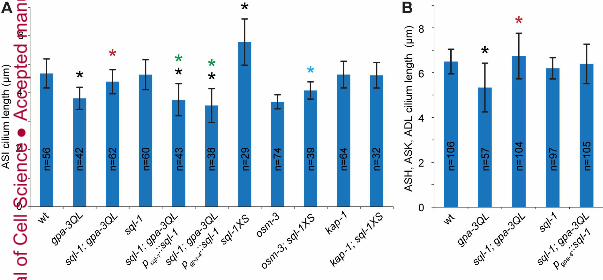
To determine if changes in sql-1 levels themselves affect cilium length we visualized cilia of
sql-1(tm2409) and sql-1XS animals. Cilium length in sql-1(tm2409) animals was comparable
to cilium length in wild type animals (Fig. 4A,B). Interestingly, the cilia of ASI neurons of
animals that overexpress SQL-1, sql-1XS, were significantly longer than those of wild-type
animals (Fig. 4A). To test if this effect on cilia length depends on either of the two kinesins
we measured ASI lengths in osm-3; sql-1XS and kap-1; sql-1XS animals. Loss of osm-3
shortened the cilia of sql-1XS animals, but these cilia were still significantly longer than those
of osm-3 animals (Fig 4A), indicating that at least the middle segments are longer in sql-1
animals. However, loss of kap-1 completely suppressed cilia lengthening as a result of sql-1
overexpression (Fig. 4A), indicating that kinesin-II is required for this effect.
To further confirm the specificity of suppression of the dye filling defective (Dyf) phenotype
in gpa-3QL animals by the loss of SQL-1 we re-introduced the sql-1 gene in sql-1(tm2409);
gpa-3QL animals. This resulted in a decreased length of all cilia, comparable to the length in
gpa-3QL animals (Fig. 4A). In addition, while expression of wild type sql-1 specifically in the
ASI neurons of sql-1; gpa-3QL animals did rescue ASI cilia length, it did not affect the length
of the cilia of ASH, ASK, and ADL (Fig 4A,B). This suggests that sql-1 acts cell-
autonomously.
Mutation of sql-1 partially uncouples kinesin-II and OSM-3
We previously hypothesized that the decreased cilia length in gpa-3QL animals was caused
by the observed partial uncoupling of kinesin-II and OSM-3 (Burghoorn et al., 2010). To
determine whether sql-1 also affects the IFT machinery, we examined the motility of the two
ciliary kinesin subunits KAP-1 (mammalian homologue KAP3) and OSM-3 (KIF17), two
complex A proteins CHE-11 (IFT140), DAF-10 (IFT22), three complex B proteins OSM-1
(IFT172), CHE-13 (IFT57), and IFT-20, and the dynein motor subunit XBX-1 (D2LIC) in
sql-1(tm2409) animals as well as in wild type animals. In wild type animals all IFT proteins
moved at approximately 0.7 µm/s in the middle segments (Table 1), and at approximately
1.15 µm/s in the distal segments (supplementary material Table S1). These speeds are
Jour
nal o
f Cel
l Sci
ence
Acc
epte
d m
anus
crip
t
11
consistent with published data (Snow et al., 2004). However, in the middle segments of sql-
1(tm2409) animals, kinesin-II moved at 0.59 µm/s, and OSM-3 at 0.89 µm/s (Table 1). Since
the two kinesins moved at different velocities it is not possible that they were in the same
complex at all times. CHE-11::GFP (complex A), OSM-1::GFP and IFT-20::GFP (both
complex B), and XBX-1::GFP (dynein) moved at similar velocities as OSM-3 in the middle
segments of sql-1(tm2409) animals (Table 1), whereas DAF-10::GFP (complex A) and CHE-
13::GFP (complex B) moved at 0.67 µm/s (Table 1). In the distal segments of sql-1(tm2409)
animals, all examined IFT markers moved at velocities similar to those in wild type animals
(supplementary material Table S1).
To determine if any of these effects were caused by changes in the intrinsic velocities of
kinesin-II or OSM-3, we decided to measure how the absence of SQL-1 affects IFT motility
in osm-3 and kap-1 mutant backgrounds. In sql-1(tm2409); osm-3 animals KAP-1::GFP
(kinesin-II), DAF-10::GFP (complex A), CHE-13::GFP (complex B), and XBX-1::GFP
(cargo) moved at 0.49-0.52 µm/s (Table 1), similar to the velocities observed in osm-3
animals. In kap-1; sql-1(tm2409) animals OSM-3::GFP, DAF-10::GFP (complex A), CHE-
13::GFP (complex B), and XBX-1::GFP (dynein) moved at the 1.00-1.19 µm/s (Table 1),
similar to velocities observed in kap-1 animals. These data indicate that the intrinsic velocities
of the kinesins are not altered in sql-1 mutant animals.
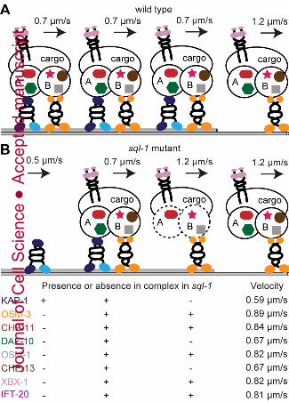
Our data indicate that in sql-1(tm2409) animals kinesin-II and OSM-3 are partially separated.
Based on the observed speeds, we calculated that approximately 38% of OSM-3 motor
proteins moved independently from kinesin-II and that 61% of kinesin-II motor proteins
moved independently from OSM-3.
Of the five non-kinesin IFT-components tested, three moved at the same speed as OSM-3,
and two moved at approximately 0.7 µm/s. This suggests that in sql-1(tm2409) animals also
the IFT complex has disintegrated, where CHE-11, OSM-1, IFT-20 and XBX-1 associate
with OSM-3 particles, and together with DAF-10 and CHE-13 associate with the kinesin-
II/OSM-3 complex. An alternative explanation would be that DAF-10 and CHE-13 associate
with all three possible particles (slow kinesin-II particles, fast OSM-3 particles and the
kinesin-II/OSM-3 complex), averaging in a velocity of approximately 0.7 µm/s. To
distinguish between these two possibilities we plotted the distributions of the velocities of
CHE-13::GFP and DAF-10::GFP IFT events, where the presence of CHE-13 and DAF-10 in
three particle types would be visible in the distribution plots as additional peaks, or at least a
Jour
nal o
f Cel
l Sci
ence
Acc
epte
d m
anus
crip
t
12
wider distribution. However, the distributions of the CHE-11::GFP and DAF-10::GFP IFT
events in wild type and sql-1(tm2409) animals were very similar (supplementary material Fig.
S5), suggesting that CHE-11 and DAF-10 were only present in the kinesin-II/OSM-3
transported particles. In addition, these results suggest that in sql-1 animals a fraction of
kinesin-II moves without further IFT components.
In summary, our data are consistent with a model in which the middle segments of the
sensory cilia of sql-1(tm2409) animals contain three types of particles: complete IFT particles,
empty kinesin-II and incomplete IFT particles consisting of OSM-3, but not kinesin-II, and at
least CHE-11, OSM-1, IFT-20, and XBX-1, but not DAF-10 and CHE-13 (Fig. 5).
OSM-3::GFP speed is affected in sql-1; gpa-3QL double mutant animals
Since in gpa-3QL, gpa-3 and sql-1(tm2409) animals IFT is affected, we examined IFT in
double mutants, to study their genetic interactions. In the middle segments of sql-1(tm2409);
gpa-3 animals KAP-1::GFP and XBX-1::GFP moved at velocities similar to those observed in
sql-1 and gpa-3 single mutant animals. OSM-3::GFP moved at the same speed as in sql-
1(tm2409) animals (Table 1). These data suggest that, genetically, sql-1 acts downstream of
gpa-3.
In the middle segments of sql-1(tm2409); gpa-3QL animals, KAP-1::GFP moved at a velocity
similar to that observed in the middle segments of sql-1(tm2409) and gpa-3QL animals,
OSM-3::GFP moved at a velocity similar to that observed in gpa-3QL animals and slower
than in sql-1(tm2409) animals, XBX-1::GFP moved significantly slower compared to the
velocities observed in sql-1(tm2409) and gpa-3QL animals (Table 1). Possibly, effects of the
mutations in sql-1(tm2409) and gpa-3QL on IFT are additive.
Interestingly, in the distal segments of sql-1(tm2409); gpa-3QL animals all measured
components of the IFT complex (OSM-3, DAF-10, CHE-13, and XBX-1) moved slower than
the expected 1.15 µm/s, the velocity of the IFT complex in the distal segments of wild type,
sql-1(tm2409), and gpa-3QL animals (supplementary material Table S1). This could be
explained by either entry of “slow” kinesin-II into the distal segment, as previously observed
in dyf-5 mutant animals (Burghoorn et al., 2007), or by a change in the intrinsic velocity of
OSM-3. First, we examined the localization of KAP-1::GFP in sql-1(tm2409); gpa-3QL
animals, but we did not observe any KAP-1::GFP in the distal segment (supplementary
material Fig. S6), suggesting that the slower velocities are not caused by entry of kinesin-II
into the distal segment.
Jour
nal o
f Cel
l Sci
ence
Acc
epte
d m
anus
crip
t
13
Second, we examined whether the intrinsic velocity of OSM-3 was affected. Therefore, we
measured the velocity of OSM-3 in kap-1, kap-1; sql-1, kap-1; gpa-3QL, and kap-1; sql-1;
gpa-3QL mutant animals. In all these animals the IFT complex is transported only by OSM-3
in both segments. As expected, OSM-3::GFP moved at a velocity of 1,17-1,23 µm/s in both
segments of kap-1, kap-1; sql-1 and kap-1; gpa-3QL animals (Fig. 6, Table 1, and
supplementary material Table S1). However, in kap-1; sql-1; gpa-3QL animals OSM-3::GFP
moved significantly slower than in kap-1, kap-1; sql-1 and kap-1; gpa-3QL animals (Fig. 6,
Table 1, and supplementary material Table S1). These data indicate that in the absence of
SQL-1 and presence of GPA-3QL the motility of OSM-3 is affected. The reduced speed of
OSM-3 in sql-1; gpa-3QL double mutant animals likely affects the speed of the whole IFT
machinery, and might explain the lower velocities observed in the sql-1(tm2409); gpa-3QL
double mutants.
Jour
nal o
f Cel
l Sci
ence
Acc
epte
d m
anus
crip
t
14
Discussion
Summary
In this study we identified and characterized SQL-1 (suppressor of gpa-3QL – 1), the C.
elegans homologue of mammalian GMAP210. Loss of sql-1 suppressed the ciliary length
defect of gpa-3QL animals and, probably as a result of that, also the dye filling defect of these
animals. Loss-of-function of sql-1 by itself did not affect cilia length, however overexpression
of sql-1 increased cilia length. We found that SQL-1 is ubiquitously expressed and localizes
to the Golgi apparatus. Loss of sql-1 affected the localization of the Golgi protein AMAN-2,
suggesting a mild effect on Golgi integrity. Importantly, mutation of sql-1 affected the IFT
machinery and partially uncoupled kinesin-II and OSM-3. Genetic epistasis analyses
suggested that sql-1 and gpa-3 function in the same genetic pathway.
SQL-1/GMAP210 functions
GMAP210 localizes to the Golgi apparatus. Two domains have been identified that mediate
this localization, the N-terminal ALPS domain, and the C-terminal GRAB domain
(Gillingham et al., 2004; Follit et al., 2008; Cardenas et al., 2009). SQL-1 also localizes to the
Golgi apparatus, as determined using immunofluorescence and GFP fusion constructs. In the
sensory neurons, SQL-1 is mostly restricted to the cell bodies, with some SQL-1 spots in the
dendrite, but never in or close to the cilium. Interestingly, not only full length SQL-1 fused to
GFP localizes to the Golgi apparatus, but also a fusion protein containing the 302 most N-
terminal amino acids localizes there. Since SQL-1 does not seem to have an ALPS domain
and the GRAB domain is not present in the shortened fragment, Golgi targeting of this
fragment must be mediated by a different, still uncharacterized SQL-1 domain.
Several functions have been ascribed to GMAP210. One of these proposed functions is
maintaining the structure of the Golgi apparatus. Fragmentation of the Golgi apparatus has
been observed in cultured HeLa cells after RNAi against GMAP210, as well as in primary
dermal fibroblasts isolated from GMAP210 -/- mice (Rios et al., 2004; Yadav et al., 2009;
Smits et al., 2010). Interestingly, others did not observe any structural Golgi defects in similar
experiments (Gillingham et al., 2004; Follit et al., 2008). We visualized the Golgi apparatus
of C. elegans specifically in the ASI neurons with AMAN-2::GFP, and observed more
dispersed or even diffuse AMAN-2::GFP fluorescence in sql-1(tm2409). This suggests that in
C. elegans SQL-1 is involved in maintaining Golgi integrity.
Jour
nal o
f Cel
l Sci
ence
Acc
epte
d m
anus
crip
t

15
Follit et al. have shown that GMAP210 interacts with complex B protein IFT20 and plays a
role in sorting and/or transport to the ciliary membrane (Follit et al., 2008). They found that
mouse embryonic kidney cells lacking GMAP210 could still form cilia, showing that
GMAP210 is not required for cilia assembly, although these cilia were slightly shorter.
Consistent with a role in vesicular transport to the cilium, it was shown that in the absence of
GMAP210 polycystin-2 levels in the cilium were lower. In C. elegans GMAP210 and IFT20
do not interact, since SQL-1 and IFT-20 did not co-immunoprecipitate and we could not
detect any IFT-20::GFP at the Golgi apparatus, where mammalian IFT20 resides in cultured
cells (Follit et al., 2006). In addition, Kaplan et al. have previously shown that the ciliary
membrane protein ODR-10 still localizes to the sensory cilia of ift-20 mutant animals (Kaplan
et al., 2010). Thus, it is very well possible that the role of IFT-20 in trafficking ciliary
membrane proteins is not conserved in C. elegans. Mutation of sql-1 does not affect cilia
length, where the absence of GMAP210 in cultured mammalian cells results in slightly
shorter cilia. Interestingly, animals that overexpress SQL-1 do show a cilia length increase,
indicating that also SQL-1 plays a role in the regulation of cilium length.
Mutation of sql-1 affects IFT
In the absence of sql-1 the velocities of different components of the IFT machinery are
affected. These changes are consistent with a model in which the middle segments of the
sensory cilia of sql-1(tm2409) animals contain three types of particles: complete IFT particles,
empty kinesin-II and incomplete IFT particles consisting of OSM-3, but not kinesin-II, and at
least CHE-11, OSM-1, IFT-20 and XBX-1, but not DAF-10 and CHE-13 (Fig. 5).
The presence of an incomplete IFT particle suggests a destabilization of the IFT complex in
sql-1(tm2409) animals. CHE-11/IFT140, one of the proteins present in the incomplete IFT
particle of sql-1(tm2409) animals, is part of the “core” of complex A (Ou et al., 2007;
Mukhopadhyay et al., 2010; Behal et al., 2012). In addition, CHE-11/IFT140 interacts with
complex B protein OSM-1/IFT172, which is also present in the incomplete IFT particle of
sql-1(tm2409) animals (Follit et al., 2009). Possibly, the destabilized IFT particles in sql-1
mutants reflect the “core” of the IFT machinery. Further analyses using dual colour imaging
to visualize the motility of additional IFT components simultaneously is necessary to reveal
the makeup of the IFT particles in sql-1 mutant animals.
At present, it is unclear how mutation of sql-1 affects the stability of the IFT complex. Our
data and those presented by Follit et al. (2008) indicate that SQL-1/GMAP210 only resides in
Jour
nal o
f Cel
l Sci
ence
Acc
epte
d m
anus
crip
t

16
the Golgi apparatus and not in the cilium. Therefore, it is likely that the IFT defect is caused
by a sorting defect of vesicles destined for the cilium. E.g. vesicles could exit the Golgi
apparatus prematurely, or fail to transport some proteins required for IFT or maintenance of
the stability of the IFT complex. Other proteins required for the stability of the IFT complex
include BBS and NPHP proteins. However, mutation of these proteins causes uncoupling of
complex A and B (BBS proteins) (Ou et al., 2005; Ou et al., 2007) or mainly affect the
complex B protein OSM-6/IFT52 (Jauregui et al., 2008). Finally, the presence of other
kinesin such as KLP-6 (Morsci and Barr, 2011) or another still unidentified ciliary kinesin,
could also explain the differences in the velocities observed in sql-1 mutant animals.
Interactions of sql-1 and gpa-3
We identified sql-1 as a suppressor of the dye filling defect of gpa-3QL animals. In agreement
with the effect on dye filing, mutation of sql-1 also suppressed the effect of gpa-3QL on
cilium length. sql-1 did not suppress the effects of gpa-3QL on the IFT machinery, but instead
introduced additional defects. Still, the suppression of the cilia length defect in gpa-3QL animals by
sql-1 can be explained by its effect on the IFT machinery. In gpa-3QL animals, kinesin II and
OSM-3 were partially separated, while complex A and B particle proteins moved at an
intermediate velocity (Burghoorn et al., 2010). These data are consistent with a model where
the middle segments of gpa-3QL animals contain complete IFT particles, IFT particles
lacking kinesin-II and IFT particle lacking OSM-3 (supplementary material Fig. S7). We
speculate that this results in a decreased delivery of particles to the distal segment and
reduced ciliary length. In sql-1 mutant animals IFT particles are predominantly transported by
OSM-3. Therefore, inactivation of sql-1 in gpa-3QL animals would cause cargo to be
transported predominantly by OSM-3, resulting in an increase of particles reaching the distal
tip, and thus suppression of the cilium length defect.
Concluding remarks
From our and previously reported data it is clear that SQL-1/GMAP210 functions at the Golgi
apparatus in routing of proteins to the cilium. Absence of SQL-1 leads to alterations in the
IFT machinery, uncoupling of the two kinesins and destabilization of complex A and B. How
these effects come about is unclear, but it seems likely that absence of SQL-1 in the Golgi
affects the routing of one or more proteins that are required in the cilium for proper IFT.
Jour
nal o
f Cel
l Sci
ence
Acc
epte
d m
anus
crip
t

17
Material and Methods
Strains and Constructs
Worms were cultured using standard methods (Brenner, 1974). Wild type animals were C.
elegans Bristol N2. Alleles used in this study were sql-1(gj202)III, sql-1(tm2440)III, sql-
1(tm4995)III, sql-1(tm2409)III, gpa-3(pk35)V, gpa-3QL(syIs25)X, gpa-3QL(syIs24)IV, ift-
20(gk548)I, osm-3(p802)IV, kap-1(ok676)II. sql-1 alleles tm2409, tm2440 and tm4955 were
obtained from NBP-Japan. Published transgenes used were: gpa-4::gfp, gpa-15::gfp, osm-
3::gfp, kap-1::gfp, che-11::gfp, osm-1::gfp, che-13::gfp, xbx-1::yfp, pglr-1::aman-2::yfp,
parl13::gfp::rab-8 (Jansen et al., 1999; Signor et al., 1999; Qin et al., 2001; Rolls et al., 2002;
Haycraft et al., 2003; Schafer et al., 2003; Snow et al., 2004; Kaplan et al., 2010). Also
several new constructs were generated. osm-3::gfp was created by fusing a 1 kb osm-3
promoter fragment to osm-3::gfp (gift from Piali Sengupta), kap-1::gfp was created by fusing
a 0,8 kb kap-1 promoter fragment to kap-1::gfp (gift from Piali Sengupta), Posm-3::daf-10::gfp
was generated by replacing the osm-3 ORF in osm-3::gfp with the daf-10 cDNA (gift from
Piali Sengupta), ift-20::gfp was generated by fusing a 3.5 kb ift-20 fragment, containing 2.5
kb upstream sequence and the ift-20 gene, in frame with gfp using fusion PCR (Hobert, 2002).
The translational sql-1::gfp and pgpa-4::sql-1::gfp constructs were made by fusing the sql-1
promoter fragment (2,2 kb) or the gpa-4 promoter fragment (2,5 kb) with the sql-1 cDNA
(obtained by combining three sql-1 cDNA fragments) in pPD95.77 (gift from Andrew Fire).
The sql-1 constructs were made by introducing a stop codon in the translational sql-1::gfp
construct, at the 3’ end of sql-1. A Pgpa-4::aman-2::yfp was made by fusing the gpa-4
promoter to the translation start site of aman-2 (gift from Tom Rapoport). A Pgpa-4::aman-
2::gfp construct was made by subcloning a PCR product of aman-2 into Pgpa-4::gpa-4::gfp in
frame with gfp. Sequences of primers used to generate these constructs are given in
supplementary material Table S2. Microinjections were performed as described (Mello and
Fire, 1995). IFT constructs were injected at concentrations ranging from 20 ng to 50 ng/μl and
analyzed for correct expression by imaging IFT particles in wild-type animals. The sql-1
construct was injected in wild type animals at concentrations up to 150 ng/μl to generate
transgenic lines that overexpress sql-1 approximately 2 – 3 fold.
Identification and characterization of sql-1
gpa-3QL(syIs25)X animals were mutagenized with 50 mM ethyl methanesulfonate (EMS) to
generate random mutations. 100 cultures were started with 12 EMS-treated animals each. The
F2 and F3 progeny of the mutagenized animals were subjected to dye filling and 12 animals
Jour
nal o
f Cel
l Sci
ence
Acc
epte
d m
anus
crip
t

18
that took up fluorescent dye were individually picked onto new culture dishes. The progeny of
these putative suppressor mutants was subjected to dye filling. This procedure identified nine
independent suppressor mutants, including sql-1(gj202). Using SNP mapping (Wicks et al.,
2001) we mapped sql-1(gj202) to a region of approximately 120 kb on chromosome III. To
identify the mutated genes, gpa-3QL(syIs25)X; sql-1(gj202) mutant animals were injected
with long range PCR fragments spanning the 35 kbp region containing the predicted genes
Y111B2A.4, Y111B2A.5 and Y111B2A.26. The dye filling defect was only restored in
animals injected with PCR fragments spanning all three genes, not with each of the genes
individually. Sequencing identified an A to T mutation in the predicted gene Y111B2A.5. We
used RT-PCR to characterize the ORF of wild type and sql-1 mutant animals. Rescue was
confirmed by injecting 5 ng/μl sql-1 or sql-1::GFP construct into gpa-3QL(syIs25)X; sql-
1(tm2409) mutant animals.
Immunofluorescence and Microscopy
Animals were permeabilized, fixed and stained according to standard methods (Finney and
Ruvkun, 1990). For SQL-1 rabbit polyclonal antibody production, the N-terminal cDNA of
sql-1 (exon 3-8) and the C-terminal cDNA of sql-1 (exon 7-18) were cloned into pGST-
Parallel (pGEX4T1 derivate, (Sheffield et al., 1999)). Polyclonal antibodies were generated
by collecting serum from rabbits immunized with either the N-terminal or the C-terminal, E.
coli produced, purified SQL-1 proteins fused to glutathione S-transferase (immunization
performed by Harlan Laboratories). Antibodies were affinity purified prior to
immunofluorescence staining (dilution was 1:200 for both antibodies). Secondary antibodies
were goat-anti-rabbit Alexa 594-conjugated (Molecular Probes, Eugen, OR; 1:800).
Specificity of the antibody was confirmed by the absence of immunoreactivity in sql-
1(tm2409) mutant animals. SQV-8 antibody (used with at a 1:100 dilution) was a gift from
Jon Audhya. GPA-3 antibody staining was performed as described (Lans et al., 2004). Dye
filling was performed using DiO (Molecular probes) as described (Perkins et al., 1986).
Antibody staining, localization of fluorescent proteins, cilia morphology and cilia lengths
were measured using a Zeiss Imager Z1 microscope with a 100x (NA 1.4) objective.
Immunoblotting
For immunoblotting lysates of 40 worms were solubilized with Laemmli loading buffer. After
electrophoresis on a SDS-PAGE gel, proteins were transferred to membranes and probed with
a primary antibody, ECL-horseradish peroxidase-conjugated secondary antibody, and
detected with ECL Plus Western blot detection system (Amersham). The following primary
Jour
nal o
f Cel
l Sci
ence
Acc
epte
d m
anus
crip
t
19
antibodies were used: rabbit polyclonal anti-SQL-1 (1:1000), anti-GPA-3 (1:1000), and anti-
GFP (NeuroMab, 1:40). The following secondary antibodies were used: ECL-horseradish
peroxidase-conjugated anti-rabbit (Dako, 1:10000) and horseradish peroxidase-conjugated
anti-mouse (GE Healthcare, 1: 10000).
IP
Strains used were Bristol N2 and gj2180[ift-20::gfp]. Worms were collected from 9 full 10
cm plates and washed 3 times with M9, once with cold 100 mM NaCl, and resuspended in
homogenization buffer (15 mM HEPES pH 7.6, 10 mM KCl, 1.5 mM MgCl2, 0.1 mM
EDTA, 0.5 mM EGTA, 44 mM sucrose, 1 mM DTT, protease inhibitor). Worms were lysed
by sonication, and after clearing of the lysates by centrifugation, they were loaded onto
antibody coupled protein A sepharose CL-4B beads (GE Healthcare). Rabbit polyclonal anti-
SQL-1 and rabbit polyclonal anti-GFP (Abcam) antibodies were bound according to
manufacturer’s instructions. Lysates were incubated overnight at 4°C, washed 5 times with IP
buffer (20 mM Tris-HCl pH 7.5, 150 mM NaCl, 5 mM MgCl2, 5 % glycerol, 0.1 % NP40,
protease inhibitor). Afterwards, lysate, supernatant, and bound fraction were analyzed by
immunoblotting.
Live imaging of IFT particles
Live imaging of the GFP-tagged IFT particles was carried out as described (Snow et al.,
2004). Images were acquired on a Zeiss confocal microscope CLSM510 with a 63x (NA1.4)
objective. Worms were mounted on an agarose pad and anaesthetized with 10 mM
levamisole. Kymographs were generated in ImageJ with the Kymograph plugin, written by J.
Rietdorf.
Calculations
The observed OSM-3::GFP and KAP-1::GFP speeds are each composed of the speed of the
fraction that moves together with the other kinesin (at 0.70 μm/seconds, OSM-3::GFP speed
in osm-3 animals, and KAP-1::GFP speed in kap-1 animals) and the fraction that moves
separately (at 1.17 μm/seconds, OSM-3::GFP speed in kap-1 animals, or 0.49 μm/seconds,
KAP-1::GFP speed in osm-3 animals). The formula used to calculate the fraction of OSM-3
that moves separately (x) in sql-1 mutants is: xosm-3 = (vsql-1–vtogether)/(vmax–vtogether), where vsql-1
is the OSM-3::GFP speed measured in a sql-1 mutant, vtogether is the OSM-3::GFP speed
measured in osm-3 animals and vmax is the OSM-3::GFP speed measured in kap-1 animals.
The formula used to calculate the fraction of KAP-1 that moves separately (x) is: xkap-1 = (vsql-
Jour
nal o
f Cel
l Sci
ence
Acc
epte
d m
anus
crip
t
20
1–vtogether)/(vmax–vtogether), where vsql-1 is the KAP-1::GFP speed measured in a sql-1 mutant,
vtogether is the KAP-1::GFP speed measured in kap-1 animals and vmax is the KAP-1::GFP
speed measured in osm-3 animals.
Acknowledgements
We thank A. van der Vaart for helpful comments on the manuscript; NBP-Japan, the
Caenorhabditis Genetics Center, P. Sengupta, J. Audhya, A. Fire, T. Rapoport, O. Blacque,
and N. Tavernarakis for C. elegans strains, constructs, and antibodies. This work was
supported by the Dutch Kidney Foundation and NWO/ALW.
Jour
nal o
f Cel
l Sci
ence
Acc
epte
d m
anus
crip
t
21
References:
Abd-El-Barr, M. M., Sykoudis, K., Andrabi, S., Eichers, E. R., Pennesi, M. E., Tan, P.
L., Wilson, J. H., Katsanis, N., Lupski, J. R. and Wu, S. M. (2007). Impaired
photoreceptor protein transport and synaptic transmission in a mouse model of Bardet-Biedl
syndrome. Vision Res. 47, 3394-3407.
Abdul-Majeed, S., Moloney, B. C. and Nauli, S. M. (2012). Mechanisms regulating cilia
growth and cilia function in endothelial cells. Cell. Mol. Life Sci. 69, 165-173.
Behal, R. H., Miller, M. S., Qin, H., Lucker, B. F., Jones, A. and Cole, D. G. (2012).
Subunit Interactions and Organization of the Chlamydomonas reinhardtii Intraflagellar
Transport Complex A Proteins. J. Biol. Chem. 287, 11689-11703.
Berbari, N. F., Lewis, J. S., Bishop, G. A., Askwith, C. C. and Mykytyn, K. (2008).
Bardet-Biedl syndrome proteins are required for the localization of G protein-coupled
receptors to primary cilia. Proc. Natl. Acad. Sci. U.S.A. 105, 4242-4246.
Berman, S. A., Wilson, N. F., Haas, N. A. and Lefebvre, P. A. (2003). A novel MAP kinase
regulates flagellar length in Chlamydomonas. Curr. Biol. 13, 1145-1149.
Besschetnova, T. Y., Kolpakova-Hart, E., Guan, Y., Zhou, J., Olsen, B. R. and Shah, J.
V. (2010). Identification of signaling pathways regulating primary cilium length and flow-
mediated adaptation. Curr. Biol. 20, 182-187.
Blacque, O. E., Reardon, M. J., Li, C., McCarthy, J., Mahjoub, M. R., Ansley, S. J.,
Badano, J. L., Mah, A. K., Beales, P. L., Davidson, W. S. et al. (2004). Loss of C. elegans
BBS-7 and BBS-8 protein function results in cilia defects and compromised intraflagellar
transport. Genes Dev. 18, 1630-1642.
Brenner, S. (1974). The genetics of Caenorhabditis elegans. Genetics 77, 71-94.
Burghoorn, J., Dekkers, M. P., Rademakers, S., de Jong, T., Willemsen, R. and Jansen,
G. (2007). Mutation of the MAP kinase DYF-5 affects docking and undocking of kinesin-2
motors and reduces their speed in the cilia of Caenorhabditis elegans. Proc. Natl. Acad. Sci.
U.S.A. 104, 7157-7162.
Burghoorn, J., Dekkers, M. P., Rademakers, S., de Jong, T., Willemsen, R., Swoboda, P.
and Jansen, G. (2010). Dauer pheromone and G-protein signaling modulate the coordination
of intraflagellar transport kinesin motor proteins in C. elegans. J. Cell Sci. 123, 2077-2084.
Cardenas-Rodriguez, M. and Badano, J. L. (2009). Ciliary biology: understanding the
cellular and genetic basis of human ciliopathies. Am. J. Med. Genet. C Semin. Med. Genet.
151C, 263-280.
Jour
nal o
f Cel
l Sci
ence
Acc
epte
d m
anus
crip
t
22
Cardenas, J., Rivero, S., Goud, B., Bornens, M. and Rios, R. M. (2009). Golgi localisation
of GMAP210 requires two distinct cis-membrane binding mechanisms. BMC Biol. 7, 56.
Clement, A., Solnica-Krezel, L. and Gould, K. L. (2011). The Cdc14B phosphatase
contributes to ciliogenesis in zebrafish. Development 138, 291-302.
Cole, D. G. and Snell, W. J. (2009). SnapShot: Intraflagellar transport. Cell 137, 784-784
e781.
Drin, G., Casella, J. F., Gautier, R., Boehmer, T., Schwartz, T. U. and Antonny, B.
(2007). A general amphipathic alpha-helical motif for sensing membrane curvature. Nat.
Struct. Mol. Biol. 14, 138-146.
Emmer, B. T., Maric, D. and Engman, D. M. (2010). Molecular mechanisms of protein and
lipid targeting to ciliary membranes. J. Cell Sci. 123, 529-536.
Finney, M. and Ruvkun, G. (1990). The unc-86 gene product couples cell lineage and cell
identity in C. elegans. Cell 63, 895-905.
Follit, J. A., Tuft, R. A., Fogarty, K. E. and Pazour, G. J. (2006). The intraflagellar
transport protein IFT20 is associated with the Golgi complex and is required for cilia
assembly. Mol. Biol. Cell 17, 3781-3792.
Follit, J. A., Xu, F., Keady, B. T. and Pazour, G. J. (2009). Characterization of mouse IFT
complex B. Cell Motil. Cytoskeleton 66, 457-468.
Follit, J. A., San Agustin, J. T., Xu, F., Jonassen, J. A., Samtani, R., Lo, C. W. and
Pazour, G. J. (2008). The Golgin GMAP210/TRIP11 anchors IFT20 to the Golgi complex.
PLoS Genet. 4, e1000315.
Gillingham, A. K., Tong, A. H., Boone, C. and Munro, S. (2004). The GTPase Arf1p and
the ER to Golgi cargo receptor Erv14p cooperate to recruit the golgin Rud3p to the cis-Golgi.
J. Cell Biol. 167, 281-292.
Golden, J. W. and Riddle, D. L. (1982). A pheromone influences larval development in the
nematode Caenorhabditis elegans. Science 218, 578-580.
Hadwiger, G., Dour, S., Arur, S., Fox, P. and Nonet, M. L. (2010). A monoclonal antibody
toolkit for C. elegans. PLoS One 5, e10161.
Haycraft, C. J., Schafer, J. C., Zhang, Q., Taulman, P. D. and Yoder, B. K. (2003).
Identification of CHE-13, a novel intraflagellar transport protein required for cilia formation.
Exp. Cell Res. 284, 251-263.
Hedgecock, E. M., Culotti, J. G., Thomson, J. N. and Perkins, L. A. (1985). Axonal
guidance mutants of Caenorhabditis elegans identified by filling sensory neurons with
fluorescein dyes. Dev. Biol. 111, 158-170.
Jour
nal o
f Cel
l Sci
ence
Acc
epte
d m
anus
crip
t
23
Hildebrandt, F., Benzing, T. and Katsanis, N. (2011). Ciliopathies. N. Engl. J. Med. 364,
1533-1543.
Hobert, O. (2002). PCR fusion-based approach to create reporter gene constructs for
expression analysis in transgenic C. elegans. Biotechniques 32, 728-730.
Infante, C., Ramos-Morales, F., Fedriani, C., Bornens, M. and Rios, R. M. (1999).
GMAP-210, A cis-Golgi network-associated protein, is a minus end microtubule-binding
protein. J. Cell Biol. 145, 83-98.
Inglis, P. N., Ou, G., Leroux, M. R. and Scholey, J. M. (2007). The sensory cilia of
Caenorhabditis elegans. WormBook, 1-22.
Jansen, G., Thijssen, K. L., Werner, P., van der Horst, M., Hazendonk, E. and Plasterk,
R. H. (1999). The complete family of genes encoding G proteins of Caenorhabditis elegans.
Nat. Genet. 21, 414-419.
Jauregui, A. R., Nguyen, K. C., Hall, D. H. and Barr, M. M. (2008). The Caenorhabditis
elegans nephrocystins act as global modifiers of cilium structure. J. Cell Biol. 180, 973-988.
Jin, H., White, S. R., Shida, T., Schulz, S., Aguiar, M., Gygi, S. P., Bazan, J. F. and
Nachury, M. V. (2010). The conserved Bardet-Biedl syndrome proteins assemble a coat that
traffics membrane proteins to cilia. Cell 141, 1208-1219.
Kaplan, O. I., Doroquez, D. B., Cevik, S., Bowie, R. V., Clarke, L., Sanders, A. A., Kida,
K., Rappoport, J. Z., Sengupta, P. and Blacque, O. E. (2012). Endocytosis Genes Facilitate
Protein and Membrane Transport in C. elegans Sensory Cilia. Curr. Biol. 22, 451-460.
Kaplan, O. I., Molla-Herman, A., Cevik, S., Ghossoub, R., Kida, K., Kimura, Y.,
Jenkins, P., Martens, J. R., Setou, M., Benmerah, A. et al. (2010). The AP-1 clathrin
adaptor facilitates cilium formation and functions with RAB-8 in C. elegans ciliary membrane
transport. J. Cell Sci. 123, 3966-3977.
Kim, J., Lee, J. E., Heynen-Genel, S., Suyama, E., Ono, K., Lee, K., Ideker, T., Aza-
Blanc, P. and Gleeson, J. G. (2010). Functional genomic screen for modulators of
ciliogenesis and cilium length. Nature 464, 1048-1051.
Lans, H., Rademakers, S. and Jansen, G. (2004). A network of stimulatory and inhibitory
Galpha-subunits regulates olfaction in Caenorhabditis elegans. Genetics 167, 1677-1687.
Mazelova, J., Astuto-Gribble, L., Inoue, H., Tam, B. M., Schonteich, E., Prekeris, R.,
Moritz, O. L., Randazzo, P. A. and Deretic, D. (2009). Ciliary targeting motif VxPx directs
assembly of a trafficking module through Arf4. EMBO J. 28, 183-192.
Mello, C. and Fire, A. (1995). DNA transformation. Methods Cell Biol. 48, 451-482.
Jour
nal o
f Cel
l Sci
ence
Acc
epte
d m
anus
crip
t
24
Miyoshi, K., Kasahara, K., Miyazaki, I. and Asanuma, M. (2009). Lithium treatment
elongates primary cilia in the mouse brain and in cultured cells. Biochem. Biophys. Res.
Commun. 388, 757-762.
Morsci, N. S. and Barr, M. M. (2011). Kinesin-3 KLP-6 Regulates Intraflagellar Transport
in Male-Specific Cilia of Caenorhabditis elegans. Curr. Biol. 21, 1239-1244.
Mukhopadhyay, S., Lu, Y., Shaham, S. and Sengupta, P. (2008). Sensory signaling-
dependent remodeling of olfactory cilia architecture in C. elegans. Dev. Cell 14, 762-774.
Mukhopadhyay, S., Wen, X., Chih, B., Nelson, C. D., Lane, W. S., Scales, S. J. and
Jackson, P. K. (2010). TULP3 bridges the IFT-A complex and membrane phosphoinositides
to promote trafficking of G protein-coupled receptors into primary cilia. Genes Dev. 24, 2180-
2193.
Nachury, M. V., Loktev, A. V., Zhang, Q., Westlake, C. J., Peranen, J., Merdes, A.,
Slusarski, D. C., Scheller, R. H., Bazan, J. F., Sheffield, V. C. et al. (2007). A core
complex of BBS proteins cooperates with the GTPase Rab8 to promote ciliary membrane
biogenesis. Cell 129, 1201-1213.
Nishimura, D. Y., Fath, M., Mullins, R. F., Searby, C., Andrews, M., Davis, R., Andorf,
J. L., Mykytyn, K., Swiderski, R. E., Yang, B. et al. (2004). Bbs2-null mice have
neurosensory deficits, a defect in social dominance, and retinopathy associated with
mislocalization of rhodopsin. Proc. Natl. Acad. Sci. U.S.A. 101, 16588-16593.
Omori, Y., Chaya, T., Katoh, K., Kajimura, N., Sato, S., Muraoka, K., Ueno, S., Koyasu,
T., Kondo, M. and Furukawa, T. (2010). Negative regulation of ciliary length by ciliary
male germ cell-associated kinase (Mak) is required for retinal photoreceptor survival. Proc.
Natl. Acad. Sci. U.S.A. 107, 22671-22676.
Ou, G., Blacque, O. E., Snow, J. J., Leroux, M. R. and Scholey, J. M. (2005). Functional
coordination of intraflagellar transport motors. Nature 436, 583-587.
Ou, G., Koga, M., Blacque, O. E., Murayama, T., Ohshima, Y., Schafer, J. C., Li, C.,
Yoder, B. K., Leroux, M. R. and Scholey, J. M. (2007). Sensory ciliogenesis in
Caenorhabditis elegans: assignment of IFT components into distinct modules based on
transport and phenotypic profiles. Mol. Biol. Cell 18, 1554-1569.
Ou, Y., Ruan, Y., Cheng, M., Moser, J. J., Rattner, J. B. and van der Hoorn, F. A.
(2009). Adenylate cyclase regulates elongation of mammalian primary cilia. Exp. Cell Res.
315, 2802-2817.
Pazour, G. J. and Bloodgood, R. A. (2008). Targeting proteins to the ciliary membrane.
Curr. Top. Dev. Biol. 85, 115-149.
Jour
nal o
f Cel
l Sci
ence
Acc
epte
d m
anus
crip
t
25
Perkins, L. A., Hedgecock, E. M., Thomson, J. N. and Culotti, J. G. (1986). Mutant
sensory cilia in the nematode Caenorhabditis elegans. Dev. Biol. 117, 456-487.
Qin, H., Rosenbaum, J. L. and Barr, M. M. (2001). An autosomal recessive polycystic
kidney disease gene homolog is involved in intraflagellar transport in C. elegans ciliated
sensory neurons. Curr. Biol. 11, 457-461.
Rios, R. M., Sanchis, A., Tassin, A. M., Fedriani, C. and Bornens, M. (2004). GMAP-210
recruits gamma-tubulin complexes to cis-Golgi membranes and is required for Golgi ribbon
formation. Cell 118, 323-335.
Rios, R. M., Tassin, A. M., Celati, C., Antony, C., Boissier, M. C., Homberg, J. C. and
Bornens, M. (1994). A peripheral protein associated with the cis-Golgi network redistributes
in the intermediate compartment upon brefeldin A treatment. J. Cell Biol. 125, 997-1013.
Rolls, M. M., Hall, D. H., Victor, M., Stelzer, E. H. and Rapoport, T. A. (2002). Targeting
of rough endoplasmic reticulum membrane proteins and ribosomes in invertebrate neurons.
Mol. Biol. Cell 13, 1778-1791.
Rosenbaum, J. L. and Witman, G. B. (2002). Intraflagellar transport. Nat. Rev. Mol. Cell
Biol. 3, 813-825.
Schafer, J. C., Haycraft, C. J., Thomas, J. H., Yoder, B. K. and Swoboda, P. (2003).
XBX-1 encodes a dynein light intermediate chain required for retrograde intraflagellar
transport and cilia assembly in Caenorhabditis elegans. Mol. Biol. Cell 14, 2057-2070.
Sheffield, P., Garrard, S. and Derewenda, Z. (1999). Overcoming expression and
purification problems of RhoGDI using a family of "parallel" expression vectors. Protein
Expr. Purif. 15, 34-39.
Short, B., Haas, A. and Barr, F. A. (2005). Golgins and GTPases, giving identity and
structure to the Golgi apparatus. Biochim. Biophys. Acta 1744, 383-395.
Signor, D., Wedaman, K. P., Orozco, J. T., Dwyer, N. D., Bargmann, C. I., Rose, L. S.
and Scholey, J. M. (1999). Role of a class DHC1b dynein in retrograde transport of IFT
motors and IFT raft particles along cilia, but not dendrites, in chemosensory neurons of living
Caenorhabditis elegans. J. Cell Biol. 147, 519-530.
Silverman, M. A. and Leroux, M. R. (2009). Intraflagellar transport and the generation of
dynamic, structurally and functionally diverse cilia. Trends Cell Biol. 19, 306-316.
Smits, P., Bolton, A. D., Funari, V., Hong, M., Boyden, E. D., Lu, L., Manning, D. K.,
Dwyer, N. D., Moran, J. L., Prysak, M. et al. (2010). Lethal skeletal dysplasia in mice and
humans lacking the golgin GMAP-210. N. Engl. J. Med. 362, 206-216.
Jour
nal o
f Cel
l Sci
ence
Acc
epte
d m
anus
crip
t
26
Snow, J. J., Ou, G., Gunnarson, A. L., Walker, M. R., Zhou, H. M., Brust-Mascher, I.
and Scholey, J. M. (2004). Two anterograde intraflagellar transport motors cooperate to build
sensory cilia on C. elegans neurons. Nat. Cell Biol. 6, 1109-1113.
Tam, L. W., Wilson, N. F. and Lefebvre, P. A. (2007). A CDK-related kinase regulates the
length and assembly of flagella in Chlamydomonas. J. Cell Biol. 176, 819-829.
Wang, Q., Pan, J. and Snell, W. J. (2006). Intraflagellar transport particles participate
directly in cilium-generated signaling in Chlamydomonas. Cell 125, 549-562.
Ward, S., Thomson, N., White, J. G. and Brenner, S. (1975). Electron microscopical
reconstruction of the anterior sensory anatomy of the nematode Caenorhabditis elegans. J.
Comp. Neurol. 160, 313-337.
Wicks, S. R., Yeh, R. T., Gish, W. R., Waterston, R. H. and Plasterk, R. H. (2001). Rapid
gene mapping in Caenorhabditis elegans using a high density polymorphism map. Nat.
Genet. 28, 160-164.
Wiens, C. J., Tong, Y., Esmail, M. A., Oh, E., Gerdes, J. M., Wang, J., Tempel, W.,
Rattner, J. B., Katsanis, N., Park, H. W. et al. (2010). Bardet-Biedl syndrome-associated
small GTPase ARL6 (BBS3) functions at or near the ciliary gate and modulates Wnt
signaling. J. Biol. Chem. 285, 16218-16230.
Yadav, S., Puri, S. and Linstedt, A. D. (2009). A primary role for Golgi positioning in
directed secretion, cell polarity, and wound healing. Mol. Biol. Cell 20, 1728-1736.
Zwaal, R. R., Mendel, J. E., Sternberg, P. W. and Plasterk, R. H. (1997). Two neuronal G
proteins are involved in chemosensation of the Caenorhabditis elegans Dauer-inducing
pheromone. Genetics 145, 715-727.
Jour
nal o
f Cel
l Sci
ence
Acc
epte
d m
anus
crip
t
27
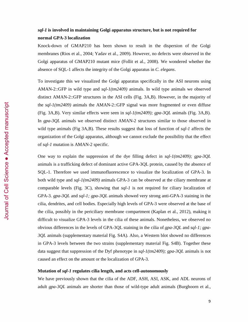
Figure legends
Fig. 1. Identification of sql-1.
(A) DiO dye filling of wild type, gpa-3QL, and sql-1(gj202); gpa-3QL animals. The dye
filling defect of gpa-3QL animals is suppressed in sql-1(gj202); gpa3QL. DiO dye filling
stains 6 pairs of amphid neurons. The cell bodies (indicated with arrowheads) and dendrites
(indicated with an arrow) of some of these are in the focal plane of this picture. Anterior is
towards the top. Outlines of the worms are indicated with a dotted line. Scale bar, 10 μm. (B)
Schematic representation of the sql-1 gene structure. Coding exons are indicated as colored
boxes. The coiled-coil region is shown in grey, the GRAB domain in blue, the GA-1 domain
in red, and regions with no predicted domains in black. The position of the missense mutation
in sql-1(gj202) animals is indicated with an arrow, and the deletions in sql-1(tm2409), sql-
1(tm2440), and sql-1(tm4995) are indicated with black bars. (C) Immunoblotting of whole
worm lysates collected from wild type, sql-1(tm2440), and sql-1(tm2409) animals, with the
antibody raised against the N-terminal half of SQL-1. * marks wild type SQL-1, ** marks
truncated SQL-1 in tm2440 animals.
Fig. 2. SQL-1 localizes to the Golgi apparatus.
(A) Representation of C. elegans indicating the location of the amphid sensory neurons
(adapted from (Inglis et al., 2007)) in green, the region where their cilia are in red and the
regions depicted in B-D in boxed areas. (B) Immunofluorescence images of an arbitrary
region of the middle of wild type animals expressing SQL-1::GFP (green) stained with anti-
SQV-8 (red) to mark the Golgi apparatus. Outline of the worm is indicated with a dotted line.
Scale bar, 10 μm. Arrowheads indicate examples of SQL-1::GFP and SQV-8 co-localization.
(C) Immunofluorescence images of the head of a wild type animal expressing the Golgi
marker AMAN-2::YFP (green) in the ASI neurons stained with anti-SQL-1 (red). Insets show
enlargements of the region containing the cell body, which is indicated with a solid line.
Anterior is towards the top. Outline of the worm is indicated with a dotted line. Scale bar, 10
μm. (D) Fluorescence images of a pgpa-4::sql-1::gfp animal. Image shows the cell body and a
part of the dendrite, the location of the cilium in the tip of the head is outside the image.
Immobile SQL-1 particles in the dendrite (indicated with a thin dotted line) are indicated with
an arrowhead. Anterior is towards the left. Scale bar, 10 μm.
Jour
nal o
f Cel
l Sci
ence
Acc
epte
d m
anus
crip
t

28
Fig. 3. SQL-1 is required to maintain Golgi organization.
(A) Percentage of wild type, sql-1(tm2409), gpa-3QL and sql-1(tm2409); gpa-3QL animals
that showed distinct AMAN-2::GFP structures (distinct), fragmentation of the structures
(fragmented), or diffuse fluorescence (diffuse). The Golgi apparatus was visualized in the ASI
neurons using AMAN-2::GFP. (B) Cell bodies of the ASI neurons of wild type, sql-
1(tm2409), gpa-3QL and sql-1(tm2409); gpa-3QL animals expressing AMAN-2::YFP.
Outline of the cells and nuclei are indicated with dotted lines. Scale bar, 5 μm. (C)
Immunofluorescence images of the tip of the head of wild type and sql-1(tm2409) animals
stained with anti-GPA-3. Sensory cilia are indicated with a bracket. Anterior is towards the
left. Outlines of the worms are indicated with a dotted line. Scale bar, 5 μm.
Fig. 4. sql-1 regulates cilia length.
Average length of sensory cilia of the ASI neurons (A), and ASH, ASK, ADL neurons (B) in
various genetic backgrounds. Statistically significant differences (p < 0.001) compared to cilia
length in wild type animals are indicated in black, compared to cilia length in gpa-3QL
animals in red, compared to cilia length in sql-1; gpa-3QL animals in green, and compared to
cilia length in osm-3 animals in blue. Statistical analysis was performed using an ANOVA,
followed by a Bonferroni post hoc test. Error bars indicate SD.
Fig. 5. Proposed model of IFT in the cilia of sql-1 mutant animals.
(A) In wild type animals IFT particles in the middle segment of the cilia are transported by
both OSM-3 and kinesin-II. (B) In sql-1 mutant animals IFT particles are destabilized,
resulting in the existence of two additional types of IFT particles: A slow particle composed
of only kinesin-II and a fast particle composed of incomplete complex A (with at least CHE-
11), incomplete complex B (with at least OSM-1 and IFT-20), and dynein, and transported by
OSM-3. Bellow the schematic model the presence (+) or absence (-) of the GFP markers in
the proposed IFT particles present in sql-1(tm2409) animals are shown, as well as the
measured velocities of the tested GFP markers in the middle segments of sql-1(tm2409)
animals (Table 1).
Jour
nal o
f Cel
l Sci
ence
Acc
epte
d m
anus
crip
t

29
Fig. 6. OSM-3::GFP speed is affected in sql-1; gpa-3QL double mutant.
Average velocity of OSM-3::GFP in kap-1, kap-1; sql-1, kap-1; gpa-3QL and kap-1; sql-1;
gpa-3QL animals. Statistically significant differences (p < 0.001) compared to kap-1 animals
are indicated in black, compared to kap-1; sql-1 animals in red and compared to kap-1; gpa-
3QL in green. Statistical analysis was performed using an ANOVA, followed by a Bonferroni
post hoc test. Error bars indicate SD.
Table 1: sql-1 affects anterograde IFT velocities in the middle segment of C. elegans’
cilia.
Average anterograde IFT velocities (in μm/s) ± standard deviation (sd) of KAP-1::GFP,
OSM-3::GFP, CHE-11::GFP, DAF-10::GFP, OSM-1::GFP, CHE-13::GFP, IFT-20::GFP and
XBX-1::GFP in wild type and mutant backgrounds. Velocities annotated with * were
previously described in (Burghoorn et al., 2010). Statistically significant differences (P <
0.001) compared to IFT velocities in wild type animals are indicated with a, compared to gpa-
3(lf) animals with b, compared to sql-1(tm2409) animals with c, compared to gpa-3QL animals
with d, compared to kap-1; sql-1(tm2409) animals with e, compared to kap-1; sql-1(tm2409)
animals with f, and compared to kap-1; gpa-3QL with g. Statistically significant differences (P
< 0.001) of IFT velocities of each of the GFP markers compared to the velocities of KAP-
1::GFP and OSM-3::GFP in the same mutant background are indicated with 1 and 2,
respectively. Statistical analysis was performed using an ANOVA, followed by a Bonferroni
post hoc test. Abbreviation: n, number of IFT particles measured. Jour
nal o
f Cel
l Sci
ence
Acc
epte
d m
anus
crip
t
Jour
nal o
f Cel
l Sci
ence
Acc
epte
d m
anus
crip
t

Jour
nal o
f Cel
l Sci
ence
Acc
epte
d m
anus
crip
t
Jour
nal o
f Cel
l Sci
ence
Acc
epte
d m
anus
crip
t
Jour
nal o
f Cel
l Sci
ence
Acc
epte
d m
anus
crip
t
Jour
nal o
f Cel
l Sci
ence
Acc
epte
d m
anus
crip
t

Jour
nal o
f Cel
l Sci
ence
Acc
epte
d m
anus
crip
t
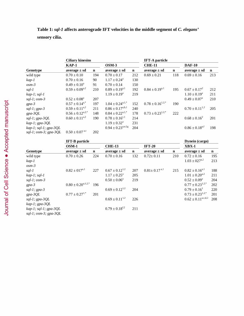
Table 1: sql-1 affects anterograde IFT velocities in the middle segment of C. elegans’
sensory cilia.
Ciliary kinesins IFT-A particle KAP-1 OSM-3 CHE-11 DAF-10 Genotype average ± sd n average ± sd n average ± sd n average ± sd n wild type 0.70 ± 0.10 194 0.70 ± 0.17 212 0.69 ± 0.21 118 0.69 ± 0.16 213 kap-1 0.70 ± 0.16 90 1.17 ± 0.24a 130 osm-3 0.49 ± 0.10a 91 0.70 ± 0.14 150 sql-1 0.59 ± 0.09a,2 210 0.89 ± 0.19a,1 192 0.84 ± 0.19a,1 195 0.67 ± 0.172 212 kap-1; sql-1 1.19 ± 0.19a 219 1.10 ± 0.19a 211 sql-1; osm-3 0.52 ± 0.08a 207 0.49 ± 0.07a 210 gpa-3 0.57 ± 0.14a,* 197 1.04 ± 0.24a,1,* 152 0.78 ± 0.161,2,* 190 sql-1; gpa-3 0.59 ± 0.11a,2 211 0.86 ± 0.17a,b,1 240 0.70 ± 0.111,2 205 gpa-3QL 0.56 ± 0.12a,2,* 148 0.84 ± 0.22a,1,* 178 0.73 ± 0.231,2,* 222 sql-1; gpa-3QL 0.60 ± 0.11a,2 190 0.78 ± 0.16c,1 214 0.68 ± 0.162 201 kap-1; gpa-3QL 1.19 ± 0.32a 231 kap-1; sql-1; gpa-3QL 0.94 ± 0.23a,e,f,g 204 0.86 ± 0.18a,f 198 sql-1; osm-3; gpa-3QL 0.50 ± 0.07 a,c 202 IFT-B particle Dynein (cargo) OSM-1 CHE-13 IFT-20 XBX-1 Genotype average ± sd n average ± sd n average ± sd n average ± sd n wild type 0.70 ± 0.26 224 0.70 ± 0.16 132 0.72± 0.11 210 0.72 ± 0.16 195 kap-1 1.03 ± 027a,2 213 osm-3 sql-1 0.82 ± 017a,1 227 0.67 ± 0.121,2 207 0.81± 0.17 a,1 215 0.82 ± 0.16a,1 188 kap-1; sql-1 1.17 ± 0.25a 205 1.01 ± 0.20a,2 211 sql-1; osm-3 0.50 ± 0.06a 219 0.52 ± 0.09a 204 gpa-3 0.80 ± 0.20a,1,2,* 196 0.77 ± 0.231,2,* 202 sql-1; gpa-3 0.69 ± 0.121,2 204 0.79 ± 0.161 220 gpa-3QL 0.77 ± 0.271,* 201 0.73 ± 0.231,2,* 201 sql-1; gpa-3QL 0.69 ± 0.111,2 226 0.62 ± 0.11a,c,d,2 208 kap-1; gpa-3QL kap-1; sql-1; gpa-3QL 0.79 ± 0.18f,2 211 sql-1; osm-3; gpa-3QL
Jour
nal o
f Cel
l Sci
ence
Acc
epte
d m
anus
crip
t