sr mcp system metacarpophalangeal arthroplastyaz621074.vo.msecnd.net/.../srmcp_st_1_en.pdfsr mcp...
TRANSCRIPT

Operative technique
SR MCP SystemMetacarpophalangeal
Arthroplasty

2
SR MCP System Metacarpophalangeal Arthroplasty | Operative technique
This publication sets forth detailed recommended procedures for using Stryker devices and instruments.
It offers guidance that you should heed, but, as with any such technical guide, each surgeon must consider the particular needs of each patient and make appropriate adjustments when and as required.
A workshop training is recommended prior to performing your first surgery.
All non-sterile devices must be cleaned and sterilized before use. Multicomponent instruments must be disassembled for cleaning. Please refer to the corresponding assembly / disassembly instructions.
Please remember that the compatibility of different product systems have not been tested unless specified otherwise in the product labeling (Instruments and Sizers V15106).
See package insert (Instructions for Use V15115) for a complete list of potential adverse effects, contraindications, warnings and precautions. The surgeon must discuss all relevant risks including the finite lifetime of the device with the patient when necessary.
SR MCP SystemMetacarpophalangeal Arthroplasty
Contents
1. Pre-operative planning and assessment . . . . . . . . . . 3
2. Indications and contraindications . . . . . . . . . . . . 4
3. Surgical procedure . . . . . . . . . . . . . . . . . . . . . . . . 6 Incision options. . . . . . . . . . . . . . . . . . . . . . . . . . . . 7 Capsular exposure . . . . . . . . . . . . . . . . . . . . . . . . . 7 Dorsal synovectomy . . . . . . . . . . . . . . . . . . . . . . . . 7 Metacarpal articular head resection . . . . . . . . . . . 8 Proximal phalanx articular surface resection . . . 8 Trial preparation . . . . . . . . . . . . . . . . . . . . . . . . . . 9 Trial placement. . . . . . . . . . . . . . . . . . . . . . . . . . . 10 Trial reduction / implant placement . . . . . . . . . . 10 Implanting the components . . . . . . . . . . . . . . . . . 11 Closure and centralization . . . . . . . . . . . . . . . . . . 11 Initial post operative treatment . . . . . . . . . . . . . 12 Rehabilitation . . . . . . . . . . . . . . . . . . . . . . . . . . . . 12 Revision or removal of MCP Prosthesis(es) . . . . . 13 Crossed intrinsic transfer procedure . . . . . . . . . 13

3
Operative technique | SR MCP System Metacarpophalangeal Arthroplasty
Pre-operative planning and assessment
Record the range of motion (ROM) for all the joints of the hand and wrist and other significant joints. Static deformities, grip strength and pinch strength should also be assessed and recorded.
X-Ray examination includes AP, lateral and such special views as may be indicated initially at surgery and at follow up intervals. Sizing templates with a 3% parallax enlargement are available. Follow-up examinations may include repetition of the above measurement as well as evaluation of positioning, alignment, cement mantle adequacy and evidence of loosening or stem erosion into bone.
Assessment of MCP palmar subluxation, ulnar drift, ulnar intrinsic contracture, extrinsic extensor tendon displacement, swan neck or boutonniere deformity, and flexor tendon displacement should be recorded.
Ulnar translation extensor
Contracture ulnar lateral band and sagittal band
Typical rheumatoid deformity
Normal
NormalTypical rheumatoid deformity
3

4
SR MCP System Metacarpophalangeal Arthroplasty | Operative technique
Indications and contraindications
IndicationsThe SR MCP system is indicated for use in arthroplasty of the MCP Joint when either the:
• Patient is in need of a revision of a failed MCP Prosthesis(es) or
• Patient expects to place his / her hands under loading situations which preclude the use of an alternative implant in the painful osteo-arthritic and post-traumatic arthritic MCP Joint
Unless otherwise specified, the Stryker GmbH non-active devices have not been evaluated for safety and compatibility in the MR environment. It has not been tested for heating, migration, or image artifact in the MR environment. The safety of the non-active implants in the MR environment is unknown. Scanning a patient who has this device may result in patient injury.
Contraindications• Bone, musculature, tendons, or adjacent soft
tissue compromised by disease, infection, or prior implantation, which cannot provide adequate support or fixation for the prosthesis
• Infection
• Skeletal immaturity
Caution Federal (United States) law restricts this device to sale, distribution, and use by or on the order of a physician.
Humanitarian use device The SR MCP is authorized by Federal law for use in the arthroplasty of the MCP joint when either the:
• Patient is in need of a revision of a failed MCP Prosthesis(es) or
• Patient expects to place his/her hands under loading situations which preclude the use of an alternative implant in the painful osteo-arthritic and post-traumatic arthritic MCP Joint
The effectiveness of this device for this use has not been demonstrated.
Please see package insert for Warnings, Precautions, Adverse Effects, and other essential product information.
Note: Intended for cement use only.

5
Operative technique | SR MCP System Metacarpophalangeal Arthroplasty
Warnings(See also the Patient Counseling Information)
• Patients should be made aware of the increased potential for device failure when excessive demands are made upon it. Strenuous loading, excessive mobility, and articular instability all may lead to accelerated wear and eventual failure by loosening, fracture, or dislocation of the device.
• Single use is defined as use of one implant or instrument on a single patient in a single surgical procedure.
• Reuse of instruments designated as single use has been associated with necrosis of bone leading to implant failure. It may also lead to sepsis and / or communication of potentially lethal viruses.
• Reuse of implants designated as single use has been associated with sepsis and / or communication of potentially lethal viruses.
Precautions• Do not resterilize. The implant is provided sterile
in an undamaged package. If either the implant or the package appears damaged, expiration date has been exceeded, or if sterility is questioned for any reason, the implant should not be used.
• Meticulous preparation of the implant site and selection of the proper size implant increase the potential for a successful outcome.
• The implant should be removed from its sterile package only after the implant site has been prepared and properly sized.
• Implants should be handled with blunt instruments to avoid scratching, cutting, or nicking the device.
Patient counseling information(See also Warnings)
A patient brochure is available for use in counseling the patient.
In addition to the patient related information contained in the Warnings and Adverse Events sections, the following information should be conveyed to the patient:
• While the expected life of total joint replacement components is difficult to estimate, it is finite. These components are made of foreign materials, which are placed within the body for the potential restoration of mobility or reduction of pain. However, due to the many biological, mechanical and physiochemical factors which affect these devices, the components cannot be expected to withstand the activity level and loads of normal healthy bone for an unlimited period of time.
• Adverse effects of this device may necessitate reoperation, revision, or fusion of the involved joint.
Indications and contraindications

6
Surgical procedure
7
Operative technique | SR MCP System Metacarpophalangeal Arthroplasty
Fig. 2
Distended dorsal capsule
Radial sagittal band
Attenuated collateral ligaments
Extensor tendon and ulnar sagittal band
Joint excisional lines
Fig. 1
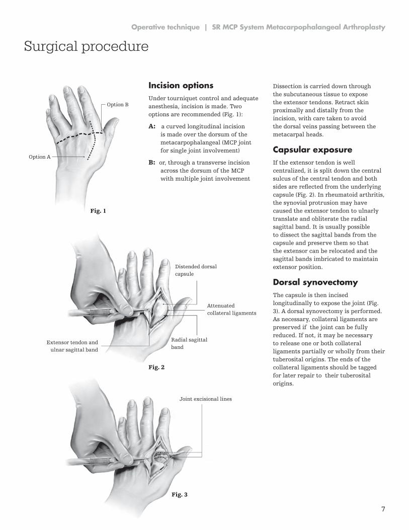
Incision optionsUnder tourniquet control and adequate anesthesia, incision is made. Two options are recommended (Fig. 1):
A: a curved longitudinal incision is made over the dorsum of the metacarpophalangeal (MCP joint for single joint involvement)
B: or, through a transverse incision across the dorsum of the MCP with multiple joint involvement
Dissection is carried down through the subcutaneous tissue to expose the extensor tendons. Retract skin proximally and distally from the incision, with care taken to avoid the dorsal veins passing between the metacarpal heads.
Capsular exposureIf the extensor tendon is well centralized, it is split down the central sulcus of the central tendon and both sides are reflected from the underlying capsule (Fig. 2). In rheumatoid arthritis, the synovial protrusion may have caused the extensor tendon to ulnarly translate and obliterate the radial sagittal band. It is usually possible to dissect the sagittal bands from the capsule and preserve them so that the extensor can be relocated and the sagittal bands imbricated to maintain extensor position.
Dorsal synovectomyThe capsule is then incised longitudinally to expose the joint (Fig. 3). A dorsal synovectomy is performed. As necessary, collateral ligaments are preserved if the joint can be fully reduced. If not, it may be necessary to release one or both collateral ligaments partially or wholly from their tuberosital origins. The ends of the collateral ligaments should be tagged for later repair to their tuberosital origins.
Surgical procedure
Option B
Option A
Fig. 3

8
SR MCP System Metacarpophalangeal Arthroplasty | Operative technique
Metacarpal articular head resectionThe metacarpal head is removed first by a vertical saw cut distal to the collateral ligaments. A second cut at 45º proximovolarly removes the remainder of the metacarpal head (Fig. 4A & 4B). Retain ligamentous integrity as much as possible.
Proximal phalanx articular surface resectionRemove the base of the proximal phalanx with a minimal slice (Fig. 5). This is to preserve the collateral ligamentous attachments in so far as possible. Marked contracture of the ulnar capsule may require detachment of the ulnar collateral ligament to assure alignment of the finger to the metacarpal.
Fig. 4A
Fig. 4B
Fig. 5
Tuberosity Joint incisional lines
Surgical procedure

9
Operative technique | SR MCP System Metacarpophalangeal Arthroplasty
Trial preparationAn awl is then inserted in the dorsal aspect of the intramedullary canal of the metacarpal (Fig. 6A). This is followed by sequential broaching until proper fit has been attained. For the index and long finger the intramedullary preparation is ulnarly displaced slightly (Fig. 6B).
This may provide a better moment arm for the radial intrinsic and extrinsic tendons to compensate for the ulnar drift tendency. The same awl and broach procedure is then followed for the proximal phalanx (Fig. 6C).
Awl
Broach
Joint removedFig. 6A
Fig. 6B
Fig. 6C
Surgical procedure

10
SR MCP System Metacarpophalangeal Arthroplasty | Operative technique
Trial placementAn impactor with a concave surface aids insertion and optimal proximal displacement of the proximal trial component (Fig. 7A). The tip of the prostheses should pass the midpoint of the metacarpal.
Avoid impaction with metallic instruments which may damage the articular surfaces of the trial components.
A convex impactor is also provided to aid insertion and seating of the distal component (Fig. 7B). Once the trial components are inserted, check for fit and position, as well as position under the image intensifier.
Trial reduction / implant preparationThe joint is reduced with both the metacarpal and phalangeal trials in place. Static position, ease of passive motion, plane of motion to full extension, and stability is checked. With multiple joints, comparison between finger alignment is checked with the trials in place. Revision trimming may be necessary to correct muscle or soft tissue tightness, and to insure a full range of motion.
If release of the collateral ligaments was previously required, two holes are drilled through the tuberosity at the dorsal lateral and dorsal medial aspect of the remaining metacarpal head for reattachment or imbrication of the ligaments (Fig. 8). Sutures for repair of the collateral ligament (4-0 Ticron /mersilene) are inserted.
When satisfied with preliminary appearances, the trials are removed using the trial extractor provided in the instrument tray.
Fig. 7A
Fig. 7B
Fig. 8
Surgical procedure

11
Operative technique | SR MCP System Metacarpophalangeal Arthroplasty
Implanting the components The intramedullary canal is then irrigated first with saline, then with 0.5% neomycin solution. The medullary cavities are dried. Both the metacarpal and phalangeal prostheses are removed from their sterile packaging and inspected. Polymethylmethacrylate adhesive (PMMA) is injected in a liquid state using a size #14 plastic intracath attached to a 10cc syringe (Fig. 9).
The distal component is inserted first. Care is taken to position the prosthetic components in proper alignment and rotation. Convex and concave plastic impactors are provided to assist implant insertion.
Avoid impaction with metallic instruments which, by compromising the surface finish, may accelerate prosthetic wear.
The joint is then extended and checked under the image intensifier before the cement hardens. Cement fixation of one finger at a time is advisable if the positioning is difficult. If multiple joints are done, it may be easier to do the distal components as a group followed by the proximal components. After the cement has cured, passive range of motion is checked to ensure a full range of motion without impingement of prosthetic binding. If satisfactory, the collateral ligaments are tightened or reattached to the tuberosity of the metacarpal head.
Closure and centralizationCapsular closure is undertaken after prosthetic fixation. Centralization of the extensor tendon and imbrication of the radial sagittal bands is usually necessary in rheumatoid hands by appropriate closure of the extensor tendon retinacular system. A “pants-over-vest” centralization may be required in cases of moderate - severe ulnar drift along with crossed -
intrinsic transfer. Snug repair of the radial collateral ligament helps to also realign the flexor assemblage (Fig. 8). Reattach radial collateral ligament to the metacarpal bone (Fig. l0A). Realign the finger before tightening sutures. With finger held in slight over correction, imbricate radial sagittal band over extensor tendon (Fig. 10B). Complete joint closure by securing radial sagittal band to the extensor apparatus (Fig. 10C).
Fig. 9
Fig. 10A Fig. 10B
Fig. 10C
Radial collateral ligament tightened
Surgical procedure

12
SR MCP System Metacarpophalangeal Arthroplasty | Operative technique
Fig. 11
Fig. 12
Pad for dorsal displacement of proximal phalanges
Support pad for flexion PIP
Pad to provide counter support
Palmar support for wrist
Velcro straps
Thermoplastic cockup splint
Set washer to adjust radial pull
Rubber band tensioner
Fish line
Eyelets
Pad to provide counter support
Coat hanger support for pulleys
Polythene pully
Leatherette slings
12
Initial post-operative treatmentPost-operative positioning is important and should place the MCP joints in slight flexion and the PIP joints in approximately 45° flexion. lf there has been preoperative ulnar deviation, or this appears following closure the fingers should be placed in 5 to 10° radial deviation. These positions must be held while applying a compressive dressing (Fig. 11).
If there has been a tendency for the fourth and fifth metacarpals to ‘droop’ the excessive metacarpal arch curvature should also be supported. The dressing may be removed at 2 to 4 days and a dynamic splint may be applied for daytime exercises. A static rest / nocturnal splint capable of holding the fingers in the corrected positions may be used at other periods for 4 to 6 weeks.
Rehabilitation The initial rehabilitation program may benefit greatly from close supervision of the physiatrist and hand therapist. Expert knowledge is recommended for the fabrication of the dynamic and static splints for correct fit as well as instruction in proper use (Fig. 12). The first week or two may best be carried out with daily or bi-daily supervision followed with repeat reassessment at the scheduled visits.
Follow-up exams should include range of motion (ROM) assessment for all the joints of the hand and wrist and other significant joints. Static deformities, grip strength and pinch strength should also be assessed and recorded. X-ray examination includes AP, lateral and such special views as may also be indicated at follow-up intervals. Assessment of MCP palmar subluxation, ulnar drift, ulnar intrinsic contracture, extrinsic extensor tendon displacement,
swan neck or boutonniere deformity, and flexor tendon displacement should also be recorded.
Low profile dynamic sling Counteracts palmer and ulnar subluxations tendencies
Surgical procedure

13
Operative technique | SR MCP System Metacarpophalangeal Arthroplasty
13
Revision or removal of MCP prostheses Should the SR MCP implants loosen, dislocate, suffer infection, or other complication requiring removal, the procedure involves retrieval of the implant under sterile conditions, tourniquet control and adequate anesthesia. The previous dorsal incision (straight or curvilinear) is incised. The soft tissue flaps are reflected down to the extensor tendon system. The extensor tendon is incised longitudinally on the radial side from the proximal phalanx base across the metacarpal head and neck. The sagittal bands are reflected from the capsule (if possible). The capsule of the joint is incised longitudinally and the implants are exposed. Release of collateral ligaments might be necessary to retrieve the implants. The metacarpal prosthesis is removed first. Small 1-2mm osteotomes are used to free the prosthesis and bone cement from within the metacarpal canal. A linear osteotomy of the metacarpal canal may be needed to remove the implant. A fine burr on a high-speed drill is used to remove the cement over the dorsal surface of the proximal component. The distal component may also be drilled out with the high speed burr. The phalangeal components can be tapped from the intramedullary canals or removed with an osteotome.
Salvaging of the joint is by soft tissue volar plate interposition or alternatively silicone interposition joint (no sepsis). Collateral ligament reconstruction and extensor tendon centralization should be performed in revision procedures similar to the index prosthetic insertion procedure. Postoperative physiotherapy is unchanged from other procedures of MCP arthroplasty.
Crossed intrinsic transfer procedureCrossed intrinsic transfers are often helpful in correcting ulnar deviation. In rheumatoid hands with ulnar capsular contracture it may be necessary to release the ulnar intrinsic and all or part of the ulnar collateral ligament, especially if there is swan neck deformity (Fig. A). Release of the ulnar intrinsic lateral band insertion midway onto the proximal phalanx allows transfer onto the radial aspect of the adjacent digit at the close of the soft tissue rebalancing (Fig. B). First palmar interosseous to radial
collateral alignment 3rd finger
Release ulnar lateral band
Radial collateral ligament tightened
Realignment of extensor and flexor tendons
Fig. A
Ulnar with deviation with contracture ulnar soft tissue
Normal
Fig. B
Surgical procedure

Manufacturer:
Stryker GmbH Bohnackerweg 1 2545 Selzach Switzerland
stryker.com
This document is intended solely for the use of healthcare professionals. A surgeon must always rely on his or her own professional clinical judgment when deciding whether to use a particular product when treating a particular patient. Stryker does not dispense medical advice and recommends that surgeons be trained in the use of any particular product before using it in surgery.
The information presented is intended to demonstrate a Stryker product. A surgeon must always refer to the package insert, product label and/or instructions for use, including the instructions for cleaning and sterilization (if applicable), before using any Stryker product. Products may not be available in all markets because product availability is subject to the regulatory and/or medical practices in individual markets. Please contact your Stryker representative if you have questions about the availability of Stryker products in your area.
Stryker Corporation or its affiliates own, use, or have applied for the following trademarks or service marks: Stryker. All other trademarks are trademarks of their respective owners or holders.
SRMCP-ST-1, Rev 2, 7-2016 Copyright © 2016 Stryker
Trauma & Extremities