stability assessment of the ankle mortise in supination- external rotation-type ankle fractures:...
TRANSCRIPT
Stability Assessment of the Ankle Mortise in Supination-External Rotation-Type Ankle Fractures: Lack of Additional
Diagnostic Value of MRI
by Simo Nortunen, Sannamari Lepojärvi, Olli Savola, Jaakko Niinimäki, Pasi Ohtonen, Tapio Flinkkilä, Iikka Lantto, Tero Kortekangas, and Harri Pakarinen
J Bone Joint Surg AmVolume 96(22):1855-1862
November 19, 2014
©2014 by The Journal of Bone and Joint Surgery, Inc.
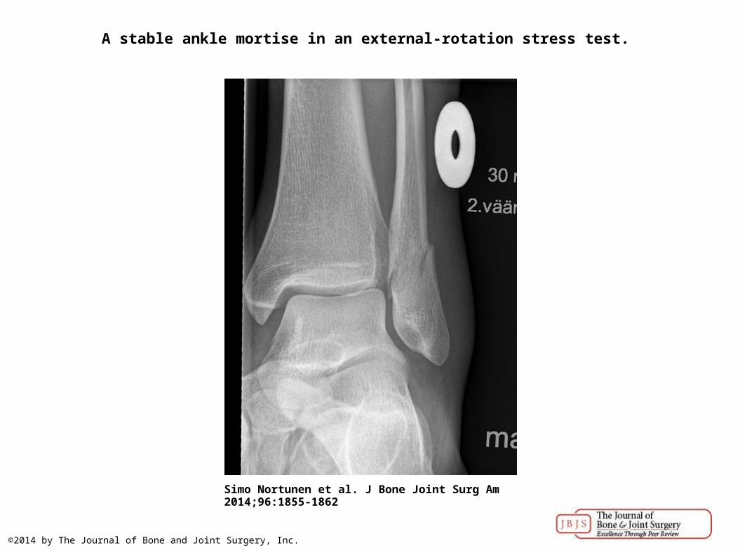
A stable ankle mortise in an external-rotation stress test.
Simo Nortunen et al. J Bone Joint Surg Am 2014;96:1855-1862
©2014 by The Journal of Bone and Joint Surgery, Inc.
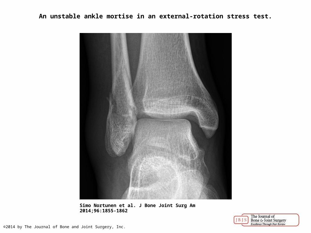
An unstable ankle mortise in an external-rotation stress test.
Simo Nortunen et al. J Bone Joint Surg Am 2014;96:1855-1862
©2014 by The Journal of Bone and Joint Surgery, Inc.

Coronal intermediate-weighted 3.0-T MRI of a normal deep deltoid ligament (white arrow) in a patient who did not participate in the study.
Simo Nortunen et al. J Bone Joint Surg Am 2014;96:1855-1862
©2014 by The Journal of Bone and Joint Surgery, Inc.
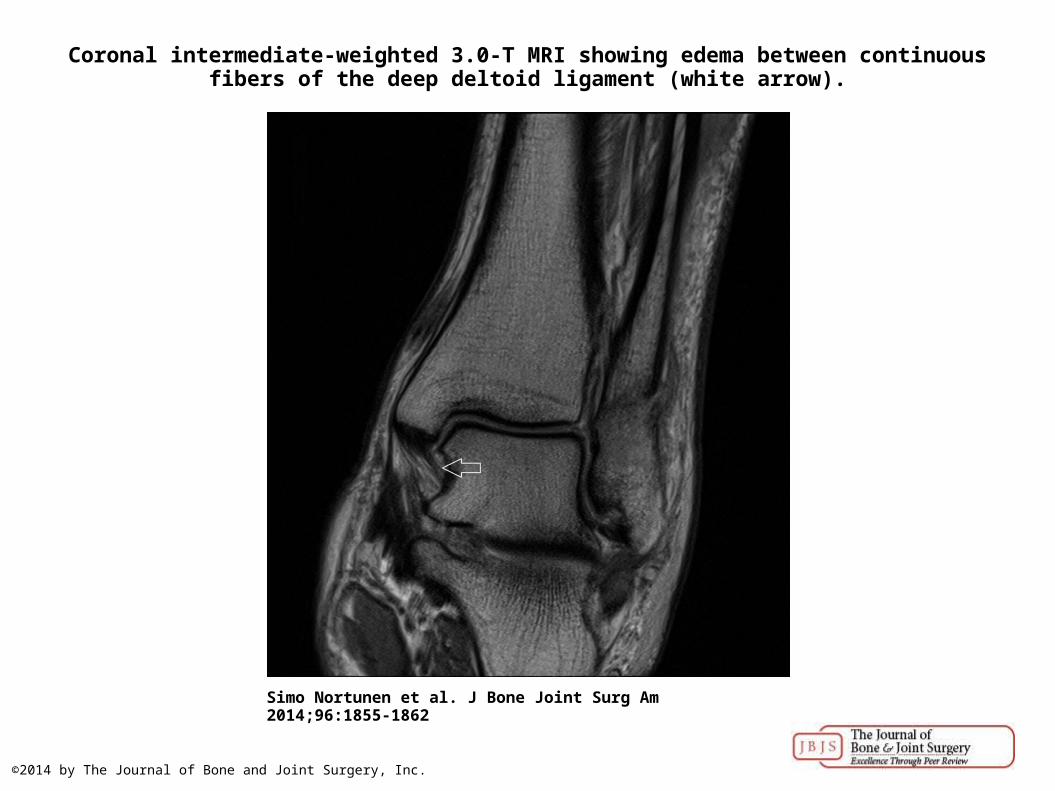
Coronal intermediate-weighted 3.0-T MRI showing edema between continuous fibers of the deep deltoid ligament (white arrow).
Simo Nortunen et al. J Bone Joint Surg Am 2014;96:1855-1862
©2014 by The Journal of Bone and Joint Surgery, Inc.

Coronal intermediate-weighted 3.0-T MRI showing a partial tear of the deep deltoid ligament; fluid signal transects some of the ligament fibers (white arrow).
Simo Nortunen et al. J Bone Joint Surg Am 2014;96:1855-1862
©2014 by The Journal of Bone and Joint Surgery, Inc.

Coronal intermediate-weighted 3.0-T MRI showing a complete tear of the deep deltoid ligament (white arrow).
Simo Nortunen et al. J Bone Joint Surg Am 2014;96:1855-1862
©2014 by The Journal of Bone and Joint Surgery, Inc.
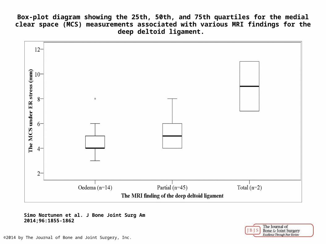
Box-plot diagram showing the 25th, 50th, and 75th quartiles for the medial clear space (MCS) measurements associated with various MRI findings for the deep deltoid ligament.
Simo Nortunen et al. J Bone Joint Surg Am 2014;96:1855-1862
©2014 by The Journal of Bone and Joint Surgery, Inc.