standard operating procedures for the collection and
TRANSCRIPT
Standard Operating Procedures
for the Collection and Analysis of Algae
June 2016 (Version 2.0)
Prepared by:
NORTH CAROLINA DEPARTMENT OF ENVIRONMENTAL QUALITY
Division of Water Resources
Water Sciences Section
Ecosystems Branch
Dow
nloaded from http://m
eridian.allenpress.com/jfw
m/article-supplem
ent/416025/pdf/10_3996082016-jfwm
-068_s16 by guest on 17 Decem
ber 2021
Dow
nloaded from http://m
eridian.allenpress.com/jfw
m/article-supplem
ent/416025/pdf/10_3996082016-jfwm
-068_s16 by guest on 17 Decem
ber 2021
Page | i
REVIEW AND REVISION LOG
Date Edited
Editor Version Edited
Section Edited Changes/updates
January 2003 Mark Vander Borgh and
Elizabeth Fensin 1.0 Original Original SOP
March 2016 Jen Schmitz 2.0 Cover Changed title, added photo, changed
department contacts
March 2016 Jen Schmitz 2.0 Entire Document Updated hyperlinks and organization names
throughout
March 2016 Jen Schmitz 2.0 Entire Document Reformatted and updated content
March 2016 Jen Schmitz 2.0 Section 1 Merged “Purpose” and Section 1:
Introduction into Section 1.0
March 2016 Jen Schmitz 2.0 Document Added Section 2.0 “Sample Analysis
Considerations”
March 2016 Jen Schmitz 2.0 Section 2 Changed section title to “Sample Processing
Procedure” and moved to Section 3.0
March 2016 Jen Schmitz 2.0 Section 3
Changed section title to “Laboratory
Techniques and Analysis” and moved to
Section 4.0
March 2016 Jen Schmitz 2.0 Section 4 Changed section title to “Data Handling and
Interpretation” and moved to Section 5.0
March 2016 Jen Schmitz 2.0 Section 5 Incorporated previous Section 5 into Sections
2.0 and 3.0
March 2016 Jen Schmitz 2.0 Section 6 Incorporated previous Section 6 and other
material into Section 2.2
March 2016 Jen Schmitz 2.0 Section 7 Moved to Section 6.0
March 2016 Jen Schmitz 2.0 Section 8 Removed
March 2016 Jen Schmitz 2.0 Section 9 Renumbered as Section 7.0
March 2016 Jen Schmitz 2.0 Appendix I Merged Appendix 1a and 1b into one form,
updated Fish Kill Form, renamed Appendix A
March 2016 Jen Schmitz 2.0 Appendix II Removed Appendix II
March 2016 Jen Schmitz 2.0 Appendix III Updated information, renamed Appendix B
March 2016 Jen Schmitz 2.0 Appendix IV Updated information, renamed Appendix C
March 2016 Jen Schmitz 2.0 Appendix V Removed Appendix V
March 2016 Jen Schmitz 2.0 Appendix VI Renamed Appendix E
March 2016 Jen Schmitz 2.0 Appendix VII Updated information, renamed Appendix D
Dow
nloaded from http://m
eridian.allenpress.com/jfw
m/article-supplem
ent/416025/pdf/10_3996082016-jfwm
-068_s16 by guest on 17 Decem
ber 2021
Page | ii
1.0 INTRODUCTION AND PURPOSE ................................................................................................................. 1
2.0 SAMPLE ANALYSIS CONSIDERATIONS ........................................................................................................ 2
2.1 SUMMARY OF THE METHOD ................................................................................................................................ 2 2.2 HEALTH AND SAFETY .......................................................................................................................................... 2 2.3 PERSONNEL QUALIFICATIONS .............................................................................................................................. 2 2.4 MICROSCOPY ................................................................................................................................................... 3
2.4.1 Microscopes ............................................................................................................................................. 3 2.4.2 Microscope Maintenance ........................................................................................................................ 3 2.4.3 Microscope Calibration and Standardization ........................................................................................... 3
3.0 SAMPLE PROCESSING PROCEDURE ............................................................................................................ 5
3.1 SAMPLE METADATA .......................................................................................................................................... 5 3.1.1 Sampling Methods ................................................................................................................................... 5 3.1.2 Sample Type ............................................................................................................................................. 5 3.1.3 Environmental Conditions ........................................................................................................................ 5
3.2 QUANTIFYING ALGAL BLOOMS ............................................................................................................................. 5 3.2.1 Chlorophyll-a Measurements ................................................................................................................... 5 3.2.2 Unit Density ............................................................................................................................................. 6 3.2.3 Cell Density............................................................................................................................................... 6 3.2.4 Biovolume ................................................................................................................................................ 6
3.3 ALGAE AND FISH KILL INVESTIGATIONS .................................................................................................................. 6 3.4 NUISANCE GROWTHS ......................................................................................................................................... 6 3.5 EPISODIC EVENTS .............................................................................................................................................. 6 3.6 SAMPLE HANDLING ........................................................................................................................................... 7 3.7 SAMPLE CONTAINERS ......................................................................................................................................... 7 3.8 SAMPLE PRESERVATION ...................................................................................................................................... 7 3.9 SAMPLE QUANTITY ............................................................................................................................................ 7 3.10 SAMPLE LABELING ............................................................................................................................................. 8 3.11 SAMPLE SHIPPING ............................................................................................................................................. 8 3.12 SAMPLE RECEIVING ........................................................................................................................................... 8 3.13 SAMPLE STORAGE ............................................................................................................................................. 9
3.13.1 Short-term sample handling ................................................................................................................ 9 3.13.2 Long-term sample handling ................................................................................................................. 9
3.14 SAMPLE DATA MANAGEMENT ............................................................................................................................. 9 3.15 SAMPLE DATA MANAGEMENT FIELDS ................................................................................................................... 9
4.0 LABORATORY TECHNIQUES AND ANALYSIS ............................................................................................. 10
4.1 SAMPLE ANALYSIS ........................................................................................................................................... 10 4.2 SUBSAMPLE PREPARATION ................................................................................................................................ 10
4.2.1 Wet mount preparation ......................................................................................................................... 10 4.2.2 Utermöhl chamber preparation ............................................................................................................. 10
4.3 VISUAL SUBSAMPLE DENSITY AND DISTRIBUTION CHECK........................................................................................... 11 4.4 RECORDING DATA ............................................................................................................................................ 12 4.5 BENCH SHEETS ................................................................................................................................................ 12 4.6 RECORDING THE NUMBER OF ALGAL CELLS ............................................................................................................ 13 4.7 QUANTITATIVE ANALYSIS .................................................................................................................................. 13 4.8 QUALITATIVE ANALYSIS ..................................................................................................................................... 14
4.8.1 Identification only .................................................................................................................................. 14
Dow
nloaded from http://m
eridian.allenpress.com/jfw
m/article-supplem
ent/416025/pdf/10_3996082016-jfwm
-068_s16 by guest on 17 Decem
ber 2021
Page | iii
4.8.2 Dominant taxa analysis ......................................................................................................................... 14
5.0 DATA HANDLING AND INTERPRETATION ................................................................................................. 15
5.1 TAXONOMIC REFERENCE TABLE ......................................................................................................................... 15 5.1.1 Table Structure and Contents ................................................................................................................ 15 5.1.2 Taxonomic Revisions .............................................................................................................................. 17 5.1.3 Adding Taxa ........................................................................................................................................... 17
5.2 DATA ENTRY ................................................................................................................................................... 17 5.3 DENSITY CALCULATIONS .................................................................................................................................... 17
5.3.1 Individual unit density calculation ......................................................................................................... 18 5.3.2 Total unit density ................................................................................................................................... 18
5.4 REFERENCE BIOVOLUME CALCULATION ................................................................................................................ 19 5.4.1 Reference biovolume calculation worksheet ......................................................................................... 19 5.4.2 Calculation of cell reference biovolume ................................................................................................. 19
5.5 CALCULATION OF AN INDIVIDUAL TAXON’S BIOVOLUME ........................................................................................... 20 5.6 CALCULATION OF THE TOTAL BIOVOLUME (BV) ..................................................................................................... 21 5.7 RECORD RETENTION ........................................................................................................................................ 22
6.0 QUALITY ASSURANCE .............................................................................................................................. 23
6.1 TAXONOMIC IDENTIFICATIONS ........................................................................................................................... 23 6.2 DATA ENTRY .................................................................................................................................................. 23
7.0 REFERENCES ............................................................................................................................................ 24
Tables
Table 1. Example of Measurements and Areas of a Whipple Disc Grid ........................................................................ 3
Table 2. Example of Ocular Reticule Grid Divisions for the Leitz Laborlux S Compound Microscope ........................... 4
Table 3. Sample Quantity and Preservative Requirements ........................................................................................... 8
Table 4. The Ten Algal Groups Listed in the Taxonomic Reference Table ................................................................... 15
Figures
Figure 1. Example of a Cell Shape ................................................................................................................................ 20
Appendices Appendix A: Field Evaluation Forms ............................................................................................................................ 25
Appendix B: Lugol’s Solution Preparation .................................................................................................................. 28
Appendix C: Phytoplankton Bench Sheet .................................................................................................................... 29
Appendix D: Example of Biovolume Calculation Worksheet/New Taxa Record Form ................................................ 30
Appendix E: List of Biovolume Equations .................................................................................................................... 31
Dow
nloaded from http://m
eridian.allenpress.com/jfw
m/article-supplem
ent/416025/pdf/10_3996082016-jfwm
-068_s16 by guest on 17 Decem
ber 2021
Page | iv
ACRONYMS AND ABBREVIATIONS
BV (or bv) biovolume
Bv ref biovolume reference
ccm cubic centimeter
cm centimeter
DO dissolved oxygen
DWR Division of Water Resources
EB Ecosystems Branch
HAB harmful algal bloom
HUC hydrologic unit code
ISB Intensive Survey Branch
m meter
µg microgram
μS/cm microsiemens/centimeter
mL milliliter
mm millimeter
NADED North American Diatom Ecological Database
NCDENR North Carolina Department of Environment and Natural Resources
NCDEQ North Carolina Department of Environmental Quality
ppt parts per thousand
QA/QC Quality Assurance/Quality Control
SDS Safety Data Sheet
SOP standard operating procedure
SU standard units
TSN taxonomic serial number
USEPA United States Environmental Protection Agency
WSS Water Sciences Section
Dow
nloaded from http://m
eridian.allenpress.com/jfw
m/article-supplem
ent/416025/pdf/10_3996082016-jfwm
-068_s16 by guest on 17 Decem
ber 2021
1
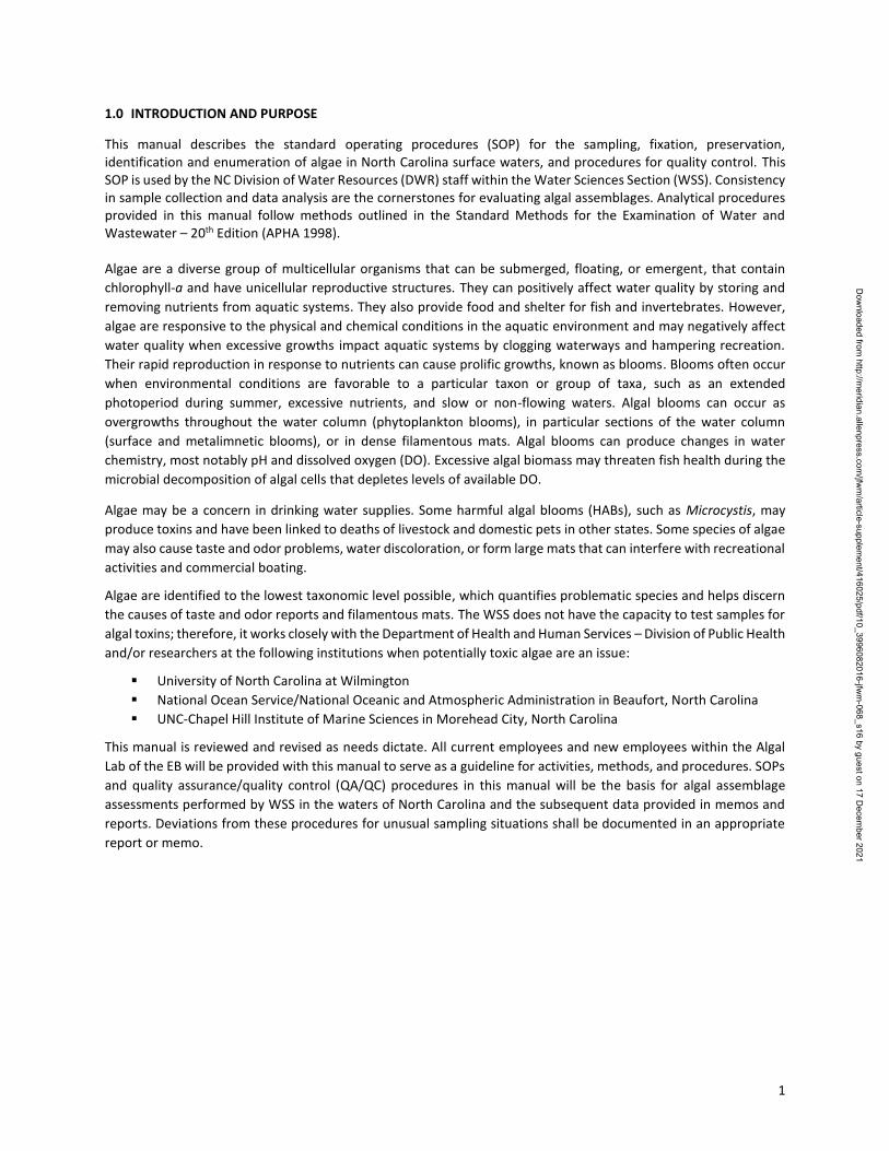
1.0 INTRODUCTION AND PURPOSE
This manual describes the standard operating procedures (SOP) for the sampling, fixation, preservation, identification and enumeration of algae in North Carolina surface waters, and procedures for quality control. This SOP is used by the NC Division of Water Resources (DWR) staff within the Water Sciences Section (WSS). Consistency in sample collection and data analysis are the cornerstones for evaluating algal assemblages. Analytical procedures provided in this manual follow methods outlined in the Standard Methods for the Examination of Water and Wastewater – 20th Edition (APHA 1998). Algae are a diverse group of multicellular organisms that can be submerged, floating, or emergent, that contain
chlorophyll-a and have unicellular reproductive structures. They can positively affect water quality by storing and
removing nutrients from aquatic systems. They also provide food and shelter for fish and invertebrates. However,
algae are responsive to the physical and chemical conditions in the aquatic environment and may negatively affect
water quality when excessive growths impact aquatic systems by clogging waterways and hampering recreation.
Their rapid reproduction in response to nutrients can cause prolific growths, known as blooms. Blooms often occur
when environmental conditions are favorable to a particular taxon or group of taxa, such as an extended
photoperiod during summer, excessive nutrients, and slow or non-flowing waters. Algal blooms can occur as
overgrowths throughout the water column (phytoplankton blooms), in particular sections of the water column
(surface and metalimnetic blooms), or in dense filamentous mats. Algal blooms can produce changes in water
chemistry, most notably pH and dissolved oxygen (DO). Excessive algal biomass may threaten fish health during the
microbial decomposition of algal cells that depletes levels of available DO.
Algae may be a concern in drinking water supplies. Some harmful algal blooms (HABs), such as Microcystis, may
produce toxins and have been linked to deaths of livestock and domestic pets in other states. Some species of algae
may also cause taste and odor problems, water discoloration, or form large mats that can interfere with recreational
activities and commercial boating.
Algae are identified to the lowest taxonomic level possible, which quantifies problematic species and helps discern
the causes of taste and odor reports and filamentous mats. The WSS does not have the capacity to test samples for
algal toxins; therefore, it works closely with the Department of Health and Human Services – Division of Public Health
and/or researchers at the following institutions when potentially toxic algae are an issue:
University of North Carolina at Wilmington
National Ocean Service/National Oceanic and Atmospheric Administration in Beaufort, North Carolina
UNC-Chapel Hill Institute of Marine Sciences in Morehead City, North Carolina
This manual is reviewed and revised as needs dictate. All current employees and new employees within the Algal
Lab of the EB will be provided with this manual to serve as a guideline for activities, methods, and procedures. SOPs
and quality assurance/quality control (QA/QC) procedures in this manual will be the basis for algal assemblage
assessments performed by WSS in the waters of North Carolina and the subsequent data provided in memos and
reports. Deviations from these procedures for unusual sampling situations shall be documented in an appropriate
report or memo.
Dow
nloaded from http://m
eridian.allenpress.com/jfw
m/article-supplem
ent/416025/pdf/10_3996082016-jfwm
-068_s16 by guest on 17 Decem
ber 2021
2
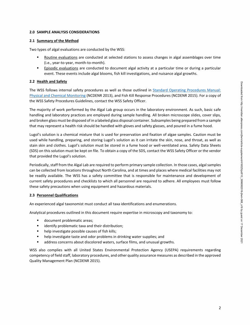
2.0 SAMPLE ANALYSIS CONSIDERATIONS
2.1 Summary of the Method
Two types of algal evaluations are conducted by the WSS:
Routine evaluations are conducted at selected stations to assess changes in algal assemblages over time
(i.e., year-to-year, month-to-month).
Episodic evaluations are conducted to document algal activity at a particular time or during a particular
event. These events include algal blooms, fish kill investigations, and nuisance algal growths.
2.2 Health and Safety
The WSS follows internal safety procedures as well as those outlined in Standard Operating Procedures Manual:
Physical and Chemical Monitoring (NCDENR 2013), and Fish Kill Response Procedures (NCDENR 2015). For a copy of
the WSS Safety Procedures Guidelines, contact the WSS Safety Officer.
The majority of work performed by the Algal Lab group occurs in the laboratory environment. As such, basic safe
handling and laboratory practices are employed during sample handling. All broken microscope slides, cover slips,
and broken glass must be disposed of in a labeled glass disposal container. Subsamples being prepared from a sample
that may represent a health risk should be handled with gloves and safety glasses, and poured in a fume hood.
Lugol’s solution is a chemical mixture that is used for preservation and fixation of algae samples. Caution must be
used while handling, preparing, and storing Lugol’s solution as it can irritate the skin, nose, and throat, as well as
stain skin and clothes. Lugol’s solution must be stored in a fume hood or well-ventilated area. Safety Data Sheets
(SDS) on this solution must be kept on file. To obtain a copy of the SDS, contact the WSS Safety Officer or the vendor
that provided the Lugol’s solution.
Periodically, staff from the Algal Lab are required to perform primary sample collection. In those cases, algal samples
can be collected from locations throughout North Carolina, and at times and places where medical facilities may not
be readily available. The WSS has a safety committee that is responsible for maintenance and development of
current safety procedures and checklists to which all personnel are required to adhere. All employees must follow
these safety precautions when using equipment and hazardous materials.
2.3 Personnel Qualifications
An experienced algal taxonomist must conduct all taxa identifications and enumerations.
Analytical procedures outlined in this document require expertise in microscopy and taxonomy to:
document problematic areas;
identify problematic taxa and their distribution;
help investigate possible causes of fish kills;
help investigate taste and odor problems in drinking water supplies; and
address concerns about discolored waters, surface films, and unusual growths.
WSS also complies with all United States Environmental Protection Agency (USEPA) requirements regarding
competency of field staff, laboratory procedures, and other quality assurance measures as described in the approved
Quality Management Plan (NCDENR 2015).
Dow
nloaded from http://m
eridian.allenpress.com/jfw
m/article-supplem
ent/416025/pdf/10_3996082016-jfwm
-068_s16 by guest on 17 Decem
ber 2021
3
2.4 Microscopy
Microscopes are needed to perform algal analyses. Measurements of an alga or a field of view are made with a
reticula (scale) or grid that has been placed within the ocular of the microscope. Determining the distance between
the lines in the reticule or the area of a grid at various magnifications requires a micrometer slide.
2.4.1 Microscopes
The WSS Algal Lab uses two styles of microscopes:
A Leitz Laborlux S compound microscope with 10X oculars; 10X, 25X, 40X, 63X, and 100X (oil
immersion) objectives (maximum magnification: 1000X) equipped with an attached three module
epifluorescence illuminator with filters; and an HBO 100 watt mercury bulb.
Two Leitz Diavert inverted microscopes: (1) one with 10X oculars and 4X, 10X, 20X, and 32X
objectives (maximum magnification: 320X); and (2) one with 15X oculars and the same objectives
(maximum magnification: 480X). Both inverted microscopes are illuminated by Leitz 100W
transformers.
2.4.2 Microscope Maintenance
Microscopes must be maintained and serviced to provide optimum visual clarity. Microscopes are covered
by a lint free cloth or plastic when not in use. Objectives and oculars are cleaned using an approved lens
cleaner, non-abrasive cotton swabs, and lens paper. A certified technician services the microscopes
annually.
2.4.3 Microscope Calibration and Standardization
All reticules (the scale inside the ocular used for measurement and area determinations) must be calibrated
periodically for all magnifications to ensure quality measurements.
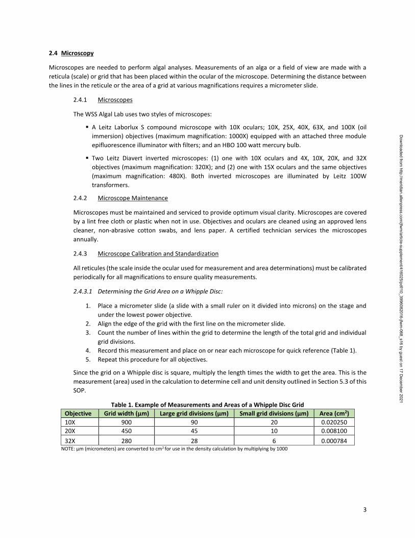
2.4.3.1 Determining the Grid Area on a Whipple Disc:
1. Place a micrometer slide (a slide with a small ruler on it divided into microns) on the stage and
under the lowest power objective.
2. Align the edge of the grid with the first line on the micrometer slide.
3. Count the number of lines within the grid to determine the length of the total grid and individual
grid divisions.
4. Record this measurement and place on or near each microscope for quick reference (Table 1).
5. Repeat this procedure for all objectives.
Since the grid on a Whipple disc is square, multiply the length times the width to get the area. This is the
measurement (area) used in the calculation to determine cell and unit density outlined in Section 5.3 of this
SOP.
Table 1. Example of Measurements and Areas of a Whipple Disc Grid
Objective Grid width (µm) Large grid divisions (µm) Small grid divisions (µm) Area (cm2)
10X 900 90 20 0.020250
20X 450 45 10 0.008100
32X 280 28 6 0.000784 NOTE: µm (micrometers) are converted to cm2 for use in the density calculation by multiplying by 1000
Dow
nloaded from http://m
eridian.allenpress.com/jfw
m/article-supplem
ent/416025/pdf/10_3996082016-jfwm
-068_s16 by guest on 17 Decem
ber 2021
4
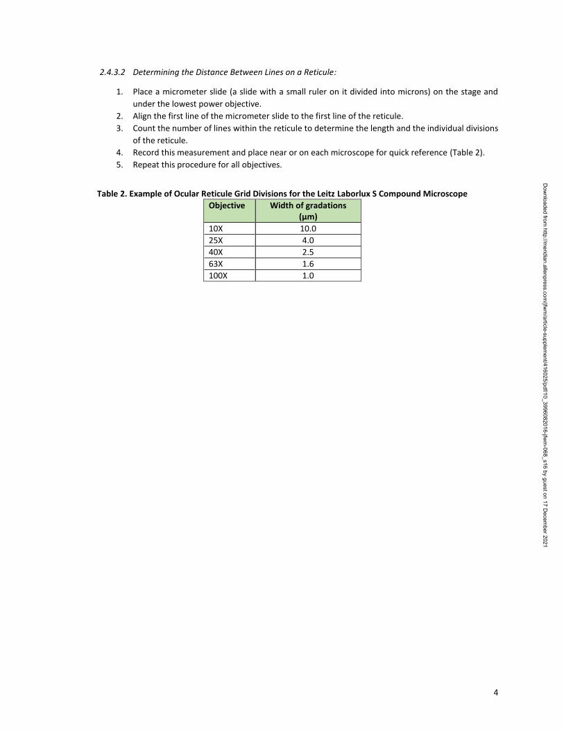
2.4.3.2 Determining the Distance Between Lines on a Reticule:
1. Place a micrometer slide (a slide with a small ruler on it divided into microns) on the stage and
under the lowest power objective.
2. Align the first line of the micrometer slide to the first line of the reticule.
3. Count the number of lines within the reticule to determine the length and the individual divisions
of the reticule.
4. Record this measurement and place near or on each microscope for quick reference (Table 2).
5. Repeat this procedure for all objectives.
Table 2. Example of Ocular Reticule Grid Divisions for the Leitz Laborlux S Compound Microscope
Objective Width of gradations (µm)
10X 10.0
25X 4.0
40X 2.5
63X 1.6
100X 1.0
Dow
nloaded from http://m
eridian.allenpress.com/jfw
m/article-supplem
ent/416025/pdf/10_3996082016-jfwm
-068_s16 by guest on 17 Decem
ber 2021
5
3.0 SAMPLE PROCESSING PROCEDURE
3.1 Sample Metadata
Sample metadata includes: 1) sampling methodology; 2) sample type; and 3) associated environmental parameters
collected.
3.1.1 Sampling Methods
There are three sampling methods that can be used to collect algae. The sample method must be noted on
the label, container, field sheet, and episodic evaluation forms.
3.1.1.1 Photic Zone Samples
These are integrated depth profiles that must be collected with a Lab-line Poly Pro Water Sampler®
(Lab-line®). A Secchi disc is first used to determine depth of visibility in the water column; the
photic zone is the depth to which ambient light penetrates and is defined as twice the Secchi depth.
A composite sample is collected by lowering the Lab-line® through the photic zone at a constant
speed. It is preferred over other sample collection methods because it provides a sample of the
water where phytoplankton (algae) may be present in the water column (approximately 2x Secchi
depth). This method should only be performed in waters that are deeper than one meter.
3.1.1.2 Grab Samples
These (also known as point samples) are collected by inverting a container, placing it below the
water surface, then turning it upright and allowing it to fill. Grab samples can also be taken from a
bridge or dock by quickly lowering a container 6 inches (15 cm) below the surface. This method
can be used during a fish kill and when a lab-line is not available or practical.
3.1.1.3 Scoop Samples
Scoops are surface skims, dips, scrapings, or any other method of collecting the sample. This
method should be used when only the identification of the algae is important.
3.1.2 Sample Type
There are three sample types:
Filamentous algae (i.e., algae in long strands that can be picked up by hand);
Periphyton (i.e., algae growing on rock, soil, or sand); and
Phytoplankton (i.e., algae growing in the water column).
3.1.3 Environmental Conditions
Physical and chemical conditions (temperature, DO, pH, Secchi depth, etc.) must be recorded and collected
according to methods outlined in the Intensive Survey Branch (ISB) SOP: Physical and Chemical Monitoring
(NCDENR 2013).
3.2 Quantifying Algal Blooms
Three measures may be used to quantify algal blooms. These measures are used independently as described below:
3.2.1 Chlorophyll-a Measurements
The WSS determines the level of chlorophyll-a with a fluorometer, according to United States
Environmental Protection Agency (USEPA) Standard Method 445.0 (Arar & Collins, 1997). These estimates
Dow
nloaded from http://m
eridian.allenpress.com/jfw
m/article-supplem
ent/416025/pdf/10_3996082016-jfwm
-068_s16 by guest on 17 Decem
ber 2021
6
of phytoplankton biovolume are used as the primary measure in phytoplankton bloom determination.
Chlorophyll-a levels that exceed 15 μg/L in trout waters or 40 μg/L in other waters are a violation of the
State water quality standard (Rule 15A NCAC 02B.02111).
3.2.2 Unit Density
Algal units are growth forms, such as colonies, filaments, or unicellular. Unit density (expressed as units/mL)
is determined by counting the number of algal units in a subsample.
3.2.3 Cell Density
Cell density, expressed as cells/mL, is the number of algal cells in a growth form, such as a colony, filament,
or unicellular, and is determined by counting the number of cells in ten growth forms of a particular alga
encountered during the analysis. These ten counts are averaged and multiplied by the total number of units
found (Section 3.2.3) in order to estimate algal cell density.
3.2.4 Biovolume
Biovolume (expressed as millimeters [mm]3/meters[m]3) is an estimate of the volume of phytoplankton
within a cubic meter of water. It is determined by counting the number of algal cells in a subsample and
multiplying that number by a reference cell biovolume. The reference biovolume (bvref) is an estimate based
upon measurements made on cells of the taxon from previous samples. The accuracy of this measurement
varies from season to season and from taxon to taxon.
3.3 Algae and Fish Kill Investigations
Phytoplankton samples collected as part of fish kill investigations are analyzed for species composition and density
to help discern whether algae were a factor in the event. Detailed information on fish kills, precautions, and
collection procedures can be found in Fish Kill Events: http://www.ncnhp.org/web/wq/ess/fishkills.
3.4 Nuisance Growths
Nuisance growths are those that people find objectionable, those that may degrade water quality with taste and
odor issues, and/or those that impact designated use(s) with large filamentous algal mats. Most nuisance growth
samples are qualitatively analyzed (identification only) for taxa composition. Taste and odor samples may be
quantitatively analyzed (identification and enumeration) for density and biovolume to determine the extent and
potential duration of the problem.
3.5 Episodic Events
Two field evaluation forms have been developed to record required data for episodic events (Appendix A):
Sample Collection and Field Evaluation Form
Fish Kill Investigation Form
These forms are used to document environmental conditions and serve as event records. Samples submitted as
phytoplankton blooms or as part of fish kill investigations are given priority. Evaluation forms should include:
Sample ID
Date/Time collected
Location (including waterbody, latitude/longitude, station, city, county, basin, and hydrologic unit code
(HUC) information)
Sample collection method
1http://reports.oah.state.nc.us/ncac/title%2015a%20-%20environmental%20quality/chapter%2002%20-%20environmental%20management/subchapter%20b/15a%20ncac%2002b%20.0211.pdf
Dow
nloaded from http://m
eridian.allenpress.com/jfw
m/article-supplem
ent/416025/pdf/10_3996082016-jfwm
-068_s16 by guest on 17 Decem
ber 2021
7
Sample type
Analytical method (quantitative of qualitative)
Collector’s name
Site photograph
Data recorded on evaluation forms should include:
DO (mg/L and % saturation)
Salinity (parts per thousand [ppt])
pH (standard units – SU)
Secchi depth (m)
Water temperature (°C)
Nutrients sampled
Air temperature (°C)
Chlorophyll-a (μg/L) sampled
Conductivity (μS/cm)
Visual description or site photograph
If possible, chlorophyll-a samples should be taken during phytoplankton blooms, fish kill investigations, and taste
and odor evaluations. Episodic evaluation forms can be submitted to the WSS electronically
([email protected] or [email protected]), faxed (919-743-8517), or sent by interoffice mail
(1623 Mail Service Center) along with the sample.
3.6 Sample Handling
Water chemistry samples must be collected, preserved, and shipped in accordance with WSS requirements 2 and the
Intensive Survey Branch Standard Operating Procedures Manual: Physical and Chemical Monitoring (NCDENR 2013).
3.7 Sample Containers
All photic zone and grab samples should be collected in a 500 mL plastic bottle. Scoop samples are not restricted to
any particular container as long as they can be sealed and are clearly labeled. Macroalgae should be wrapped in a
wet paper towel or newspaper and placed in a labeled, sealed plastic bag.
3.8 Sample Preservation
Adding a preservative is required to maintain algal cell structure, reduce microbial decomposition, halt grazing
activity by zooplankton, and stop cell division. All samples should be kept cool and out of direct sunlight.
The recommended preservative for phytoplankton and scraped periphyton samples is a modified Lugol’s solution
(Vollenweider 1974). Lugol’s solution should remain chemically stable for up to 5 years when kept in a dark container
in a cool dry place. Lugol’s solution can be obtained by contacting the WSS or prepared by following the directions
outlined in Appendix B.
Approximately 3 to 4 mL of Lugol's solution is required for 500 mL of sample. The exact quantity will depend on the
algal density and particulate matter in the sample. A well-preserved sample should be a dark straw or tea-like color.
3.9 Sample Quantity
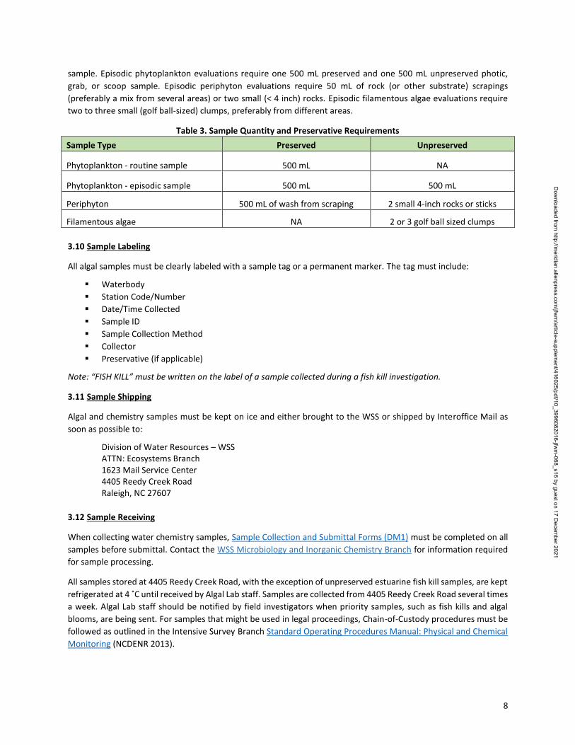
The quantity of sample required (Table 3) varies according to the evaluation (episodic or routine), sample type, and
sample method. Episodic and routine phytoplankton evaluations require one 500 mL preserved photic or grab
2https://ncdenr.s3.amazonaws.com/s3fs-public/Water%20Quality/Chemistry%20Lab/Operations/Staff%20Resources/PreservationHoldTime-SurfaceWaterSamples-July232015-WSSChemLab-FINAL.pdf
Dow
nloaded from http://m
eridian.allenpress.com/jfw
m/article-supplem
ent/416025/pdf/10_3996082016-jfwm
-068_s16 by guest on 17 Decem
ber 2021
8
sample. Episodic phytoplankton evaluations require one 500 mL preserved and one 500 mL unpreserved photic,
grab, or scoop sample. Episodic periphyton evaluations require 50 mL of rock (or other substrate) scrapings
(preferably a mix from several areas) or two small (< 4 inch) rocks. Episodic filamentous algae evaluations require
two to three small (golf ball-sized) clumps, preferably from different areas.
Table 3. Sample Quantity and Preservative Requirements
Sample Type Preserved Unpreserved
Phytoplankton - routine sample 500 mL NA
Phytoplankton - episodic sample 500 mL 500 mL
Periphyton 500 mL of wash from scraping 2 small 4-inch rocks or sticks
Filamentous algae NA 2 or 3 golf ball sized clumps
3.10 Sample Labeling
All algal samples must be clearly labeled with a sample tag or a permanent marker. The tag must include:
Waterbody
Station Code/Number
Date/Time Collected
Sample ID
Sample Collection Method
Collector
Preservative (if applicable)
Note: “FISH KILL” must be written on the label of a sample collected during a fish kill investigation.
3.11 Sample Shipping
Algal and chemistry samples must be kept on ice and either brought to the WSS or shipped by Interoffice Mail as
soon as possible to:
Division of Water Resources – WSS ATTN: Ecosystems Branch 1623 Mail Service Center 4405 Reedy Creek Road Raleigh, NC 27607
3.12 Sample Receiving
When collecting water chemistry samples, Sample Collection and Submittal Forms (DM1) must be completed on all
samples before submittal. Contact the WSS Microbiology and Inorganic Chemistry Branch for information required
for sample processing.
All samples stored at 4405 Reedy Creek Road, with the exception of unpreserved estuarine fish kill samples, are kept
refrigerated at 4 ˚C until received by Algal Lab staff. Samples are collected from 4405 Reedy Creek Road several times
a week. Algal Lab staff should be notified by field investigators when priority samples, such as fish kills and algal
blooms, are being sent. For samples that might be used in legal proceedings, Chain-of-Custody procedures must be
followed as outlined in the Intensive Survey Branch Standard Operating Procedures Manual: Physical and Chemical
Monitoring (NCDENR 2013).
Dow
nloaded from http://m
eridian.allenpress.com/jfw
m/article-supplem
ent/416025/pdf/10_3996082016-jfwm
-068_s16 by guest on 17 Decem
ber 2021
9
3.13 Sample Storage
3.13.1 Short-term sample handling
Preserved algal samples should be stored in a dark, dry, cool place prior to analysis.
3.13.2 Long-term sample handling
Phytoplankton samples that have been processed and analyzed are archived in the WSS lab until the study
is completed. For example, basinwide samples are discarded prior to the next round of sampling. Estuarine
samples are kept for at least one month in case any questions about particular samples arise. All
unpreserved samples are discarded after analysis.
3.14 Sample Data Management
Samples analyzed by Algal Lab staff are given a unique tracking number when they are entered into a Microsoft
Access® database. The nine-digit number is automatically generated with the first five numbers being the line
number of the database Entry Log Table followed by the four-digit year. For example, the sample id number 10245-
2015 was given to the 10245th sample entered into the database during the year 2015.
3.15 Sample Data Management Fields
The following sample data are entered into a database:
Date/time collected
Waterbody
Station code/number
Station description
Latitude/longitude
County
EcoType (Estuary, River/Stream, Lake/Reservoir, Other)
Basin
Sample type (phyto-, peri-, fila-)
Collector
ID method (quan/qual)
Sample method (Photic, Grab, Scoop)
Assessment type (Episodic, Routine)
Episode description (Fishkill, Bloom, Other)
Date received
Entered by
Chemistry lab number (if available)
Physical data (if available)
Chemical data (if available)
HUC
Dow
nloaded from http://m
eridian.allenpress.com/jfw
m/article-supplem
ent/416025/pdf/10_3996082016-jfwm
-068_s16 by guest on 17 Decem
ber 2021
10
4.0 LABORATORY TECHNIQUES AND ANALYSIS
4.1 Sample Analysis
Microscopes are necessary to conduct algal/phytoplankton analyses, which can be quantitative or qualitative.
A quantitative analysis identifies taxa to the lowest taxonomic level possible and enumerates a sample providing
information on:
algal density
estimates of biovolume
assemblage composition
dominant taxa
A qualitative analysis identifies taxa to the lowest taxonomic level possible and provides information on:
assemblage composition
dominant taxa
The sample analysis process includes:
preparation of a subsample
a visual check of the subsample for density and distribution (quantitative analysis only)
recording required data about the sample (e.g., location) and subsample (e.g., taxon) on a bench sheet
4.2 Subsample preparation
Two types of subsample preparation techniques are used for algal analysis:
4.2.1 Wet mount preparation
Wet mounts are used on compound microscopes. They should be used when high magnification (> 400X) is
needed for taxa identification and cell or unit density is not important. Prepare a wet mount by placing a
small aliquot (≈ 0.05 mL) of sample on a glass microscope slide or Palmer slide and then covering it with a
coverslip.
4.2.2 Utermöhl chamber preparation
Utermöhl chambers are used on inverted microscopes. They are particularly useful for samples with a wide
range of salinity because the salts remain in suspension. They must be used for all quantitative analyses.
Utermöhl chambers have four parts:
A threaded metal base.
A threaded Plexiglas chamber that comes in 5 cubic centimeters (ccm) and 10 ccm sizes.
A replaceable bottom coverslip.
A circular glass top plate.
Note: 1 ccm = 1 cm3 = 1 mL.
Utermöhl chambers must be thoroughly cleaned before each use. To clean an Utermöhl chamber:
1. Rinse out the chamber with tap water.
2. Wash the inside of the chamber and both sides of the glass coverslip with a nonabrasive, non-
residue soap and a cotton swab. Rinse out the chamber at least three times with distilled water.
3. Dry with a nonabrasive, no-lint paper towel.
4. Visually inspect the cleaned chamber to ensure that no oils or residues are remaining.
Cleaned chambers can be stored by placing them upside down on a clean surface.
Dow
nloaded from http://m
eridian.allenpress.com/jfw
m/article-supplem
ent/416025/pdf/10_3996082016-jfwm
-068_s16 by guest on 17 Decem
ber 2021
11
Cracked or broken bottom coverslips must be replaced. To replace a bottom coverslip on an Utermöhl
chamber:
1. Unscrew the chamber cylinder from the metal base.
2. Wash the chamber with a non-abrasive, non-residue soap, and water.
3. Dry the chamber with a non-abrasive, no-lint paper towel.
4. Apply a light coat of petroleum jelly on the threads of the cylinder.
5. Place a new coverslip in metal base.
6. Screw cylinder into ring.
7. Remove any excess petroleum jelly from chamber and coverslip with a non-abrasive, no-lint paper
towel.
8. Wash the chamber.
To prepare a subsample using an Utermöhl chamber:
1. Bring the bottled sample to room temperature to equalize gas pressure to reduce air bubble
formation after the subsample has been poured in the chamber.
2. Determine the size of chamber to use by evaluating the amount of particulates on the bottom of
the sample container. Use a 5 ccm chamber for heavy (easily visible) particulates or a 10 ccm
chamber for light (barely visible) particulates.
3. Check the bottom coverslip on the chamber for any particulates, oils, or films that would obscure
the algae or affect an even distribution during settling.
4. Re-suspend the algae in the 500 mL polyethylene container by gently inverting it nine or ten times.
Do not vigorously shake the sample container, as care must be taken to reduce aeration that can
cause air bubble formation in the chambers. If samples have not been analyzed within a few weeks
of collection, a spinner plate may be used to break up any clumping within the sample.
5. Slowly pour a subsample from the sample container into the chamber until it is completely filled
and a convex meniscus forms at the top of the chamber.
6. Slide the circular glass plate onto the top of the chamber removing the convex meniscus and sealing
the chamber.
7. Allow subsamples to settle for a minimum of 8 hours on a clean level surface.
8. The preparation may last up to a week without desiccation. A few drops of water may be added
into settled samples to remove bubbles; however, care is required not to disturb the settled
material. If more than a few drops of water are needed to remove bubbles, another subsample
should be poured and allowed to settle. If deionized water is used in a sample it may disturb the
ionic balance of a subsample.
4.3 Visual subsample density and distribution check
The quality of the analysis depends on the density and distribution of the subsample in the prepared Utermöhl
chamber. A visual inspection must be performed on all Utermöhl chamber preparations for subsample density and
random distribution. In general, the chamber should not have algae overlapping or obscuring the identification or
enumeration of other algae. The algae should also be equally distributed throughout the Utermöhl chamber.
To check for proper density and uniform distribution:
1. Place the prepared Utermöhl chamber on the microscope stage.
2. Briefly scan the prepared Utermöhl chamber for distribution at low magnification (≈100X).
3. The subsample should be evenly distributed throughout the chamber.
4. Non-evenly distributed subsamples or samples that are clumped together should not be quantitatively
analyzed (either discard the sample or pour another subsample).
5. Adjust the microscope to a higher magnification (≈320X).
Dow
nloaded from http://m
eridian.allenpress.com/jfw
m/article-supplem
ent/416025/pdf/10_3996082016-jfwm
-068_s16 by guest on 17 Decem
ber 2021
12
6. The ideal density of algal units within the field of view is between 10 and 40 units.
a. Dense subsamples (> 40 units within a field of view at 320X) should be re-poured using a smaller
chamber or diluted. Dilution can be done by extracting 50 mL of the sample, placing it into an
unused polyethylene container and adding a known quantity of filtered (0.45 μm) water. It is
important to use dilution water with a similar salinity to the sample as to maintain the osmotic
balances of the algal cells.
b. Sparse subsamples should be re-poured using a larger chamber or concentrated. Concentration
can be done by siphoning a known amount of water (i.e., ½) from a settled sample. Caution, this
technique is prone to error. When siphoning, the nozzle must not touch the sides or get near the
bottom.
c. Deionized water or other solution may be used to adjust salinity of samples collected from
estuarine, marine, or brackish water environments.
d. Any alterations to the volume of the sample or the subsample must be recorded on the sample
container and on the bench sheet.
4.4 Recording data
A quantitative analysis requires more detailed data to be recorded about a sample than a qualitative analysis. These
data are required to calculate the density and biovolume of the algae in the sample. If these data are not known or
unavailable, then the sample cannot be fully quantified. All data on the analysis must be recorded on an approved
bench sheet.
4.5 Bench sheets
The bench sheets for quantitative or qualitative analysis are kept in accordance with North Carolina public records
statutes. Data on the bench sheets must be written legibly so that others may read and understand notes,
identifications, and counts. An example of the bench sheets can be found in Appendix C.
Bench sheet data must include:
sample tracking number
analyst name
date the sample was analyzed
taxa codes of the algae identified
Bench sheets used for quantitative analysis must also contain:
quantity of the subsample analyzed
area of the Utermöhl chamber analyzed
number of algal cells in the area analyzed
number of algal units in the area analyzed
The total number of cells and units must be calculated and recorded in the appropriate box on the bench sheet when
a quantitative analysis is completed. These totals are used later after the data are entered to verify that the numbers
of units and cells entered are equal to the number of units and cells on the bench sheet.
Qualitative analysis may require recording data on the number of cells and units of the taxa identified during the
analysis. The number of cells and units can be used to determine the relative abundance of those taxa. Relative
abundance is the abundance of a particular taxon in relationship to other taxa and is used to provide an estimate of
what taxon is dominant in the sample.
Dow
nloaded from http://m
eridian.allenpress.com/jfw
m/article-supplem
ent/416025/pdf/10_3996082016-jfwm
-068_s16 by guest on 17 Decem
ber 2021
13
4.6 Recording the number of algal cells
Algal cell counts are required to calculate algal biovolume in a sample. However, counting all the algal cells of all the
algal units found during an analysis is impractical. Therefore, an average number of cells in a taxon’s unit can be
determined and then applied to the total number of units counted when the analysis is completed. This procedure
can be performed before, during, or after the analysis using any style of microscope with the appropriate
magnification.
To obtain an average number of cells in a taxon’s unit:
1. Enumerate the number of cells in at least 10 units (n>10) of the selected taxon.
2. Record the number of cells of each unit in the margin of the bench sheet.
3. When the analysis is completed, determine the average number of cells in a unit using the formula below.
4. Multiply the average number of cells per unit by the total number of units counted and round to the nearest
whole number.
5. Record this number in the appropriate box on the bench sheet.
unitpercellsofnumberAverageUnitsTotal
CellsTotal
Where:
Total Cells is the total number of the particular taxon’s cells that were counted to be averaged.
Total Units is the total number of the particular taxon’s units where cells were counted.
Example of obtaining an average number of cells in a unit.
Pseudanabaena is a filamentous blue green alga.
Ten filaments (units) in sample A were counted (Total units = 10).
The numbers of cells in each of the 10 units were: 11, 8, 10, 2, 5, 6, 11, 4, 10, 8.
The total number of cells counted was 75 (total cells = 75).
The average number of cells per unit was 7.5 (75/10 = 7.5), rounding = 8.
Thus, there were eight cells per unit (8 cells/unit) of Pseudanabaena in sample A.
4.7 Quantitative analysis
All quantitative analyses are performed on Leitz Diavert inverted microscopes as described in Section 2.4.1; a
Whipple disc in the ocular; and an Utermöhl settling chamber filled with the subsample to be analyzed. A Whipple
disc, described in Standard Methods, is inscribed with an accurately-ruled grid that is subdivided into 100 squares.
A grid is defined as the total area enclosed within the most outside lines-the square perimeter of the Whipple disc;
the size of the grid will vary based upon the level of magnification. It is the responsibility of the analyst to know the
area of the grid he/she is using. (Refer to Section 2.4 to determine the area of the grid on a Whipple disc and other
microscope calibration procedures.)
To identify and enumerate an algal assemblage in a sample:
1. Place a prepared Utermöhl chamber on the microscope stage.
2. Record sample data on a phytoplankton bench sheet (i.e., tracking code and size of chamber being used).
3. Randomly select an area to be analyzed.
4. Identify (to the lowest taxonomic unit possible) all taxa and the number of their cells and units within the entire
Whipple disc area or touching the right and bottom grid lines.
5. Only count units that contain cytoplasm (i.e., disregard empty cells and disintegrating filaments or colonies).
Dow
nloaded from http://m
eridian.allenpress.com/jfw
m/article-supplem
ent/416025/pdf/10_3996082016-jfwm
-068_s16 by guest on 17 Decem
ber 2021
14
6. Continue to analyze randomly selected discs until 100 units of the most abundant taxon is reached. 3
7. Record the number of discs analyzed, the algae found, and the number of their cells and units on the bench
sheet.
8. Tabulate the total number of units and cells for each taxon identified and record the totals into the space
provided on the bench sheet.
9. Tabulate the total number of cells and units for the entire analysis and write it in the space provided on the
bench sheet.
10. Enter bench sheet data into the database.
4.8 Qualitative analysis
Two types of qualitative analyses are performed:
Identification only ('What is it?').
Identification and enumeration for taxa dominance ('Which is the most common?').
These analyses are done by various means and not restricted to a particular microscope, slide preparation technique,
or magnification.
4.8.1 Identification only
1. Place prepared slide or chamber on the microscope stage.
2. Record the sample data on a bench sheet (i.e., tracking code and date of analysis).
3. Identify the algae in the sample.
4. Record identifications on a bench sheet.
5. The bench sheet data are now ready to be entered into the database.
4.8.2 Dominant taxa analysis
1. Place prepared slide or chamber on the stage of microscope.
2. Record all sample data on a bench sheet (i.e., tracking code, date of analysis).
3. Identify the algae and count their number of cells and units in a field. (A field can be the grid on a
Whipple disc or the complete field of view of the objective.)
4. Continue analyzing fields until at least 10 units of 10 taxa are reached.
5. Tabulate the total number of cells and units for each taxon.
6. Tabulate the total number of all cells and units of all taxa and record the total on the bottom of
the bench sheet.
7. The bench sheet data are now ready to be entered into the database.
8. The dominant taxa can be defined either by greater number of units or by the largest biovolume
depending upon the type of information needed.
3 Exceptions to Step #6 are: Sparse samples (i.e., few algal cells present) -- analyze at least 0.5% of the Utermöhl chamber (i.e., 40 grids at 300X magnification). Diverse samples with no dominant taxon -- analyze until a total of 300 units is reached.
Dow
nloaded from http://m
eridian.allenpress.com/jfw
m/article-supplem
ent/416025/pdf/10_3996082016-jfwm
-068_s16 by guest on 17 Decem
ber 2021
15
5.0 DATA HANDLING AND INTERPRETATION
5.1 Taxonomic Reference Table
A taxonomic reference table of the algal taxa known to be present in North Carolina was developed by WSS staff
and is utilized by the Algal Lab for taxonomic identification and is available upon request. This table provides a
uniform structure for assigning taxonomic codes with scientific nomenclature. The taxa list is limited to
phytoplankton at this time, but it is designed to incorporate other forms of algae in the future.
A taxonomic reference table requires periodic maintenance to update nomenclature and add new taxa.
5.1.1 Table Structure and Contents
5.1.1.1 Taxonomic Designation
The taxonomic designation of algae listed in the taxonomic reference table is based upon widely accepted
botanical classification conventions as defined in two primary reference books: Freshwater Algae of North
America (Wehr et. al. 2015) and Identifying Marine Phytoplankton (Tomas et al. 1997). Algal groups are
listed instead of phyla or divisions because “algae” is a collective term that refers to a number of loosely-
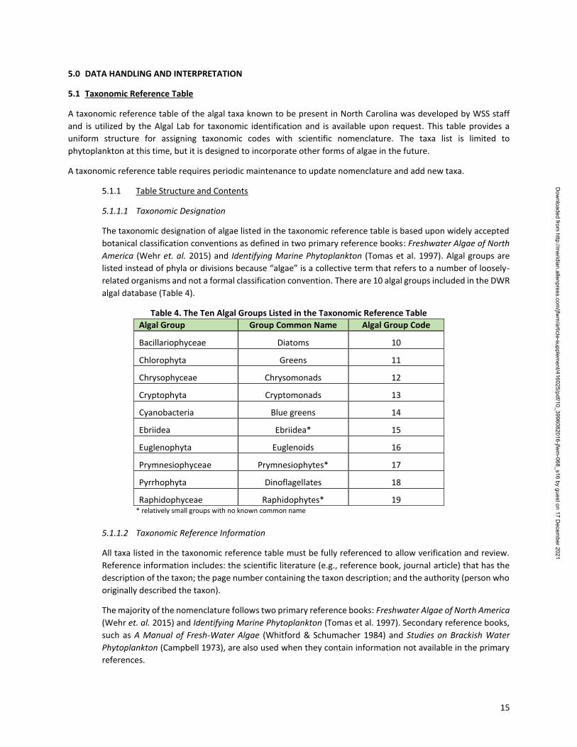
related organisms and not a formal classification convention. There are 10 algal groups included in the DWR
algal database (Table 4).
Table 4. The Ten Algal Groups Listed in the Taxonomic Reference Table
Algal Group Group Common Name Algal Group Code
Bacillariophyceae Diatoms 10
Chlorophyta Greens 11
Chrysophyceae Chrysomonads 12
Cryptophyta Cryptomonads 13
Cyanobacteria Blue greens 14
Ebriidea Ebriidea* 15
Euglenophyta Euglenoids 16
Prymnesiophyceae Prymnesiophytes* 17
Pyrrhophyta Dinoflagellates 18
Raphidophyceae Raphidophytes* 19 * relatively small groups with no known common name
5.1.1.2 Taxonomic Reference Information
All taxa listed in the taxonomic reference table must be fully referenced to allow verification and review.
Reference information includes: the scientific literature (e.g., reference book, journal article) that has the
description of the taxon; the page number containing the taxon description; and the authority (person who
originally described the taxon).
The majority of the nomenclature follows two primary reference books: Freshwater Algae of North America
(Wehr et. al. 2015) and Identifying Marine Phytoplankton (Tomas et al. 1997). Secondary reference books,
such as A Manual of Fresh-Water Algae (Whitford & Schumacher 1984) and Studies on Brackish Water
Phytoplankton (Campbell 1973), are also used when they contain information not available in the primary
references.
Dow
nloaded from http://m
eridian.allenpress.com/jfw
m/article-supplem
ent/416025/pdf/10_3996082016-jfwm
-068_s16 by guest on 17 Decem
ber 2021
16
5.1.1.3 Taxonomic Serial Numbers
The taxonomic reference table contains two versions of taxonomic serial numbers (TSNs): both DWR and
North American Diatom Ecological Database (NADED) numbers. These are also referred to as “reference
codes” that are unique to a specific taxon. Codes are commonly used in place of scientific names to improve
accuracy and efficiency in data entry, data storage, and data retrieval. An abbreviated version of the taxon’s
scientific name (i.e., Anacystis cyanea = anac cya) was used previously by NCDWR staff. However,
abbreviations become problematic when multiple names are spelled similarly or when new codes have to
be created to accommodate changes in nomenclature. Therefore, TSNs were developed to avoid these
problems. The TSNs are in the form of a 6-digit sequence. The first two digits designate the algal group,
such as diatom or green algae. The third and fourth digits designate the genus. The fifth and sixth digits
designate the species.
5.1.1.4 Assigning WSS Taxonomic Serial Numbers
The WSS uses the following steps to assign new TSNs:
1. Assign a TSN to a new taxon beginning with the algal group’s number (10 through 99). If the algal
group has not been previously assigned a number, then use the next consecutive number after the
last group number used.
2. Use the two-digit genus code if one exists (01 through 99). If the taxon is a new genus in the group,
then use the next consecutive number after the last genus number used.
3. Use the next consecutive number after the last number used for a species in that genus (1 through
99). If the species has not been determined, then use double zeroes (00) to signify that the identity
of the species is not known.
Example A. The TSN for Anabaena spp.
- The first two digits, the number 14, designate that it is a blue green alga.
- The third and fourth digits, the number 01, designate that it is the genus Anabaena.
- The fifth and sixth digits, the number 00, designates that it is an undetermined species of
Anabaena.
- The completed TSN for Anabaena spp. is 140100.
Example B. The TSN for Anabaena circinalis.
- The first two digits, the number 14, designates that it is a blue green alga.
- The third and fourth digits, the number 01, designates that it is the genus Anabaena.
- The fifth and sixth digits, the number 02, designates that it is the species A. circinalis.
- The completed TSN for A. circinalis is 140102.
Example C. Assigning a TSN to the new taxon Anabaena planctonica
- The first two digits, the number 14, designate that it is a blue green alga
- The third and fourth digits, the number 01, designate that it is the genus Anabaena.
- The fifth and sixth digits, 04 (the next consecutive number after the A. flos-aquae), designate
that it is the species A. planctonica
- The completed TSN for A. planctonica is 140104
5.1.1.5 North American Diatom Ecological Database (NADED) Numbers
The North American Diatom Ecological Database numbers (NADEDs) are also listed in the taxonomic
reference table. The NADED numbers were originally created for a database on diatoms at the Academy of
Natural Sciences. The database was later expanded to include all forms of algae. The NADED numbers listed
on the table have been created by the Academy of Natural Sciences of Philadelphia and are used to
integrate WSS data with outside laboratories.
Dow
nloaded from http://m
eridian.allenpress.com/jfw
m/article-supplem
ent/416025/pdf/10_3996082016-jfwm
-068_s16 by guest on 17 Decem
ber 2021
17
5.1.1.6 Cell Volume
Cell volumes, commonly called “biovolumes,” are used to roughly estimate algal biomass and provide an
alternative way of expressing the quantity of algae present as opposed to unit or cell density. Cell volumes
are determined by measuring the size of an algal cell and applying the measurements to the geometrical
equation that best fits the algal cell shape. Cell volumes listed on the table have either been calculated by
NCDWR staff, obtained through literature, or by consulting outside laboratories. The cell volumes listed are
conservative and based upon the smallest measures or calculations so as to not overestimate biomass. The
procedure to calculate the volume of an algal cell can be found in Section 5.6.
The accuracy of cell volume measurements is directly related to the number of measurements taken. If the
cell volume for a particular taxon is not known, then a zero (0) should be listed on the table. Cell volumes
should be updated as needed.
5.1.2 Taxonomic Revisions
The taxonomic reference list must reflect periodic changes in scientific nomenclature, thus it should be
periodically reviewed and updated and when nomenclature has changed, biovolume information has been
recalculated, or new taxa need to be added.
5.1.3 Adding Taxa
A taxon must be identified more than once and verified by a second qualified taxonomist before it can be
placed on the table. Rare, obscure or tentative identifications, even with outside consultation, should not
be placed on the table. Under no circumstances should a taxon be placed on the table without the
consultation of other DWR taxonomists. The table should only contain taxa that are commonly found, easily
referenced and relevant to the mission of the WSS.
5.2 Data entry
All data on samples and their analyses are entered into and stored in a Microsoft Access® database.
The final total from the database should be compared to the final total on the bench sheet. If the totals are not
equal, then there is an error in either the bench sheet calculations or the entered data and any discrepancies must
be rectified. Mark the box on the bench sheet labeled “Totals checked” when the two totals are equal.
5.3 Density calculations
Density can be expressed as unit density or cell density (Section 3.2). Unit densities are used in reports and cell
densities are used to calculate biovolume estimates. Both density calculations are performed using the same
equation. Only one calculation, the calculation for unit density, will be used in the example below. To calculate cell
density, substitute cells for units in the equation.
Unit density is calculated for each individual taxon in the assemblage and for the entire assemblage. The unit density
of each individual taxon is calculated first, and then the density of all the taxa are summed together to provide total
density. The same procedure and equations are used to calculate individual and total cell density. Individual cell
density is used to calculate sample biovolume.
Dow
nloaded from http://m
eridian.allenpress.com/jfw
m/article-supplem
ent/416025/pdf/10_3996082016-jfwm
-068_s16 by guest on 17 Decem
ber 2021
18
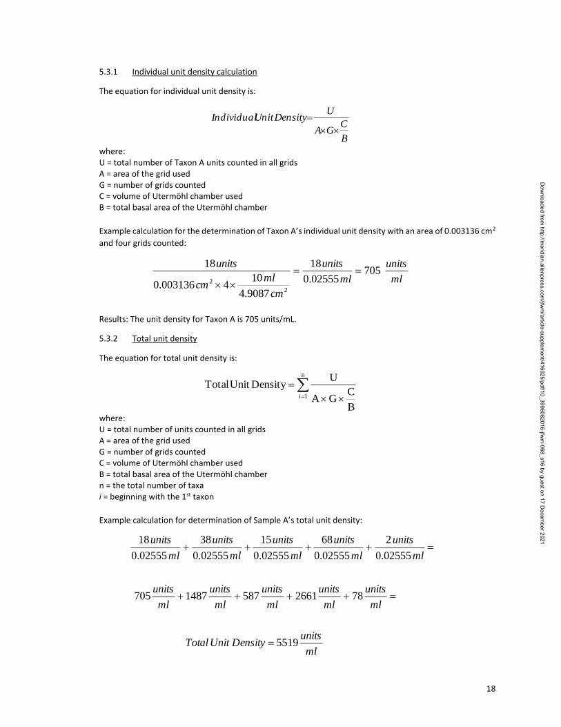
5.3.1 Individual unit density calculation
The equation for individual unit density is:
B
CGA
UDensityUnitIndividual
where: U = total number of Taxon A units counted in all grids A = area of the grid used G = number of grids counted C = volume of Utermöhl chamber used B = total basal area of the Utermöhl chamber
Example calculation for the determination of Taxon A’s individual unit density with an area of 0.003136 cm2
and four grids counted:
ml
units
ml
units
cm
mlcm
units705
02555.0
18
9087.4
104003136.0
18
2
2
Results: The unit density for Taxon A is 705 units/mL.
5.3.2 Total unit density
The equation for total unit density is:
n
1i
B
CGA
UDensityUnitTotal
where: U = total number of units counted in all grids A = area of the grid used G = number of grids counted C = volume of Utermöhl chamber used B = total basal area of the Utermöhl chamber n = the total number of taxa i = beginning with the 1st taxon
Example calculation for determination of Sample A’s total unit density:
ml
unitsDensityUnitTotal
ml
units
ml
units
ml
units
ml
units
ml
units
ml
units
ml
units
ml
units
ml
units
ml
units
5519
7826615871487705
02555.0
2
02555.0
68
02555.0
15
02555.0
38
02555.0
18
Dow
nloaded from http://m
eridian.allenpress.com/jfw
m/article-supplem
ent/416025/pdf/10_3996082016-jfwm
-068_s16 by guest on 17 Decem
ber 2021
19
Results: The total unit density for sample A is 5519 units/mL; however, scientific convention calls for
rounding or truncating values based on significant digit usage. In this example, which is the typical approach
to unit density calculations, the result would include only two significant digits (5,500 units/mL).
5.4 Reference biovolume calculation
Biovolumes are calculated for each individual taxon (individual biovolume) in the assemblage and for the assemblage
as a whole (total biovolume). These calculations are based upon the average biovolume (reference biovolume) of a
taxon’s cell and the taxon’s cell density. The total biovolume of a sample is determined by adding the biovolumes of
each individual taxon together.
A biovolume calculation worksheet documents how a particular taxon’s biovolume was determined. Once
determined, the taxon’s biovolume is entered into the database and used as the reference biovolume for
calculations.
5.4.1 Reference biovolume calculation worksheet
A reference biovolume calculation worksheet (Appendix D) must be completed on any new alga
being added to the WSS taxa reference list or when reevaluating an existing biovolume. The
worksheet must be stored in the filing cabinet designated for taxa information in the 4401 Algae
Lab.
The worksheet should include:
Division Authority
Family Sample location
Genus Date collected
Species (if applicable) Variety (if applicable)
Digital images or drawings of the alga
Equations used to calculate biovolumes of different cell morphologies are in Appendix E.
5.4.2 Calculation of cell reference biovolume
All cells should be measured (µm) with an ocular reticule at 400X magnification or greater.
To determine the cell’s reference biovolume:
1. Select the equation or set of equations that best fits the morphology of the algal cell from
Appendix E.
2. Record the equation on the reference biovolume calculation worksheet.
3. Prepare a wet mount of the sample that contains the alga being evaluated.
4. Locate a specimen on the microscope slide.
5. Take the necessary measurements (µm) using the Whipple grid.
6. Specimen position can be changed by lightly tapping the cover slip with the tip of a pencil
or pen. Practice is required to reposition the specimen correctly.
7. Record the measurements on the reference biovolume calculation worksheet.
8. Measure the cells of at least 15 specimens (preferably from more than one sample and
during different seasons, since this calculation will ultimately represent an “ideal” or
reference biovolume).
9. Determine the average size of a cell by adding all related measurements together then
dividing by the number of measurements taken.
Dow
nloaded from http://m
eridian.allenpress.com/jfw
m/article-supplem
ent/416025/pdf/10_3996082016-jfwm
-068_s16 by guest on 17 Decem
ber 2021
20
10. Use the average of these measurements in the selected equation to determine the
reference biovolume of the cell.
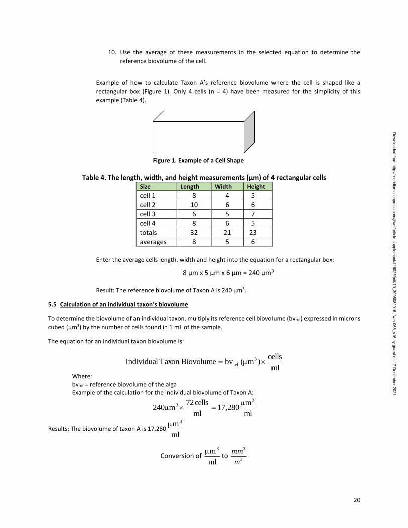
Example of how to calculate Taxon A’s reference biovolume where the cell is shaped like a
rectangular box (Figure 1). Only 4 cells (n = 4) have been measured for the simplicity of this
example (Table 4).
Table 4. The length, width, and height measurements (µm) of 4 rectangular cells Size Length Width Height
cell 1 8 4 5
cell 2 10 6 6
cell 3 6 5 7
cell 4 8 6 5
totals 32 21 23
averages 8 5 6
Enter the average cells length, width and height into the equation for a rectangular box:
8 µm x 5 µm x 6 µm = 240 µm3 Result: The reference biovolume of Taxon A is 240 µm3.
5.5 Calculation of an individual taxon’s biovolume
To determine the biovolume of an individual taxon, multiply its reference cell biovolume (bvref) expressed in microns
cubed (μm3) by the number of cells found in 1 mL of the sample.
The equation for an individual taxon biovolume is:
ml
cells)m(bvBiovolumeTaxonIndividual 3
ref
Where: bvref = reference biovolume of the alga Example of the calculation for the individual biovolume of Taxon A:
ml
m280,17
ml
cells72m240
33
Results: The biovolume of taxon A is 17,280ml
m3
Conversion of ml
m3to
3
3
m
mm
Figure 1. Example of a Cell Shape
Dow
nloaded from http://m
eridian.allenpress.com/jfw
m/article-supplem
ent/416025/pdf/10_3996082016-jfwm
-068_s16 by guest on 17 Decem
ber 2021
21
The result of the equation for an individual taxon biovolume is expressed in microns cubed per milliliter (μm3/mL)
and must be converted to into millimeters cubed per meters cubed (mm3/m3) to be consistent with historic data.
To convert μm3/mL to mm3/m3:
Multiply the individual taxon biovolume expressed in microns cubed per mL (μm3/mL) by the number of
millimeters cubed per microns cubed (mm3/(1 x109 μm3)).
Multiply the individual taxon biovolume in one milliliter (mm3/mL) by the number of milliliters in one liter
(1000 mL/L).
Multiply the individual taxon biovolume in one liter (mm3/L) by the number of liters in one meter cubed
(1000 L/m3).
The conversion equation is:
339
33
3
3
m
L1000
L
ml1000
m101
mm
ml
mbv
m
mmbv
Example for converting Taxon A’s biovolume from μm3/mL to mm3/m3:
3
3
339
33
m
mm28.17
m
L1000
L
ml1000
m101
mm
ml
m280,17
Results: The biovolume for Taxon A is 17.28 mm3/m3
*Note: Dividing the bvml
m3 by 1000 provides the same result.
1000ml
mbv
m
mmbv
3
3
3
5.6 Calculation of the total biovolume (BV)
The total biovolume of a sample is calculated by adding all the individual taxon biovolumes together.
The equation for the total biovolume is:
n
1i
3
ml
mbvBV
Where: bv = biovolume of an individual taxon in 1 m3. BV = total biovolume in 1 m3. n = the total number of algae identified in the sample. i = beginning with the 1st taxon.
Example of calculating the total biovolume for Sample A:
Dow
nloaded from http://m
eridian.allenpress.com/jfw
m/article-supplem
ent/416025/pdf/10_3996082016-jfwm
-068_s16 by guest on 17 Decem
ber 2021
22
3
3
3
3
3
3
3
3
3
3
74.7779.889.3678.1428.17m
mm
m
mm
m
mm
m
mm
m
mm
BiovolumeTotalDTaxonCTaxonBTaxonATaxon
Results: The total biovolume in Sample A is = 783
3
m
mm
5.7 Record Retention
Information pertaining to an episodic evaluation consists of, but is not limited to:
Sample submittal form
Bench sheet
Summary report
Information pertaining to a routine evaluation consists of, but is not limited to:
Study plan
Bench sheet
Summary report or final dataset/workbook
Related information, such as photos, maps, or news articles, are also archived along with the above items. The
Division has modernized their records retention policy, including documents produced and used by the WSS.
Hardcopies of episodic and routine evaluation records prior to 2010 are filed in individual folders in a file cabinet
located in the lab. Subsequent years include both hardcopies, filed appropriately in the lab, and electronic sample
submittal reports which are placed in folders per regional office on the WSS shared network drive. Study designs,
reports, or dataset workbooks of routine evaluations are kept in folders organized by study.
Dow
nloaded from http://m
eridian.allenpress.com/jfw
m/article-supplem
ent/416025/pdf/10_3996082016-jfwm
-068_s16 by guest on 17 Decem
ber 2021
23
6.0 QUALITY ASSURANCE
Current practices provide quality assurance on taxonomic identifications and data entry only.
6.1 Taxonomic Identifications
Staff expertise and a well-developed reference taxa list are the basis for accurate algal identifications over time. A
reference taxa list maintains continuity between/among analysts and reduces problems associated with changes in
nomenclature.
The current reference taxa list can be found in a unique table within the WSS algal database.
The individual taxa list should contain:
Complete name and classification:
- division
- family
- genus
- species
- variety
Taxa code
The reference(s) used to make the identification, along with the page number
Size ranges
Reference biovolume
Habitat (freshwater or estuarine)
Digital image (if available)
Only those taxa on the WSS reference taxa list may be entered into the database. A list of the primary reference
materials used by the WSS can be found in the Reference section of this document.
6.2 Data Entry
Data are entered into the database on two occasions: 1) during the sample login procedure and 2) after analysis
(bench sheet data). These data are checked for accuracy by a comparison of original records to entered data. Every
entry must be reviewed by the lead taxonomist to check that the required data has been recorded correctly. The
total number of cells and units must be tabulated and written on the bench sheet. These numbers must be compared
to entered data and a check mark placed in the designated line to verify that the totals have been checked. Any
discrepancies between original and entered data must be rectified (i.e., chamber volume, fields counted, dates
collected and analyzed, and cell and unit calculations).
Dow
nloaded from http://m
eridian.allenpress.com/jfw
m/article-supplem
ent/416025/pdf/10_3996082016-jfwm
-068_s16 by guest on 17 Decem
ber 2021
24
7.0 REFERENCES
American Public Health Association (APHA). 1998. Standard Methods for the Examination of Water and
Wastewater, 20th Edition. Washington, DC. Arar, E. and Gary Collins 1997. In Vitro Determination of Chlorophyll a and Pheophytin-a in Marine and Freshwater
Algae by Fluorescence. EPA Method 445.0-1. Revision 1.2. Campbell, Peter H. 1973. Studies on Brackish Water Phytoplankton. University of North Carolina, School of Public
Health. Sea Grant Publication. UNC-SG-73-07. North Carolina Department of Environmental Quality. Division of Water Resources (DWR). 2015. Fish Kill Response
Procedures. Biological Assessment Unit. http://www.ncnhp.org/web/wq/ess/fishkills
North Carolina Department of Environmental Quality. 2015. Sample Submission Guidance. Division of Water Resources (DWR). Laboratory Section. http://deq.nc.gov/about/divisions/water-resources/water-resources-data/water-sciences-home-page/microbiology-inorganics-branch/sample-submission
North Carolina Department of Environment and Natural Resources. 2013. Standard Operating Procedures
Manual: Physical and Chemical Monitoring. Intensive Survey Unit. http://portal.ncdenr.org/c/document_library/get_file?uuid=516f1b7b-fbb6-419f-83c8-0c981b2e1f78&groupId=38364
North Carolina Department of Environment and Natural Resources. 2015. Quality Management Plan, Version 4.1.
Original document submitted in 2011. Tomas, C., et al. 1997. Identifying Marine Phytoplankton. Academic Press, San Diego, CA. Vollenweider, R.A. 1974. A Manual for Measuring Primary Production in Aquatic Environments. IBP Handbook No.
12. F.A. Davis Company, Philadelphia Wehr, J. D., R. G. Sheath, Kociolek, P.J. (Eds). 2015. Freshwater Algae of North America: Ecology and Classification.
Second Edition. Academic Press, San Diego, CA Wetzel, R.G. and G.E. Likens. 1991. Limnological Analyses, Second Edition Springer Verlag, New York, NY. 391 pp. Whitford, L.A. and G.J. Schumacher. 1984. A Manual of Freshwater Algae. Sparks Press, Raleigh, NC.
Dow
nloaded from http://m
eridian.allenpress.com/jfw
m/article-supplem
ent/416025/pdf/10_3996082016-jfwm
-068_s16 by guest on 17 Decem
ber 2021
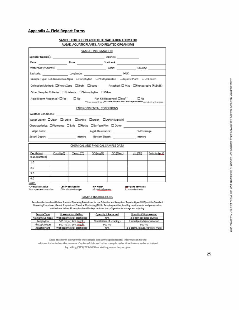
25
Appendix A. Field Report Forms
Dow
nloaded from http://m
eridian.allenpress.com/jfw
m/article-supplem
ent/416025/pdf/10_3996082016-jfwm
-068_s16 by guest on 17 Decem
ber 2021
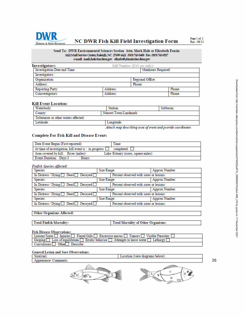
26
Dow
nloaded from http://m
eridian.allenpress.com/jfw
m/article-supplem
ent/416025/pdf/10_3996082016-jfwm
-068_s16 by guest on 17 Decem
ber 2021
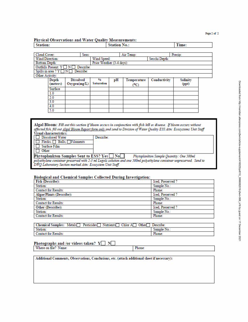
27
Dow
nloaded from http://m
eridian.allenpress.com/jfw
m/article-supplem
ent/416025/pdf/10_3996082016-jfwm
-068_s16 by guest on 17 Decem
ber 2021
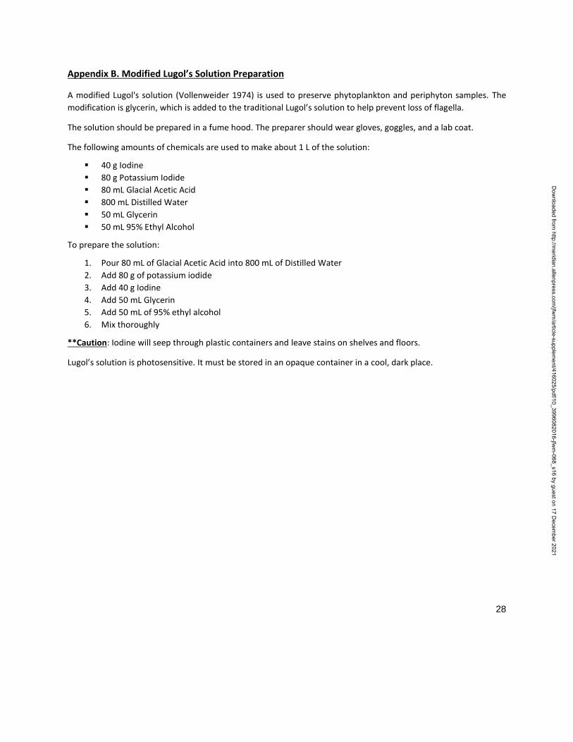
28
Appendix B. Modified Lugol’s Solution Preparation
A modified Lugol's solution (Vollenweider 1974) is used to preserve phytoplankton and periphyton samples. The
modification is glycerin, which is added to the traditional Lugol’s solution to help prevent loss of flagella.
The solution should be prepared in a fume hood. The preparer should wear gloves, goggles, and a lab coat.
The following amounts of chemicals are used to make about 1 L of the solution:
40 g Iodine
80 g Potassium Iodide
80 mL Glacial Acetic Acid
800 mL Distilled Water
50 mL Glycerin
50 mL 95% Ethyl Alcohol
To prepare the solution:
1. Pour 80 mL of Glacial Acetic Acid into 800 mL of Distilled Water
2. Add 80 g of potassium iodide
3. Add 40 g Iodine
4. Add 50 mL Glycerin
5. Add 50 mL of 95% ethyl alcohol
6. Mix thoroughly
**Caution: Iodine will seep through plastic containers and leave stains on shelves and floors.
Lugol’s solution is photosensitive. It must be stored in an opaque container in a cool, dark place.
Dow
nloaded from http://m
eridian.allenpress.com/jfw
m/article-supplem
ent/416025/pdf/10_3996082016-jfwm
-068_s16 by guest on 17 Decem
ber 2021
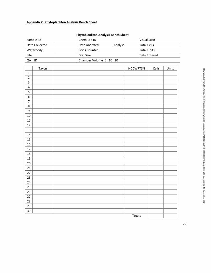
29
Appendix C. Phytoplankton Analysis Bench Sheet
Phytoplankton Analysis Bench Sheet
Sample ID Chem Lab ID Visual Scan
Date Collected Date Analyzed Analyst Total Cells
Waterbody Grids Counted Total Units
Site Grid Size Date Entered
QA ID Chamber Volume 5 10 20
Taxon NCDWRTSN Cells Units
1
2
3
4
5
6
7
8
9
10
11
12
13
14
15
16
17
18
19
20
21
22
23
24
25
26
27
28
29
30
Totals
Dow
nloaded from http://m
eridian.allenpress.com/jfw
m/article-supplem
ent/416025/pdf/10_3996082016-jfwm
-068_s16 by guest on 17 Decem
ber 2021
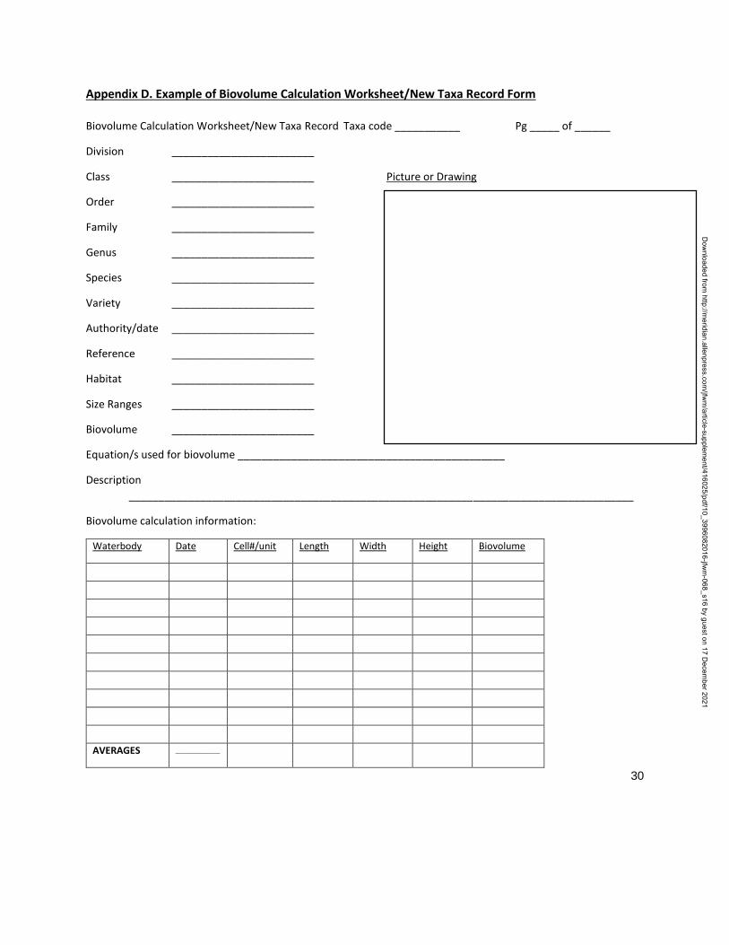
30
Appendix D. Example of Biovolume Calculation Worksheet/New Taxa Record Form
Biovolume Calculation Worksheet/New Taxa Record Taxa code ___________ Pg _____ of ______
Division ________________________
Class ________________________ Picture or Drawing
Order ________________________
Family ________________________
Genus ________________________
Species ________________________
Variety ________________________
Authority/date ________________________
Reference ________________________
Habitat ________________________
Size Ranges ________________________
Biovolume ________________________
Equation/s used for biovolume _____________________________________________
Description
_____________________________________________________________________________________
Biovolume calculation information:
Waterbody Date Cell#/unit Length Width Height Biovolume
AVERAGES _______________
Dow
nloaded from http://m
eridian.allenpress.com/jfw
m/article-supplem
ent/416025/pdf/10_3996082016-jfwm
-068_s16 by guest on 17 Decem
ber 2021
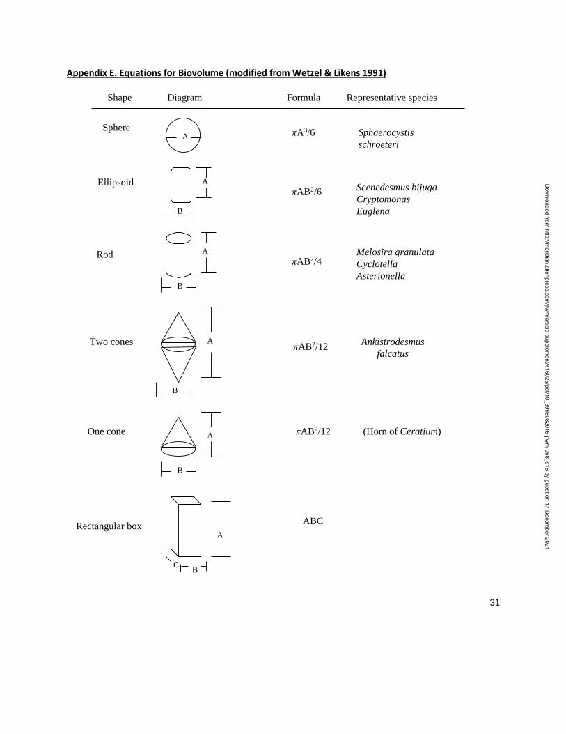
31
Appendix E. Equations for Biovolume (modified from Wetzel & Likens 1991)
Shape Diagram Formula Representative species
SphereA πA3/6 Sphaerocystis
schroeteri
Ellipsoid A
B
πAB2/6 Scenedesmus bijuga
Cryptomonas
Euglena
Rod A
B
πAB2/4Melosira granulata
Cyclotella
Asterionella
Two cones A
B
πAB2/12Ankistrodesmus
falcatus
One cone A
B
πAB2/12 (Horn of Ceratium)
Rectangular box
C B
A
ABC
Dow
nloaded from http://m
eridian.allenpress.com/jfw
m/article-supplem
ent/416025/pdf/10_3996082016-jfwm
-068_s16 by guest on 17 Decem
ber 2021
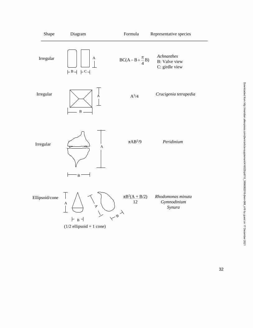
32
Shape Diagram Formula Representative species
Irregular A
CB
Achnanthes
B: Valve view
C: girdle view
Irregular A
B
A3/4Crucigenia tetrapedia
A
B
IrregularπAB2/9 Peridinium
Ellipsoid/cone
A
B
A
B
(1/2 ellipsoid + 1 cone)
πB2(A + B/2)
12
Rhodomonas minuta
Gymnodinium
Synura
)B4
BA(BC
Dow
nloaded from http://m
eridian.allenpress.com/jfw
m/article-supplem
ent/416025/pdf/10_3996082016-jfwm
-068_s16 by guest on 17 Decem
ber 2021