standardization proposal of soft tissue artefact …standardization proposal of soft tissue artefact...
TRANSCRIPT

Seediscussions,stats,andauthorprofilesforthispublicationat:https://www.researchgate.net/publication/313881031
Standardizationproposalofsofttissueartefactdescriptionfordatasharinginhumanmotionmeasurements
ArticleinJournalofBiomechanics·February2017
DOI:10.1016/j.jbiomech.2017.02.004
CITATIONS
0
READS
80
40authors,including:
Someoftheauthorsofthispublicationarealsoworkingontheserelatedprojects:
FARSEEING(FAllRepositoryforthedesignofSmartandsElf-adaptiveEnvironmentsprolonging
IndependentlivinG)Viewproject
Gazestrategiesduringobstaclenegotiationinthepresenceofdistractors:avirtualrealitybased
assessmentViewproject
AndreaCereatti
UniversitàdegliStudidiSassari
133PUBLICATIONS734CITATIONS
SEEPROFILE
DanielLBenoit
UniversityofOttawa
64PUBLICATIONS759CITATIONS
SEEPROFILE
CaeciliaCharbonnier
Artanim
62PUBLICATIONS238CITATIONS
SEEPROFILE
RitaStagni
UniversityofBologna
176PUBLICATIONS948CITATIONS
SEEPROFILE
AllcontentfollowingthispagewasuploadedbyAndreaCereattion22February2017.
Theuserhasrequestedenhancementofthedownloadedfile.Allin-textreferencesunderlinedinblueareaddedtotheoriginaldocumentandarelinkedtopublicationsonResearchGate,lettingyouaccessandreadthemimmediately.
Accepted Manuscript
Standardization proposal of soft tissue artefact description for data sharing in
human motion measurements
Andrea Cereatti, Tecla Bonci, Massoud Akbarshahi, Kamiar Aminian, Arnaud
Barré, Mickael Begon, Daniel L. Benoit, Caecilia Charbonnier, Fabien Dal
Maso, Silvia Fantozzi, Cheng-Chung Lin, Tung-Wu Lu, Marcus G. Pandy, Rita
Stagni, Antonie J. van den Bogert, Valentina Camomilla
PII: S0021-9290(17)30100-8
DOI: http://dx.doi.org/10.1016/j.jbiomech.2017.02.004
Reference: BM 8124
To appear in: Journal of Biomechanics
Accepted Date: 11 February 2017
Please cite this article as: A. Cereatti, T. Bonci, M. Akbarshahi, K. Aminian, A. Barré, M. Begon, D.L. Benoit, C.
Charbonnier, F.D. Maso, S. Fantozzi, C-C. Lin, T-W. Lu, M.G. Pandy, R. Stagni, A.J. van den Bogert, V. Camomilla,
Standardization proposal of soft tissue artefact description for data sharing in human motion measurements, Journal
of Biomechanics (2017), doi: http://dx.doi.org/10.1016/j.jbiomech.2017.02.004
This is a PDF file of an unedited manuscript that has been accepted for publication. As a service to our customers
we are providing this early version of the manuscript. The manuscript will undergo copyediting, typesetting, and
review of the resulting proof before it is published in its final form. Please note that during the production process
errors may be discovered which could affect the content, and all legal disclaimers that apply to the journal pertain.
Standardization proposal of soft tissue artefact description for data sharing in human
motion measurements
Andrea Cereatti1,2,3
*, Tecla Bonci3,4
, Massoud Akbarshahi5, Kamiar Aminian
6, Arnaud Barré
6, Mickael
Begon7, Daniel L. Benoit
8, Caecilia Charbonnier
9, Fabien Dal Maso
7, Silvia Fantozzi
10, Cheng-Chung
Lin11,12
, Tung-Wu Lu11,13
, Marcus G. Pandy5, Rita Stagni
10, Antonie J. van den Bogert
14, Valentina
Camomilla3,15
1 POLCOMING Department, Information Engineering Unit, University of Sassari, Sassari, Italy
2 Dept. of Electronics and Telecommunications, Politecnico di Torino, Torino, Italy
3 Interuniversity Centre of Bioengineering of the Human Neuromusculoskeletal system, University of
Rome “Foro Italico”, Rome, Italy 4 Life and Health Sciences, Aston University, Birmingham, United Kingdom
5 Department of Mechanical Engineering, University of Melbourne, Victoria, Australia
6 Laboratory of Movement Analysis and Measurement, Ecole Polytechnique Fédérale de Lausanne,
Lausanne, Switzerland 7 Laboratory of Simulation and Movement Modeling, Department of Kinesiology, University of
Montreal, Montreal, Canada 8 Faculty of Health Sciences, University of Ottawa, Ottawa, Canada
9 Artanim Foundation, Medical Research Department, Geneva, Switzerland
10 Department of Electric, Electronic and Information Engineering "Guglielmo Marconi" – DEI,
University of Bologna, Italy 11
Institute of Biomedical Engineering, National Taiwan University, Taiwan, ROC 12
Department of Electronic Engineering, Fu-Jen Catholic University, Taiwan, ROC 13
Department of Orthopaedic Surgery, School of Medicine, National Taiwan University, Taiwan, ROC
14 Department of Mechanical Engineering, Cleveland State University, Cleveland, Ohio, USA
15 Department of Movement, Human and Health Sciences, University of Rome “Foro Italico”, Rome,
Italy
* Tel: +39-3387854455, E-mail: [email protected]
Word Count: 3944
Keywords: Human Movement Analysis, Kinematics, Soft Tissue Artefact, Stereophotogrammetry,
Open Data
Abstract
Soft tissue artefact (STA) represents one of the main obstacles for obtaining accurate and
reliable skeletal kinematics from motion capture. Many studies have addressed this issue, yet
there is no consensus on the best available bone pose estimator and the expected errors
associated with relevant results. Furthermore, results obtained by different authors are
difficult to compare due to the high variability and specificity of the phenomenon and the
different metrics used to represent these data. Therefore, the aim of this study was twofold:
firstly, to propose standards for description of STA; and secondly, to provide illustrative STA
data samples for body segments in the upper and lower extremities and for a range of motor
tasks specifically, level walking, stair ascent, sit-to-stand, hip- and knee-joint functional
movements, cutting motion, running, hopping, arm elevation and functional upper-limb
movements. The STA dataset includes motion of the skin markers measured in vivo and ex
vivo using stereophotogrammetry as well as motion of the underlying bones measured using
invasive or bio-imaging techniques (i.e., X-ray fluoroscopy or MRI). The data are
accompanied by a detailed description of the methods used for their acquisition, with
information given about their quality as well as characterization of the STA using the
proposed standards. The availability of open-access and standard-format STA data will be
useful for the evaluation and development of bone pose estimators thus contributing to the
advancement of three-dimensional human movement analysis and its translation into the
clinical practice and other applications.
1. Introduction
The analysis of joint mechanics requires the estimation of both position and orientation
(pose) of the bones which meet at a joint. However, due to muscle contraction, wobbling of
soft tissues and skin stretching/sliding, the relative positions between the skin and the
underlying bones changes over time during the execution of a given motor task. The relative
movement between the skin and underlying bone is commonly referred to as soft tissue
artefact (STA) and represents one of the main obstacles for obtaining accurate and reliable
measurements of skeletal kinematics using skin-mounted markers and stereophotogrammetry
or wearable sensors (Leardini et al., 2005; Peters et al., 2010). Various bone pose estimators
have been proposed to reduce the impact of STA on estimates of joint kinematics, including
least square methods (Camarn and Milburn, 2005), inertia methods (Andriacchi et al., 1998;
Alexander and Andriacchi, 2001), optimal cluster model procedures (Chèze et al., 1995;
Taylor et al., 2005), methods incorporating STA calibration procedures (Lucchetti et al.,
1998; Cappello et al., 2005) and global optimization approaches (Andersen et al., 2009; Lu
and O’Connor, 1999; Reinbolt et al., 2005). However, no consensus has been reached either
on the best available estimator or on the maximum errors associated with these different
methods (Barré et al., 2015; Benoit et al., 2007; Cereatti et al., 2006; Stagni et al., 2009).
There are several reasons for the lack of a consensus. First, STA quantification is a
cumbersome, expensive, and time-consuming process which requires the determination of a
virtually error-free bone pose using either invasive techniques such as pins inserted into the
bones (Benoit et al., 2006; Cereatti et al., 2009; Dal Maso et al., 2015; Lafortune et al., 1992;
Reinschmidt et al., 1997) or bio-imaging techniques such as fluoroscopy and magnetic
resonance (MR) imaging (Bey et al., 2008; Garling et al., 2008; Guan et al., 2016; Stagni et
al., 2005). The need for complex experimental set-ups and procedures (e.g. simultaneous
recordings using different instrumentation and surgical intervention for insertion of bone
pins), expensive measurement systems (e.g. single/dual-plane fluoroscopy, MR imaging, and
high resolution multi-camera systems) and highly-specific, multidisciplinary expertise
(bioengineers, orthopaedics, and physiotherapists) may explain the relatively small sample
sizes and diverse experimental datasets available in the literature. Second, differences
observed in the STA characteristics may be due to experimental inaccuracies resulting from
intrinsic measurement limitations that have affected both the spatial and temporal resolution
of the measured error-free bone pose (Peters et al., 2010; Ramsey et al., 2003; Tersi et al.,
2013). Third, there is ample evidence in the literature to suggest that STA depends on several
factors such as subject anthropometry, the body segment on which a particular marker is
located, the location of that marker, and the type of activity performed (Barré et al., 2013;
Cappello et al., 2005; Cappozzo et al., 1996; Peters et al., 2010). These factors result in the
high variability and specificity observed in the STA patterns and amplitudes. Therefore, when
STA data are used, for example, to assess the performance of a specific bone pose estimator
or to perform comparative evaluations, it is crucial to provide a thorough description of the
experimental data used as input for the analysis (e.g. number of markers forming the cluster,
marker location, description of the motor task analysed and of the subject characteristics). It
should be noted that the aforementioned STA variability and specificity have impeded the
development of subject-specific models for STA compensation, applicable and effective
under different experimental conditions. Lastly, different metrics have been used in the
literature to describe the STA amplitude making it difficult to direct compare the results
(Dumas et al., 2014; Peters et al., 2010).
The present study addresses the aforementioned limitations by proposing a standardization
of the metrics for STA description at the marker level and providing an exemplar STA dataset
organized in a standardized format for STA data exchange. This dataset is comprised of STA
data relative to different body segments in the upper and lower extremities, different subjects,
and motor tasks (walking, step ascent, sit-to-stand, hip and knee joint functional movements,
cutting motion, running, hopping, arm elevation and functional upper limb movements). The
dataset was created by compiling the STA data published by various investigators from
different laboratories using different techniques. It includes the motion of the skin marker
measured using stereophotogrammetry in vivo and ex vivo as well as that of the underlying
bones using invasive or bio-imaging techniques (i.e., X-ray fluoroscopy or MRI) for various
motor tasks in single trials of single selected subjects or specimens (data sample) for various
motor tasks. Each data sample is accompanied with a thorough description of the material and
methods used, information about the data quality when available in the original studies, and a
characterization of the STA characteristics using the proposed standards.
2. Material and Methods
2.1 Metrics for STA description
Consider a skin marker attached to a generic body segment, and let be its position
vector in the relevant bone-embedded anatomical coordinate system (ACS) at a given
sampled instant of time i. During the motion of the body segment, will change due to the
deformation of the soft tissues. The variation of over time represents the STA affecting the
skin marker. In other words, the problem of the STA characterization is equivalent to the
description of the change over time of a vector in a 3D Euclidean space.
An effective statistical description of the STA should include information on both
amplitude and direction. For each given skin marker during a given motor task, the following
quantities are defined over the N available observations over time:
Mean position vector: Ni ,..,1 (1)
Instantaneous displacement vector: (2)
ip
ip
ip
N
i
iN 1
1pp
ppd iiziyixi ddd ,,,
Root mean square amplitude: N
d
N
ii
1
2
)(rms
d
(3)
Root mean square amplitude components: N
rmsd
N
i
ic
c
1
2
,d
zyxc ,, ; (4)
Peak-to-peak amplitude: jip pp maxmax (5)
Peak-to peak components:
)min()max( ,, jcicc ppp zyxcNjNi ,,;,..,1;,..,1 (6)
The parameter rmsd provides a mean description of the STA amplitude in 3D space
whereas the parameters rmsdc (with c=x,y,z) describe the mean STA amplitude along each
axis of the ACS. In addition, and cp (with c=x,y,z) represent the maximum variation
of in 3D space and along each axis of the ACS, respectively. Both the root mean square
(RMS) amplitude rmsd and the peak-to-peak amplitude do not depend on the definition
of the ACS (Grimpampi et al., 2014) whereas rmsdc and cp do.
In summary, the STA affecting a selected skin marker during a given motor task can be
described by the following eight parameters:
- rmsd and which describe the “mean” and “maximum” STA amplitude.
- rmsdc and cp (with c=x,y,z) which provide information about the STA direction (6
parameters).
2.2 Description of the experimental data samples
The present study incorporates the largest number of STA data available in the literature;
whenever possible, we have included data collected by different authors for the same motor
task to increase the heterogeneity and completeness of the database. The inclusion criteria
were as follows: a) sufficiently detailed description of the experimental methodology
maxp
ip
maxp
maxp

employed for data collection; b) use of technically sound and validated techniques to obtain
the ground truth bone pose; c) dynamic trials; d) availability of the time-variant anatomical
coordinate system (ACS) pose and of the skin marker trajectories with respect to the ACS
during the analysed motor task; e) willingness to share data.
For the sake of completeness, the data sample provided by Akbarshahi et al. (2010) was
included, even though it does not fully satisfy the aforementioned criteria (only the relevant
joint angle histories are reported but not the ACS poses during time).
For the convenience of data users, the following information is summarized and provided
for each STA data samples as Supplementary Material A (section 2).
a) Data sample name and scientific article(s) of reference;
b) Subject or specimen characteristics: information about sex, age, mass, height, body mass
index;
c) Motor task description: information aimed at describing the motor task analyzed (e.g. type
of motion, gait speed, range of joint motion, tread and rise when step or seat are used, type of
footwear.);
d) Experimental data description: list of the body segments analysed, skin marker locations,
and anatomical landmarks used;
e) Anatomical coordinate systems definitions (ACS);
f) Measurement specifications: description of the measurement systems and techniques used
to process the position data (e.g. number of cameras, capture volume, sample frequency,
measurement accuracy);
g) Ground truth: description of the technique used to determine the ground truth bone pose
(e.g. measurement accuracy, procedures for calibration, registration, and synchronization
between instruments);
h) STA characterization: for each marker, a description of the relevant STA is provided
according to the proposed metrics. The dispersion of each STA parameter over all available
markers is described using a five-number summary technique (minimum, lower quartile,
median, upper quartile, and maximum).
Data are presented according to a lexicon described in Supplementary Material A (section 3).
The lexicon was devised to store the data in a common data format, relative to position and
orientation of upper or lower limb body segments while aiming at a complete description of
the kinematics of a motor task. This choice allows a user to obtain a final data representation
according to his/her interests, without knowing the experimental set-up of the laboratory
where data were acquired. The lexicon is detailed in terms of:
Data set storing description (Dataset name; Data information; Measurement Units).
Subject description (Subject name; Subject information; Warning; Subject data).
Legend tables (owner, motor task, footwear, pathology, side, segment, anatomical
landmarks)
For some variables and parameters, to be included in the file, standard names were used
(listed in ad hoc tables). The data structure is depicted in Figure 1. For the sake of usability,
each data sample is organized using both a MATLAB structure (MathWorks) and an open
textual data format (XML) and made available as Supplementary Material “dataSample”.
Further details on the structure of the data can be found in Supplementary Material A (section
3).
FIGURE 1 ABOUT HERE
An overview of the 31 data samples available, grouped in terms of body segment and
motor task, is given in Table 1. A detailed description of each data sample is provided in
Supplementary Material A (section 2).
TABLE 1 ABOUT HERE
2.3 Data processing
Skin marker trajectories of each data sample were represented in the relevant ACS. The
coordinates of the anatomical landmarks in the ACS were also provided when these data were
available. A minimal amount of adjustments were made to the original raw data as described
below:
- Gap filling: marker trajectories with gaps smaller than 0.35 s were filled using a partial
Procrustes superimposition approach (Grimpampi et al., 2014), while trajectories showing
gaps larger than 0.35 s were removed (gap filling not reliable). For these data samples, both
original data and data after gap filling were provided.
For the data sample Overground walking no gap filling was implemented since all the skin
markers showed a gap of 0.43 s due to overlapping of the knee joints on the fluoroscopic
image during the gait cycle.
- Data Filtering: no further data processing was performed in addition to the original filtering
specified, if performed, in the Supplementary material;
- Coordinate system transformations: the original ACSs were rotated whenever necessary to
consistently express the skin marker trajectories with respect to the anatomical directions in
accordance to the proposed Lexicon (x: anterior (+)-posterior, y: superior (+)-inferior, z: right
(+)-left anatomical directions; supplementary material A, section 3).
After these preliminary data processing steps, the skin marker trajectories represented in
the relevant ACSs were used to compute the eight parameters proposed as a metrics for STA
description. Relevant descriptive statistics were summarized with the five–number summary
technique (minimum, lower quartile, median, upper quartile, and maximum).
3. Results
An overall description of the STA mean and maximum amplitude computed over the
available skin markers, as obtained from the different data samples according to the proposed
metrics, is given in Table 2. The total number of skin markers varied greatly over the different
data samples, between 4 and 35 for the thigh, between 3 and 26 for the shank, between 4 and
7 for the arm and between 8 and 57 for the scapula. A detailed description of the STA
affecting each skin markers can be found in the Supplementary Material A (section 2).
All data samples described in Table 2 are made available for download and include
information about the positions of the skin markers in the relevant ACS and the position and
orientation of the ACS during the dynamic trials. Each data sample is thoroughly described
and organized according to a well-documented structure to facilitate data sharing (Figure 1).
TABLE 2 ABOUT HERE
4. Discussion
The aim of this work was to propose standards for the description of STA and for data
exchange and to provide an exemplar dataset that can support the development and evaluation
of methods used to accurately estimate bone pose.
According to the proposed metrics, STA affecting each single marker is described through
eight parameters, specifically, the mean and maximum amplitudes (rmsd and ) and their maxp
relevant variations along ACS directions (rmsdc and cp , with c=x,y,z). It is important to
note that the parameter rmsd does not depend on the definition of the ACS because it
represents the RMS of the marker instantaneous displacement with respect to its mean
position in the ACS for the specific motor task analyzed. In contrast, in previous studies the
STA affecting the skin marker trajectory has often been defined as its local displacement from
a reference position fixed in the ACS. This reference position was possibly chosen as the
position of the marker at a given time, for example, at the beginning of an experiment or
while the subject assumes a standard static posture (Grimpampi et al., 2014). The latter
description can be useful and practical when applying methods for STA compensation based
on the identification of the anatomical landmarks in given configurations (e.g. double
anatomical calibration technique) (Cappello et al., 1996). However, when the primary aim is
to characterize STA amplitude, the mean position is preferred since it is independent of the
choice of initial reference position, thus facilitating a comparison of the STA amplitude
among different experiments (Fig. 2).
FIGURE 2 ABOUT HERE
The STA dataset made available for download was created from selected data samples
obtained from previously published studies (Supplementary Material “dataSample”). STA
measurements obtained in different studies on various body segments and motor tasks using
different techniques are presented here for the first time using standardized metrics, thus
eliminating inconsistencies arising from the selection of different descriptions or reference
positions for the STA definition.
It is important to note that each data sample made available for download refers to single
subject/specimen performing a single trial. Due to the arbitrary selection of the experimental
data sample, the limited number of subjects/specimens for each motor task and the variability
in the marker locations, the present dataset is not intended to provide a statistical description
of the STA characteristics. Consequently, a comparison of the STA characteristics among the
different data samples (Table 2) is only adequate for preliminary analysis, and for verifying
the internal consistency of STA observation in human subjects.
In accordance with previous investigations (Cappozzo et al., 1996; Peters et al., 2010),
STAs affecting the thigh markers were highly variable for the different motor tasks analysed,
but in general were larger than those affecting the shank. The only exceptions related to the
task of knee extension against gravity, performed in an up-right posture with the hip flexed at
approximately 45 deg (Stagni et al., 2005), and the task of knee flexion (Akbarshahi et al.,
2010). In these studies, both STA rmsd and values were slightly, but consistently,
larger for the shank compared to the thigh (knee extension against gravity: median rmsd
values = 11.5 mm and 9.2 mm for shank and thigh, respectively; knee-flexion: median rmsd
values = 8.6 mm and 7.4 mm for shank and thigh, respectively). These results suggest that
differences in experimental settings and in the motor task analyzed may have a strong
influence in determining STA magnitude. In fact, the aforementioned tasks involved large
rotations at the knee joint with the hip joint locked. This circumstance could cause substantial
sliding of the skin markers in proximity of the knee joint, regardless of whether they belong to
the thigh or the shank.
Thigh STA amplitudes observed during walking (over-ground and treadmill walking) in
three out of four different data samples (Barré et al., 2013; Benoit et al., 2006; Tsai et al.,
2009) were consistent, exhibiting median rmsd values in the range of 7.6-8.4 mm and median
values in the range of 23.4-28.4 mm. Larger STA amplitudes were observed for the
treadmill walking data collected by Akbarshahi et al., 2010 (median rmsd = 13.7 mm and
median equal to 41.2 mm). A larger variability was observed for the STA affecting the
maxp
maxp
maxp
shank markers during walking (median rmsd in the range of 2.4-7.5 mm and median
values in the range of 8.4-26.3 mm).
With respect to the lower limb, the largest STAs were observed for the thigh markers
during the Sit-to-stand and Step-up exercises investigated by Stagni et al., (2005) and Tsai et
al., (2009) (median values up to 72.3 mm and 46.5 mm for Sit-to-stand and Step-up,
respectively). These results may be explained by the effects of the skin sliding and muscle
contraction components, which are expected to be considerable during tasks involving large
and simultaneous joint excursion at the hip and knee joints. Furthermore, during Sit-to-stand,
soft tissue deformation due to the compression of the seat during the sitting phase may cause
an increase of the STA amplitude affecting the markers proximally and posteriorly located on
the thigh segment.
The differences observed in STA amplitudes for the data samples may also be explained by
the different numbers of markers and their disparate locations on the body segment, together
with differences in age and body mass index of the subjects.
STAs affecting the upper limb during basic arm movements (flexion-extension and ab-
adduction) highly varied between the two studies analysed (median rmsd in the range of 4.1-
15.3 mm and median in the range of 11.8-41.2 mm for the arm) (Charbonnier et al.,
2014; Dal Maso et al., 2015, 2014). Substantially larger STA amplitudes were observed for
the scapula, which exhibited median rmsd values in the range 8.5-21.0 mm and median
values in the range 27.8-56.8 mm (Charbonnier et al., 2014; Dal Maso et al., 2015, 2014). The
discrepancies observed between the STA amplitudes reported here may be due to the
anthropometric differences in height, mass, and muscle volume between the two subjects
analyzed (Cereatti et al., 2015; Charbonnier et al., 2014; Dal Maso et al., 2015, 2014). The
largest STAs were observed for the markers attached to the scapula during the execution of
maxp
maxp
maxp
maxp
sport activities such as Ball throwing (median rmsd = 34.8 mm; median = 111.3 mm)
and Punching (median rmsd = 19.8 mm; median = 63.4 mm) (Dal Maso et al., 2015).
These results confirmed the well-known difficulties related to the measurement of scapular
motion (Anglin and Wyss, 2000), especially in sporting related activities such as throwing
(Myers et al., 2015).
The present database includes STA measurements for the thigh, shank, arm and scapula.
We acknowledge that markers positioned on the ASISs are susceptible to large STA (Hara et
al., 2014), however it was not possible to include data on the pelvic STA because of the
absence of data recorded under dynamic conditions.
The STA data included in the open dataset were obtained using a variety of gold standard
methods. Each technique has its own limitations (e.g., pins may constrain skin movement;
scapula is difficult to track using fluoroscopy) which involve measurement errors, the
magnitudes of which are difficult to predict and quantify (Peters et al., 2010). For this reason,
we have included in the final dataset the largest number of STA data samples from different
sources, even if these data were collected during similar or identical motor tasks.
The data presented in the Supplementary Material includes, for each sampled time instant,
both the positions of the skin markers in the relevant ACS and the position and orientation of
the ACS with respect to a global coordinate system during related dynamic trials. Through
simple rigid body transformations, it is possible to express the marker data in any arbitrary
selected coordinate system and to compute joint angular kinematics according to any
preferred rotation sequence (Senk et al., 2006; Wu et al., 2002; Wu et al:, 2005). Whilst
representing the data in this way makes it applicable to a wide range of applications, the one
exception to the aforementioned data format is the study by Akbarshahi et al. (2010) which
contains the marker trajectories in the ACS and the relevant joint angle histories, not the ACS
poses over time. The latter description, although partial, could be useful for instance to
maxp
maxp
investigate possible correlation between STA and joint angular kinematics (Camomilla et al.,
2013; Cappozzo et al., 1996).
There are limitations of this study that must be acknowledged. First, the present work is
concerned only with STA affecting individual markers, but not at marker cluster level.
However, a similar analysis could be performed, using the provided skin marker data, to
describe the effect of the STA on cluster position and orientation (rigid motion), and size and
shape (non-rigid motion) (Andersen et al., 2012; Benoit et al., 2015; De Rosario et al., 2012;
Dumas et al., 2014; Dumas and Chèze, 2009; Grimpampi et al., 2014). Second, we did not
include data on STA effect on measures performed with wearable measurement units markers
attached to a rigid shell (Manal et al., 2000). To authors’ knowledge no information is
available in the literature about STA affecting wearable measurement units. Nevertheless, the
STA data provided in this study could be used to preliminary devise simulations of the rigid
motion component due to STA affecting the segment location where the wearable
measurement units or rigid shells would be attached. However, these data do not allow to
describe the inertial effects of the mass of the wearable measurement units or rigid shells and
the effects of the different fixing techniques. These factors would surely affect the magnitude
and frequency content of the STA. Future work in this direction is recommended since
wearable measurement units are becoming increasingly popular for kinematic measurements.
We expect that the verification of STA data performed in the present study together with
the proposed standardization and sharing of the data will promote the following outcomes:
first, it will enable a more effective and reliable comparison of existing methods for STA
compensation (Alexander and Andriacchi, 2001; Cappello et al., 1996; Chéze et al., 1995;
Peters et al., 2010; Solav et al., 2015; Stagni et al., 2009); second, it will facilitate the creation
and validation of novel bone pose estimators eventually embedding models of the STA that
can capture its specificity (Richard et al., 2012); and third, it will lead to an evidence-based
consensus on the level of accuracy of the marker-based stereophotogrammetry methods for
estimating the pose of different bony segments.
Furthermore, when more sample trials and subjects will be collected and made available, it
could be possible to develop statistical parametric model of the STA in humans and to
validate these for different motor tasks and anthropometric differences to partially remove
STA from skin markers or sensors (Andersen et al., 2012; Bonci et al., 2014; Camomilla et
al., 2015; van Weeren et al., 1992).
Finally, we hope that by providing easy access to data describing the deformation of body
segments during movement, researchers from different backgrounds and disciplines will be
better motivated to challenge the STA issue with new ideas and methods for the advancement
of three-dimensional human movement analysis and its translation into the clinical practice
and other applications (Hicks et al., 2015).
Conflicts of interest
There are no conflicts of interest.
Data use policy
We request that the present study be specifically and clearly acknowledged when data sets
or data samples are used for data analyses and visualizations in publications, posters, oral
presentations, reports, Web pages, and any other types of scientific media. Please cite also the
relevant original studies of each used specific data samples
Acknowledgements
This work was partly supported by a Grant from the Università di Roma “Foro Italico”
(call PR_15) and the “Fondation de soutien à la recherche dans le domaine de l’othorpédie-
traumatologie”.
References
Akbarshahi, M., Schache, A.G., Fernandez, J.W., Baker, R., Banks, S., Pandy, M.G., 2010.
Non-invasive assessment of soft-tissue artifact and its effect on knee joint kinematics
during functional activity. J. Biomech. 43, 1292–1301.
Alexander, E.J., Andriacchi, T.P., 2001. Correcting for deformation in skin-based marker
systems. J. Biomech. 34, 355–361.
Andersen, M.S., Benoit, D.L., Damsgaard, M., Ramsey, D.K., Rasmussen, J., 2010. Do
kinematic models reduce the effects of soft tissue artefacts in skin marker-based motion
analysis? An in vivo study of knee kinematics. J. Biomech. 43, 268-273.
Andersen M.S., Damsgaard, M., Rasmussen J., 2009. Kinematic analysis of over-determinate
biomechanical systems. Comput Methods Biomech Biomed Engin. 12, 371-84.
Andersen, M.S., Damsgaard, M., Rasmussen, J., Ramsey, D.K., Benoit, D.L., 2012. A linear
soft tissue artefact model for human movement analysis: Proof of concept using in vivo
data. Gait Posture 35, 606–611.
Andriacchi, T.P., Alexander E.J., Toney M.K., Dyrby C., Sum, J., 1998. A point cluster
method for in vivo motion analysis: applied to a study of knee kinematics. J. Biomech.
Eng. 120, 743-9.
Anglin, C., Wyss, U.P., 2000. Review of arm motion analyses. Proc. Inst. Mech. Eng. H. 214,
541–555.
Barré, A., Jolles, B.M., Theumann, N., Aminian, K., 2015. Soft tissue artifact distribution on
lower limbs during treadmill gait: Influence of skin markers’ location on cluster design.
J. Biomech. 1–7.
Barré, A., Thiran, J.P., Jolles, B.M., Theumann, N., Aminian, K., 2013. Soft tissue artifact
assessment during treadmill walking in subjects with total knee arthroplasty. IEEE
Trans. Biomed. Eng. 60, 3131–3140.
Benoit, D.L., Ramsey, D.K., Lamontagne, M., Xu, L., Wretenberg, P., Renström, P., 2006.
Effect of skin movement artifact on knee kinematics during gait and cutting motions
measured in vivo. Gait Posture 24, 152–164.
Benoit, D.L., Ramsey, D.K., Lamontagne, M., Xu, L., Wretenberg, P., Renström, P., 2007. In
vivo knee kinematics during gait reveals new rotation profiles and smaller translations.
Clin. Orthop. Relat. Res. 454, 81–88.
Benoit, D.L., Damsgaard, M., Andersen, M.S., 2015. Surface marker cluster translation,
rotation, scaling and deformation: Their contribution to soft tissue artefact and impact on
knee joint kinematics, J. Biomech. 48(10), 2124-2129.
Bey, M.J., Kline, S.K., Zauel, R., Lock, T.R., Kolowich, P.A., 2008. Measuring dynamic in-
vivo glenohumeral joint kinematics: Technique and preliminary results. J. Biomech. 41,
711–714.
Bonci, T., Camomilla,V., Dumas, R., Chèze, L., Cappozzo, A., 2014. A soft tissue artefact
model driven by proximal and distal joint kinematics. J.Biomech. 47(10), 2354–2361.
Carman, A., Milburn, P., 2006. Determining rigid body transformation parameters from ill
conditioned spatial marker co-ordinates. J. Biomech. 39(10), 1778-86.
Camomilla, V., Cereatti, A., Chèze, L., Cappozzo, A., 2013. A hip joint kinematics driven
model for the generation of realistic thigh soft tissue artefacts. J. Biomech. 46, 625–630.
Camomilla, V., Bonci, T., Dumas, R., Chèze, L., Cappozzo, A., 2015. A model of the soft
tissue artefact rigid component. J. Biomech. 48, 1752–1759.
Cappello, A., Cappozzo, A., Palombara, P.F. La, Leardini, A., Bertani, A., 1996. Skin artefact
compensation by double calibration in bone motion reconstruction. Proc. 18th Annu. Int.
Conf. IEEE Eng. Med. Biol. Soc.
Cappello, A., Stagni, R., Fantozzi, S., Leardini, A., 2005. Soft tissue artifact compensation in
knee kinematics by double anatomical landmark calibration: Performance of a novel
method during selected motor tasks. IEEE Trans. Biomed. Eng. 52, 992–998.
Cappozzo, A., Catani, F., Leardini, A., Benedetti, M.G., Della Croce, U., 1996. Position and
orientation in space of bones during movement: Experimental artefacts. Clin. Biomech.
11, 90–100.
Cereatti, A., Della Croce, U., Cappozzo, A., 2006. Reconstruction of skeletal movement using
skin markers: comparative assessment of bone pose estimators. J. Neuroeng. Rehabil. 3,
7.
Cereatti, A., Donati, M., Camomilla, V., Margheritini, F., Cappozzo, A., 2009. Hip joint
centre location: An ex vivo study. J. Biomech. 42, 818–823.
Cereatti, A., Rosso, C., Nazarian, A., DeAngelis, J.P., Ramappa, A.J., Croce, U. Della, 2015.
Scapular motion tracking using acromion skin marker cluster: In vitro accuracy
assessment. J. Med. Biol. Eng. 35, 94–103.
Charbonnier, C., Chagué, S., Kolo, F.C., Chow, J.C.K., Ladermann, A., 2014. A patient-
specific measurement technique to model shoulder joint kinematics. Orthop. Traumatol.
Surg. Res. 100, 715–719.
Chéze, L., Fregly, B.J., Dimnet, J., 1995. A solidification procedure to facilitate kinematic
analyses based on video system data. J. Biomech. 28, 879–884.
Dal Maso, F., Blache, Y., Raison, M., Lundberg, A., Begon, M., 2015. Glenohumeral joint
kinematics measured by intracortical pins, reflective markers, and computed
tomography: A novel technique to assess acromiohumeral distance. J. Electromyogr.
Kinesiol. 1–8.
Dal Maso, F., Raison, M., Lundberg, A., Arndt, A., Begon, M., 2014. Coupling between 3D
displacements and rotations at the glenohumeral joint during dynamic tasks in healthy
participants. Clin. Biomech. 29, 1048–1055.
De Rosario, H., Page, A., Besa, A., Mata, V., Conejero, E. 2012. Kinematic description of
soft tissue artifacts: quantifying rigid versus deformation components and their relation
with bone motion. Med. Biol. Eng. Comput., 50(11), 1173-1181.
Dumas, R., Camomilla, V., Bonci, T., Chéze, L., Cappozzo, A., 2014. Generalized
mathematical representation of the soft tissue artefact. J. Biomech. 47, 476–481.
Dumas, R., Chéze, L., 2009. Soft tissue artifact compensation by linear 3D interpolation and
approximation methods. J. Biomech. 42, 2214–2217.
Garling, E.H., Kaptein, B.L., Mertens, B., Barendregt, W., Veeger, H.E.J., Nelissen,
R.G.H.H., Valstar, E.R., 2008. Soft-tissue artefact assessment during step-up using
fluoroscopy and skin-mounted markers. J Biomech. 41, 2332-5.
Grimpampi, E., Camomilla, V., Cereatti, A., De Leva, P., Cappozzo, A., 2014. Metrics for
describing soft-tissue artefact and its effect on pose, size, and shape of marker clusters.
IEEE Trans. Biomed. Eng. 61, 362–367.
Grood, E.S., Suntay, W.J., 1983. A joint coordinate system for the clinical description of
three-dimensional motions: application to the knee. J. Biomech. Eng. 105, 136–44.
Guan, S., Gray, H., Keynejad, F., Pandy, M.G., 2016. Mobile biplane X-ray imaging system
for measuring 3D dynamic joint motion during overground gait. IEEE Transactions on
Medical Imaging 35: 326-336.
Hara, R., Sangeux, M., Baker, R., McGinley, J., 2014. Quantification of pelvic soft tissue
artifact in multiple static positions. Gait Posture 39, 712–717.
Hicks, J.L., Uchida, T.K., Seth, A., Rajagopal, A., Delp, S., 2015. Is my model good enough?
Best practices for verification and validation of musculoskeletal models and simulations
of human movement. J. Biomech. Eng. 137, 20905.
Lafortune, M.A., Cavanagh, P.R., Sommer, H.J., Kalenak, A., 1992. Three-dimensional
kinematics of the human knee during walking. J. Biomech. 25, 347–357.
Leardini, A., Chiari, L., Della Croce, U., Cappozzo, A., 2005. Human movement analysis
using stereophotogrammetry - Part 3. Soft tissue artifact assessment and compensation.
Gait Posture 21, 212–225.
Lucchetti, L., Cappozzo A., Cappello, A,. Della Croce U., 1998. Skin movement artefact
assessment and compensation in the estimation of knee-joint kinematics. J Biomech. 31,
977-84.
Lu, T.W., O’Connor, J.J., 1999. Bone position estimation from skin marker co-ordinates
using global optimisation with joint constraints. J. Biomech. 32, 129–134.
Manal, K., McClay, I., Stanhope, S., Richards, J., Galinat, B., 1997. Comparison of surface
mounted markers and attachment methods in estimating tibial rotations during walking:
an in vivo study. Gait Posture 11, 38-45.
Myers, C.A., Laz, P.J., Shelburne, K.B., Davidson, B.S., 2015. A Probabilistic Approach to
Quantify the Impact of Uncertainty Propagation in Musculoskeletal Simulations. Ann.
Biomed. Eng. 43, 1098–1111.
Peters, A., Galna, B., Sangeux, M., Morris, M., Baker, R., 2010. Quantification of soft tissue
artifact in lower limb human motion analysis: A systematic review. Gait Posture 31, 1–8.
Ramsey, D.K., Wretenberg, P.F., Benoit, D.L., Lamontagne, M., Németh, G., 2003.
Methodological concerns using intra-cortical pins to measure tibiofemoral kinematics.
Knee Surg. Sports Traumatol. Arthrosc. 11, 344–349.
Reinbolt, J.A., Schutte, J.F., Fregly, B.J., Koh, B.I., Haftka, R.T., George, A.D., Mitchell,
K.H.,. 2005. Determination of patient-specific multi-joint kinematic models though two-
level optimization. J Biomech. 38:621–626.
Reinschmidt,C., VanDenBogert, A.J., Nigg, B.M., Lundberg, A., Murphy,N., 1997. Effect of
skin movement on the analysis of skeletal knee joint motion during running. J.Biomech.
30(7),729–732.
Richard V., Camomilla V., Cheze L., Cappozzo A., Dumas, R, 2012. Feasibility of
incorporating a soft tissue artefact model in multi-body optimisation. Computer methods
in biomechanics and biomedical engineering 15 (sup1), 194-196.
Senk M, Chèze L., 2006. Rotation sequence as an important factor in shoulder kinematics.
Clin Biomech. 21, 1:S3-8.
Solav, D., Rubin, M.B., Cereatti, A., Camomilla, V., Wolf, A., 2015. Bone Pose Estimation in
the Presence of Soft Tissue Artifact Using Triangular Cosserat Point Elements. Ann.
Biomed. Eng.
Stagni, R., Fantozzi, S., Cappello, A., 2009. Double calibration vs. global optimisation:
Performance and effectiveness for clinical application. Gait Posture 29, 119–122.
Stagni, R., Fantozzi, S., Cappello, A., Leardini, A., 2005. Quantification of soft tissue artefact
in motion analysis by combining 3D fluoroscopy and stereophotogrammetry: A study on
two subjects. Clin. Biomech. 20, 320–329.
Taylor, W.R., Ehrig, R.M., Duda, G.N., Schell, H., Seebeck, P., Heller, M.O., 2005. On the
influence of soft tissue coverage in the determination of bone kinematics using skin
markers. J. Orthop. Res. 23, 726–734.
Tersi, L., Barré, A., Fantozzi, S., Stagni, R., 2013. In vitro quantification of the performance
of model-based mono-planar and bi-planar fluoroscopy for 3D joint kinematics
estimation. Med. Biol. Eng. Comput. 51, 257–265.
Tsai, T.-Y., Lu, T.-W., Kuo, M.-Y., Hsu, H.-C., 2009. Quantification of Three-Dimensional
Movement of skin markers relative to the underlying bones during functional activities.
Biomed. Eng. Appl. Basis Commun. 21, 223–232.
van Weeren, P.R., van den Bogert, A.J., Barneveld A., 1992. Correction models for skin
displacement in equine kinematics gait analysis, J. Equine Vet. Sci., 12, 178-192.
Wu, G., Siegler S., Allard P., Kirtley C., Leardini A., Rosenbaum D., Whittle M., D'Lima
D.D., Cristofolini L., Witte H., Schmid O., Stokes I. 2002. ISB recommendation on
definitions of joint coordinate system of various joints for the reporting of human joint
motion--part I: ankle, hip, and spine. J. Biomech 35, 543-8.
Wu, G., van der Helm, F.C.T., Veeger, H.E.J., Makhsous, M., Van Roy, P., Anglin, C.,
Nagels, J., Karduna, A.R., McQuade, K., Wang, X., Wernerl, F.W., Buchholz, B., 2005.

ISB recommendation on definitions of joint coordinate systems of various joints for the
reporting of human joint motion—Part II: shoulder, elbow, wrist and hand. J. Biomech
38, 981–992.
Captions to figures
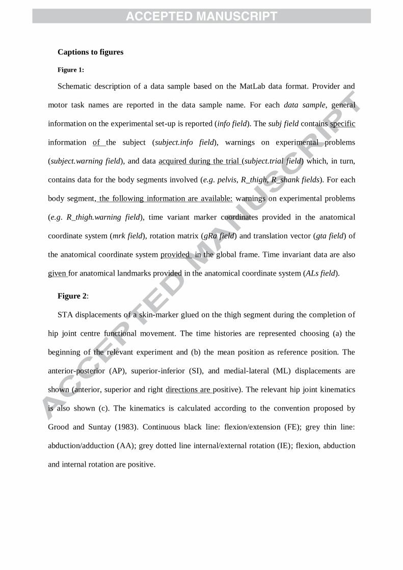
Figure 1:
Schematic description of a data sample based on the MatLab data format. Provider and
motor task names are reported in the data sample name. For each data sample, general
information on the experimental set-up is reported (info field). The subj field contains specific
information of the subject (subject.info field), warnings on experimental problems
(subject.warning field), and data acquired during the trial (subject.trial field) which, in turn,
contains data for the body segments involved (e.g. pelvis, R_thigh, R_shank fields). For each
body segment, the following information are available: warnings on experimental problems
(e.g. R_thigh.warning field), time variant marker coordinates provided in the anatomical
coordinate system (mrk field), rotation matrix (gRa field) and translation vector (gta field) of
the anatomical coordinate system provided in the global frame. Time invariant data are also
given for anatomical landmarks provided in the anatomical coordinate system (ALs field).
Figure 2:
STA displacements of a skin-marker glued on the thigh segment during the completion of
hip joint centre functional movement. The time histories are represented choosing (a) the
beginning of the relevant experiment and (b) the mean position as reference position. The
anterior-posterior (AP), superior-inferior (SI), and medial-lateral (ML) displacements are
shown (anterior, superior and right directions are positive). The relevant hip joint kinematics
is also shown (c). The kinematics is calculated according to the convention proposed by
Grood and Suntay (1983). Continuous black line: flexion/extension (FE); grey thin line:
abduction/adduction (AA); grey dotted line internal/external rotation (IE); flexion, abduction
and internal rotation are positive.
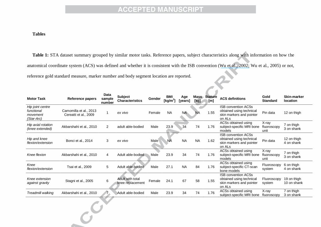
Tables
Table 1: STA dataset summary grouped by similar motor tasks. Reference papers, subject characteristics along with information on how the
anatomical coordinate system (ACS) was defined and whether it is consistent with the ISB convention (Wu et al., 2002; Wu et al., 2005) or not,
reference gold standard measure, marker number and body segment location are reported.
Motor Task Reference papers Data
sample number
Subject Characteristics
Gender BMI
[kg/m2]
Age [years]
Mass [kg]
Stature [m]
ACS definitions Gold Standard
Skin-marker location
Hip joint centre functional movement (Star-Arc)
Camomilla et al., 2013 Cereatti et al., 2009
1 ex vivo Female NA NA NA 1.55
ISB convention ACSs obtained using technical skin markers and pointer on ALs
Pin data 12 on thigh
Hip axial rotation (knee extended)
Akbarshahi et al., 2010 2 adult able-bodied Male 23.9 34 74 1.76 ACSs obtained using subject-specific MRI bone models
X-ray fluoroscopy unit
7 on thigh 3 on shank
Hip and knee flexion/extension
Bonci et al., 2014 3 ex vivo Male NA NA NA 1.62
ISB convention ACSs obtained using technical skin markers and pointer on ALs
Pin data 12 on thigh 4 on shank
Knee flexion Akbarshahi et al., 2010 4 Adult able-bodied Male 23.9 34 74 1.76 ACSs obtained using subject-specific MRI bone models
X-ray fluoroscopy unit
7 on thigh 3 on shank
Knee flexion/extension
Tsai et al., 2009 5 Adult able-bodied Male 27.1 NA 84 1.76 ACSs obtained using subject-specific CT-scan bone models
Fluoroscopy system
6 on thigh 4 on shank
Knee extension against gravity
Stagni et al., 2005 6 Adult with total knee replacement
Female 24.1 67 58 1.55
ISB convention ACSs obtained using technical skin markers and pointer on ALs
Fluoroscopy system
19 on thigh 10 on shank
Treadmill walking Akbarshahi et al., 2010 7 Adult able-bodied Male 23.9 34 74 1.76 ACSs obtained using subject-specific MRI bone
X-ray fluoroscopy
7 on thigh 3 on shank
models unit
Treadmill walking Barré et al., 2014 8 Postero-stabilized total knee prosthesis patient
Female 23.3 75 65 1.67 ACSs defined on the knee prosthesis
Fluoroscopy system
80 over one lower limb
Overground walking Tsai et al., 2009 9 Adult able-bodied Male 27.4 NA 83 1.74 ACSs obtained using subject-specific CT-scan bone models
Fluoroscopy system
6 on thigh 4 on shank
Overground walking Benoit et al., 2006 10 Adult able-bodied Male 20.6 22 63 1.75 ACSs obtained using ALs identified on recorded RSA
Pin data 4 on thigh 4 on shank
Lateral cutting manoeuvres
Benoit et al., 2006 11 Adult able-bodied Male 20.6 22 63 1.75 ACSs obtained using ALs identified on recorded RSA
Pin data 4 on thigh 4 on shank
Sit-to-stand Tsai et al., 2009 12 Adult able-bodied Male 27.4 NA 83 1.74 ACSs obtained using subject-specific CT-scan bone models
Fluoroscopy system
6 on thigh 4 on shank
Sit-to-stand Kuo et al., 2011 13
Adult with posterior cruciate ligament retaining mobile bearing total knee replacement
Female 33.5 NA 87 1.61
ACSs obtained using subject-specific computer-aided design models of the knee prosthesis
Fluoroscopy system
6 on thigh 4 on shank
Sit-to-stand/stand-to-sit
Stagni et al., 2005 14 Adult with total knee replacement
Female 24.1 67 58 1.55
ISB convention ACSs obtained using technical skin markers and pointer on ALs
Fluoroscopy system
19 on thigh 10 on shank
Step-up Akbarshahi et al., 2010 15 Adult able-bodied Male 23.9 34 74 1.76 ACSs obtained using subject-specific MRI bone models
X-ray fluoroscopy unit
7 on thigh 3 on shank
Step-up Tsai et al., 2011 16 Adult able-bodied Male 27.4 NA 83 1.74 ACSs obtained using subject-specific CT-scan bone models .
Fluoroscopy system
6 on thigh 4 on shank
Step-up/down Stagni et al., 2005 17 Adult with total knee replacement
Female 24.1 67 58 1.55
ISB convention ACSs obtained ACSs defined using technical skin markers and pointer on ALs
Fluoroscopy system
19 on thigh 10 on shank
Running Reinschmidt et al.,1997 18 Adult able-bodied Male
ACSs assumed to be parallel to the global frame during a standing trial
Pin data 5 on thigh 6 on shank
Hopping Benoit et al., 2006
Andersen et al., 2012 19 Adult able-bodied Male 20.6 22 63 1.75
ACSs obtained using ALs identified on recorded
Pin data 4 on thigh 4 on shank
RSA
Arm adduction Dal Maso et al., 2015 20 Adult able-bodied Male 20.9 27 57 1.65 ISB convention ACSs obtained using skin markers located on ALs.
Pin data 9 on scapula 7 on humerus 6 on thorax
Arm abduction Dal Maso et al., 2015 21 Adult able-bodied Male 20.9 27 57 1.65 ISB convention ACSs obtained using skin markers located on ALs.
Pin data 9 on scapula 7 on humerus, 6 on thorax
Arm abduction Charbonnier et al.,
2014 22 Adult able-bodied Male 24.7 25 80 1.80
ISB convention ACSs obtained using ALs identified on the reconstructed bone models and MR images
Fluoroscopy at 30Hz
4 on upper arm 57 on the shoulder blade
Arm flexion Dal Maso et al., 2015 23 Adult able-bodied Male 20.9 27 57 1.65 ISB convention ACSs obtained using skin markers located on ALs.
Pin data 9 on scapula 7 on humerus, 6 on thorax
Arm flexion Charbonnier et al.,
2014 24 Adult able-bodied Male 24.7 25 80 1.80
ISB convention ACSs obtained using ALs identified on the reconstructed bone models and MR images
Fluoroscopy at 30Hz
4 on upper arm 57 on the shoulder blade
Arm extension Dal Maso et al., 2015 25 Adult able-bodied Male 20.9 27 57 1.65 ISB convention ACSs obtained using skin markers located on ALs.
Pin data 9 on scapula 7 on humerus, 6 on thorax
Hair combing Dal Maso et al., 2015 26 Adult able-bodied Male 20.9 27 57 1.65 ISB convention ACSs obtained using skin markers located on ALs.
Pin data 9 on scapula 7 on humerus, 6 on thorax
Ball throwing Dal Maso et al., 2015 27 Adult able-bodied Male 20.9 27 57 1.65 ISB convention ACSs obtained using skin markers located on ALs.
Pin data 9 on scapula 7 on humerus, 6 on thorax
Eating Dal Maso et al., 2015 28 Adult able-bodied Male 20.9 27 57 1.65 ISB convention ACSs obtained using skin markers located on ALs.
Pin data 9 on scapula 7 on humerus, 6 on thorax
Gleno-humeral functional movement
Dal Maso et al., 2015 29 Adult able-bodied Male 20.9 27 57 1.65 ISB convention ACSs obtained using skin markers located on ALs.
Pin data 9 on scapula 7 on humerus, 6 on thorax
Punching Dal Maso et al., 2015 30 Adult able-bodied Male 20.9 27 57 1.65 ISB convention ACSs obtained using skin markers located on ALs.
Pin data 9 on scapula 7 on humerus, 6 on thorax
Reaching the back Dal Maso et al., 2015 31 Adult able-bodied Male 20.9 27 57 1.65 ISB convention ACSs obtained using skin markers located on ALs.
Pin data 9 on scapula 7 on humerus, 6 on thorax
32
Table 2: First, second and third quartile of the standardized and common metrics used for STA characterization (i.e., “mean” and “maximum”
STA amplitude, rmsd and , respectively). Statistics performed over n skin-markers glued on the relevant segment. Values are calculated for
the available data samples and grouped in terms of body segment and motor task.
THIGH
SHANK
rmsd
Δpmax
n rmsd
Δpmax
n 1
st 2
nd 3
rd
1st 2
nd 3
rd
1st 2
nd 3
rd
1st 2
nd 3
rd
Lo
wer
Lim
b
Hip joint centre functional movement (Star-Arc)
Camomilla et al., 2013;Cereatti et al., 2009
4.4 (5.9) 7.1
17.6 (21.6) 25.9 12 – – –
– – – –
Akbarshahi et al., 2010 5.4 (6.7) 8.3 13.8 (17.7) 23.9 7
3.6 (3.7) 5.0 9.7 (10.1) 14.8 3
Hip and knee flexion/extension Bonci et al., 2014 5.7 (6.1) 6.4 15.1 (16.4) 17.7 12
1.2 (1.3) 1.4 4.1 (4.3) 4.4 4
Knee flexion/extension Akbarshahi et al., 2010 6.9 (7.4) 9.4
23.8 (25.5) 29.3 7
7.4 (8.6) 9.1
26.6 (31.7) 33.7 3
Tsai et al., 2009 8.6 (9.2) 13.1 26.4 (27.6) 41.4 6
3.6 (5.0) 7.1 15.3 (18.7) 22.4 4
Treadmill walking Akbarshahi et al., 2010 10.1 (13.7) 14.9
30.0 (41.2) 44.7 7
7.1 (7.5) 8.2
18.0 (18.9) 20.1 3
Barrè et al., 2014 6.8 (8.4) 9.7 23.4 (28.4) 32.7 35
2.5 (2.7) 2.9 10.0 (12.0) 14.1 26
Overground walking Tsai et al., 2009 7.3 (8.0) 9.3
21.3 (23.4) 27.3 6
2.4 (2.4) 2.4
7.9 (8.4) 8.7 4
Benoit et al., 2006 6.3 (7.6) 8.6 22.0 (24.0) 24.3 4
3.9 (4.5) 5.0 22.2 (26.3) 28.7 4
Lateral cutting manoeuvres Benoit et al., 2006 6.5 (7.3) 8.1 19.5 (22.2) 26.5 4
2.1 (2.4) 2.6 6.2 (7.1) 7.9 4
Sit-to-stand
Tsai et al., 2009 12.0 (12.9) 13.9
32.2 (35.3) 38.6 6
1.8 (2.2) 3.5
5.4 (7.2) 11.1 4
Stagni et al., 2005 22.9 (25.3) 27.7
65.4 (72.3) 75.7 19
7.3 (7.6) 8.0
27.7 (29.1) 29.5 10
Kuo et al., 2011 7.4 (8.0) 12.0 21.7 (22.2) 33.3 6
2.6 (3.0) 3.9 9.6 (11.4) 13.7 4
Step-up
Akbarshahi et al., 2010 12.0 (12.4) 12.7
33.5 (34.8) 37.2 7
6.6 (7.6) 8.1
22.4 (26.4) 27.6 3
Tsai et al., 2011 14.8 (15.1) 18.6
39.7 (46.5) 47.3 6
4.5 (5.0) 6.1
13.4 (15.6) 18.2 4
Stagni et al., 2005 12.1 (14.9) 16.1 32.7 (41.8) 46.9 19
7.4 (7.5) 8.0 27.2 (28.5) 29.4 10
Knee extension against gravity Stagni et al., 2005 7.3 (9.2) 10.4 24.0 (26.7) 31.4 19
10.8 (11.5) 12.2 31.3 (32.5) 33.3 10
Running Reinschmidt et al.,1997 6.0 (6.9) 7.7 15.4 (21.1) 25.6 5
4.2 (4.7) 5.4 11.9 (12.8) 14.4 6
Hopping Andersen et al., 2012 3.5 (4.1) 4.7 16.1 (18.3) 19.7 4 1.4 (1.7) 1.8 11.9 (15.2) 17.9 4
maxp
33
ARM
SCAPULA
rmsd
Δpmax
n rmsd
Δpmax
n 1st 2
nd 3
rd
1
st 2
nd 3
rd
1
st 2
nd 3
rd
1
st 2
nd 3
rd
Up
per
Lim
b
Arm adduction Dal Maso et al., 2015 3.9 (4.1) 6.0
11.2 (11.8) 20.0 7
18.2 (19.9) 21.4
50.9 (54.0) 57.7 8
Arm abduction Dal Maso et al., 2015 3.8 (4.5) 6.3
10.8 (12.3) 15.9 7
12.2 (15.2) 21.7
35.7 (45.6) 62.7 8
Charbonnier et al., 2014 14.0 (15.3) 15.5
35.8 (41.2) 46.7 4
7.2 (10.2) 15.2
23.9 (28.7) 42.7 57
Arm flexion Dal Maso et al., 2015 3.7 (5.2) 7.5
11.4 (16.3) 20.7 7
9.3 (12.5) 17.4
24.9 (33.6) 47.5 8
Charbonnier et al., 2014 7.3 (8.6) 11.5
20.5 (22.3) 30.7 4
14.6 (21.0) 27.7
41.0 (56.8) 68.6 57
Arm extension Dal Maso et al., 2015 3.7 (5.0) 7.3
11.8 (15.0) 20.6 7
7.5 (8.5) 9.2
24.8 (27.8) 29.5 8
Hair combing Dal Maso et al., 2015 4.8 (5.2) 7.7
16.2 (20.7) 27.4 7
6.8 (10.0) 16.3
22.2 (34.3) 55.0 8
Ball throwing Dal Maso et al., 2015 4.3 (4.8) 6.8
15.3 (20.6) 26.6 7
34.4 (34.8) 36.9
110.5 (111.3) 111.7 8
Eating Dal Maso et al., 2015 5.2 (6.0) 6.5
16.0 (16.8) 18.2 7
6.8 (7.9) 12.8
20.9 (23.9) 37.6 8
Gleno-humeral functional movement
Dal Maso et al., 2015 2.8 (2.9) 4.7
12.6 (13.0) 16.6 7
12.4 (13.5) 18.1
53.4 (54.3) 58.6 8
Punching Dal Maso et al., 2015 3.8 (4.5) 4.7
16.2 (18.5) 19.5 7
18.8 (19.0) 19.9
62.5 (63.4) 64.5 8
Reaching the back Dal Maso et al., 2015 4.8 (6.8) 9.2
13.6 (17.5) 25.9 7
5.5 (5.6) 7.1
18.3 (19.7) 23.7 8
View publication statsView publication stats