stem cell-derived clade f aavs mediate high-efficiency ... · stem cell-derived clade f aavs...
TRANSCRIPT
Stem cell-derived clade F AAVs mediate high-efficiencyhomologous recombination-based genome editingLaura J. Smitha, Jason Wrightb, Gabriella Clarka, Taihra Ul-Hasana, Xiangyang Jina, Abigail Fonga, Manasa Chandraa,Thia St Martinb, Hillard Rubinb, David Knowltonb, Jeff L. Ellsworthb, Yuman Fonga, Kamehameha K. Wong Jr.c,and Saswati Chatterjeea,1
aDepartment of Surgery, Beckman Research Institute, City of Hope Medical Center, Duarte, CA 91010; bHomology Medicines, Inc., Bedford, MA 01730;and cDepartment of Hematology and Stem Cell Transplantation, City of Hope Medical Center, Duarte, CA 91010
Edited by Kenneth I. Berns, University of Florida College of Medicine, Gainesville, FL, and approved June 14, 2018 (received for review February 7, 2018)
The precise correction of genetic mutations at the nucleotide levelis an attractive permanent therapeutic strategy for human disease.However, despite significant progress, challenges to efficient andaccurate genome editing persist. Here, we report a genome edit-ing platform based upon a class of hematopoietic stem cell (HSC)-derived clade F adeno-associated virus (AAV), which does not re-quire prior nuclease-mediated DNA breaks and functions exclusivelythrough BRCA2-dependent homologous recombination. Genomeediting is guided by complementary homology arms and is highlyaccurate and seamless, with no evidence of on-target mutations,including insertion/deletions or inclusion of AAV inverted terminalrepeats. Efficient genome editing was demonstrated at different lociwithin the human genome, including a safe harbor locus, AAVS1,and the therapeutically relevant IL2RG gene, and at the murineRosa26 locus. HSC-derived AAV vector (AAVHSC)-mediated genomeediting was robust in primary human cells, including CD34+ cells,adult liver, hepatic endothelial cells, and myocytes. Importantly,high-efficiency gene editing was achieved in vivo upon a single i.v.injection of AAVHSC editing vectors in mice. Thus, clade F AAV-mediated genome editing represents a promising, highly efficient,precise, single-component approach that enables the developmentof therapeutic in vivo genome editing for the treatment of a multi-tude of human gene-based diseases.
adeno-associated virus | genome editing | homologous recombination |hematopoietic stem cells | in vivo genome editing
The ultimate goal of genetic medicine is to precisely and ef-ficiently edit the human genome to correct somatic mutations
or insert therapeutic sequences at targeted chromosomal loca-tions. Despite significant recent progress, current genome edit-ing technologies have yet to attain therapeutically relevant levelsof efficiency and precision, particularly in vivo. The majority ofcurrent gene editing platforms utilize nuclease-mediated DNAcleavage as the first step in editing events. The efficiency andaccuracy of nuclease-mediated cleavage are often variable (1),and repair of double-stranded DNA breaks by homologous re-combination (HR) is low (2–11). These nuclease-induced DNAbreaks are then repaired by the error-prone nonhomologousend-joining (NHEJ) pathway, which results in random nucleo-tide insertions/deletions (indels) at the cleavage site, potentiallycreating undesirable on-target mutations (12) or insertion of theentire vector genome, including the inverted terminal repeats(ITRs) (13). In contrast, DNA break repair by HR is moreprecise and can be engineered to accurately insert specific se-quences at precise chromosomal locations, but it occurs at alower frequency than NHEJ-mediated repair (2–11, 14–16).While nuclease-based genome editing is effective for gene dis-ruption, the predictable correction of mutations and targetedinsertion of therapeutic sequences are harder to achieve due tothe combination of the low frequency of HR repair coupled withthe requirement to deliver multiple components to target cells.Another significant challenge to the therapeutic translation ofgenome editing is that classic HR is restricted to dividing cells,
thus excluding most cells in vivo, which exist in a postmitoticstate (17). Nuclease-based editing platforms also carry the bur-den of promiscuous off-target cleavage by which double-strandedDNA breaks are created at unintended sites, resulting in thepotential for genome-wide mutagenesis (18–23). Targeting ofnucleases to specific genomic sites is accomplished by the use ofDNA-binding domains or guide RNA, which can place constraintson the exact location of cleavage sites (23). Finally, efficient invivo delivery of genome-editing components to target tissues re-mains challenging. Most platforms consist of multiple compo-nents, which are often too large to fit into a single delivery vector.To achieve genome editing in vivo, each target cell would requirethe delivery of multiple editing components, further reducing ef-ficiency. Thus, while much effort has been focused upon trans-lating genome editing to therapeutic applications, significantchallenges persist. An accurate, efficient, and predictable genomeediting technology based solely on HR pathways would thusrepresent a significant advance in the field and would allow pre-cise targeted insertion of therapeutic sequences and gene cor-rection without the risk of on-target mutagenesis or promiscuousoff-target effects. If such an editing platform functioned efficientlyin postmitotic cells in vivo and consisted of a single component
Significance
Precise genomic correction of pathogenic mutations is an at-tractive therapeutic strategy. Here, we show that a uniqueclass of stem cell-derived nonpathogenic virus, hematopoieticstem cell-derived adeno-associated virus vector (AAVHSC),mediates precise genome editing at unprecedented efficienciesin primary human cells, including postmitotic cells, and in vivo.Unlike the majority of current editing platforms, genomeediting by AAVHSC is uniquely based on homologous re-combination (HR), requiring no exogenous nucleases. AAVHSC-mediated editing is seamless, with no evidence of on-targetinsertion/deletion mutations common to nuclease-based plat-forms. Efficient in vivo editing was achieved by i.v. injection ofAAVHSC editing vectors. The combination of efficient andprecise HR-based genome editing coupled with the superior invivo transduction properties of AAV facilitates progress towardin vivo therapeutic gene editing.
Author contributions: J.W., Y.F., K.K.W., and S.C. designed research; L.J.S., J.W., G.C.,T.U.-H., X.J., A.F., M.C., T.S.M., H.R., D.K., K.K.W., and S.C. performed research; L.J.S.,J.W., X.J., J.L.E., K.K.W., and S.C. analyzed data; and L.J.S., J.W., and S.C. wrote the paper.
Conflict of interest statement: S.C. and L.J.S. are cofounders of Homology Medicines, Inc.,and S.C., L.J.S., J.W., T.S.M., H.R., J.L.E., and D.K. hold equity in Homology Medicines, Inc.
This article is a PNAS Direct Submission.
Published under the PNAS license.1To whom correspondence should be addressed. Email: [email protected].
This article contains supporting information online at www.pnas.org/lookup/suppl/doi:10.1073/pnas.1802343115/-/DCSupplemental.
www.pnas.org/cgi/doi/10.1073/pnas.1802343115 PNAS Latest Articles | 1 of 10
MED
ICALSC
IENCE
S

amenable to in vivo use, it would further resolve the significantchallenges currently facing therapeutic genome editing.Adeno-associated virus (AAV)–based vectors have previously
been shown to mediate genome editing without the requirementfor exogenous nucleases (24–29). However, the frequencies ofgenome editing reported were very low, generally well below0.1%. In this study, we interrogated a class of CD34+ hemato-poietic stem cell-derived AAV vectors (AAVHSCs) (30), fortheir genome editing capacity. The AAVHSCs represent a familyof natural, human, nonpathogenic, single-stranded, replication-defective AAVs that infect both dividing and nondividing cellsand display broad systemic tropism in vivo (30–36). Here, weshow that AAVHSCs mediate precise, efficient on-target ge-nome editing in primary human cells and at multiple genomiclocations, including therapeutically relevant genes in vitro and invivo, without the requirement for prior nucleolytic DNA cleav-age. Importantly, AAVHSC-based genome editing occurs ex-clusively via the HR pathway and is highly efficient in primaryhuman cells. We further show that the precision of on-targetediting is highly accurate, with no evidence of genomic scar-ring, indels, or incorporation of a residual viral footprint. Wedemonstrate a highly precise, exclusively HR-based, nuclease-free genome editing platform that functions at unparalleled ef-
ficiencies in primary cells and in vivo and is composed of a singlecomponent with built-in in vivo delivery properties.
ResultsAAVHSCs Mediate Efficient Genome Editing in Human Cells in theAbsence of Exogenous Nucleases. We designed a reporter-basedediting assay to measure the targeted genomic insertion of apromoterless green fluorescent protein (GFP) into the safe harborlocus, AAVS1, located on human chromosome 19 (37), such thatexpression would be dependent upon accurate insertion down-stream of a chromosomal promoter (Fig. 1A). The editing vectorgenome was bounded at both ends by AAV ITRs, consistedof a promoterless GFP ORF immediately downstream of asplice acceptor (SA) and T2A (SA/T2A) sequence followed by apolyadenylation (pA) signal, and was flanked bilaterally by 800-bphomology arms (HAs) complementary to the PPP1R12C genesequence on human chromosome 19 within the safe harbor locusAAVS1 (37). This vector, termed PPP1R12C-GFP, was designedto insert the GFP ORF into intron 1 of the PPP1R12C gene (Fig.1A and SI Appendix, Table S1). We reasoned that accurate editingof the promoterless GFP cassette into the intron downstream ofthe PPP1R12C promoter would result in GFP expression drivenby the endogenous PPP1R12C promoter (Fig. 1A).
B
% G
FP
po
siti
ve
3530
25
20
15
10
5
0
1.5E0.75E1.0E
30
20
10
0
40
50
60
1.5E 3.0E 4.5E0.0MOI:
C
AAV6AAV2
AAV8AAVHSC1
AAVHSC4
AAVHSC5
AAVHSC7
AAVHSC9
AAVHSC12
AAVHSC13
AAVHSC15
AAVHSC16
AAVHSC17
AAV9
GFP+ < 5% GFP+ > 50%
D
% G
FP
po
siti
ve
0
20
40
60
80 ***p = 10-7
***p = 10-9
Untd
Clade A
Clade B
Clade E
Clade F
VG/CD34
nucleus:5.50 0.19 4.27 149.30n/a
**p = 10-5
**p = 10-5AAV6AAVHSC7AAVHSC17
HepG2
AAVHSC7AAVHSC5
AAV6AAVHSC17
CD34 +
1
32
A
5’3’
5’3’
PPP1R12C locus
EditedEx1 Ex2 Ex3
5’3’
P
P
Editing vector
PPP1R12C locus
PPP1R12C-GFP (ssDNA)
ITR GFP ITRSA/2A PA
HARHAL
HARHAL
GFP
PPP1R12C locus
Fig. 1. Genome editing of a promoterless GFP into intron 1 of the human PPP1R12C gene. (A) Schema for genome editing assay. (Upper) Map of thePPP1R12C-GFP editing vector genome. In line 1, the insert cassette consists of a GFP ORF (green) preceded by SA/T2A (SA/2A) sequences and followed by a pA(PA) signal (cyan), and it is flanked on either side by 800-bp HAs (blue) complementary to chromosomal sequences in PPP1R12C. HAL, left HA; HAR, right HA.The entire editing construct is bounded by AAV2 ITRs (purple). In line 2, the PPP1R12C gene is depicted. Promoter (P) is shown in yellow, exons (Ex) are shownin dark green, and introns are shown in pink. Line 3 depicts a hypothetical HR schema. Line 4 depicts the edited PPP1R12C gene showing the insertion of theGFP cassette into intron 1. (B) Dose–response curves correlating genome editing with the MOI of the PPP1R12C-GFP editing vector. The editing vector genomewas packaged in the capsids noted within each box. (Left) Editing of primary human CD34+ cells. (Right) Editing of the human hepatocellular carcinoma cellline HepG2. (C) Heat map showing genome editing efficiency of targeted insertion of GFP into PPP1R12C by AAV serotype (columns) in different human celltypes (rows) as follows: (1) transformed cell lines, (2) primary human cells, and (3) immortalized LCLs carrying genetic mutations. Values represent specific GFPexpression as assessed by flow cytometry. GFP expression was assessed only in viable cells, as determined by DAPI exclusion. Data shown are aggregatesof >906 individual measurements. Cell type details are shown in SI Appendix, Fig. S1. (D) Comparison of median genome editing efficiency across AAV clade A(AAV6), clade B (AAV2), clade E (AAV8), and clade F (AAV9) (AAVHSCs). Editing efficiencies were measured by GFP expression by flow cytometry. Cells weretransduced at a MOI of 1.5E5 and evaluated by flow cytometry at 48 h. Sample sizes for the experimental groups are as follows: untreated (Untd), n = 25;AAV6, n = 33; AAV2, n = 17; AAV8, n = 19; AAV9, n = 30; and AAVHSCs combined, n = 390. AAVHSC represents data compiled from AAVHSC1, AAVHSC4,AAVHSC5, AAVHSC7, AAVHSC9, AAVHSV12, AAVHSC13, AAVHSV15, AAVHSV16, and AAVHSC17. Outliers are represented by individual circles. Significancewas determined by a paired two-tailed t test using AAVHSCs as the comparison reference. The vector genomes (VG) were quantitated in nuclei purified fromAAV-treated CD34+ cells 48 h posttreatment. The number of VG per nucleus was determined by real-time PCR for GFP and the housekeeping gene hApoB.Values shown are averages of three replicates per transduction and three transductions with each AAV vector.
2 of 10 | www.pnas.org/cgi/doi/10.1073/pnas.1802343115 Smith et al.
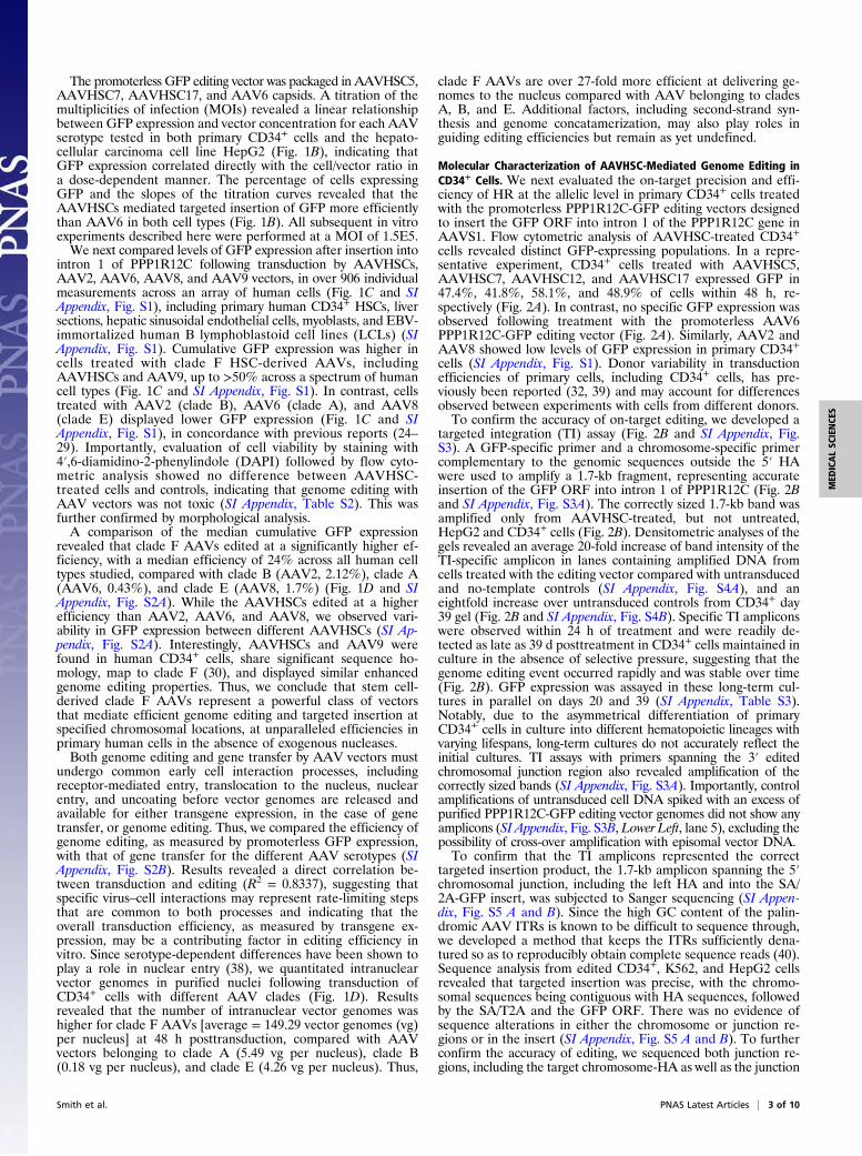
The promoterless GFP editing vector was packaged in AAVHSC5,AAVHSC7, AAVHSC17, and AAV6 capsids. A titration of themultiplicities of infection (MOIs) revealed a linear relationshipbetween GFP expression and vector concentration for each AAVserotype tested in both primary CD34+ cells and the hepato-cellular carcinoma cell line HepG2 (Fig. 1B), indicating thatGFP expression correlated directly with the cell/vector ratio ina dose-dependent manner. The percentage of cells expressingGFP and the slopes of the titration curves revealed that theAAVHSCs mediated targeted insertion of GFP more efficientlythan AAV6 in both cell types (Fig. 1B). All subsequent in vitroexperiments described here were performed at a MOI of 1.5E5.We next compared levels of GFP expression after insertion into
intron 1 of PPP1R12C following transduction by AAVHSCs,AAV2, AAV6, AAV8, and AAV9 vectors, in over 906 individualmeasurements across an array of human cells (Fig. 1C and SIAppendix, Fig. S1), including primary human CD34+ HSCs, liversections, hepatic sinusoidal endothelial cells, myoblasts, and EBV-immortalized human B lymphoblastoid cell lines (LCLs) (SIAppendix, Fig. S1). Cumulative GFP expression was higher incells treated with clade F HSC-derived AAVs, includingAAVHSCs and AAV9, up to >50% across a spectrum of humancell types (Fig. 1C and SI Appendix, Fig. S1). In contrast, cellstreated with AAV2 (clade B), AAV6 (clade A), and AAV8(clade E) displayed lower GFP expression (Fig. 1C and SIAppendix, Fig. S1), in concordance with previous reports (24–29). Importantly, evaluation of cell viability by staining with4′,6-diamidino-2-phenylindole (DAPI) followed by flow cyto-metric analysis showed no difference between AAVHSC-treated cells and controls, indicating that genome editing withAAV vectors was not toxic (SI Appendix, Table S2). This wasfurther confirmed by morphological analysis.A comparison of the median cumulative GFP expression
revealed that clade F AAVs edited at a significantly higher ef-ficiency, with a median efficiency of 24% across all human celltypes studied, compared with clade B (AAV2, 2.12%), clade A(AAV6, 0.43%), and clade E (AAV8, 1.7%) (Fig. 1D and SIAppendix, Fig. S2A). While the AAVHSCs edited at a higherefficiency than AAV2, AAV6, and AAV8, we observed vari-ability in GFP expression between different AAVHSCs (SI Ap-pendix, Fig. S2A). Interestingly, AAVHSCs and AAV9 werefound in human CD34+ cells, share significant sequence ho-mology, map to clade F (30), and displayed similar enhancedgenome editing properties. Thus, we conclude that stem cell-derived clade F AAVs represent a powerful class of vectorsthat mediate efficient genome editing and targeted insertion atspecified chromosomal locations, at unparalleled efficiencies inprimary human cells in the absence of exogenous nucleases.Both genome editing and gene transfer by AAV vectors must
undergo common early cell interaction processes, includingreceptor-mediated entry, translocation to the nucleus, nuclearentry, and uncoating before vector genomes are released andavailable for either transgene expression, in the case of genetransfer, or genome editing. Thus, we compared the efficiency ofgenome editing, as measured by promoterless GFP expression,with that of gene transfer for the different AAV serotypes (SIAppendix, Fig. S2B). Results revealed a direct correlation be-tween transduction and editing (R2 = 0.8337), suggesting thatspecific virus–cell interactions may represent rate-limiting stepsthat are common to both processes and indicating that theoverall transduction efficiency, as measured by transgene ex-pression, may be a contributing factor in editing efficiency invitro. Since serotype-dependent differences have been shown toplay a role in nuclear entry (38), we quantitated intranuclearvector genomes in purified nuclei following transduction ofCD34+ cells with different AAV clades (Fig. 1D). Resultsrevealed that the number of intranuclear vector genomes washigher for clade F AAVs [average = 149.29 vector genomes (vg)per nucleus] at 48 h posttransduction, compared with AAVvectors belonging to clade A (5.49 vg per nucleus), clade B(0.18 vg per nucleus), and clade E (4.26 vg per nucleus). Thus,
clade F AAVs are over 27-fold more efficient at delivering ge-nomes to the nucleus compared with AAV belonging to cladesA, B, and E. Additional factors, including second-strand syn-thesis and genome concatamerization, may also play roles inguiding editing efficiencies but remain as yet undefined.
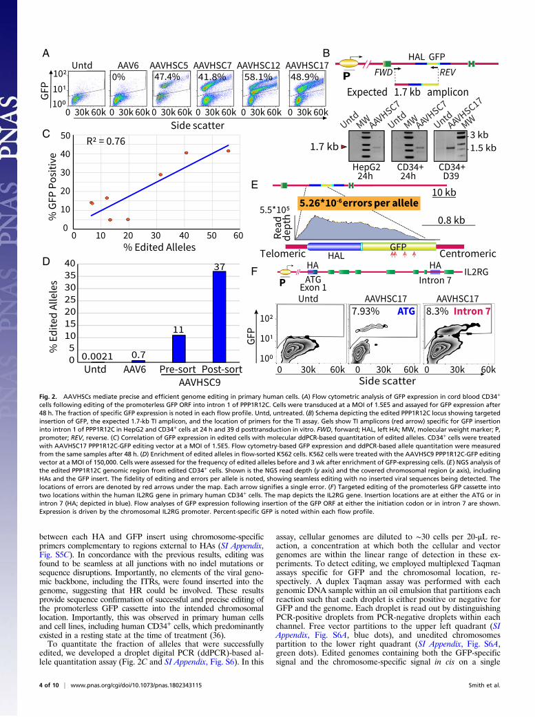
Molecular Characterization of AAVHSC-Mediated Genome Editing inCD34+ Cells. We next evaluated the on-target precision and effi-ciency of HR at the allelic level in primary CD34+ cells treatedwith the promoterless PPP1R12C-GFP editing vectors designedto insert the GFP ORF into intron 1 of the PPP1R12C gene inAAVS1. Flow cytometric analysis of AAVHSC-treated CD34+cells revealed distinct GFP-expressing populations. In a repre-sentative experiment, CD34+ cells treated with AAVHSC5,AAVHSC7, AAVHSC12, and AAVHSC17 expressed GFP in47.4%, 41.8%, 58.1%, and 48.9% of cells within 48 h, re-spectively (Fig. 2A). In contrast, no specific GFP expression wasobserved following treatment with the promoterless AAV6PPP1R12C-GFP editing vector (Fig. 2A). Similarly, AAV2 andAAV8 showed low levels of GFP expression in primary CD34+cells (SI Appendix, Fig. S1). Donor variability in transductionefficiencies of primary cells, including CD34+ cells, has pre-viously been reported (32, 39) and may account for differencesobserved between experiments with cells from different donors.To confirm the accuracy of on-target editing, we developed a
targeted integration (TI) assay (Fig. 2B and SI Appendix, Fig.S3). A GFP-specific primer and a chromosome-specific primercomplementary to the genomic sequences outside the 5′ HAwere used to amplify a 1.7-kb fragment, representing accurateinsertion of the GFP ORF into intron 1 of PPP1R12C (Fig. 2Band SI Appendix, Fig. S3A). The correctly sized 1.7-kb band wasamplified only from AAVHSC-treated, but not untreated,HepG2 and CD34+ cells (Fig. 2B). Densitometric analyses of thegels revealed an average 20-fold increase of band intensity of theTI-specific amplicon in lanes containing amplified DNA fromcells treated with the editing vector compared with untransducedand no-template controls (SI Appendix, Fig. S4A), and aneightfold increase over untransduced controls from CD34+ day39 gel (Fig. 2B and SI Appendix, Fig. S4B). Specific TI ampliconswere observed within 24 h of treatment and were readily de-tected as late as 39 d posttreatment in CD34+ cells maintained inculture in the absence of selective pressure, suggesting that thegenome editing event occurred rapidly and was stable over time(Fig. 2B). GFP expression was assayed in these long-term cul-tures in parallel on days 20 and 39 (SI Appendix, Table S3).Notably, due to the asymmetrical differentiation of primaryCD34+ cells in culture into different hematopoietic lineages withvarying lifespans, long-term cultures do not accurately reflect theinitial cultures. TI assays with primers spanning the 3′ editedchromosomal junction region also revealed amplification of thecorrectly sized bands (SI Appendix, Fig. S3A). Importantly, controlamplifications of untransduced cell DNA spiked with an excess ofpurified PPP1R12C-GFP editing vector genomes did not show anyamplicons (SI Appendix, Fig. S3B, Lower Left, lane 5), excluding thepossibility of cross-over amplification with episomal vector DNA.To confirm that the TI amplicons represented the correct
targeted insertion product, the 1.7-kb amplicon spanning the 5′chromosomal junction, including the left HA and into the SA/2A-GFP insert, was subjected to Sanger sequencing (SI Appen-dix, Fig. S5 A and B). Since the high GC content of the palin-dromic AAV ITRs is known to be difficult to sequence through,we developed a method that keeps the ITRs sufficiently dena-tured so as to reproducibly obtain complete sequence reads (40).Sequence analysis from edited CD34+, K562, and HepG2 cellsrevealed that targeted insertion was precise, with the chromo-somal sequences being contiguous with HA sequences, followedby the SA/T2A and the GFP ORF. There was no evidence ofsequence alterations in either the chromosome or junction re-gions or in the insert (SI Appendix, Fig. S5 A and B). To furtherconfirm the accuracy of editing, we sequenced both junction re-gions, including the target chromosome-HA as well as the junction
Smith et al. PNAS Latest Articles | 3 of 10
MED
ICALSC
IENCE
S
between each HA and GFP insert using chromosome-specificprimers complementary to regions external to HAs (SI Appendix,Fig. S5C). In concordance with the previous results, editing wasfound to be seamless at all junctions with no indel mutations orsequence disruptions. Importantly, no elements of the viral geno-mic backbone, including the ITRs, were found inserted into thegenome, suggesting that HR could be involved. These resultsprovide sequence confirmation of successful and precise editing ofthe promoterless GFP cassette into the intended chromosomallocation. Importantly, this was observed in primary human cellsand cell lines, including human CD34+ cells, which predominantlyexisted in a resting state at the time of treatment (36).To quantitate the fraction of alleles that were successfully
edited, we developed a droplet digital PCR (ddPCR)-based al-lele quantitation assay (Fig. 2C and SI Appendix, Fig. S6). In this
assay, cellular genomes are diluted to ∼30 cells per 20-μL re-action, a concentration at which both the cellular and vectorgenomes are within the linear range of detection in these ex-periments. To detect editing, we employed multiplexed Taqmanassays specific for GFP and the chromosomal location, re-spectively. A duplex Taqman assay was performed with eachgenomic DNA sample within an oil emulsion that partitions eachreaction such that each droplet is either positive or negative forGFP and the genome. Each droplet is read out by distinguishingPCR-positive droplets from PCR-negative droplets within eachchannel. Free vector partitions to the upper left quadrant (SIAppendix, Fig. S6A, blue dots), and unedited chromosomespartition to the lower right quadrant (SI Appendix, Fig. S6A,green dots). Edited genomes containing both the GFP-specificsignal and the chromosome-specific signal in cis on a single
A B
C
D
E
F
Fig. 2. AAVHSCs mediate precise and efficient genome editing in primary human cells. (A) Flow cytometric analysis of GFP expression in cord blood CD34+
cells following editing of the promoterless GFP ORF into intron 1 of PPP1R12C. Cells were transduced at a MOI of 1.5E5 and assayed for GFP expression after48 h. The fraction of specific GFP expression is noted in each flow profile. Untd, untreated. (B) Schema depicting the edited PPP1R12C locus showing targetedinsertion of GFP, the expected 1.7-kb TI amplicon, and the location of primers for the TI assay. Gels show TI amplicons (red arrow) specific for GFP insertioninto intron 1 of PPP1R12C in HepG2 and CD34+ cells at 24 h and 39 d posttransduction in vitro. FWD, forward; HAL, left HA; MW, molecular weight marker; P,promoter; REV, reverse. (C) Correlation of GFP expression in edited cells with molecular ddPCR-based quantitation of edited alleles. CD34+ cells were treatedwith AAVHSC17 PPP1R12C-GFP editing vector at a MOI of 1.5E5. Flow cytometry-based GFP expression and ddPCR-based allele quantitation were measuredfrom the same samples after 48 h. (D) Enrichment of edited alleles in flow-sorted K562 cells. K562 cells were treated with the AAVHSC9 PPP1R12C-GFP editingvector at a MOI of 150,000. Cells were assessed for the frequency of edited alleles before and 3 wk after enrichment of GFP-expressing cells. (E) NGS analysis ofthe edited PPP1R12C genomic region from edited CD34+ cells. Shown is the NGS read depth (y axis) and the covered chromosomal region (x axis), includingHAs and the GFP insert. The fidelity of editing and errors per allele is noted, showing seamless editing with no inserted viral sequences being detected. Thelocations of errors are denoted by red arrows under the map. Each arrow signifies a single error. (F) Targeted editing of the promoterless GFP cassette intotwo locations within the human IL2RG gene in primary human CD34+ cells. The map depicts the IL2RG gene. Insertion locations are at either the ATG or inintron 7 (HA; depicted in blue). Flow analyses of GFP expression following insertion of the GFP ORF at either the initiation codon or in intron 7 are shown.Expression is driven by the chromosomal IL2RG promoter. Percent-specific GFP is noted within each flow profile.
4 of 10 | www.pnas.org/cgi/doi/10.1073/pnas.1802343115 Smith et al.
molecule of DNA are represented in the partition to the upperright quadrant (SI Appendix, Fig. S6A, orange dots), as well asthe possible coincidence of genome and free vector. To controlfor the coincidence of GFP and genome probes, the number ofexpected double-positive droplets generated by chance wassubtracted from the observed number of double-positive drop-lets. A standard curve generated for this assay (SI Appendix, Fig.S5B) shows a correlation coefficient of 0.9717. SI Appendix,Table S4 provides the complete statistical analysis of the allelequantitation assay. To confirm the quantitation of editing and tocontrol for error due to sampling, each DNA sample was alsoanalyzed following restriction digestion with an enzyme thatcleaves between the GFP and chromosome-specific probe-bindingsites. This artificial separation of the editing signal from the ge-nome signal results in the independent partitioning of each probetarget (SI Appendix, Fig. S7A). SI Appendix, Fig. S7B and Table S5show that following restriction digestion, the signal in the upperright quadrant is fully resolved into the free vector and free locussignals, indicating that the edited allele signal (SI Appendix, Fig.S7B, orange dots) accurately represented the binding of bothvector and locus probes to the same piece of DNA (SI Appendix,Fig. S7B and Table S5). Thus, we conclude that this ddPCR-basedallele quantitation assay accurately measures edited chromosomes.To determine if GFP expression in edited cells correlated with
the frequency of edited alleles detected by ddPCR, we treatedCD34+ cells with the AAVHSC17 PPP1R12C-GFP editing vector.Results revealed that GFP expression, as measured by flowcytometry, was highly correlated with edited alleles (R2 = 0.76)(Fig. 2C). A detailed statistical analysis is shown in SI Appendix,Table S6. These results validated that GFP detection by flowcytometry was an accurate measure of genome editing. To furtherconfirm this, we tested whether flow-sorted, GFP-positive,AAVHSC-treated cells would display an enrichment of editedalleles. K562 erythroleukemia cells were treated with theAAVHSC9 PPP1R12C-GFP editing vector. Allele quantitation byddPCR before and after GFP-based flow sorting revealed an en-richment of edited alleles, with 37% of alleles being edited 6 wkafter transduction compared with 11% before sorting, indicatingthe stability of the AAVHSC-mediated genome editing over time(Fig. 2D). Given that K562 represents an aneuploid cell line, andthe possibility of monoallelic editing, these results confirmed thatGFP expression correlated well with the frequency of edited al-leles and was stable over time in culture without selective pressure.
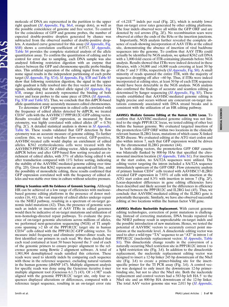
Editing Is Seamless with No Evidence of Genomic Scarring. AlthoughHR can be achieved at a low range of efficiencies with nuclease-based genome editing platforms in the presence of donor mol-ecules (16, 41), the vast majority of edited alleles undergo repairvia the NHEJ pathway, resulting in a spectrum of on-target ge-nomic indel mutations (42). Thus, the presence of genomic scarssuch as indels or insertion of AAV ITRs in edited genomeswould then be indicative of on-target mutations and utilization ofnon–homology-directed repair pathways. To evaluate the pres-ence of on-target genomic alterations across millions of alleles,we performed next-generation sequencing (NGS) of TI ampli-cons spanning >2 kb of the PPP1R12C target site in humanCD34+ cells edited with the PPP1R12C-GFP editing vector. Tomeasure indel frequency and eliminate primer-dimer reads, wefirst identified our primers in each read. We then checked thateach read contained at least 50 bases beyond the 3′ end of eachof the genomic primers to ensure proper alignment to the ref-erence genome using Bowtie 2 alignment software. A total of1,901,904 demultiplexed reads satisfied this criterion. Thesereads were used to identify indels by comparing each sequencewith those in the reference sequence, excluding natural variantsin the human genome (dbSNP 147). Multiple alignment analysisfor specific reads was done using the Geneious iterative k-mermultiple alignment tool (Geneious 6.1.7) (43). Of >1.9E6 readsaligned with the genomic PPP1R12C target site, only four se-quences displayed alterations or differences, compared with areference target sequence, resulting in an on-target error rate
of <4.21E−6 indels per read (Fig. 2E), which is notably lowerthan on-target error rates generated by other editing platforms.The four indels observed were all within the GFP ORF and aredenoted by red arrows (Fig. 2E). No recombination scars wereobserved at either the ends of the HAs or the insertion junctions.Importantly, NGS analysis further revealed the complete ab-
sence of reads showing incorporation of AAV ITRs at the editedsite, demonstrating the absence of insertion of viral backbonesequences into the genome. To confirm that AAV ITRs couldactually be identified by NGS analysis, we spiked K562 cell DNAwith a 1,000-fold excess of ITR-containing plasmids before NGSanalysis. Results showed that ITRs were indeed detected in theselibraries, with >34,000 and >8,000 reads covering at least 50 bpof the 5′ and 3′ ITRs, respectively (SI Appendix, Fig. S8). A smallminority of reads spanned the entire ITR, with the majority ofsequences dropping off after ∼60 bp. Thus, if ITRs were indeedincorporated at editing sites, at least 50 bp of each ITR sequencewould have been detectable in the NGS analysis. NGS analysisalso confirmed the findings of accurate and seamless editing asdetermined by Sanger sequencing (SI Appendix, Fig. S5). Theseresults established that AAVHSC-mediated genome editing ishighly precise and occurs without any concurrent on-target mu-tations commonly associated with DNA strand breaks and isconsistent with the utilization of an HR editing pathway.
AAVHSCs Mediate Genome Editing at the Human IL2RG Locus. Toconfirm that AAVHSC-mediated genome editing was not lim-ited to the single PPP1R12C locus, we tested editing of other lociwithin the human genome. We evaluated targeted insertion ofthe promoterless GFP ORF within two locations in the clinicallyrelevant human IL2RG locus, mutations of which cause X-linkedSCID disease. We evaluated editing at the ATG start codon andalso within intron 7, such that GFP expression would be drivenby the chromosomal IL2RG promoter (44).In both editing vectors, the promoterless GFP ORF cassette
was bilaterally flanked by 800-bp HAs that specified the chro-mosomal insertion location (SI Appendix, Table S1). For insertionat the start codon, no SA/T2A sequences were utilized. Theediting vector targeting the intron included a SA/T2A sequenceimmediately upstream of the GFP ORF. Flow cytometric analysisof primary human CD34+ cells treated with AAVHSC17-IL2RGrevealed GFP expression in 7.93% of cells with insertion at theATG start codon and 8.3% with insertion in intron 7 (Fig. 2F).Locus-dependent differences in gene editing efficiencies havebeen described and likely account for the differences in efficiencyobserved between the PPP1R12C and IL2RG loci (45). Thus, weconclude that AAVHSC-mediated editing of the IL2RG gene ispotentially therapeutic. Similarly, we have also observed successfulediting at two locations within the human factor VIII gene.
AAVHSCs Mediate Nucleotide Replacement. With current genomeediting technologies, correction of point mutations is challeng-ing. Instead of correcting mutations, DNA breaks repaired viathe NHEJ pathway result in unpredictable on-target indels andthe possible introduction of new mutations. Hence, we tested thepotential of AAVHSC vectors to accurately correct point mu-tations at the nucleotide level. A dinucleotide editing vector wasused to alter a wild-type “TA” sequence to an “AT” in intron 1 ofPPP1R12C (nucleotide replacement vector; SI Appendix, TableS1). This dinucleotide change results in the conversion of anaturally occurring NheI restriction site in PPP1R12C intron 1 toa SphI restriction site (Fig. 3A). In addition to the dinucleotidereplacement, the nucleotide replacement editing vector wasdesigned to insert a 12-bp linker 245 bp downstream of the NheIsite (Fig. 3A) to create a primer-binding site for the insert-specific primer for the TI PCR assay (Fig. 3A). A control vec-tor was designed to only insert the downstream 12-bp primer-binding site, but not to alter the NheI site. Both the nucleotidereplacement and control vectors had a 583-bp left HA 5′ to theNheI site and an 800-bp HA downstream of the 12-bp linker.The total AAV vector genome size was 2,011 bp (SI Appendix,
Smith et al. PNAS Latest Articles | 5 of 10
MED
ICALSC
IENCE
S
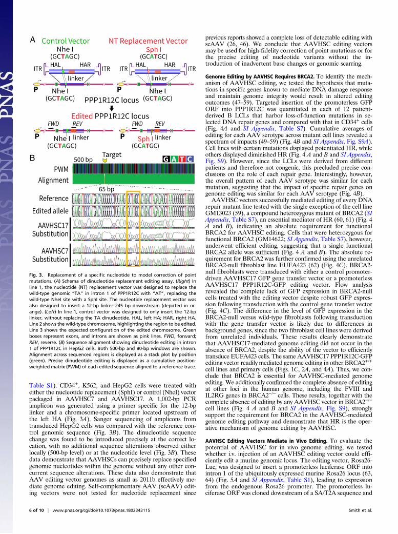
Table S1). CD34+, K562, and HepG2 cells were treated witheither the nucleotide replacement (SphI) or control (NheI) vectorpackaged in AAVHSC7 and AAVHSC17. A 1,002-bp PCRamplicon was generated using a primer specific for the 12-bplinker and a chromosome-specific primer located upstream ofthe left HA (Fig. 3A). Sanger sequencing of amplicons fromtransduced HepG2 cells was compared with the reference con-trol genomic sequence (Fig. 3B). The dinucleotide sequencechange was found to be introduced precisely at the correct lo-cation, with no additional sequence alterations observed eitherlocally (500-bp level) or at the nucleotide level (Fig. 3B). Thesedata demonstrate that AAVHSCs can precisely replace specifiedgenomic nucleotides within the genome without any other con-current sequence alterations. These data also demonstrate thatAAV editing vector genomes as small as 2011b effectively me-diate genome editing. Self-complementary AAV (scAAV) edit-ing vectors were not tested for nucleotide replacement since
previous reports showed a complete loss of detectable editing withscAAV (26, 46). We conclude that AAVHSC editing vectorsmay be used for high-fidelity correction of point mutations or forthe precise editing of nucleotide variants without the in-troduction of inadvertent base changes or genomic scarring.
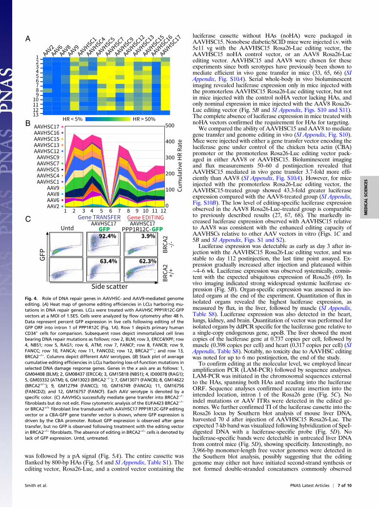
Genome Editing by AAVHSC Requires BRCA2. To identify the mech-anism of AAVHSC editing, we tested the hypothesis that muta-tions in specific genes known to mediate DNA damage responseand maintain genome integrity would result in altered editingoutcomes (47–59). Targeted insertion of the promoterless GFPORF into PPP1R12C was quantitated in each of 12 patient-derived B LCLs that harbor loss-of-function mutations in se-lected DNA repair genes and compared with that in CD34+ cells(Fig. 4A and SI Appendix, Table S7). Cumulative averages ofediting for each AAV serotype across mutant cell lines revealed aspectrum of impacts (49–59) (Fig. 4B and SI Appendix, Fig. S9A).Cell lines with certain mutations displayed potentiated HR, whileothers displayed diminished HR (Fig. 4 A and B and SI Appendix,Fig. S9). However, since the LCLs were derived from differentpatients and therefore not congenic, this precluded precise con-clusions on the role of each repair gene. Interestingly, however,the overall pattern of each AAV serotype was similar for eachmutation, suggesting that the impact of specific repair genes ongenome editing was similar for each AAV serotype (Fig. 4B).AAVHSC vectors successfully mediated editing of every DNA
repair mutant line tested with the single exception of the cell lineGM13023 (59), a compound heterozygous mutant of BRCA2 (SIAppendix, Table S7), an essential mediator of HR (60, 61) (Fig. 4A and B), indicating an absolute requirement for functionalBRCA2 for AAVHSC editing. Cells that were heterozygous forfunctional BRCA2 (GM14622; SI Appendix, Table S7), however,underwent efficient editing, suggesting that a single functionalBRCA2 allele was sufficient (Fig. 4 A and B). The absolute re-quirement for BRCA2 was further confirmed using the unrelatedBRCA2-null fibroblast line EUFA423 (62) (Fig. 4C). BRCA2-null fibroblasts were transduced with either a control promoter-driven AAVHSC17 GFP gene transfer vector or a promoterlessAAVHSC17 PPP1R12C-GFP editing vector. Flow analysisrevealed the complete lack of GFP expression in BRCA2-nullcells treated with the editing vector despite robust GFP expres-sion following transduction with the control gene transfer vector(Fig. 4C). The difference in the level of GFP expression in theBRCA2-null versus wild-type fibroblasts following transductionwith the gene transfer vector is likely due to differences inbackground genes, since the two fibroblast cell lines were derivedfrom unrelated individuals. These results clearly demonstratethat AAVHSC17-mediated genome editing did not occur in theabsence of BRCA2, despite the ability of the vector to efficientlytransduce EUFA423 cells. The same AAVHSC17 PPP1R12C-GFPediting vector readily mediated genome editing in other BRCA2+/+cell lines and primary cells (Figs. 1C, 2A, and 4A). Thus, we con-clude that BRCA2 is essential for AAVHSC-mediated genomeediting. We additionally confirmed the complete absence of editingat other loci in the human genome, including the FVIII andIL2RG genes in BRCA2−/− cells. These results, together with thecomplete absence of editing by any AAVHSC vector in BRCA2−/−
cell lines (Fig. 4 A and B and SI Appendix, Fig. S9), stronglysupport the requirement for BRCA2 in the AAVHSC-mediatedgenome editing pathway and demonstrate that HR is the oper-ative mechanism of genome editing by AAVHSC.
AAVHSC Editing Vectors Mediate in Vivo Editing. To evaluate thepotential of AAVHSC for in vivo genome editing, we testedwhether i.v. injection of an AAVHSC editing vector could effi-ciently edit a murine genomic locus. The editing vector, Rosa26-Luc, was designed to insert a promoterless luciferase ORF intointron 1 of the ubiquitously expressed murine Rosa26 locus (63,64) (Fig. 5A and SI Appendix, Table S1), leading to expressionfrom the endogenous Rosa26 promoter. The promoterless lu-ciferase ORF was cloned downstream of a SA/T2A sequence and
Substitution
B
Reference
Substitution
PWM
Alignment
Edited allele
AAVHSC7
AAVHSC17
G A T C500 bp
65 bp
Target
A
PPP1R12C locus
Edited PPP1R12C locus
Control Vector
P
ITRHARHAL
ITR
Nhe I(GCTAGC)
linker
REV FWD
linker
ITR
Sph I(GCATGC)
HARHALITR
NT Replacement Vector
linker
REV FWD
linker
P
P P
Nhe I(GCTAGC)
Nhe I(GCTAGC)
Nhe I(GCTAGC)
Sph I(GCATGC)
Fig. 3. Replacement of a specific nucleotide to model correction of pointmutations. (A) Schema of dinucleotide replacement editing assay. (Right) Inline 1, the nucleotide (NT) replacement vector was designed to replace thewild-type genomic “TA” in intron 1 of PPP1R12C with “AT”, replacing thewild-type NheI site with a SphI site. The nucleotide replacement vector wasalso designed to insert a 12-bp linker 245 bp downstream (depicted in or-ange). (Left) In line 1, control vector was designed to only insert the 12-bplinker, without replacing the TA dinucleotide. HAL, left HA; HAR, right HA.Line 2 shows the wild-type chromosome, highlighting the region to be edited.Line 3 shows the expected configuration of the edited chromosome. Greenboxes represent exons, and introns are shown as pink lines. FWD, forward;REV, reverse. (B) Sequence alignment showing dinucleotide editing in intron1 of PPP1R12C in HepG2 cells. Both 500-bp and 80-bp windows are shown.Alignment across sequenced regions is displayed as a stack plot by position(green). Precise dinucleotide editing is displayed as a cumulative position-weighted matrix (PWM) of each edited sequence aligned to a reference trace.
6 of 10 | www.pnas.org/cgi/doi/10.1073/pnas.1802343115 Smith et al.
was followed by a pA signal (Fig. 5A). The entire cassette wasflanked by 800-bp HAs (Fig. 5A and SI Appendix, Table S1). Theediting vector, Rosa26-Luc, and a control vector containing the
luciferase cassette without HAs (noHA) were packaged inAAVHSC15. Nonobese diabetic/SCID mice were injected i.v. with5e11 vg with the AAVHSC15 Rosa26-Luc editing vector, theAAVHSC15 noHA control vector, or an AAV8 Rosa26-Lucediting vector. AAVHSC15 and AAV8 were chosen for theseexperiments since both serotypes have previously been shown tomediate efficient in vivo gene transfer in mice (33, 65, 66) (SIAppendix, Fig. S10A). Serial whole-body in vivo bioluminescentimaging revealed luciferase expression only in mice injected withthe promoterless AAVHSC15 Rosa26-Luc editing vector, but notin mice injected with the control noHA vector lacking HAs, andonly nominal expression in mice injected with the AAV8 Rosa26-Luc editing vector (Fig. 5B and SI Appendix, Figs. S10 and S11).The complete absence of luciferase expression in mice treated withnoHA vectors confirmed the requirement for HAs for targeting.We compared the ability of AAVHSC15 and AAV8 to mediate
gene transfer and genome editing in vivo (SI Appendix, Fig. S10).Mice were injected with either a gene transfer vector encoding theluciferase gene under control of the chicken beta actin (CBA)promoter or the promoterless Rosa26-Luc editing vector pack-aged in either AAV8 or AAVHSC15. Bioluminescent imagingand flux measurements 50–60 d postinjection revealed thatAAVHSC15 mediated in vivo gene transfer 3.7-fold more effi-ciently than AAV8 (SI Appendix, Fig. S10A). However, for miceinjected with the promoterless Rosa26-Luc editing vector, theAAVHSC15-treated group showed 43.3-fold greater luciferaseexpression compared with the AAV8-treated group (SI Appendix,Fig. S10B). The low level of editing-specific luciferase expressionobserved in the AAV8 Rosa26-Luc–treated group is comparableto previously described results (27, 67, 68). The markedly in-creased luciferase expression observed with AAVHSC15 relativeto AAV8 was consistent with the enhanced editing capacity ofAAVHSCs relative to other AAV vectors in vitro (Figs. 1C and5B and SI Appendix, Figs. S1 and S2).Luciferase expression was detectable as early as day 3 after in-
jection with the AAVHSC15 Rosa26-Luc editing vector, and wasstable to day 112 postinjection, the last time point assayed. Ex-pression gradually increased after injection and plateaued within∼4–6 wk. Luciferase expression was observed systemically, consis-tent with the expected ubiquitous expression of Rosa26 (69). Invivo imaging indicated strong widespread systemic luciferase ex-pression (Fig. 5B). Organ-specific expression was assessed in iso-lated organs at the end of the experiment. Quantitation of flux inisolated organs revealed the highest luciferase expression, asmeasured by flux, in the liver, followed by muscle (SI Appendix,Table S8). Luciferase expression was also detected in the heart,lungs, kidney, and brain. Quantitation of vector was performed forisolated organs by ddPCR specific for the luciferase gene relative toa single-copy endogenous gene, apoB. The liver showed the mostcopies of the luciferase gene at 0.737 copies per cell, followed bymuscle (0.398 copies per cell) and heart (0.317 copies per cell) (SIAppendix, Table S8). Notably, no toxicity due to AAVHSC editingwas noted for up to 6 mo postinjection, the end of the study.To confirm editing at the molecular level, we employed linear
amplification PCR (LAM-PCR) followed by sequence analyses.LAM-PCR was initiated in the chromosomal sequences externalto the HAs, spanning both HAs and reading into the luciferaseORF. Sequence analyses confirmed accurate insertion into theintended location, intron 1 of the Rosa26 gene (Fig. 5C). Noindel mutations or AAV ITRs were detected in the edited ge-nomes. We further confirmed TI of the luciferase cassette into theRosa26 locus by Southern blot analysis of mouse liver DNA,harvested 70 d after injection of AAVHSC15 Rosa26-Luc. Theexpected 7-kb band was visualized following hybridization of SpeI-digested DNA with a luciferase-specific probe (Fig. 5D). Noluciferase-specific bands were detectable in untreated liver DNAfrom control mice (Fig. 5D), showing specificity. Interestingly, no3,966-bp monomer-length free vector genomes were detected inthe Southern blot analysis, possibly suggesting that the editinggenome may either not have initiated second-strand synthesis ornot formed double-stranded concatamers commonly observed
CUntd
GF
P BR
CA
2-/
-B
RC
A2
+/+
Side scatter
AAVHSC17PPP1R12C-GFP
3.9%
62.3%
Gene EDITINGAAVHSC17GFP
92.4%
63.4%
Gene TRANSFER
B
A
HR < 5% HR > 50%
AAV2AAV6
AAV8AAV9
AAVHSC1
AAVHSC4
AAVHSC5
AAVHSC7
AAVHSC9
AAVHSC12
AAVHSC13
AAVHSC15
AAVHSC16
AAVHSC17
123456789
10111213
Cu
mu
lati
ve H
R R
ate
0
100
200
300
400
500AAVHSC17AAVHSC16AAVHSC15AAVHSC13AAVHSC12
AAVHSC9AAVHSC7AAVHSC5AAVHSC4AAVHSC1
AAV9AAV8AAV6AAV2
1 2 3 4 5 6 7 8 9 10 11 12
Fig. 4. Role of DNA repair genes in AAVHSC- and AAV9-mediated genomeediting. (A) Heat map of genome editing efficiencies in LCLs harboring mu-tations in DNA repair genes. LCLs were treated with AAVHSC PPP1R12C-GFPvectors at a MOI of 1.5E5. Cells were analyzed by flow cytometry after 48 h.Data represent percent GFP expression in live cells following editing of theGFP ORF into intron 1 of PPP1R12C (Fig. 1A). Row 1 depicts primary humanCD34+ cells for comparison. Subsequent rows depict immortalized cell linesbearing DNA repair mutations as follows: row 2, BLM; row 3, ERCC4/XPF; row4, NBS1; row 5, RAG1; row 6, ATM; row 7, FANCF; row 8, FANCB; row 9,FANCC; row 10, FANCA; row 11, FANCD2; row 12, BRCA2+/−; and row 13,BRCA2−/−. Columns depict different AAV serotypes. (B) Stack plot of averagecumulative editing efficiencies in LCLs harboring loss-of-function mutations inselected DNA damage response genes. Genes in the x axis are as follows: 1,GM04408 (BLM); 2, GM08437 (ERCC4); 3, GM15818 (NBS1); 4, ID00078 (RAG1);5, GM03332 (ATM); 6, GM13023 (BRCA2−/−); 7, GM13071 (FANCB); 8, GM14622(BRCA2+/−); 9, GM12794 (FANCC); 10, GM16749 (FANCA); 11, GM16756(FANCD2); and 12, GM16757 (FANCF). Each AAV serotype is denoted by aspecific color. (C) AAVHSCs successfully mediate gene transfer into BRCA2−/−
fibroblasts but do not edit. Flow cytometric analysis of the EUFA423 BRCA2−/−
or BRCA2+/+ fibroblast line transduced with AAVHSC17 PPP1R12C-GFP editingvector or a CBA-GFP gene transfer vector is shown, where GFP expression isdriven by the CBA promoter. Robust GFP expression is observed after genetransfer, but no GFP is observed following treatment with the editing vectorin BRCA2−/− fibroblasts. The absence of editing in BRCA2−/− cells is denoted bylack of GFP expression. Untd, untreated.
Smith et al. PNAS Latest Articles | 7 of 10
MED
ICALSC
IENCE
S
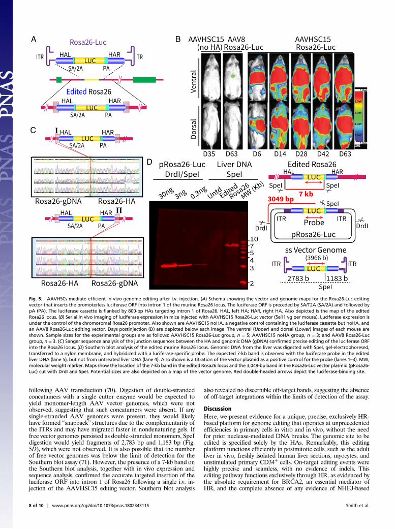
following AAV transduction (70). Digestion of double-strandedconcatamers with a single cutter enzyme would be expected toyield monomer-length AAV vector genomes, which were notobserved, suggesting that such concatamers were absent. If anysingle-stranded AAV genomes were present, they would likelyhave formed “snapback” structures due to the complementarity ofthe ITRs and may have migrated faster in nondenaturing gels. Iffree vector genomes persisted as double-stranded monomers, SpeIdigestion would yield fragments of 2,783 bp and 1,183 bp (Fig.5D), which were not observed. It is also possible that the numberof free vector genomes was below the limit of detection for theSouthern blot assay (71). However, the presence of a 7-kb band onthe Southern blot analysis, together with in vivo expression andsequence analysis, confirmed the accurate targeted insertion of theluciferase ORF into intron 1 of Rosa26 following a single i.v. in-jection of the AAVHSC15 editing vector. Southern blot analysis
also revealed no discernible off-target bands, suggesting the absenceof off-target integrations within the limits of detection of the assay.
DiscussionHere, we present evidence for a unique, precise, exclusively HR-based platform for genome editing that operates at unprecedentedefficiencies in primary cells in vitro and in vivo, without the needfor prior nuclease-mediated DNA breaks. The genomic site to beedited is specified solely by the HAs. Remarkably, this editingplatform functions efficiently in postmitotic cells, such as the adultliver in vivo, freshly isolated human liver sections, myocytes, andunstimulated primary CD34+ cells. On-target editing events werehighly precise and seamless, with no evidence of indels. Thisediting pathway functions exclusively through HR, as evidenced bythe absolute requirement for BRCA2, an essential mediator ofHR, and the complete absence of any evidence of NHEJ-based
BA
Do
rsa
lV
en
tra
l
Rosa26-LucAAV8
(no HA)
D35 D63 D6 D14 D28 D42 D63
AAVHSC15 AAVHSC15Rosa26-Luc
Rosa26-Luc
Edited Rosa26
SA/2A PA
ITR ITRHAL HARLUC
SA/2A PA
HAL HARLUC
C
Rosa26-gDNA Rosa26-HA
ILUC
SA/2A PA
HAL HAR
Rosa26-HA Rosa26-gDNA
IILUC
SA/2A PA
HAL HAR
D
30ng3ng
0.3ngUntd
MW (Kb)
Liver DNA
SpeI
pRosa26-Luc
DrdI/SpeI
Rosa26
Edited
10
4
75
3
2SpeI
ss Vector Genome
LUCITRITR
2783 b 1183 b
(3966 b)
ITRProbe
ITR
SpeI
DrdIDrdI
�
��
3049 bp
pRosa26-Luc
LUC
Edited Rosa26
SpeI� �
7 kb
HARHAL
SpeILUC
Fig. 5. AAVHSCs mediate efficient in vivo genome editing after i.v. injection. (A) Schema showing the vector and genome maps for the Rosa26-Luc editingvector that inserts the promoterless luciferase ORF into intron 1 of the murine Rosa26 locus. The luciferase ORF is preceded by SA/T2A (SA/2A) and followed bypA (PA). The luciferase cassette is flanked by 800-bp HAs targeting intron 1 of Rosa26. HAL, left HA; HAR, right HA. Also depicted is the map of the editedRosa26 locus. (B) Serial in vivo imaging of luciferase expression in mice injected with AAVHSC15 Rosa26-Luc vector (5e11 vg per mouse). Luciferase expression isunder the control of the chromosomal Rosa26 promoter. Also shown are AAVHSC15 noHA, a negative control containing the luciferase cassette but noHA, andan AAV8 Rosa26-Luc editing vector. Days postinjection (D) are depicted below each image. The ventral (Upper) and dorsal (Lower) images of each mouse areshown. Sample sizes for the experimental groups are as follows: AAVHSC15 Rosa26-Luc group, n = 5; AAVHSC15 noHA group, n = 3; and AAV8 Rosa26-Lucgroup, n = 3. (C) Sanger sequence analysis of the junction sequences between the HA and genomic DNA (gDNA) confirmed precise editing of the luciferase ORFinto the Rosa26 locus. (D) Southern blot analysis of the edited murine Rosa26 locus. Genomic DNA from the liver was digested with SpeI, gel-electrophoresed,transferred to a nylon membrane, and hybridized with a luciferase-specific probe. The expected 7-kb band is observed with the luciferase probe in the editedliver DNA (lane 5), but not from untreated liver DNA (lane 4). Also shown is a titration of the vector plasmid as a positive control for the probe (lanes 1–3). MW,molecular weight marker. Maps show the location of the 7-kb band in the edited Rosa26 locus and the 3,049-bp band in the Rosa26-Luc vector plasmid (pRosa26-Luc) cut with DrdI and SpeI. Potential sizes are also depicted on a map of the vector genome. Red double-headed arrows depict the luciferase-binding site.
8 of 10 | www.pnas.org/cgi/doi/10.1073/pnas.1802343115 Smith et al.
repair, including indels and incorporation of AAV ITRs. Thisgenome editing modality may be used not only for efficient editingof therapeutic sequences at specified chromosomal locations butalso for accurate correction of disease-causing point mutations.While AAV-mediated genome editing in the absence of nucleaseshas been previously reported (24–29, 67, 68), efficiencies were toolow for most therapeutic applications (29). Here, we present datasupporting genome editing efficiencies >50% in primary humancells as measured by expression of a reporter gene following tar-geted chromosomal insertion. This property of enhanced genomeediting efficiency is uniquely shared by members of stem cell-derived AAV clade F, including AAVHSCs and AAV9, and pro-vides a distinctive alternative to current genome editing platforms.Recently, AAV6 has been used in conjunction with nuclease-
based genome editing platforms for the delivery of donor DNAfor homology-dependent repair of double-stranded DNA breaks(72–76). While AAV6-mediated donor delivery was efficient andediting was observed following the creation of DNA breaks, noAAV6-mediated editing was observed in the absence of nucle-ases (72–75), in concordance with our findings.Our results indicate that vector genomes ranging from ∼2 to
4 kb were found to mediate enhanced genome editing, suggest-ing that genome size may not be critical. The editing vectors usedhere were designed with 800b symmetrical HAs, with the ex-ception of the nucleotide substitution vector, which had asym-metrical HAs of 583b and 1045b, indicating that flexibility in HAdesign is feasible.This distinctive editing platform is based upon the naturally
occurring AAVs present in human HSCs, including AAVHSCsand AAV9 (30). The unique single-stranded AAV genomesbounded by the palindromic G/C-rich ITRs likely activate acellular HR pathway, which leads to genome editing. This issupported by previous observations that scAAV genomes, whichare fully double-stranded, do not mediate recombination (26,46). Results presented here suggest that in addition to the uniqueAAV genome structure, the capsid sequences of clade F AAVplay an essential role in potentiating HR and genome editing.Identical editing vector genomes packaged in non-clade F cap-sids, including AAV2, AAV6, or AAV8, mediate editing at sig-nificantly lower efficiencies both in vitro and in vivo.Our results indicate that clade F AAVs were over 27-fold
more efficient at nuclear entry than AAVs belonging to clades A,B, and E. Since nuclear entry is essential for both genome editingand transduction, it is possible that more efficient nuclear entrymight be partially responsible for the higher level of genomeediting observed with clade F viruses, potentially accounting forthe observed correlation between transduction and editing effi-ciencies in vitro. However, in vivo comparisons of gene transferwith genome editing via AAVHSC15 and AAV8 suggested thatediting efficiency did not directly correlate with transduction effi-ciency in vivo. Although both AAV8 and AAVHSC15 transducedmice efficiently, AAVHSC15 was more efficient at editing thanAAV8. Whether specific HR pathways are additionally initiatedupon receptor engagement by AAVHSC or other cellular mech-
anisms are activated upon interaction of target cells with thecapsids is under investigation.Upon nuclear entry and uncoating, single-stranded AAV ge-
nomes undergo second-strand synthesis and often exist as cir-cular concatamers. Interestingly, under certain conditions, suchas in the absence of ATM, AAV genomes were observed to re-main in a linear configuration, inaccessible to the NHEJ pathway(70). Notably, no free vector genomes were detected in ourSouthern blot analysis, consistent with the possibility that circu-lar concatamers were not generated. It is conceivable that aprolonged single-stranded phase of editing vector genomes mayadditionally lead to enhanced editing. While single-strandedvector genomes could potentially be unstable in the nuclearenvironment, association with a cascade of repair proteins mayserve to protect them from degradation and promote HR.Importantly, the ability of AAVHSC and AAV9 to mediate
HR efficiently in resting and postmitotic cells was evidenced bysuccessful editing of freshly isolated liver sections, myocytes, andunstimulated CD34+ cells, as well as in vivo, including the liverand muscle in adult mice. The ability of AAVHSC and AAV9 tosupport genome editing in postmitotic adult tissue is unique (17)and has both mechanistic as well as therapeutic implications.Thus, AAV clade F, including AAVHSC and AAV9, repre-
sents a uniquely HR-based, highly precise, and efficient seamlessgenome editing modality in a single-component, nuclease-freeplatform, with a built-in delivery component that overcomesmany of the challenges currently facing therapeutic translation ofgenome editing. Thus, together with its potent in vivo editingability, clade F AAVs represent a distinct platform for the de-velopment of therapeutic in vivo genome editing for the treat-ment and potential cure of human diseases.
Materials and MethodsAll animal care and experiments were performed under protocols approvedby the City of Hope Institutional Animal Care and Use Committee. Deiden-tified, cytokine-primed, peripheral blood CD34+ cells were obtained withinformed consent from healthy donors under a City of Hope InstitutionalReview Board (IRB)-approved protocol as previously described (29). Dei-dentified liver samples were obtained from the City of Hope operating roomunder an IRB-approved protocol for the use of discard material. Recombi-nant DNA work was performed according to NIH guidelines. Tissue culture,transductions, flow cytometry, Southern blot analysis, and PCR reactionswere performed using standard procedures unless otherwise specified. Amore detailed description of treatments and analyses is provided in SI Ap-pendix, SI Materials and Methods.
ACKNOWLEDGMENTS. We thank Dr. Indra M. Newman for valuable adviceand assistance with the manuscript; Drs. Stephen Elledge, Kush Parmar,Albert Seymour, Arthur Tzianabos, and Sam Rasty for critical scientificfeedback; and Drs. James McSwiggen and Mike O’Callaghan for stimulatingdiscussions, scientific feedback, and helpful interactions. City of Hope corefacilities utilized for this study include the Analytical Cytometry, DNA Se-quencing, and Small Animal Imaging Cores. This research was supportedby the National Heart, Lung, and Blood Institute and National Cancer In-stitute of the NIH through Grant HL087285 (to S.C.) and by Cancer CenterSupport Grant P30CA033572.
1. Jasin M (1996) Genetic manipulation of genomes with rare-cutting endonucleases.
Trends Genet 12:224–228.2. Szostak JW, Orr-Weaver TL, Rothstein RJ, Stahl FW (1983) The double-strand-break
repair model for recombination. Cell 33:25–35.3. Pâques F, Haber JE (1999) Multiple pathways of recombination induced by double-
strand breaks in Saccharomyces cerevisiae. Microbiol Mol Biol Rev 63:349–404.4. Bibikova M, Golic M, Golic KG, Carroll D (2002) Targeted chromosomal cleavage and
mutagenesis in Drosophila using zinc-finger nucleases. Genetics 161:1169–1175.5. Zhang F, et al. (2011) Efficient construction of sequence-specific TAL effectors for
modulating mammalian transcription. Nat Biotechnol 29:149–153.6. Miller JC, et al. (2011) A TALE nuclease architecture for efficient genome editing. Nat
Biotechnol 29:143–148.7. Mali P, et al. (2013) RNA-guided human genome engineering via Cas9. Science 339:
823–826.8. Cong L, et al. (2013) Multiplex genome engineering using CRISPR/Cas systems. Science
339:819–823.9. Jinek M, et al. (2012) A programmable dual-RNA-guided DNA endonuclease in
adaptive bacterial immunity. Science 337:816–821.
10. Zetsche B, et al. (2015) Cpf1 is a single RNA-guided endonuclease of a class 2 CRISPR-
Cas system. Cell 163:759–771.11. Lieber MR (2010) The mechanism of double-strand DNA break repair by the non-
homologous DNA end-joining pathway. Annu Rev Biochem 79:181–211.12. Jeggo PA (1998) DNA breakage and repair. Adv Genet 38:185–218.13. Daya S, Cortez N, Berns KI (2009) Adeno-associated virus site-specific integration is me-
diated by proteins of the nonhomologous end-joining pathway. J Virol 83:11655–11664.14. Rudin N, Haber JE (1988) Efficient repair of HO-induced chromosomal breaks in
Saccharomyces cerevisiae by recombination between flanking homologous se-
quences. Mol Cell Biol 8:3918–3928.15. Capecchi MR (1989) Altering the genome by homologous recombination. Science 244:
1288–1292.16. Rouet P, Smih F, Jasin M (1994) Expression of a site-specific endonuclease stimulates
homologous recombination in mammalian cells. Proc Natl Acad Sci USA 91:6064–6068.17. Hustedt N, Durocher D (2016) The control of DNA repair by the cell cycle. Nat Cell Biol
19:1–9.18. Cox DB, Platt RJ, Zhang F (2015) Therapeutic genome editing: Prospects and chal-
lenges. Nat Med 21:121–131.
Smith et al. PNAS Latest Articles | 9 of 10
MED
ICALSC
IENCE
S
19. Fu Y, et al. (2013) High-frequency off-target mutagenesis induced by CRISPR-Casnucleases in human cells. Nat Biotechnol 31:822–826.
20. Kuscu C, Arslan S, Singh R, Thorpe J, Adli M (2014) Genome-wide analysis revealscharacteristics of off-target sites bound by the Cas9 endonuclease. Nat Biotechnol 32:677–683.
21. Tsai SQ, et al. (2015) GUIDE-seq enables genome-wide profiling of off-target cleavageby CRISPR-Cas nucleases. Nat Biotechnol 33:187–197.
22. Ran FA, et al. (2015) In vivo genome editing using Staphylococcus aureus Cas9. Nature520:186–191.
23. Komor AC, Badran AH, Liu DR (2017) CRISPR-based technologies for the manipulationof eukaryotic genomes. Cell 169:559, erratum (2017) 168:20–36.
24. Russell DW, Hirata RK (1998) Human gene targeting by viral vectors. Nat Genet 18:325–330.
25. Porteus MH, Cathomen T, Weitzman MD, Baltimore D (2003) Efficient gene targetingmediated by adeno-associated virus and DNA double-strand breaks. Mol Cell Biol 23:3558–3565.
26. Vasileva A, Linden RM, Jessberger R (2006) Homologous recombination is required forAAV-mediated gene targeting. Nucleic Acids Res 34:3345–3360.
27. Barzel A, et al. (2015) Promoterless gene targeting without nucleases ameliorateshaemophilia B in mice. Nature 517:360–364.
28. Miller DG, et al. (2006) Gene targeting in vivo by adeno-associated virus vectors. NatBiotechnol 24:1022–1026.
29. Khan IF, Hirata RK, Russell DW (2011) AAV-mediated gene targeting methods forhuman cells. Nat Protoc 6:482–501.
30. Smith LJ, et al. (2014) Gene transfer properties and structural modeling of humanstem cell-derived AAV. Mol Ther 22:1625–1634.
31. Podsakoff G, Wong KK, Jr, Chatterjee S (1994) Efficient gene transfer into non-dividing cells by adeno-associated virus-based vectors. J Virol 68:5656–5666.
32. Santat L, et al. (2005) Recombinant AAV2 transduction of primitive human hemato-poietic stem cells capable of serial engraftment in immune-deficient mice. Proc NatlAcad Sci USA 102:11053–11058.
33. Zincarelli C, Soltys S, Rengo G, Rabinowitz JE (2008) Analysis of AAV serotypes 1-9 mediated gene expression and tropism in mice after systemic injection.Mol Ther 16:1073–1080.
34. Chatterjee S, Johnson PR, Wong KK, Jr (1992) Dual-target inhibition of HIV-1 in vitroby means of an adeno-associated virus antisense vector. Science 258:1485–1488.
35. Fisher-Adams G, Wong KK, Jr, Podsakoff G, Forman SJ, Chatterjee S (1996) Integrationof adeno-associated virus vectors in CD34+ human hematopoietic progenitor cellsafter transduction. Blood 88:492–504.
36. Paz H, et al. (2007) Quiescent subpopulations of human CD34-positive hematopoieticstem cells are preferred targets for stable recombinant adeno-associated virus type2 transduction. Hum Gene Ther 18:614–626.
37. Kotin RM, et al. (1990) Site-specific integration by adeno-associated virus. Proc NatlAcad Sci USA 87:2211–2215.
38. Zhong L, et al. (2004) Impaired nuclear transport and uncoating limit recombinantadeno-associated virus 2 vector-mediated transduction of primary murine hemato-poietic cells. Hum Gene Ther 15:1207–1218.
39. Ponnazhagan S, et al. (1997) Adeno-associated virus type 2-mediated transduction inprimary human bone marrow-derived CD34+ hematopoietic progenitor cells: Donorvariation and correlation of transgene expression with cellular differentiation. J Virol71:8262–8267.
40. Mroske C, Rivera H, Ul-Hasan T, Chatterjee S, Wong KK (2012) A capillary electro-phoresis sequencing method for the identification of mutations in the inverted ter-minal repeats of adeno-associated virus. Hum Gene Ther Methods 23:128–136.
41. Sargent RG, Brenneman MA, Wilson JH (1997) Repair of site-specific double-strandbreaks in a mammalian chromosome by homologous and illegitimate recombination.Mol Cell Biol 17:267–277.
42. Pipiras E, Coquelle A, Bieth A, Debatisse M (1998) Interstitial deletions and intra-chromosomal amplification initiated from a double-strand break targeted to amammalian chromosome. EMBO J 17:325–333.
43. Sherry ST, et al. (2001) dbSNP: The NCBI database of genetic variation. Nucleic AcidsRes 29:308–311.
44. Genovese P, et al. (2014) Targeted genome editing in human repopulating haema-topoietic stem cells. Nature 510:235–240.
45. Miyaoka Y, et al. (2016) Systematic quantification of HDR and NHEJ reveals effects oflocus, nuclease, and cell type on genome-editing. Sci Rep 6:23549.
46. Hirata RK, Russell DW (2000) Design and packaging of adeno-associated virus genetargeting vectors. J Virol 74:4612–4620.
47. Powell SN, Kachnic LA (2003) Roles of BRCA1 and BRCA2 in homologous re-combination, DNA replication fidelity and the cellular response to ionizing radiation.Oncogene 22:5784–5791.
48. Oettinger MA, Schatz DG, Gorka C, Baltimore D (1990) RAG-1 and RAG-2, adjacentgenes that synergistically activate V(D)J recombination. Science 248:1517–1523.
49. Liu P, et al. (1993) Regional mapping of human DNA excision repair gene ERCC4 tochromosome 16p13.13-p13.2. Mutagenesis 8:199–205.
50. Meyn MS (1995) Ataxia-telangiectasia and cellular responses to DNA damage. CancerRes 55:5991–6001.
51. Tauchi H, et al. (2002) Nbs1 is essential for DNA repair by homologous recombinationin higher vertebrate cells. Nature 420:93–98.
52. Ellis NA, et al. (1995) The Bloom’s syndrome gene product is homologous to RecQhelicases. Cell 83:655–666.
53. Joenje H, et al. (1997) Evidence for at least eight Fanconi anemia genes. Am J HumGenet 61:940–944.
54. Joenje H, et al. (2000) Complementation analysis in Fanconi anemia: Assignment ofthe reference FA-H patient to group A. Am J Hum Genet 67:759–762.
55. Meetei AR, et al. (2004) X-linked inheritance of Fanconi anemia complementationgroup B. Nat Genet 36:1219–1224.
56. Ikeda H, et al. (2003) Genetic reversion in an acute myelogenous leukemia cell linefrom a Fanconi anemia patient with biallelic mutations in BRCA2. Cancer Res 63:2688–2694.
57. Takata M, et al. (2006) The Fanconi anemia pathway promotes homologous re-combination repair in DT40 cell line. Subcell Biochem 40:295–311.
58. Michl J, Zimmer J, Tarsounas M (2016) Interplay between Fanconi anemia and ho-mologous recombination pathways in genome integrity. EMBO J 35:909–923.
59. Strathdee CA, Gavish H, Shannon WR, Buchwald M (1992) Cloning of cDNAs forFanconi’s anaemia by functional complementation. Nature 356:763–767.
60. Moynahan ME, Pierce AJ, Jasin M (2001) BRCA2 is required for homology-directedrepair of chromosomal breaks. Mol Cell 7:263–272.
61. Davies AA, et al. (2001) Role of BRCA2 in control of the RAD51 recombination andDNA repair protein. Mol Cell 7:273–282.
62. Feng Z, et al. (2011) Rad52 inactivation is synthetically lethal with BRCA2 deficiency.Proc Natl Acad Sci USA 108:686–691.
63. Friedrich G, Soriano P (1991) Promoter traps in embryonic stem cells: A genetic screento identify and mutate developmental genes in mice. Genes Dev 5:1513–1523.
64. Kisseberth WC, Brettingen NT, Lohse JK, Sandgren EP (1999) Ubiquitous expression ofmarker transgenes in mice and rats. Dev Biol 214:128–138.
65. Wang L, et al. (2015) Comparative study of liver gene transfer with AAV vectors basedon natural and engineered AAV capsids. Mol Ther 23:1877–1887.
66. Smith L, et al. (2011) Functional mapping of tissue tropism of naturally occurringadeno-associated virus isolates from human hematopoietic stem cells. Mol Ther19(Suppl 1):S127–S128.
67. Paulk NK, et al. (2010) Adeno-associated virus gene repair corrects a mouse model ofhereditary tyrosinemia in vivo. Hepatology 51:1200–1208.
68. Borel F, et al. (2017) Survival advantage of both human hepatocyte xenografts andgenome-edited hepatocytes for treatment of alpha-1 antitrypsin deficiency.Mol Ther25:2477–2489.
69. Giel-Moloney M, Krause DS, Chen G, Van Etten RA, Leiter AB (2007) Ubiquitous anduniform in vivo fluorescence in ROSA26-EGFP BAC transgenic mice. Genesis 45:83–89.
70. Cataldi MP, McCarty DM (2010) Differential effects of DNA double-strand break re-pair pathways on single-strand and self-complementary adeno-associated virus vectorgenomes. J Virol 84:8673–8682.
71. Brown T (1993) Southern blotting. Curr Protoc Mol Biol 21:2.9.1–2.9.20.72. Sather BD, et al. (2015) Efficient modification of CCR5 in primary human hemato-
poietic cells using a megaTAL nuclease and AAV donor template. Sci Transl Med 7:307ra156.
73. Wang J, et al. (2015) Homology-driven genome editing in hematopoietic stem andprogenitor cells using ZFN mRNA and AAV6 donors. Nat Biotechnol 33:1256–1263.
74. Wang J, et al. (2016) Highly efficient homology-driven genome editing in humanT cells by combining zinc-finger nuclease mRNA and AAV6 donor delivery. NucleicAcids Res 44:e30.
75. De Ravin SS, et al. (2016) Targeted gene addition in human CD34(+) hematopoieticcells for correction of X-linked chronic granulomatous disease. Nat Biotechnol 34:424–429.
76. Ling C, et al. (2016) High-efficiency transduction of primary human hematopoieticstem/progenitor cells by AAV6 vectors: Strategies for overcoming donor-variation andimplications in genome editing. Sci Rep 6:35495.
10 of 10 | www.pnas.org/cgi/doi/10.1073/pnas.1802343115 Smith et al.