steps towards smarter solutions in optometry and …€¦ · subjective measurements using a...
TRANSCRIPT

healthcare
Article
Steps towards Smarter Solutions in Optometry andOphthalmology—Inter-Device Agreement ofSubjective Methods to Assess the Refractive Errors ofthe Eye
Arne Ohlendorf 1,2,*, Alexander Leube 1 and Siegfried Wahl 1,2
1 Institute for Ophthalmic Research, University of Tuebingen, Geschwister-Scholl-Platz, 72074 Tübingen,Germany; [email protected] (A.L.); [email protected] (S.W.)
2 Carl Zeiss Vision International GmbH, 73430 Aalen, Germany* Correspondence: [email protected]; Tel.: +49-7071-2984509
Academic Editor: Sampath ParthasarathyReceived: 23 February 2016; Accepted: 6 July 2016; Published: 13 July 2016
Abstract: Purpose: To investigate the inter-device agreement and mean differences between a newlydeveloped digital phoropter and the two standard methods (trial frame and manual phoropter).Methods: Refractive errors of two groups of participants were measured by two examiners (examiner1 (E1): 36 subjects; examiner 2 (E2): 38 subjects). Refractive errors were assessed using a trial frame, amanual phoropter and a digital phoropter. Inter-device agreement regarding the measurement ofrefractive errors was analyzed for differences in terms of the power vector components (sphericalequivalent (SE) and the cylindrical power vector components J0 and J45) between the used methods.Intraclass correlation coefficients (ICC’s) were calculated to evaluate correlations between the usedmethods. Results: Analyzing the variances between the three methods for SE, J0 and J45 using atwo-way ANOVA showed no significant differences between the methods (SE: p = 0.13, J0: p = 0.58and J45: p = 0.96) for examiner 1 and for examiner 2 (SE: p = 0.88, J0: p = 0.95 and J45: p = 1). Meandifferences and ˘95% Limits of Agreement for each pair of inter-device agreement regarding theSE for both examiners were as follows: Trial frame vs. digital phoropter: +0.10 D ˘ 0.56 D (E1) and+0.19 D ˘ 0.60 D (E2), manual phoropter vs. trial frame: ´0.04 D ˘ 0.59 D (E1) and ´0.12 D ˘ 0.49 D(E2) and for manual vs. digital phoropter: +0.06 D ˘ 0.65 D (E1) and +0.08 D ˘ 0.45 D (E2). ICCsrevealed high correlations between all methods for both examiner (p < 0.001). The time to assessthe subjective refraction was significantly smaller with the digital phoropter (examiner 1: p < 0.001;examiner 2: p < 0.001). Conclusion: “All used subjective methods show a good agreement betweeneach other terms of ICC (>0.9). Assessing refractive errors using different subjective methods, resultsin similar mean differences and 95% limits of agreement, when compared to those reported in studiescomparing subjective refraction non-cylcoplegic retinoscopy or autorefraction”.
Keywords: public health; optometry; subjective refraction; refractive errors; agreement
1. Background
Uncorrected refractive errors, such as myopia, hyperopia and astigmatism, have a high impact onthe prevalence of visual impairment or blindness, as recently reviewed by Naidoo and colleagues [1].Since it is known that the prevalence of refractive errors is increasing [2], its assessment will becomeone of the major tasks in the public health sector worldwide. Digitalization is already affecting ourlives, and its influence will increase in the future; the aim is to develop smart products that are able toassess refractive errors in order to provide adequate correction for people living in developing, as wellas in industrial, countries. Several “smart” solutions are already available that assess refractive errors
Healthcare 2016, 4, 41; doi:10.3390/healthcare4030041 www.mdpi.com/journal/healthcare
Healthcare 2016, 4, 41 2 of 11
objectively and subjectively. For example, the company EyeNetra (EyeNetra Inc., Somerville, MA,USA) developed a smartphone-based refraction for mobile measurements of refractive errors [3–5] thatuses a pinhole optic to display a stripe pattern on the participant’s retina, where the task of the subject isto align a red and green stimulus [3]. Another smartphone-based autorefractor is SVOne (Smart VisionLabs, New York, NY, USA), where a portable Hartmann-Shack wavefront aberrometer is attachedto a smartphone [6]. In contradiction, Opternative (Opternative Inc., Chicago, IL, USA) is an onlinesolution that aims to measure the refraction of the eye in a self-directed way, using a computer-basedresponse to presented stimuli (https://www.opternative.com/) [7]. Thus far, the performance of theseproducts is limited. Using Opternative, the range of refractive errors that can be measured is between0 D and ´4 D [7]. EyeNetra claims to have an extended range of refractive errors that can be measured(´12.5 D to +5.5 D) [8]. While the assessment of the refractive errors is the core competence of eye careprofessionals (ECPs), all used methods have to agree between each other and have to be reproducible,as well as repeatable. Published data on the variability of refraction, either assessed subjectively orobjectively, were mainly evaluated during studies that assessed the variability and repeatability ofauto-refractors. Most of the studies [9–11] used two repetitive measurements of subjective refractionper subject, only Rosenfield and Chiu, 1995 [12] assessed the subjective refraction with five repeatedmeasurements. The reported 95% limit of agreement for the subjective measurement of the sphericalequivalent refractive error, which was ˘0.29 D, suggesting that the subjective refraction is accurateto about a quarter diopter [12]. In contradiction, Zadnik, Mutti and Adams, 1992 [10] reported a95% limit of agreement for the SE of ˘0.63 D for cycloplegic and of ˘0.72 D for non-cycloplegicrefraction. Studies regarding the agreement between the earlier mentioned “smart” products, suchas EyeNetra or SVOne, with the traditional methods, showed good agreement between traditionaland smartphone-based technologies. Clinical results in 27 subjects (54 eyes) with refractive errorsranging from 0 D to ´6 D for the EyeNetra (Netra G) device showed that the difference between theEyeNetra and the subjective refraction for spherical equivalent error SE was 0.31 ˘ 0.37 D [4]. Later,it was shown that two different versions of the EyeNetra device overestimated myopia (sphericalerror) by 0.48 ˘ 0.66 D (Netra G #243) in 24 subjects and by 0.64 ˘ 0.71 D (Netra G #244 with a smallerpupillary distance) in 19 subjects [3]. In 50 visually-normal, young subjects with an average sphericalequivalent refractive error of ´2.87 D, the 95% limit of agreement for the assessment of the powervector components (J0 and J45) was highest for the SVOne device, when compared to retinoscopy andtwo autorefractors (Topcon KR-1W and Righton Retinomax-3) under cycloplegic and non-cylcoplegicconditions [6]. In case of the SE, no significant differences were found between the different methodsand devices and the 95% limit of agreement for the SVOne was comparable to Retinoscopy undernon-cycloplegic conditions [6]. Clinical data for 60 eyes from 30 subjects (aged between 18 and 40 yearsand with spherical refractive errors of up to ´4 D and astigmatism of up to ´2 D) for the Opternativesoftware are available on their webpage [7], but is not yet published. In summary, they report that adifference in the spherical equivalent difference of 0.25 diopter was apparent in 70% of the eyes, anda spherical equivalent difference of 0.50 diopter or less was found in 90% of the eyes. Nevertheless,in their review, Goss and Grosvenor, 1996 [13] concluded that the assessment of refractive errorsusing subjective methods, such as a trial frame or a phoropter, are far better compared to any othermethod, and that the agreement of the measurement of the SE (either intra-examiner or inter-examiner)was close to ˘0.25 D in 80% of the measurements, while the agreement was ˘0.50 D for 95% of themeasurements for the SE, the sphere and the cylinder power.
The purpose of the current study was to investigate the inter-device agreement and meandifferences between a newly developed digital phoropter and the two standard methods (trial frameand manual phoropter) currently used to assess the refractive error of the eye.

Healthcare 2016, 4, 41 3 of 11
2. Methods
2.1. Subjects
Inclusion criterion for participation was a refractive error of less than ˘8.0 D of sphericalametropia, ď´4.0 D of astigmatism and best corrected visual acuity of minimum 0.0 logMAR. Subjectswith known ocular diseases were not allowed to participate in the course of the study. Two examiners(examiner 1: Author AO and examiner 2: Author AL, both certified optometrists) measured therefractive errors in two independent studies, using the same devices/methods but different subjects.Examiner 1 measured refractive errors in 36 subjects, aged 23–47 years (mean: 36.4 ˘ 7.4 years), with amean spherical refractive error (S) of ´0.71 ˘ 1.62 D (range +2 D to ´5.75 D and a mean astigmaticrefractive error of ´0.59 ˘ 0.45 D (range 0 D to ´2 D)). The study group for examiner 2 included38 subjects, aged 22–47 years (mean: 36.7 ˘ 7.1 years), with a mean S of ´0.83 ˘ 1.39 D (range +1.75 to´3.5 D and an average astigmatic error of ´0.67 ˘ 0.49 D (range 0 D to ´2 D)). All subjects were naïveto the purpose of the experiment. The study course was approved from the Ethics Commission of theMedical Faculty of the University of Tuebingen. The research followed the tenets of the Declaration ofHelsinki, informed consent was obtained from all subjects after explanation of the nature and possibleconsequences of the study.
2.2. Equipment
To assess the refractive errors of an individual’s eye objectively, a wavefront-based autorefractorwas used (ZEISS i.Profiler plus, Carl Zeiss Vision GmbH, Aalen, Germany). Subjective refraction wasassessed using a Subjective Refraction Unit (SRU), a trial frame (UB4, Oculus, Wetzlar, Germany) incombination with trial lenses (BK1, Oculus, Wetzlar, Germany) and a manual phoropter (AmericanOptical Phoropter M/N 11320, American Optics, Buffalo, NY, USA). The SRU includes a digitalphoropter (ZEISS Visuphor 500, Carl Zeiss Vision GmbH, Aalen, Germany) and a screen to displayoptotypes (ZEISS Visuscreen 500). The digital phoropter covers spherical refractive errors from ´19 Dto +16.75 D and astigmatic errors from 0 D to ˘8.75 D. A Tablet PC (iPad3, Apple, Cupertinoj, CA,USA) was used that controlled the mentioned devices from the SRU, using an application called i.Commobile (Carl Zeiss Vision GmbH). All optotypes (SLOAN Letters) to subjectively measure the refractiveerrors were displayed on a digital visual acuity chart (ZEISS Visuscreen 500, Carl Zeiss Vision GmbH)at a distance of 6 m with a minimum luminance of 250 cd/m2.
2.3. Experimental Procedures
Objective measurements of refractive errors were obtained three times for each eye prior to thesubjective measurements using a wavefront-based autorefractor (ZEISS i.Profiler plus) and the mostpositive reading served as the starting value for the subjective refraction (sphere, cylinder, axis). Bothexaminers measured the refractive errors under monocular as well as binocular conditions. Theprocedure was as follows: Objective refraction was measured prior to the subjective refraction by atechnician in order to test if the subject meets the exclusion and inclusion criteria and both examinerswere masked to the results of the autorefraction measurement. Following, the examiners conductedthe subjective refraction, starting either with the manual phoropter or the digital phoropter (theprocedure was randomized), since it was possible to mask the “workflow” (the power of the usedlenses and the results) from the examiner. In case of the manual, as well as the digital, phoropter,both examiners were masked from the results of the subjective refraction. The trial frame refractionwas performed at the end of the study. The SRU provides a preconfigured refraction workflow thatguides the eye care professional through the process of the subjective refraction procedure. Thisworkflow contains the following steps to determine the monocular refractive error, starting with theright eye: (a) determination of the best sphere; (b) determination of a cylindrical error, using a Jacksoncross cylinder (if a cylindrical error was measured using the objective method, this step is skipped);(c) determination of the axis of the existing cylindrical refractive error; (d) determination of the power

Healthcare 2016, 4, 41 4 of 11
of the existing cylindrical refractive error; (e) fine adjustment of the sphere (monocular). The sameworkflow is used to assess the subjective refraction of the left eye, after measurements of the righteye are finished. After assessment of the monocular prescription, the following binocular tests aredone: (a) polarized duochrome test to achieve binocular balance and (b) polarized optotypes for theassessment of the best sphere under binocular conditions. Lighting conditions during the experimentsfollowed the international standards DIN EN ISO 8596 and 8597, which defines an ambient luminanceof 80–320 cd/m2.
2.4. Analysis
Both examiners used three methods of subjective refraction to assess the refractive errors in twoseparate study cohorts. Examiner 1 measured refractive errors in a group of 36 subjects, while examiner2 assessed the refractive errors in a group of 38 subjects. Data about the refractive errors of the left eyesafter the monocular refraction was used to assess the inter-device agreement between the differentmethods. Refractive errors were analyzed for the power vector components spherical equivalentrefractive error (SE) and J0 and J45 that were introduced by Thibos, Wheeler and Horner, 1997 [14].Time needed for each measurement of refraction including the tests under binocular conditions, wasdirectly saved by the i.Com software. The time durations for each used method were analyzed in orderto assess differences between the three methods.
2.5. Statistics
Statistical analyses were performed with the statistics software package JMP 11.1.1 (SAS Institute,Cary, NC, USA) and IBM SPSS Statistics 22 (IBM, Armonk, NY, USA). JMP was used to investigatethe presence of normality of the data using the Shapiro-Wilk test. An ANOVA was performed toanalyze differences between all three methods, when inter-device agreement was assessed. SPSS wasused to calculate Intraclass Correlation Coefficients [15] and Bland Altman [16] plots were used toinvestigate the inter-device agreement and the differences, when refraction was assessed using thethree different methods.
3. Results
3.1. Descriptive Statistics
Table 1 gives an overview on the mean refractive data (˘standard deviation) of the left eye for themonocular correction of SE, J0 and J45, when refraction was assessed with all of the used methods,separated for the two examiners. Standard errors were calculated from the standard deviation dividedbe the square root of the sample size. The mean values and standard deviations of all three powervector components of refraction showed similar values across all three methods.
Table 1. Mean values, ˘standard deviation and ˘standard error for each refractive parameter (SE, J0and J45) for examiner 1 and 2, when refraction was assessed with all methods.
Examiner 1 for n = 36 Subjects Examiner 2 for n = 38 Subjects
MeanValue (D)
˘1 StandardDeviation (D)
˘1 StandardError (D)
MeanValue (D)
˘1 StandardDeviation (D)
˘1 StandardError (D)
trial frameSE ´0.92 1.57 0.26 ´1.07 1.38 0.22J0 0.01 0.31 0.05 0.03 0.35 0.06J45 0.00 0.21 0.04 0.02 0.22 0.04
manualphoropter
SE ´0.99 1.59 0.27 ´1.17 1.38 0.22J0 0.02 0.34 0.06 0.00 0.36 0.06J45 ´0.01 0.20 0.03 0.00 0.26 0.04
digitalphoropter
SE ´1.02 1.57 0.26 ´1.24 1.36 0.22J0 0.01 0.32 0.05 0.02 0.36 0.06J45 0.01 0.20 0.03 0.00 0.24 0.04

Healthcare 2016, 4, 41 5 of 11
3.2. Bland-Altman Analysis for Inter-Device Agreement
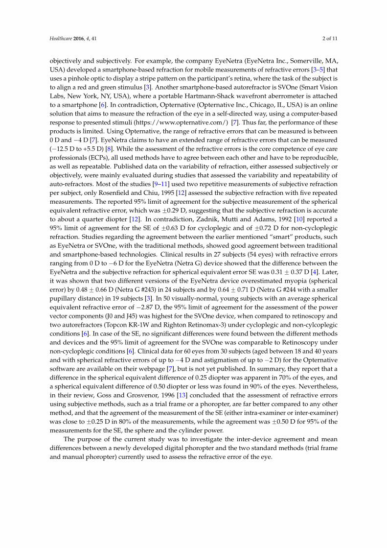
Bland-Altman analysis for inter-device agreement represents the level of agreement betweenseveral measurements of the refractive error of one subject that is refracted by the same examinerunder the same conditions but with different methods. Results for examiners 1 and 2 are summarizedin Figure 1 for the measurement of the SE for each pair-by-pair comparison of the three methods,using a Bland-Altmann plot and 95% Limits of Agreement (LoA), calculated as 1.96 multiplied by thestandard deviation of the difference [15]. Comparison of trial frame vs. digital phoropter are shown in(a) and (d), manual phoropter vs. digital phoropter in (b) and (e) and manual phoropter vs. trial framein (c) and (f).
Healthcare 2016, 4, 41 5 of 11
3.2. Bland-Altman Analysis for Inter-Device Agreement
Bland-Altman analysis for inter-device agreement represents the level of agreement between several measurements of the refractive error of one subject that is refracted by the same examiner under the same conditions but with different methods. Results for examiners 1 and 2 are summarized in Figure 1 for the measurement of the SE for each pair-by-pair comparison of the three methods, using a Bland-Altmann plot and 95% Limits of Agreement (LoA), calculated as 1.96 multiplied by the standard deviation of the difference [15]. Comparison of trial frame vs. digital phoropter are shown in (a) and (d), manual phoropter vs. digital phoropter in (b) and (e) and manual phoropter vs. trial frame in (c) and (f).
Figure 1. (a–f) Difference versus mean plot to compare the three subjective methods to determine the spherical equivalent refractive error (SE) of the left eye measured by examiner 1 (n = 36, Figure 1a–c) and 2 (n = 38, Figure 1d–f). (a) and (d) trial frame vs. digital phoropter; (b) and (e) manual phoropter vs. digital phoropter and (c) and (f) manual phoropter vs. trial frame. Solid line indicates the mean difference, while dashed lines represent the upper and lower limit (±95% limit of agreement). MD = mean difference and s = standard deviation. Shaded areas present 95% confidence interval limits for the mean difference and 95% limits of agreement.
Figure 1. (a–f) Difference versus mean plot to compare the three subjective methods to determine thespherical equivalent refractive error (SE) of the left eye measured by examiner 1 (n = 36, Figure 1a–c)and 2 (n = 38, Figure 1d–f). (a) and (d) trial frame vs. digital phoropter; (b) and (e) manual phoroptervs. digital phoropter and (c) and (f) manual phoropter vs. trial frame. Solid line indicates themean difference, while dashed lines represent the upper and lower limit (˘95% limit of agreement).MD = mean difference and s = standard deviation. Shaded areas present 95% confidence interval limitsfor the mean difference and 95% limits of agreement.
Healthcare 2016, 4, 41 6 of 11
For examiner 1, agreement between measurements of the spherical equivalent of the refractiveerror and both power vector components J0 and J45 (see Table 2) was similar for all used subjectivemethods. A statistically significant difference was found in the measurement of the SE between thetrial frame and the digital phoropter, while the trial frame showed more positive readings. Ninety-fivepercent LoA for the measurement of the SE was smaller between trial frame and automated phoropterfor exaimer 1 (˘0.56 D), followed by manual phoropter vs. trial frame (˘0.59 D) and manual phoroptervs. digital phoropter (˘0.65 D). Ninety-five percent CI of the lower and upper 95% LoA for the SEin case of examiner 1 were ˘0.17 D (trial frame vs. digital phoropter, Figure 1a), ˘0.19 D (manualphoropter vs. digital phoropter, Figure 1b) and ˘0.18 D (manual phoropter vs. trial frame, Figure 1c).In the case of examiner 2, measurement of the spherical equivalent refractive error was more positiveusing the trial frame, when compared to either the digital (mean difference = 0.19 D, Figure 1d) or themanual phoropter (mean difference = 0.12 D, Figure 1f). When comparing the manual and the digitalphoropter, the manual phoropter showed more positive measurements for the spherical equivalent(Figure 1e). Ninety-five percent LoA for the assessment of the SE was smallest for the comparison ofmanual vs. digital phoropter (˘0.45 D), followed by the manual phoropter vs. trial frame (˘0.49 D)and the trial frame vs. the digital phoropter (˘0.56 D). Calculated 95% CI of the upper and lowerlimit of the 95% LoA for the measured SE were ˘0.18 D (trial frame vs. digital phoropter, Figure 1d),˘0.13 D (manual phoropter vs. digital phoropter, Figure 1e) and ˘0.10 D (manual phoropter vs. trialframe, Figure 1f). For both examiners, no influence of the refractive error of the subject’s eye onthe difference between the used methods was observed. An ANOVA that analyzed the variancesbetween the three methods for SE, J0 and J45 showed no significant differences between the methods(SE: p = 0.13, J0: p = 0.58 and J45: p = 0.96, two-way ANOVA) for examiner 1 and for examiner 2(SE: p = 0.88, J0: p = 0.95 and J45: p = 1, two-way ANOVA).
Table 2. Descriptive analysis for the comparison of the three subjective methods thatassess the subjective refractive error, separated for the two examiners. Data represents theBland-Altmann analysis.
Examiner 1 (n = 36)
Mean Difference(D)
95% Limit ofAgreement (D)
95% CI forUpper Limit (D)
95% CI forLower Limit (D)
trial frame vs.digital phoropter
SE 0.10 ˘ 0.56 0.49 to 0.82 ´0.29 to ´0.62J0 0.00 ˘ 0.14 0.11 to 019 ´0.10 to ´0.18J45 ´0.01 ˘ 0.14 0.09 to 0.17 ´0.11 to ´0.19
manualphoropter vs.
digital phoropter
SE 0.06 ˘ 0.65 0.63 to 1.01 ´0.43 to ´0.82J0 0.01 ˘ 0.21 0.16 to 0.29 ´0.14 to ´0.27J45 ´0.02 ˘ 0.18 0.11 to 0.22 ´0.14 to ´0.25
manualphoropter vs.
trial frame
SE ´0.04 ˘ 0.59 0.56 to 0.91 ´0.64 to ´0.99J0 0.01 ˘ 0.19 0.14 to 0.26 ´0.12 to ´0.24J45 ´0.01 ˘ 0.15 0.10 to 0.19 ´0.11 to ´0.20
Examiner 2 (n = 38)
Mean Difference(D)
95% Limit ofAgreement (D)
95% CI forUpper Limit (D)
95% CI forLower Limit (D)
trial frame vs.digital phoropter
SE 0.19 ˘ 0.60 0.62 to 0.97 ´0.23 to ´0.58J0 0.00 ˘ 0.20 0.14 to 0.25 ´0.14 to ´0.25J45 0.01 ˘ 0.18 0.14 to 0.24 ´0.11 to ´0.21
manualphoropter vs.
digital phoropter
SE 0.08 ˘ 0.45 0.40 to 0.66 ´0.24 to ´0.51J0 ´0.02 ˘ 0.16 0.10 to 0.19 ´0.14 to ´0.23J45 0.00 ˘ 0.17 0.11 to 0.22 ´0.12 to ´0.22
manualphoropter vs.
trial frame
SE ´0.12 ˘ 0.49 0.23 to 0.52 ´0.47 to ´0.75J0 ´0.02 ˘ 0.22 0.13 to 0.26 ´0.18 to ´0.30J45 ´0.02 ˘ 0.18 0.11 to 0.21 ´0.15 to 0.25

Healthcare 2016, 4, 41 7 of 11
Calculations regarding the 95% LoA were also done for both power vector components J0 and J45to evaluate differences between the three subjective methods and the data are summarized in Table 2for both for examiners. Additionally, the 95% CI for the upper and lower limit of the 95% LoA [15]were calculated and are presented in the same table.
3.3. Intra Class Correlation Analysis
In addition to the use of a Bland Altman plot and the calculation of the 95% LoA, Intra-ClassCorrelation coefficients (ICC) [17] were also used for the analysis of the inter-device agreement. Inthe conducted analysis, a two-way random absolute agreement calculation ICC(2,k) was performedand the ICCs were calculated for both examiners separately with pairwise correlations of each deviceand for the three power vector components of refraction (SE, J0 and J45). Results can be obtainedfrom Table 3.
Table 3. Intra-Class Correlation, their lower and upper 95% CI interval, for the pairwise correlation ofeach device, separated for the two examiners and for the power vector components of refraction SE, J0and J45.
Examiner 1 (n = 36)
ICC95% CI p
Lower Lower
trial frame vs. digitalphoropter
SE 0.992 0.984 0.996 <0.01J0 0.985 0.97 0.992 <0.01J45 0.965 0.932 0.982 <0.01
manual phoropter vs.digital phoropter
SE 0.989 0.978 0.994 <0.01J0 0.967 0.935 0.963 <0.01J45 0.932 0.866 0.965 <0.01
manual phoropter vs.trial frame
SE 0.991 0.982 0.995 <0.01J0 0.974 0.949 0.987 <0.01J45 0.958 0.917 0.978 <0.01
Examiner 2 (n = 38)
ICC95% CI p
Lower Lower
trial frame vs.digital phoropter
SE 0.987 0.975 0.993 <0.01J0 0.978 0.958 0.989 <0.01J45 0.953 0.91 0.976 <0.01
manual phoropter vs.digital phoropter
SE 0.993 0.986 0.996 <0.01J0 0.985 0.971 0.992 <0.01J45 0.963 0.928 0.981 <0.01
manual phoropter vs.trial frame
SE 0.991 0.84 0.996 <0.01J0 0.971 0.944 0.985 <0.01J45 0.953 0.909 0.975 <0.01
The intra class correlation revealed high correlations for the assessment of the refractive errors between all threeused methods for both examiners. Additionally, all correlations showed high significant values (p < 0.001).
3.4. Time to Assess Subjective Refraction under Binocular Conditions
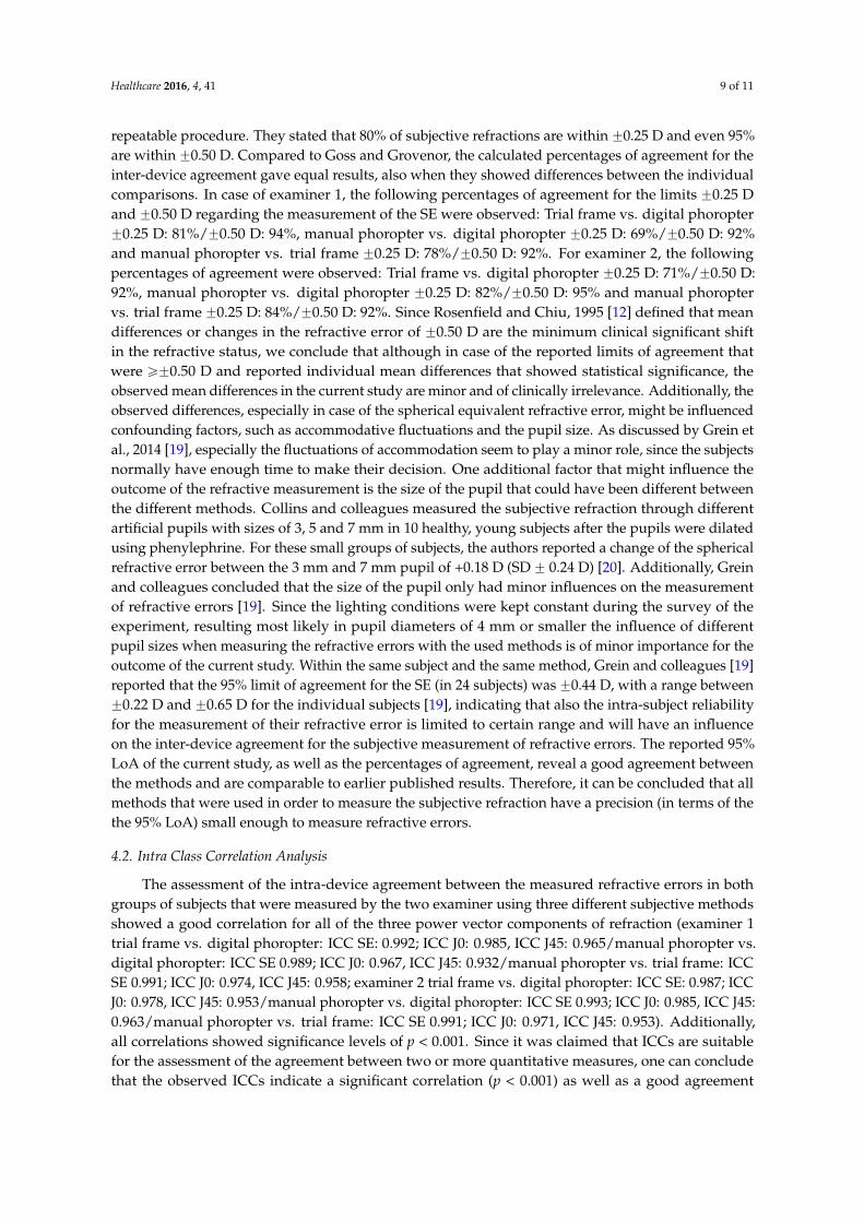
The time needed to assess the subjective refraction under binocular conditions was savedautomatically by the i.Com software. Analysis was conducted for each measurement with all threemethods and for each examiner (for examiner 1, 36; and for examiner 2, 38 measurements). Figure 2arepresents the time for each subject, as well as the associated mean values ˘1 standard deviations(seconds) for examiner 1, while Figure 2b represents results for examiner 2.

Healthcare 2016, 4, 41 8 of 11
For both examiners, subjective refraction with the digital phoropter was significantly fastercompared to the assessment when the trial frame and the manual phoropter were used.Healthcare 2016, 4, 41 8 of 11
Figure 2. Individual and average data (±1 standard deviation) for the time of the assessment of the subjective refraction for each method, separated for examiner 1 (a) and examiner 2 (b). * = p < 0.05; ** = p < 0.01; *** = p < 0.001.
4. Discussion
Many studies [8–12] on the reproducibility, the repeatability, and the level of agreement between different methods to measure the subjective refraction of the eye have been conducted and have resulted in different estimates for the various methods.
4.1. Bland-Altman Analysis for Inter-Device Agreement
For the subjective measurement of sphero-cylindrical refractive errors for repeated measures by the same examiner, previously reported 95% limit of agreement range from ±0.94 D for cycloplegic subjective refraction to ±0.63 D for the non-cycloplegic assessment of refractive errors [10]. In the case of retinoscopy, 95% limit of agreement was reported to be ±0.95 D for cycloplegic retinoscopy and ±0.78 D for non-cycloplegic retinoscopy, for repeated measures of refractive errors by the same examiner [10]. In the current study, we compared the 95% limit of agreement when refractive errors were assessed by the same examiner but with three different subjective methods for non-cycloplegic refraction in either 36 or 38 participants for examiner 1 and examiner 2, respectively. Similar 95% levels of agreement where observed, when comparing the measurement of non-cycloplegic measurement of the spherical equivalent refractive error with the three used methods for examiner 1 (95% LoA trial frame vs. digital phoropter: ±0.56 D, 95% LoA manual phoropter vs. digital phoropter: ±0.65 D, 95% LoA manual phoropter vs. trial frame: ±0.59 D) and examiner 2 (95% LoA trial frame vs. digital phoropter: ±0.60 D, 95% LoA manual phoropter vs. digital phoropter: ±0.45 D, 95% LoA manual phoropter vs. trial frame: ±0.49 D). Rosenfield and Chiu, 1995 [12] showed that the 95% limits of agreement for repeated measurements of the subjective refractive is ±0.25 D, Bullimore [18] found an 95% limit of agreement regarding the repeatability of the spherical equivalent refractive error, measured by two optometrists of ±0.78 D with a mean difference of −0.12 D.
Nevertheless, one still has to keep in mind that differences in the measurement of refractive errors would lead to different results for the correction of refractive errors. For example for examiner 2, statistically significant mean differences were found for the SE, while more positive values were measured using the trial frame (mean difference 0.19 D compared to the digital phoropter and 0.12 D compared with the manual phoropter) and the manual phoropter vs. the digital phoropter (mean difference 0.08 D). In case of examiner 1, a difference in the measurement of the SE was only observed between the trial frame and the digital phoropter (mean difference 0.10 D). As stated already in the background section, Goss and Grovenor [13] concluded that the subjective refraction is a valid and repeatable procedure. They stated that 80% of subjective refractions are within ±0.25 D and even 95% are within ±0.50 D. Compared to Goss and Grovenor, the calculated percentages of agreement for the inter-device agreement gave equal results, also when they showed differences between the individual comparisons. In case of examiner 1, the following percentages of agreement for the limits ±0.25 D and ±0.50 D regarding the measurement of the SE were observed: Trial frame vs. digital phoropter ±0.25 D: 81%/±0.50 D: 94%, manual phoropter vs. digital phoropter ±0.25 D:
Figure 2. Individual and average data (˘1 standard deviation) for the time of the assessment of thesubjective refraction for each method, separated for examiner 1 (a) and examiner 2 (b). * = p < 0.05;** = p < 0.01; *** = p < 0.001.
4. Discussion
Many studies [8–12] on the reproducibility, the repeatability, and the level of agreement betweendifferent methods to measure the subjective refraction of the eye have been conducted and haveresulted in different estimates for the various methods.
4.1. Bland-Altman Analysis for Inter-Device Agreement
For the subjective measurement of sphero-cylindrical refractive errors for repeated measures bythe same examiner, previously reported 95% limit of agreement range from ˘0.94 D for cycloplegicsubjective refraction to ˘0.63 D for the non-cycloplegic assessment of refractive errors [10]. In thecase of retinoscopy, 95% limit of agreement was reported to be ˘0.95 D for cycloplegic retinoscopyand ˘0.78 D for non-cycloplegic retinoscopy, for repeated measures of refractive errors by the sameexaminer [10]. In the current study, we compared the 95% limit of agreement when refractive errorswere assessed by the same examiner but with three different subjective methods for non-cycloplegicrefraction in either 36 or 38 participants for examiner 1 and examiner 2, respectively. Similar 95% levelsof agreement where observed, when comparing the measurement of non-cycloplegic measurement ofthe spherical equivalent refractive error with the three used methods for examiner 1 (95% LoA trialframe vs. digital phoropter: ˘0.56 D, 95% LoA manual phoropter vs. digital phoropter: ˘0.65 D,95% LoA manual phoropter vs. trial frame: ˘0.59 D) and examiner 2 (95% LoA trial frame vs. digitalphoropter: ˘0.60 D, 95% LoA manual phoropter vs. digital phoropter: ˘0.45 D, 95% LoA manualphoropter vs. trial frame: ˘0.49 D). Rosenfield and Chiu, 1995 [12] showed that the 95% limits ofagreement for repeated measurements of the subjective refractive is ˘0.25 D, Bullimore [18] foundan 95% limit of agreement regarding the repeatability of the spherical equivalent refractive error,measured by two optometrists of ˘0.78 D with a mean difference of ´0.12 D.
Nevertheless, one still has to keep in mind that differences in the measurement of refractiveerrors would lead to different results for the correction of refractive errors. For example for examiner2, statistically significant mean differences were found for the SE, while more positive values weremeasured using the trial frame (mean difference 0.19 D compared to the digital phoropter and 0.12 Dcompared with the manual phoropter) and the manual phoropter vs. the digital phoropter (meandifference 0.08 D). In case of examiner 1, a difference in the measurement of the SE was only observedbetween the trial frame and the digital phoropter (mean difference 0.10 D). As stated already in thebackground section, Goss and Grovenor [13] concluded that the subjective refraction is a valid and
Healthcare 2016, 4, 41 9 of 11
repeatable procedure. They stated that 80% of subjective refractions are within ˘0.25 D and even 95%are within ˘0.50 D. Compared to Goss and Grovenor, the calculated percentages of agreement for theinter-device agreement gave equal results, also when they showed differences between the individualcomparisons. In case of examiner 1, the following percentages of agreement for the limits ˘0.25 Dand ˘0.50 D regarding the measurement of the SE were observed: Trial frame vs. digital phoropter˘0.25 D: 81%/˘0.50 D: 94%, manual phoropter vs. digital phoropter ˘0.25 D: 69%/˘0.50 D: 92%and manual phoropter vs. trial frame ˘0.25 D: 78%/˘0.50 D: 92%. For examiner 2, the followingpercentages of agreement were observed: Trial frame vs. digital phoropter ˘0.25 D: 71%/˘0.50 D:92%, manual phoropter vs. digital phoropter ˘0.25 D: 82%/˘0.50 D: 95% and manual phoroptervs. trial frame ˘0.25 D: 84%/˘0.50 D: 92%. Since Rosenfield and Chiu, 1995 [12] defined that meandifferences or changes in the refractive error of ˘0.50 D are the minimum clinical significant shiftin the refractive status, we conclude that although in case of the reported limits of agreement thatwere ě˘0.50 D and reported individual mean differences that showed statistical significance, theobserved mean differences in the current study are minor and of clinically irrelevance. Additionally, theobserved differences, especially in case of the spherical equivalent refractive error, might be influencedconfounding factors, such as accommodative fluctuations and the pupil size. As discussed by Grein etal., 2014 [19], especially the fluctuations of accommodation seem to play a minor role, since the subjectsnormally have enough time to make their decision. One additional factor that might influence theoutcome of the refractive measurement is the size of the pupil that could have been different betweenthe different methods. Collins and colleagues measured the subjective refraction through differentartificial pupils with sizes of 3, 5 and 7 mm in 10 healthy, young subjects after the pupils were dilatedusing phenylephrine. For these small groups of subjects, the authors reported a change of the sphericalrefractive error between the 3 mm and 7 mm pupil of +0.18 D (SD ˘ 0.24 D) [20]. Additionally, Greinand colleagues concluded that the size of the pupil only had minor influences on the measurementof refractive errors [19]. Since the lighting conditions were kept constant during the survey of theexperiment, resulting most likely in pupil diameters of 4 mm or smaller the influence of differentpupil sizes when measuring the refractive errors with the used methods is of minor importance for theoutcome of the current study. Within the same subject and the same method, Grein and colleagues [19]reported that the 95% limit of agreement for the SE (in 24 subjects) was ˘0.44 D, with a range between˘0.22 D and ˘0.65 D for the individual subjects [19], indicating that also the intra-subject reliabilityfor the measurement of their refractive error is limited to certain range and will have an influenceon the inter-device agreement for the subjective measurement of refractive errors. The reported 95%LoA of the current study, as well as the percentages of agreement, reveal a good agreement betweenthe methods and are comparable to earlier published results. Therefore, it can be concluded that allmethods that were used in order to measure the subjective refraction have a precision (in terms of thethe 95% LoA) small enough to measure refractive errors.
4.2. Intra Class Correlation Analysis
The assessment of the intra-device agreement between the measured refractive errors in bothgroups of subjects that were measured by the two examiner using three different subjective methodsshowed a good correlation for all of the three power vector components of refraction (examiner 1trial frame vs. digital phoropter: ICC SE: 0.992; ICC J0: 0.985, ICC J45: 0.965/manual phoropter vs.digital phoropter: ICC SE 0.989; ICC J0: 0.967, ICC J45: 0.932/manual phoropter vs. trial frame: ICCSE 0.991; ICC J0: 0.974, ICC J45: 0.958; examiner 2 trial frame vs. digital phoropter: ICC SE: 0.987; ICCJ0: 0.978, ICC J45: 0.953/manual phoropter vs. digital phoropter: ICC SE 0.993; ICC J0: 0.985, ICC J45:0.963/manual phoropter vs. trial frame: ICC SE 0.991; ICC J0: 0.971, ICC J45: 0.953). Additionally,all correlations showed significance levels of p < 0.001. Since it was claimed that ICCs are suitablefor the assessment of the agreement between two or more quantitative measures, one can concludethat the observed ICCs indicate a significant correlation (p < 0.001) as well as a good agreement

Healthcare 2016, 4, 41 10 of 11
(ICC > 0.75) between the compared subjective methods for both examiners, independent from theanalyzed refractive error.
The small 95% confidence intervals for all three power vector components of the refractive errorsreveals the good agreement between the tested methods. Studies regarding the ICCs for the refractiveerrors of the eye (sphere, spherical equivalent, cylinder, axis, J0 and J45) compared subjective refraction(mainly using trial frames) with objective refraction (using autorefractors or aberrometers) but have notcompared different methods of subjective refraction so far. For example, De Carlo and colleagues [21]found high ICCs regarding the agreement for the spherical equivalent error (0.92), but not for the twoastigmatic vectors J0 (0.08) and J45 (0.15) when they compared trial frame refraction and autorefraction,using the Nikon Retinomax K+ (Nikon, Inc., Tokyo, Japan) in 440 low-vision patients. The observeddifference from De Carlo to the current study regarding the power vector components J0 and J45 aremost likely caused by the fact that they compared trial frame refraction to autorefraction. Segura [22]investigated the repeatability of different spherocylindrical corrections in 42 eyes, obtained during asubjective refraction, with an autorefractor (WAM-5500, Shigiya Machinery Works Ltd., Fukuyama City,Hiroshima, Japan) and an aberrometer (iTrace, Tracey Technologies, Houston, TX, USA). They reportedICC’s regarding the repeatability for the spherical refraction of the autorefractor of 0.999 and 0.998 forthe aberrometer. Comparing the intra-observer reliability of the SE obtained with classical trial framerefraction and autorefraction (using the WAM-5500, Grand Seiko Co. Ltd), Pujol [23] reported ICCs forthe SE regarding the intra-observer reliability, when refractive errors were measured in 104 eyes of 52subjects. In case of trial frame refraction, ICC was 0.993, while it was 0.991 for the used autorefractor(WAM-5500, Grand Seiko Co. Ltd). The reported ICCs of the current study are comparable to ICCsfrom previous studies investigating the intra-examiner reliability for the assessment of refractive errors.
5. Conclusions
The agreement of the measurement of the refractive error of the eye between different subjectivemethods results in comparable values to non-cylcoplegic retinoscopy or autorefraction measurements,in case of mean differences and 95% limits of agreement, as well as intra-class correlation coefficients(ICC > 0.9). All used subjective methods showed a good agreement and correlation between eachother, while the refraction using the digital phoropter was significantly faster.
Acknowledgments: This work was done in an industry-on-campus-cooperation between the UniversityTuebingen and Carl Zeiss Vision International GmbH. The work was supported by third-party-funding. AlexanderLeube is scientists at the University Tuebingen, Arne Ohlendorf and Siegfried Wahl are employed by Carl ZeissVision International GmbH and are scientists at the University Tuebingen.
Author Contributions: This study was conducted by the ZEISS Vision Science Lab, Institute for OphthalmicResearch, University of Tuebingen. Arne Ohlendorf and Siegfried Wahl are employees of Carl Zeiss VisionInternational GmbH. All authors were involved in the design, the reasoning, interpretation, and summarizing ofthe study and the key contributions were as follows: Arne Ohlendorf and Alexander Leube developed the studyprotocol and did the data recording and Siegfried Wahl was the principal investigator.
Conflicts of Interest: The authors declare no conflict of interest.
References
1. Naidoo, K.S.; Leasher, J.; Bourne, R.R.; Flaxman, S.R.; Jonas, J.B.; Keeffe, J.; Limburg, H.; Pesudovs, K.;Price, H.; White, R.A.; et al. Global vision impairment and blindness due to uncorrected refractive error,1990–2010. Ophthalmol. Vis. Sci. 2016, 93, 227–234. [CrossRef] [PubMed]
2. Vitale, S.; Ellwein, L.; Cotch, M.F.; Ferris, F.L.; Sperduto, R. Prevalence of refractive error in the United States,1999–2004. Arch. Ophthalmol. 2008, 126, 1111–1119. [CrossRef] [PubMed]
3. Solaka, N.; Modi, R.; Gaiser, H.; Pamplona, V.; Schafran, D.; He, R.; Moore, B.D. Comparison of aNew Prototype of Netra-G Cell Phone-Based Refraction with Subjective Refraction. Available online:http://iovs.arvojournals.org/article.aspx?articleid=2268075 (accessed on 24 March 2016).

Healthcare 2016, 4, 41 11 of 11
4. Gaiser, H.; Moore, B.; Pamplona, V.; Solaka, N.; Schafran, D.; Merrill, D.; Sharpe, N.; Geringer, J.; Raskar, R.Comparison of a Novel Cell Phone-Based Refraction Technique (Netra-G) with Subjective Refraction.Available online: http://iovs.arvojournals.org/article.aspx?articleid=2147023 (accessed on 24 March 2016).
5. Bastawrous, A.; Leak, C.; Howard, F.; Kumar, V. Validation of near eye tool for refractive assessment(NETRA)—Pilot study. J. Mob. Technol. Med. 2012, 1, 6–16. [CrossRef]
6. Ciuffreda, K.J.; Rosenfield, M. Evaluation of the SVOne: A handheld, smartphone-based autorefractor.Optom. Vis. Sci. 2015, 92, 1133–1139. [CrossRef] [PubMed]
7. Opternative. Eye Exams from Home. Available online: https://www.opternative.com/ (accessed on24 March 2016).
8. EyeNetra Inc. NETRA—Refraction for Anyone Anywere. Available online: https://eyenetra.com/product-netra.html (accessed on 11 May 2016).
9. Elliott, M.; Simpson, T.; Richter, D.; Fonn, D. Repeatability and accuracy of automated refraction: Acomparison of the Nikon NRK-8000, the Nidek AR-1000, and subjective refraction. Optom. Vis. Sci.1997, 74, 434–438. [CrossRef] [PubMed]
10. Zadnik, K.; Mutti, D.O.; Adams, A.J. The repeatability of measurement of the ocular components.Investig. Ophthalmol. Vis. Sci. 1992, 33, 2325–2333.
11. Sheedy, J.; Schanz, P.; Bullimore, M. Evaluation of an automated subjective refractor. Optom. Vis. Sci. 2004,81, 334–340. [CrossRef] [PubMed]
12. Rosenfield, M.; Chiu, N.N. Repeatability of subjective and objective refraction. Optom. Vis. Sci. 1995, 72,577–579. [CrossRef] [PubMed]
13. Goss, D.A.; Grosvenor, T. Reliability of refraction—A literature review. J. Am. Optom. Assoc. 1996, 67, 619–630.[PubMed]
14. Thibos, L.N.; Wheeler, W.; Horner, D. Power vectors: An application of fourier analysis to the descriptionand statistical analysis of refractive error. Optom. Vis. Sci. 1997, 74, 367–375. [CrossRef] [PubMed]
15. McAlinden, C.; Khadka, J.; Pesudovs, K. Statistical methods for conducting agreement (comparison ofClinical Tests) and precision (repeatability or Reproducibility) Studies in optometry and ophthalmology.Ophthalmic Physiol. Opt. 2011, 31, 330–338. [CrossRef] [PubMed]
16. Bland, J.M.; Altman, D.G. Statistical methods for assessing agreement between two methods of clinicalmeasurement. Lancet 1986, 1, 307–310. [CrossRef]
17. Bland, J.M.; Altman, D.G. Measurement error and correlation coefficients. BMJ 1996, 313, 41–42. [CrossRef][PubMed]
18. Bullimore, M.A.; Fusaro, R.E.; Adams, C.W. The repeatability of automated and clinician refraction.Optom. Vis. Sci. 1998, 75, 617–622. [CrossRef] [PubMed]
19. Grein, H.-J.; Schmidt, O.; Ritsche, A. Reproducibility of subjective refraction measurement. Ophthalmologe2014, 111, 1057–1064. [CrossRef] [PubMed]
20. Collins, M.; Shaw, A.; Menkens, E.; Davis, B.; Franklin, R. The Effect of Pupil Size on Subjective Refractionwith Irregular Corneas. Available online: http://iovs.arvojournals.org/article.aspx?articleid=2419314(accessed on 11 May 2016).
21. DeCarlo, D.K.; McGwin, G.; Searcey, K.; Gao, L.; Snow, M.; Waterbor, J.; Owsley, C. Trial frame refractionversus autorefraction among new patients in a low-vision clinic. Investig. Ophthalmol. Vis. Sci. 2013, 54,19–24. [CrossRef] [PubMed]
22. Segura, F.; Sanchez-Cano, A.; Lopez de la Fuente, C.; Fuentes-Broto, L.; Pinilla, I. Evaluation of patient visualcomfort and repeatability of refractive values in non-presbyopic healthy eyes. Int. J. Ophthalmol. 2015, 8,1031–1036. [PubMed]
23. Pujol, J.; Ondategui-Parra, J.C.; Badiella, L.; Otero, C.; Vilaseca, M.; Aldaba, M. Spherical subjective refractionwith a novel 3D virtual reality based system. J. Optom. 2016. [CrossRef] [PubMed]
© 2016 by the authors; licensee MDPI, Basel, Switzerland. This article is an open accessarticle distributed under the terms and conditions of the Creative Commons Attribution(CC-BY) license (http://creativecommons.org/licenses/by/4.0/).