stereotactic biopsy - asrt and arrt category a · pdf file• targeting errors ......
TRANSCRIPT

STEREOTACTIC STEREOTACTIC BIOPSYBIOPSY
Maria T. Maria T. MontemayorMontemayor, BSRT (R)(M)(QM), BSRT (R)(M)(QM)St. LukeSt. Luke’’s Episcopal Health Systems Episcopal Health System
OBJECTIVESOBJECTIVES
• DEFINING BREAST BIOPSIES• PRINCIPLES OF STEREOTACTIC BIOPSY• DIFFERENCES BETWEEN SYSTEMS• TARGETING ERRORS• PATIENT PREPARATION• QUALITY CONTROL • ACR ACCREDITATION
What is Image Guided Breast BiopsyWhat is Image Guided Breast Biopsy
The use of an imaging modality to guide a sampling device into the
breast to remove tissue
IMAGE GUIDED BREAST BIOPSY TYPESIMAGE GUIDED BREAST BIOPSY TYPES
• Stereotactic Mammography• Prone Dedicated Tables• Upright Add-on Units
• Ultrasound• MRI
Benefits of Core Breast Biopsy• • Reduced complications
• No post-procedure disability• Improved cosmesis• Eliminates distortion of breast tissue
that might make interpretation of a mammogram difficult.
• Reduced costs• Permits optimal preoperative planning
when there is a definitive diagnosis of cancer.
• Can be performed in an outpatient setting or doctor’s office.
• Local anesthesia• No stitches• Single needle insertion to collect
a sufficient amount of breast tissue for an accurate diagnosis (uses vacuum-assistance).
• • Ability to mark biopsy site.

STEREOTACTIC BIOPSY STEREOTACTIC BIOPSY PRONE UNITSPRONE UNITS
UPRIGHT UNITSUPRIGHT UNITS
System DifferencesSystem Differences
Prone System Upright SystemRequires extras space No additional SpaceProne Sitting/recumbentRadiologist work under Radiologist next to
(eye level with Pt) patientProcedure Time 30 min Procedure Time 15 minUnit costly 140K Unit cost 20-100K
System DifferencesSystem Differences
• 360/270 Access No limitations• DICOM Compatible DICOM Compatible• Patient Comfort Patient Comport• Less likely for patient More likely for
to Vasovagal patient to vasovagal• Does not tie up a Tie up mammogram
mammogram unit unit/cause sch.delays

Principles of Stereotactic
The science of Stereology deals with the transformation of 2-D observations to 3-D information using mathematical, statistical and geometrical tools.
StereotacticStereotactic PrinciplesPrinciples
Through the measurement of the parallax shift of the targeted area from the first to the second stereotactic image, the computer software allows for the calculation of the three dimensional position within the breast- the three axis.
Each eye captures its own view. The two separate images are sent to the brain for processing. The two images arrive simultaneously and are united into one picture.

The coordinates are determined from reference point.
• Breast Support (Polar Coordinates):Determines the depth from the distal aspect of
the breast.
• Compression Paddle (Cartesian Coordinates): Determines the depth from the proximal aspect of the breast.
Reference PointReference Point POLAR /FISCHER CARTESIAN/HOLOGIC
ImageReceptor
ImageReceptor
Compression Paddle
Compression Paddle
0 1 2 3 44 3 2 1 0
D = Depth Z = Depth
Scout ImageScout Image
X-Ray Tube
Parallax ShiftParallax Shift
15º
Parallax ShiftParallax Shift
15º
Parallax ShiftParallax Shift
X-Ray Tube

Stereo PairStereo Pair Stereo Pair
Two images acquired with the X-ray tube at two different positions.
Stereo Images
ImageReceptor
Breast SupportLesion
Compression Paddle
15º0º
15º
COORDINATE SYSTEMS PRONE TABLESCOORDINATE SYSTEMS PRONE TABLES
Coordinate systems are used to target a precise location in the breast for the biopsy needle.
Stereotactic localizations are accurate to within 1mm.
Two types of coordinate systems are used in stereotactic guidance
Cartesian (Hologic) Polar (Siemens/mammotest/Fischer)
CARTESIANCARTESIAN
• The Cartesian coordinate system defines the target using distances from the 3 axes that intersect at the right angles x,y,z.
• X =Horizontal• Y=Vertical• Z=Depth
• This system is advantageous because it is intuitive and familiar to operators.– It is simple to use– The needle position can be easily adjusted– Stereo Image Pair along x axis– Positive Stereo View (+) is the technologist
right– Negative Stereo View (-) is the technologist
left
CartesianCartesian
Non-fixed image receptor
• Lens coupling device
• Bi-directional patient positioning

CartesianCartesian
• Lesion “Shift”
• • If the lesion “shifts” a small amount, then the lesion is an anterior lesion.
• • If the lesion shifts a large amount, then the lesion is a posterior lesion.
Cartesian Parallax ShiftCartesian Parallax Shift
CartesianCartesian
POLAR POLAR
• The polar coordinate system defines a target by distances from a fixed point and the angular distances from a reference line.
• Coordinates are represented by H,V and D.– H Horizontal plane– V Vertical Plane– D Depth
• The polar system is accurate• More difficult for the user to notice errors• Needle position not as easily adjusted may
required a second set of targets• The stereo image pair is represented by H
(horizontal )– Positive (+) stereo image: Technologist’s left– Negative (-) stereo image: Technologist’s
right

Polar SystemPolar System
• Fixed image receptor
• Fiber-optic coupling device.
• Uni-directional patient position.
Polar Polar
• Lesion “Shift”
• • If the lesion “shifts” a large amount, then the lesion is an anterior lesion.
• • If the lesion shifts a small amount, then the lesion is a posterior lesion.
Polar Parallax ShiftPolar Parallax Shift
+15º -15º -15º+15º
Polar Parallax ShiftPolar Parallax Shift
Two Coordinate SystemsPolar Coordinates
H=HorizontalV=VerticalD=Depth
Cartesian CoordinatesX=HorizontalY=VerticalZ=Depth
Side ViewFrontal View
D or Z
H or X
V or Y

TARGETING ERRORSTARGETING ERRORS
PrePre--Fire PositioningFire Positioning
Correct Probe Position
Post FirePost Fire -X/Horizontal Error
• Probe aperture is to the left of the center of the lesion on both (-) and (+) 15o images. Sampling should be increased
• between 12:00 –6:00 going through 3:00
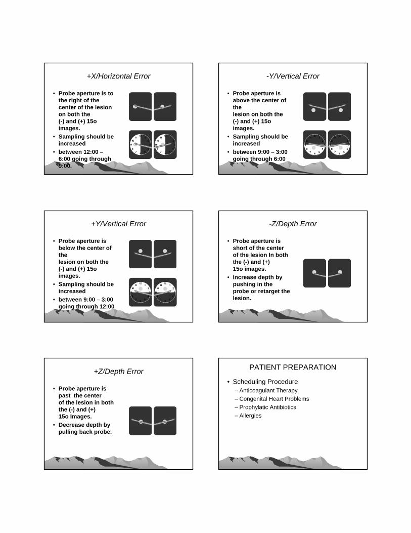
+X/Horizontal Error
• Probe aperture is to the right of the center of the lesion on both the (-) and (+) 15o images.
• Sampling should be increased
• between 12:00 –6:00 going through 9:00.
-Y/Vertical Error
• Probe aperture is above the center of the lesion on both the (-) and (+) 15o images.
• Sampling should be increased
• between 9:00 – 3:00 going through 6:00
+Y/Vertical Error
• Probe aperture is below the center of the lesion on both the (-) and (+) 15o images.
• Sampling should be increased
• between 9:00 – 3:00 going through 12:00
-Z/Depth Error
• Probe aperture is short of the center of the lesion In both the (-) and (+) 15o images.
• Increase depth by pushing in the probe or retarget the lesion.
+Z/Depth Error
• Probe aperture is past the center of the lesion in both the (-) and (+) 15o Images.
• Decrease depth by pulling back probe.
PATIENT PREPARATIONPATIENT PREPARATION
• Scheduling Procedure– Anticoagulant Therapy– Congenital Heart Problems– Prophylatic Antibiotics– Allergies

Office (832) 355-8130 Fax (832) 355-8123
St. Luke’s Women’s CenterBREAST BIOPSY
PROTOCOL SHEET►PATIENT GENERAL INFORMATIONPatient’s Name:______________________________________SLEH Medical Record No:____________________DOB:_______________________Age:______________Height_______________Weight:____________________Home Phone No: ____________________Work Phone No:________________ Cell Phone No:________________Clinical Diagnosis______________________________________________________________________________Hospital (facility) of Previous Mammogram:_________________________________________________________Referring Physician:___________________________Phone/Fax No: ____________________________________Date Films Received for Approval: _____________________________________________________________________________________________________________________________________________________________► APPROVED PROCEDURE□ Stereotactic Breast Biopsy/Mammotome Left Right Bilateral □ Ultrasound Core Biopsy Left Right Bilateral □ Ultrasound Aspiration/ FNA Left Right Bilateral □ Ultrasound Breast HH/Mammotome Biopsy Left Right Bilateral □ MRI Guided Core Biopsy Left Right BilateralPlease Specify: □ Calcifications □ Mass No. of Lesions: _____________--------_____________________________________________________________________________________________Ultrasound Prior to Procedure: □ Yes □ No Mammogram Prior to Procedure: □ Yes □ NoPlease Specify (re: mammography views): ___________________________________________________________________________________________________________________________________________________► APPROVALApproved by _________________________________MD. Date: ________________________________Schedule by: _____________________________________ Date: ________________________________Scheduled Date/Time _________________________________________________________________________DENIED Reason: ____________________________________________________________________________Referring MD Notify/Date: ____________________________________________________________________
• ► ADDITIONAL RADIOLOGIST INSTRUCTIONS: _________________________________________________________________________________________________________________________________________________________________________________________________________________________________________________________________________________
• ►NURSING • Responsibilities/ Medication prior to
Procedure:________________________________________________________________________________________________________________________________________________________________________________________________________________________________________________________________________
• ___________________________________________________________________________________________• ►PATIENT HISTORY (To be reviewed by Scheduler, Nurse, Mammographer ♦ Initials: __________)• Current Medication:
____________________________________________________________________________________• Allergies:
____________________________________________________________________________________• History of Blood Clots: □Yes □No• High Blood Pressure: □Yes □No• Kidney Disease: □Yes □No• Heart Disease: □Yes □No• Heart Valve Repair: □Yes □No• Artificial Valve □Yes □No• History of IEC □Yes □No• ___________________________________________________________________________________________• To be communicated to Patient:• Special Instructions:• PLEASE ASK THE PATIENT IF THEY ARE TAKING ANY BLOOD THINNERS; ASPIRIN, PLAVIX, COMADIN,
OR ANY OTHER BLOOD THINNING MEDICATIONS.• IF YES, PLEASE CONSULT WITH THE SPERVISOR/MANAGER FOR FURTHER INSTRUCTIONS PRIOR TO
SCHEDULING.• ® Patient will be given additional instructions if they are on Blood Thinning Medication• Schedulers Initials: ___________________• ___________________________________________________________________________________________• Additional Remarks:
______________________________________________________________________________________________________________________________________________________________________________________________________________________________________________________________________________________________________________________________________________________________________________________________________________________________________________________________________
PATIENT PREPARATIONPATIENT PREPARATIONPre-Procedure (nurse)
– Review patient history– Base line vital Signs– Consent by Physician – Prophylatic antibiotics
• Post Procedure– Wound Care/Pressure Dressing– Medications– 24-36 hour check up phone call
INSTRUMENTATIONINSTRUMENTATION

INSTRUMENTATIONINSTRUMENTATION
COMPLICATIONSCOMPLICATIONS

ACR STEREO ACCREDIATION PROCESSACR STEREO ACCREDIATION PROCESS
• Established in 1996• Model after the Mammography Program• Program assessment:
– Personnel training/qualification– Equipment maintenance/q.c.– Clinical Performance
Personnel QualificationsPersonnel Qualifications
• Physicians – Radiologist– Group Practice– Independent Practice– Surgeon– Group Practice (radiologist)– Independent Practice
• Technologists• Medical Physicist
Clinical Image QualityClinical Image Quality
• Stereotactic Biopsy Procedure Only– 2 view orthogonal mammogram showing
calcifications (one set) (cc/ml or mlo)– Stereotactic Procedure
• Scout image• Scout stereo Pair• Pre-Fire stereotactic set• Post-fire stereotactic set
Phantom Image QualityPhantom Image Quality
• Phantom Image Criteria• Screen film • Digital
– Mini-Phantom– Full size ACR phantom
• Image Dose Criteria– Less than 300mrads
ASSESSING IMAGE QUALITYASSESSING IMAGE QUALITY• MAP (Mammography Accreditation Phantom)
S/F DIGITALFibers 4.0 5.0Specks 3.0 4.0Masses 3.0 3.5
• Mini PhantomS/F DIGITAL
Fibers 2.0 3.0Specks 2.0 3.0Masses 2.0 2.5

SCOUTSCOUT
PREPRE--FIRE STEREOFIRE STEREO POST FIREPOST FIRE

SPECIMEN RADIOGRAPHSPECIMEN RADIOGRAPH

REFERENCESREFERENCES• Bolmgren J, Jacobson B, Nordenstrom B. Stereotaxic
instrument for needle biopsy of the mamma. AJR Am J Roentgenol 1977; 129:121-125.[Abstract]
• Burbank F. Stereotactic breast biopsy: its history, its present, and its future. Am Surg 1996; 62:128-150.[Medline]
• Lorad. Operators manual Rev 2. Danbury, Conn: Lorad, 1994; 1-50.
• Lorad. User’s guide Rev 2. Danbury, Conn: Lorad, 1995; 1-82.
• Fine RE, Boyd BA. Stereotactic breast biopsy: a practical approach. Am Surg 1996; 62:96-102.[Medline]
• Parker SH. Stereotactic large-core breast biopsy. In: Parker SH, Jobe WE, eds. Percutaneous breast biopsy. New York, NY: Raven, 1993; 61-79
• J&J Ethicon Endo-surgery Corp.