stimulating and blocking thyroid-stimulating hormone (tsh) …graves.pandapoop.net › wp-content...
TRANSCRIPT
J. Clin. Endocrinol. Metab. 2007 92:1058-1065 originally published online Dec 19, 2006; , doi: 10.1210/jc.2006-2213
Nils G. Morgenthaler, Su Chin Ho and Waldemar B. Minich
Binding SitesHypothyroidism Have Very Similar Concentration, TSH Receptor Affinity, and
Autoantibodies from Patients with Graves’ Disease and Autoimmune Stimulating and Blocking Thyroid-Stimulating Hormone (TSH) Receptor
Society please go to: http://jcem.endojournals.org//subscriptions/ or any of the other journals published by The EndocrineJournal of Clinical Endocrinology & Metabolism To subscribe to
Copyright © The Endocrine Society. All rights reserved. Print ISSN: 0021-972X. Online

Stimulating and Blocking Thyroid-Stimulating Hormone(TSH) Receptor Autoantibodies from Patients withGraves’ Disease and Autoimmune Hypothyroidism HaveVery Similar Concentration, TSH Receptor Affinity, andBinding Sites
Nils G. Morgenthaler, Su Chin Ho, and Waldemar B. Minich
Institut fur Experimentelle Endokrinologie und Endokrinologisches Forschungszentrum EnForCe (N.G.M.), Charite,Universitatsmedizin Berlin, 10117 Berlin, Germany; Department of Endocrinology (S.C.H.), Thyroid Unit, SingaporeGeneral Hospital, Republic of Singapore 169608; and Bioassays GmbH (N.G.M., W.B.M.), Biotechnology CentreHennigsdorf/Berlin, D-16761 Hennigsdorf/Berlin, Germany
Objective: The distinct biological properties of TSH receptor (TSH-R)autoantibodies (TRAbs) from patients with Graves’ disease (GD) areyet unexplained on the molecular level. Here we compare serumconcentration, affinity to the TSH-R, and binding sites on the TSH-Rof stimulating (TSAb) and blocking (TBAb) TRAbs.
Methods and Patients: Four-step affinity purification using humanrecombinant TSH-R was performed with 22 TRAb-positive sera fromGD patients (11 with only TSAb and 11 with only TBAb) and fivecontrol sera. Antibody concentration, TSH binding inhibition (TBII),and TSAb/TBAb activity of the purified TRAb were assessed. Labeledpurified TRAbs were used for displacement studies with TRAb and anadditional 30 patients and 10 control sera.
Results: TRAbs could be purified to 80–93% purity with recovery ofthe TBII and TSAb and TBAb activity. No TRAbs could be purified
from healthy individuals. The mean � SD concentration of TRAb was17.3 � 5.4 �g/IU for the TSAb sera (range, 9.6–25.9) and 18.2 � 8.5�g/IU for the TBAb sera (range, 4.6–29.2), respectively (P � 0.79).Affinity was in the picomolar range for both TRAb subtypes withmean � SD dissociation constant of 167 � 109 pM (60–410 pM) forTSAb and 253 � 132 pM (80–410 pM) for TBAb (P � 0.12). Purifiedand labeled TSAb and TBAb showed a very similar binding patternto the TSH-R in displacement studies with unlabeled TSAb/TBAb orunpurified patients sera, indicating binding sites on the TSH-R inclose proximity to each other.
Conclusion: TSAbs and TBAbs in the serum of patients with GDhave similar characteristics. They are of low concentration with highaffinity and have also similar binding epitopes on the TSH-R. (J ClinEndocrinol Metab 92: 1058–1065, 2007)
TSH RECEPTOR (TSH-R) autoantibodies (TRAbs) in pa-tients with Graves’ disease (GD) are functionally het-
erogeneous; they include mainly thyroid-stimulating anti-bodies (TSAbs) and occasionally also thyroid-blockingantibodies (TBAbs). TSAbs activate the receptor and causehyperthyroidism, whereas receptor occupancy by nonstimu-latory TBAbs inhibits TSH action and may cause hypothy-roidism. Together TSAbs and TBAbs comprise the TSH bind-ing inhibition (TBII) activity detected by TRAb assays.
Despite a plethora of information over the last decades, itis yet unclear what renders a TRAb a stimulator or a blocker.Although of interest in the assessment of the patient, thedifference between a TSAb and a TBAb on the molecularlevel is still unsolved. Potential discriminators include anti-
body concentration, affinity to the TSH-R, and (most fa-vored) binding epitopes on the TSH-R. Here TSAbs andTBAbs have been discussed for a long time to be directed todifferent and distinct parts of the TSH-R (1). However, recentevidence from monoclonal antibodies (2–5) suggests similarbinding sites for both subtypes of TRAb (6).
We recently developed a method for the affinity purifi-cation of TRAb from human serum and the subsequent usageof this purified TRAb for affinity studies and diagnostic assayformats (7).
In the present study, we apply this technology to purifyTRAb from sera from patients with GD or autoimmune hy-pothyroidism (AH) with strong TSAb or TBAb activity. Wethen compared the purified subtypes with respect to auto-antibody concentration, affinity to the TSH-R and competi-tion with autoantibodies in serum.
Patients and MethodsPatient sera
GD sera and sera from patients with AH were obtained from selectedpatients on the basis of their high TRAb levels and their TSAb or TBAbantibody subtypes. Patients with a history of GD were selected fromblood donors recruited for the development of in vitro diagnostics (In-vent GmbH, Hennigsdorf/GmbH, Hennigsdorf, Germany). This blood
First Published Online December 19, 2006Abbreviations: AH, Autoimmune hypothyroidism; ATD, antithyroid
drug; GD, Graves’ disease; InI, inhibition index; Kd, dissociation con-stant; RLU, relative light unit; SI, stimulation index; TBAb, thyroid-blocking autoantibody; TBII, TSH binding inhibition; TRAb, TSH-R au-toantibodies; TSAb, thyroid-stimulating autoantibody; TSH-R, TSHreceptor.JCEM is published monthly by The Endocrine Society (http://www.endo-society.org), the foremost professional society serving the en-docrine community.
0021-972X/07/$15.00/0 The Journal of Clinical Endocrinology & Metabolism 92(3):1058–1065Printed in U.S.A. Copyright © 2007 by The Endocrine Society
doi: 10.1210/jc.2006-2213
1058
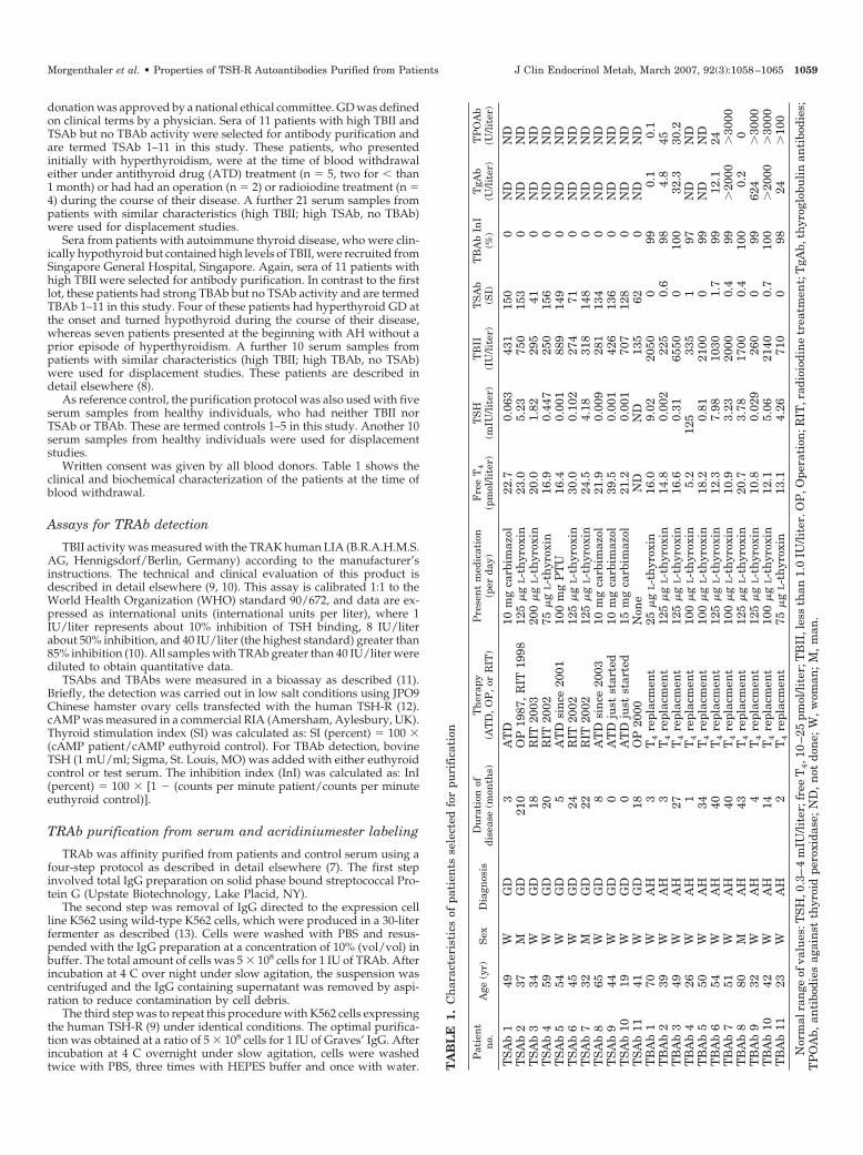
donation was approved by a national ethical committee. GD was definedon clinical terms by a physician. Sera of 11 patients with high TBII andTSAb but no TBAb activity were selected for antibody purification andare termed TSAb 1–11 in this study. These patients, who presentedinitially with hyperthyroidism, were at the time of blood withdrawaleither under antithyroid drug (ATD) treatment (n � 5, two for � than1 month) or had had an operation (n � 2) or radioiodine treatment (n �4) during the course of their disease. A further 21 serum samples frompatients with similar characteristics (high TBII; high TSAb, no TBAb)were used for displacement studies.
Sera from patients with autoimmune thyroid disease, who were clin-ically hypothyroid but contained high levels of TBII, were recruited fromSingapore General Hospital, Singapore. Again, sera of 11 patients withhigh TBII were selected for antibody purification. In contrast to the firstlot, these patients had strong TBAb but no TSAb activity and are termedTBAb 1–11 in this study. Four of these patients had hyperthyroid GD atthe onset and turned hypothyroid during the course of their disease,whereas seven patients presented at the beginning with AH without aprior episode of hyperthyroidism. A further 10 serum samples frompatients with similar characteristics (high TBII; high TBAb, no TSAb)were used for displacement studies. These patients are described indetail elsewhere (8).
As reference control, the purification protocol was also used with fiveserum samples from healthy individuals, who had neither TBII norTSAb or TBAb. These are termed controls 1–5 in this study. Another 10serum samples from healthy individuals were used for displacementstudies.
Written consent was given by all blood donors. Table 1 shows theclinical and biochemical characterization of the patients at the time ofblood withdrawal.
Assays for TRAb detection
TBII activity was measured with the TRAK human LIA (B.R.A.H.M.S.AG, Hennigsdorf/Berlin, Germany) according to the manufacturer’sinstructions. The technical and clinical evaluation of this product isdescribed in detail elsewhere (9, 10). This assay is calibrated 1:1 to theWorld Health Organization (WHO) standard 90/672, and data are ex-pressed as international units (international units per liter), where 1IU/liter represents about 10% inhibition of TSH binding, 8 IU/literabout 50% inhibition, and 40 IU/liter (the highest standard) greater than85% inhibition (10). All samples with TRAb greater than 40 IU/liter werediluted to obtain quantitative data.
TSAbs and TBAbs were measured in a bioassay as described (11).Briefly, the detection was carried out in low salt conditions using JPO9Chinese hamster ovary cells transfected with the human TSH-R (12).cAMP was measured in a commercial RIA (Amersham, Aylesbury, UK).Thyroid stimulation index (SI) was calculated as: SI (percent) � 100 �(cAMP patient/cAMP euthyroid control). For TBAb detection, bovineTSH (1 mU/ml; Sigma, St. Louis, MO) was added with either euthyroidcontrol or test serum. The inhibition index (InI) was calculated as: InI(percent) � 100 � [1 � (counts per minute patient/counts per minuteeuthyroid control)].
TRAb purification from serum and acridiniumester labeling
TRAb was affinity purified from patients and control serum using afour-step protocol as described in detail elsewhere (7). The first stepinvolved total IgG preparation on solid phase bound streptococcal Pro-tein G (Upstate Biotechnology, Lake Placid, NY).
The second step was removal of IgG directed to the expression cellline K562 using wild-type K562 cells, which were produced in a 30-literfermenter as described (13). Cells were washed with PBS and resus-pended with the IgG preparation at a concentration of 10% (vol/vol) inbuffer. The total amount of cells was 5 � 108 cells for 1 IU of TRAb. Afterincubation at 4 C over night under slow agitation, the suspension wascentrifuged and the IgG containing supernatant was removed by aspi-ration to reduce contamination by cell debris.
The third step was to repeat this procedure with K562 cells expressingthe human TSH-R (9) under identical conditions. The optimal purifica-tion was obtained at a ratio of 5 � 108 cells for 1 IU of Graves’ IgG. Afterincubation at 4 C overnight under slow agitation, cells were washedtwice with PBS, three times with HEPES buffer and once with water. T
AB
LE
1.C
har
acte
rist
ics
ofpa
tien
tsse
lect
edfo
rpu
rifi
cati
on
Pat
ien
tn
o.A
ge(y
r)S
exD
iagn
osis
Du
rati
onof
dise
ase
(mon
ths)
Th
erap
y(A
TD
,O
P,
orR
IT)
Pre
sen
tm
edic
atio
n(p
erda
y)F
ree
T4
(pm
ol/li
ter)
TS
H(m
IU/li
ter)
TB
II(I
U/li
ter)
TS
Ab
(SI)
TB
Ab
InI
(%)
TgA
b(U
/lite
r)T
PO
Ab
(U/li
ter)
TS
Ab
149
WG
D3
AT
D10
mg
carb
imaz
ol22
.70.
063
431
150
0N
DN
DT
SA
b2
37M
GD
210
OP
1987
,R
IT19
9812
5�
gL-t
hyr
oxin
23.0
5.23
750
153
0N
DN
DT
SA
b3
34W
GD
18R
IT20
0320
0�
gL-t
hyr
oxin
20.0
1.82
295
410
ND
ND
TS
Ab
459
WG
D20
RIT
2002
75�
gL-t
hyr
oxin
16.9
0.44
725
015
60
ND
ND
TS
Ab
554
WG
D5
AT
Dsi
nce
2001
100
mg
PT
U16
.40.
001
889
149
0N
DN
DT
SA
b6
45W
GD
24R
IT20
0212
5�
gL-t
hyr
oxin
30.0
0.10
227
471
0N
DN
DT
SA
b7
32M
GD
22R
IT20
0212
5�
gL-t
hyr
oxin
24.5
4.18
318
148
0N
DN
DT
SA
b8
65W
GD
8A
TD
sin
ce20
0310
mg
carb
imaz
ol21
.90.
009
281
134
0N
DN
DT
SA
b9
44W
GD
0A
TD
just
star
ted
10m
gca
rbim
azol
39.5
0.00
142
613
60
ND
ND
TS
Ab
1019
WG
D0
AT
Dju
stst
arte
d15
mg
carb
imaz
ol21
.20.
001
707
128
0N
DN
DT
SA
b11
41W
GD
18O
P20
00N
one
ND
ND
135
620
ND
ND
TB
Ab
170
WA
H3
T4
repl
acm
ent
25�
gL-t
hyr
oxin
16.0
9.02
2050
099
0.1
0.1
TB
Ab
239
WA
H3
T4
repl
acm
ent
125
�g
L-t
hyr
oxin
14.8
0.00
222
50.
698
4.8
45T
BA
b3
49W
AH
27T
4re
plac
men
t12
5�
gL-t
hyr
oxin
16.6
0.31
6550
010
032
.330
.2T
BA
b4
26W
AH
1T
4re
plac
men
t10
0�
gL-t
hyr
oxin
5.2
125
335
197
ND
ND
TB
Ab
550
WA
H34
T4
repl
acm
ent
100
�g
L-t
hyr
oxin
18.2
0.81
2100
099
ND
ND
TB
Ab
654
WA
H40
T4
repl
acm
ent
125
�g
L-t
hyr
oxin
12.3
7.98
1030
1.7
9912
.124
TB
Ab
751
WA
H40
T4
repl
acm
ent
100
�g
L-t
hyr
oxin
10.9
3.23
2000
0.4
99�
2000
�30
00T
BA
b8
80M
AH
43T
4re
plac
men
t12
5�
gL-t
hyr
oxin
20.7
3.78
1700
0.4
100
0.2
0T
BA
b9
32W
AH
4T
4re
plac
men
t12
5�
gL-t
hyr
oxin
10.8
0.02
926
00
9962
4�
3000
TB
Ab
1042
WA
H14
T4
repl
acm
ent
100
�g
L-t
hyr
oxin
12.1
5.06
2140
0.7
100
�20
00�
3000
TB
Ab
1123
WA
H2
T4
repl
acm
ent
75�
gL-t
hyr
oxin
13.1
4.26
710
098
24�
100
Nor
mal
ran
geof
valu
es:T
SH
,0.3
–4
mIU
/lite
r;fr
eeT
4,1
0–2
5pm
ol/li
ter;
TB
II,l
ess
than
1.0
IU/li
ter.
OP
,Ope
rati
on;R
IT,r
adio
iodi
ne
trea
tmen
t;T
gAb,
thyr
oglo
buli
nan
tibo
dies
;T
PO
Ab,
anti
bodi
esag
ain
stth
yroi
dpe
roxi
dase
;N
D,
not
don
e;W
,w
oman
;M
,m
an.
Morgenthaler et al. • Properties of TSH-R Autoantibodies Purified from Patients J Clin Endocrinol Metab, March 2007, 92(3):1058–1065 1059

Supernatant was removed by aspiration. TRAbs were eluted three timesby centrifugation with elution buffer [25 mm citric acid, 100 mm NaCl(pH 2.1)]. Fractions were combined, neutralized with 200 mm Tris-HCl(pH 7.5) and centrifuged at 100,000 � g to remove any cellular debris.To prevent subsequent adsorption of TRAb to surfaces, 1% (wt/vol) BSA(Sigma) was added, and the preparation was stored at �80 C.
The fourth and final purification round was performed using recom-binant TSH-R bound to a specific monoclonal antibody [BA8 (9)] onCarbolink gel (Pierce Biotechnology, Rockford, IL). Coupling was per-formed overnight according to the manual with a yield of 70–90%.TSH-R extract from K562 cells was obtained as described (9), and 200 mlmembrane extract were incubated with 2.5 ml gel in batch for 5 h at 4C under mild agitation. Two hundred milliliters membrane extract rep-resent the detergent preparation of around 5 � 109 cells. Gel material wasthree times resuspended with washing buffer and centrifuged. After thelast washing step, the gel material was resuspended in the TRAb-enriched buffer from the third purification step at a ratio of 1 ml gel to15 IU TRAbs and incubated overnight at 4 C under mild agitation,allowing the TRAb to bind specifically to the solid-phase bound humanTSH-R. The gel material was washed three times with 50 ml washingbuffer, and acidic elution was done with 15 ml freshly prepared elutionbuffer [50 mm citric acid, 1% BSA, 150 mm NaCl (pH 2.1)]. Immediatelyafter elution, the fraction was neutralized by adding 15 ml PBS con-taining 1% BSA and as additional neutralization buffer 200 mm phos-phate buffer (pH 10) to a final pH of 6.8. Before freezing, the buffer wascentrifuged at 100,000 � g to remove precipitated TSH-R and othercellular debris.
Purified TRAbs were diluted with buffer [20 mm HEPES-NaOH (pH7.5), 150 mm NaCl, 1% BSA, 10% glycerol] to a concentration of 20IU/liter TRAbs and incubated over night at 4 C with an equal amountof acridinium ester (MACN; Invent GmbH, Hennigsdorf, Germany)labeled goat antihuman antibodies. At the end of the incubation, 1mg/ml total human IgG was added to block unbound tracer.
Calculation of dissociation constant (Kd) of TRAb
Different concentrations (5–3000 pm) of indirectly labeled TRAbswere added in 0.3 ml buffer [20 mm HEPES-NaOH (pH 7.5), 150 mmNaCl, 1% BSA, 10% glycerol, 1 mg/ml human IgG] to TSH-R-coatedtubes (TRAK human RIA). Tubes were incubated for 24 h at roomtemperature, washed four times with 2 ml washing buffer, and boundrelative light units (RLUs) were measured in a luminometer. Nonspecificbinding, determined in excess of unlabeled purified TRAbs, was sub-tracted. The Kd was calculated at 50% saturation by nonlinear regressionanalysis (Prism 4; GraphPad Software, San Diego, CA). Data are shownas saturation curve and in the conventional Scatchard plot (14).
Displacement studies
For antibody displacement studies, 0.125 mU of purified and labeledTRAbs were added in duplicate to 0.3 ml of buffer [20 mm HEPES-NaOH(pH 7.5), 100 mm NaCl, 1% BSA, 10% glycerol] together with increasingamounts of unlabeled purified TRAbs to TSH-R-coated tubes(B.R.A.H.M.S. AG). Tubes were incubated overnight at 4 C, washed fourtimes with 2 ml washing buffer (B.R.A.H.M.S. AG), and bound RLUswere measured in a luminometer (Berthold, Bad Wildbad, Germany).The total RLUs added were 126,000–240,000, depending on the purifiedand labeled TRAb used.
For serum displacement studies, 100 �l of patients’ serum or TRAKhuman standards (B.R.A.H.M.S. AG) were added in duplicate to TSH-R-coated tubes (B.R.A.H.M.S. AG), followed by 100 �l buffer [100 mmHEPES-KOH (pH 7.5), 20 mm EDTA, 0.5 mm N-ethyl-maleimide, 1%BSA, 0.5% Triton X-100] and 50 �l PBS containing 0.25 mIU of labeledTRAbs as a tracer (80,000–300.000 RLU). Tubes were incubated over-night at 4 C, washed four times with 2 ml washing buffer (B.R.A.H.M.S.AG), and bound RLUs were measured in a luminometer (Berthold).Results were expressed in international units per liter according to theWHO standard used for calibration.
Results
TRAb were purified from 22 selected patients with GD orAH. All sera were tested for TBII, TSAbs, and TBAbs. All
patients had very high levels of TBII, ranging from 135 to6550 IU/liter based on the WHO standard 90/672 calibra-tion. In terms of inhibition of TSH binding, all patients hadnear total inhibition of binding, even at high dilutions. All 11patients with a history of hyperthyroidism had high levels ofTSAbs (mean � sd stimulation index 121 � 41.7) but noTBAbs. Inversely, all 11 patients with hypothyroidism hadhigh levels of TBAbs (mean � sd inhibition: 98.9 � 0.9%) butwere negative for TSAbs. Thus, the patients selected for thisstudy represent both extreme ends of the autoimmune re-sponse to the TSH-R. Table 2 lists all sera grouped accordingto their bioactivity. The mean � sd TBII activity in the TBAbpatients was 1736 � 1778 IU/liter, which is significantlyhigher (P � 0.03) than the mean TBII activity in the TSAb-positive patients (432 � 242 IU/liter). Serum obtained fromthe five healthy blood donors had no TBII or TSAb/TBAbactivity.
Four-step affinity purification was performed with all 22patients and five control sera (Table 2). Purification wassuccessful with 21 of 22 patients sera. The purified TRAbs ofpatient TBAb 7 were lost due to technical difficulties with theelution buffer. This sample was excluded from further cal-culations. After affinity purification, TBII and TSAb/TBAbactivity of the TRAbs was still present. Figure 1 shows dose-response curves with increasing TRAb concentration in acAMP bioassay for two purified TSAb (Fig. 1A) and onepurified TBAb sample (Fig. 1B). The complete cAMP bioas-say data of all purified TRAbs is shown in Table 2 at a TRAbconcentration of 10 IU/liter.
The total TRAb international units recovered based onTBII activity was 26.9 � 17.2% for TSAb samples and 21.9 �15.2% for TBAbs, respectively. Measurement of total IgG inpurified samples of patients and controls showed a cleardifference. Patients had mean � sd total IgG concentrationsof 375 � 151 �g/liter for TSAbs and 458 � 88 �g/liter forTBAbs, respectively, whereas controls had only 47.6 � 15.6�g/liter. However, the purified material from both TSAband TBAb patients still had strong TBII activity, whereas thecontrol material did not show any interaction with theTSH-R. The IgG still present in control samples after the samepurification protocol indicates nonspecific background car-ried through the process and allows the estimation of thepurity of IgG from patients samples to be 80–93% pureTRAbs.
A comparison of total IgG and TRAb international unitsallowed calculation of the amount of IgG per internationalunit of TRAb. This was very similar for each individualserum purified and showed no difference between TSAbsand TBAbs. The mean � sd was 17.3 � 5.4 �g/IU for theTSAb sera (range 9.6–25.9), and 18.2 � 8.5 �g/IU for theTBAb sera (range 4.6–29.2), respectively (P � 0.78). There-fore, the concentration of TRAbs in the serum of patients withGD is between 5 and 30 �g/liter for 1 international WHO unit(IU), irrespective of the TSAb or TBAb subtype. The patientsin this study with very high levels of TRAbs had antibodiesin the milligram per liter range, whereas the average patientwith GD (usually with TRAb/TBII � 40 IU/liter) is likely tohave antibodies in the medium to high microgram per literrange.
Affinity of the purified TRAbs to the TSH-R was measured
1060 J Clin Endocrinol Metab, March 2007, 92(3):1058–1065 Morgenthaler et al. • Properties of TSH-R Autoantibodies Purified from Patients

by saturation experiments, in which increasing amounts ofindirectly labeled purified TRAbs were incubated on TSH-R-coated tubes. Figure 2 shows the Scatchard plot for tworepresentative TSAb and two TBAb patients. The Kd for allTRAb samples is shown in Table 3, again grouped according
to TSAb or TBAb activity. Affinity was in the picomolarrange for both TRAb subtypes, and there was no differencebetween TSAb and TBAb samples. Mean � sd Kd for TSAbwas 167 � 109 pm (60–410 pm) and 253 � 132 pm (80–410pm) for TBAb, respectively (P � 0.12).
TABLE 2. Affinity purification of TSH-R autoantibodies from patients’ serum
Patient serum used for purification After affinity purification on recombinant TSH-R
Serumno.
TBII(IU/liter)
TSAb(SI)
TBAb InI(%)
TRAbadded(mIU)
Recovery(mIU)
Recovery(%)
TBII(IU/liter)
TSAb(SI)
TBAb InI(%)
Total IgG(�g/liter)
TRAb(�g/liter)
TRAbpurity
(%)
TRAb per IU(�g/IU)
TSAb1 431 150 0 431 66 15.3 18.8 162 3 252 204 80.9 10.82 750 153 0 750 178 23.7 19.8 145 8 239 191 79.9 9.63 295 41 0 295 80 27.1 21.8 67 13 459 411 89.5 18.94 250 156 0 250 46 18.4 11.5 113 10 266 218 82.0 19.05 889 149 0 889 196 22.0 21.8 69 0 520 472 90.8 21.76 274 71 0 274 37 13.5 10.5 73 17 320 272 85.0 25.97 318 148 0 318 48 15.1 13.8 176 14 253 205 81.0 14.98 281 134 0 281 176 62.6 19.5 191 17 341 293 85.9 15.09 426 136 0 426 78 18.3 22.3 96 11 302 254 84.1 11.410 707 128 0 707 147 20.8 21.0 84 0 444 396 89.2 18.811 135 62 0 1193 700 58.7 28.0 39.7 0 730 682 93.4 24.4Mean 432 121 0,0 529 159 26.9 19.0 111 8.5 375 327 85.6 17.3SD 242 41.7 0,0 312 188.8 17.2 5.2 50.6 6.7 151 151 4.6 5.4
TBAb1 2050 0 99 2000 329 16.5 36.5 0.1 96 403 355 88.1 9.72 225 0.6 98 250 44 17.6 12.5 0.1 98 393 345 87.8 27.63 6550 0 100 3000 749 25.0 83.3 0.2 94 434 386 88.9 4.64 335 1 97 335 51 15.2 14.5 0.2 97 382 334 87.4 23.05 2100 0 99 2000 421 21.1 46.8 0.6 94 486 438 90.1 9.36 1030 1.7 99 1100 83 7.5 16.5 0.1 98 434 386 88.9 23.47 2000 0.4 99 1000 1.58 1700 0.4 100 850 128 15.1 23.3 0.1 97 486 438 90.1 18.89 260 0 99 260 58 22.3 14.5 0.1 98 387 339 87.6 23.410 2140 0.7 100 750 118 15.7 21.5 0.3 82 675 627 92.9 29.211 710 0 98 1428 900 63.0 36.0 0.1 99 500 452 90.4 12.6Mean 1736 0.4 98.9 1179 262 21.9 30.5 0.2 95.3 458 410 89.2 18.2SD 1778 0.5 0.9 865 308 15.2 21.8 0.2 5.0 88.0 88.0 1.7 8.5
Controls1 0.7 0 0 0.1 48.02 0.2 0 0 0.2 64.03 0.4 0 0 0.2 54.04 0.1 0 0 0.1 22.05 0.4 0 0 0.1 50.0Mean 0.4 0.0 0.0 0.1 47.6SD 0.2 0.0 0.0 0.1 15.6
FIG. 1. cAMP bioassay with purified TSAbs and TBAbs. Increasing concentrations of purified TRAb were incubated in a Chinese hamster ovarycell bioassay with cAMP readout. Shown is the dose-response curve for two purified TSAbs and one TBAb.
Morgenthaler et al. • Properties of TSH-R Autoantibodies Purified from Patients J Clin Endocrinol Metab, March 2007, 92(3):1058–1065 1061

Finally, we tested purified TRAb material in competitionstudies to see whether a different displacement pattern in-dicates differences in the binding sites of TSAbs and TBAbs.First, competition was done among the purified samples ina cross-over design. Different TSAb samples as tracer werecompeted with increasing concentration of other TSAbs (Fig.3A) or TBAbs (Fig. 3B). The same was done with labeledTBAb samples, which were brought into competition withTSAb samples (Fig. 3C) or other TBAb samples (Fig. 3D). Thedose-response curves shown are examples of several exper-iments and demonstrate similar displacement among all pu-rified TRAb and no difference between TSAbs and TBAbs.
Then competition studies were done with labeled TSAb orTBAb samples and unpurified sera from additional patients(20 patients with GD, 10 patients with autoimmune thyroiddisease containing only TBAbs, and 10 controls). Figure 4shows six representative experiments (three with labeledTSAbs and three with labeled TBAbs), which demonstrate avery similar binding pattern of both TSAbs and TBAbs. In allexperiments, purified TSAbs competed with patients seracontaining only TSAbs but also with sera containing onlyTBAbs. The identical pattern was seen for purified TBAbs,which competed with not only TBAb-containing sera but alsoTSAb-positive GD sera. In all experiments, the interactionwith TBAb-containing sera was higher than with TSAb sera,irrespective of the tracer used. No interaction was seen withcontrol sera. Using the standard bovine TSH tracer from theTRAb kit gave very similar results (Fig. 5).
Discussion
This study took advantage of a recently established puri-fication method for TRAbs (7) and examined the differencesbetween TSAbs and TBAbs purified from patients with GDand hypothyroid autoimmune disease. The results can besummarized as follows. First, the concentration of TRAbs inpatients with GD is about 18 �g/liter IgG (5–30 �g/liter) perinternational WHO unit, with no difference between TSAband TBAb. Second, the affinity of both TSAbs and TBAbs tothe TSH-R is in the picomolar range (60–410 pm), againwithout a significant difference between TSAbs and TBAbs.Third, both TSAbs and TBAbs show a very similar bindingpattern to the TSH-R in displacement studies with purifiedTRAbs and patients’ sera, indicating binding sites on theTSH-R in very close proximity. Because the binding of TSHshows the same pattern, this binding site of TSAbs and TB-Abs is most likely in the TSH binding part of the TSH-R,which according to present models of the receptor lies withinthe horseshoe of the ectodomain (15–17).
This study confirms our preliminary report, that TRAbsare of low concentration with high affinity (7). The 14–28�g/liter per international unit reported initially are well inconcordance with the figures in this study. Although therewere earlier estimates of TRAb concentration based on neu-tralizing studies with soluble TSH-R (18, 19), the concentra-tion (and also affinity) of TRAb is a constant matter of debate.It should be pointed out that the TRAb concentration per 1
FIG. 2. Saturation curve and Scatchard plot for purified and labeled TRAbs. The Kd was calculated with nonlinear regression analysis. Shownare two representative purified TSAbs and two TBAbs. The complete affinity data are listed in Table 3.
1062 J Clin Endocrinol Metab, March 2007, 92(3):1058–1065 Morgenthaler et al. • Properties of TSH-R Autoantibodies Purified from Patients

IU is relatively low, and most GD patients have less than 40IU/liter TRAbs. However, a few patients may have several
thousand international units per liter TRAbs, which is equiv-alent to a TRAb concentration in the milligram per liter range.
The affinity of TRAbs to the TSH-R in this study confirmsour preliminary report. It is noteworthy that the linear Scat-chard plot indicates the presence of TRAbs with very similarKd in each patient preparation. This is also supported by thecomparable results in all experiments. TRAbs may be het-erogeneous within and between patients, but all TRAbs havea very similar affinity to the TSH-R. This affinity is compa-rable with that found for bovine TSH (7, 19) and with ahuman TSAb monoclonal antibody (5).
The identification of the binding sites of TSAbs and TBAbsto the TSH-R is still controversial. Over decades data werepresented that supported a rather simple concept: TSAbsbind to the N-terminal part of the TSH-R ectodomain andTBAbs to the C-terminal part close to the membrane (20–22).Following this concept, we tried to develop different in vitroassays for the discrimination of TSAbs and TBAbs by chang-ing either the receptor (23) or tracer (24), albeit with moderateresults. After the establishment of monoclonal antibodieswith TSAb or TBAb activity (2–5), the model of distinct anddistant TSAb and TBAb epitopes could no longer be sup-ported. Three groups examined independently the interac-tion of labeled monoclonal antibodies with autoantibodiesfrom patients to the TSH-R and reported similar results:monoclonal TSAbs seem to interact with binding sites forTSH, TSAbs, and TBAbs (2, 5, 6, 25), whereas monoclonalTBAbs likewise do exactly the same (6). Our study offersindependent proof for this with purified TRAbs from pa-
FIG. 3. TSAb tracer and TBAb tracer displacement by purified autoantibodies. Shown are representative examples of a purified TSAb sampleas tracer in competition with increasing concentration of other purified TSAbs (A) or purified TBAbs (B). Inversely, a labeled TBAb samplecompeted with purified TSAb samples (C) or other purified TBAb samples (D).
TABLE 3. Affinity of purified TRAb to the TSH-R
Purified TRAb Kd (10�12 M) 95% CI of Kd Bmax (10�12 M)
TSAb1 80.8 50.7–111 20.02 78.5 59.5–97.4 18.73 260 174–346 31.84 228 152–303 55.05 119 84.2–154 20.66 410 194–627 44.37 92.1 40.6–144 35.48 81.0 38.0–124 27.69 59.7 14.8–105 22.910 192 105–278 34.011 240 200–265 30.0Mean � SD 167 � 109 30.9 � 11.1
TBAb1 195 109–281 55.22 370 318–422 35.43 128 87.12–169.0 36.94 155 113–198 24.55 303 182–424 72.66 109 57.8–161 13.278 381 221–542 85.49 397 149–645 41.510 410 266–554 58.111 80 60.3–92.8 12.0Mean � SD 253 � 132 43.5 � 24.3
CI, Confidence interval; Bmax, maximal binding capacity.
Morgenthaler et al. • Properties of TSH-R Autoantibodies Purified from Patients J Clin Endocrinol Metab, March 2007, 92(3):1058–1065 1063

tients. We also found no differences between TSAb and TBAbinteraction with the TSH-R in competition studies with pu-rified TRAb or patient sera.
If concentration, affinity for, and binding site on the TSH-Rare similar for TSAbs and TBAbs, what makes the biologicaldifference? A possible explanation was proposed by a recentstudy on the interaction of TSAb and TBAb monoclonalantibodies and TSH-R mutants with single amino acid sub-stitutions in the crucial leucine-rich horseshoe part of thereceptor (26). This study showed that TSAb and TBAbepitopes are in very close steric proximity, but they are nev-ertheless distinct on the molecular level (6). Single amino aciddifferences in the binding site could be the reason for thedramatic difference in bioactivity. However, alternative ex-planations may exist. The recent crystal structure of theFSH-R together with its ligand FSH (27) suggests that similar
approaches for the TSH-R and TSH or TSAb/TBAb could behelpful in solving this riddle.
Two limitations of our study should be addressed. First,the patients selected for this study had high levels of TRAbs,some even after treatment, which may not be representativefor the average patient with GD. Second, the stringent pu-rification conditions might have been selective for high-af-finity TRAbs. Whereas the average recovery of TRAbs afterpurification was reasonable (27%), low-affinity TRAbs couldhave been lost during the process. Therefore, our study doesnot exclude the existence of TRAb with lower affinity thanreported here.
Finally, it should be noted that we were unable to purifyTRAbs from healthy individuals. This is in contrast to a studyon affinity enrichment of TRAbs from GD patients andhealthy individuals (28). In this study the evidence for TRAbsin healthy individuals was (besides fluorescence-activatedcell sorter analysis) based on a weak interaction of the en-riched material in a competitive TBII system. It is importantto keep in mind that the detection of TRAbs by any com-petitive radioreceptor assay is an indirect method and there-fore subject to artifacts. Any substance (i.e. residual chemicalsfrom the purification protocol or a high salt concentration)that negatively influences the TSH-R TSH interaction willreduce the binding of the tracer, which results in a positivereadout. However, it is also possible that this discrepancybetween the two studies is due to differences in the affinitypurification protocol, particularly the loss of low-affinityTRAbs in our system.
As we have pointed out before (7), the strongest evidencefor a successful purification of TRAbs by our protocol is not
FIG. 4. TSAb tracer and TBAb tracer displacement by sera from patients. Total tracer added to TSH-R-coated tubes 80,000–300,000 RLU.Control, Healthy blood donors without thyroid disease; TSAb, patients with GD containing only TSAbs; TBAb, patients with AH. The line showsthe median for each group.
FIG. 5. TBII assay with bovine TSH as tracer with sera from patients.The same patients shown in Fig. 4 together with purified TSAbs orTBAbs as tracer had a similar displacement pattern in a coated tubesecond-generation TBII assay with bovine TSH as tracer.
1064 J Clin Endocrinol Metab, March 2007, 92(3):1058–1065 Morgenthaler et al. • Properties of TSH-R Autoantibodies Purified from Patients

necessarily the recovery of TBII activity but a clear stimu-lation in the bioassay and the fact that we are able to label thepurified TRAbs for saturation and displacement studies.Here the purified autoantibodies from patients behave sim-ilar to studies we did with monoclonal TSH-R antibodies (6,24), indicating a high level of purity and functionality.
Acknowledgments
The authors thank Dr. W. Weglohner, S. Karasch, and C. Schwiebert(InVivo GmbH, Hennigsdorf/Berlin, Germany) for the production oflarge amounts of TSH-R-expressing K562 cells and Dr. Daphne Khoo(Singapore General Hospital), Dr. Andreas Bergmann, and Dr. JoachimStruck (B.R.A.H.M.S. AG, Hennigsdorf/Berlin, Germany) for helpfuldiscussions.
Received October 11, 2006. Accepted December 12, 2006.Address all correspondence and requests for reprints to: Dr. Nils G.
Morgenthaler, Institut fur Experimentelle Endokrinologie und Endokri-nologisches Forschungs-Centrum, EnForCe der Charite, CampusCharite Mitte, Chariteplatz 1, 10117 Berlin, Germany. E-mail:[email protected].
Present address for W.B.M.: KreLo GmbH Medical Diagnostics, Ulm,Germany.
Disclosure Summary: N.G.M., S.C.H., and W.B.M. have no conflict ofinterest related to this study.
References
1. Rapoport B, Chazenbalk GD, Jaume JC, McLachlan SM 1998 The thyrotropin(TSH) receptor: interaction with TSH and autoantibodies. Endocr Rev 19:673–716
2. Sanders J, Jeffreys J, Depraetere H, Richards T, Evans M, Kiddie A, BreretonK, Groenen M, Oda Y, Furmaniak J, Rees Smith B 2002 Thyroid-stimulatingmonoclonal antibodies. Thyroid 12:1043–1050
3. Ando T, Latif R, Pritsker A, Moran T, Nagayama Y, Davies TF 2002 Amonoclonal thyroid-stimulating antibody. J Clin Invest 110:1667–1674
4. Costagliola S, Franssen JD, Bonomi M, Urizar E, Willnich M, Bergmann A,Vassart G 2002 Generation of a mouse monoclonal TSH receptor antibody withstimulating activity. Biochem Biophys Res Commun 299:891–896
5. Sanders J, Evans M, Premawardhana LD, Depraetere H, Jeffreys J, RichardsT, Furmaniak J, Rees Smith B 2003 Human monoclonal thyroid stimulatingautoantibody. Lancet 362:126–128
6. Costagliola S, Bonomi M, Morgenthaler NG, Van Durme J, Panneels V,Refetoff S, Vassart G 2004 Delineation of the discontinuous-conformationalepitope of a monoclonal antibody displaying full in vitro and in vivo thyro-tropin activity. Mol Endocrinol 18:3020–3034
7. Morgenthaler NG, Minich WB, Willnich M, Bogusch T, Hollidt JM, We-glohner W, Lenzner C, Bergmann A 2003 Affinity purification and diagnosticuse of TSH receptor autoantibodies from human serum. Mol Cell Endocrinol212:73–79
8. Khoo DH, Eng PH, Ho SC, Fok AC 1999 Differences in the levels of TSH-binding inhibitor immunoglobulins in goitrous and agoitrous autoimmunethyroiditis after twelve months of l-thyroxine therapy. Clin Endocrinol (Oxf)51:73–79
9. Costagliola S, Morgenthaler NG, Hoermann R, Badenhoop K, Struck J,Freitag D, Poertl S, Weglohner W, Hollidt JM, Quadbeck B, Dumont JE,Schumm-Draeger PM, Bergmann A, Mann K, Vassart G, Usadel KH 1999Second generation assay for thyrotropin receptor antibodies has superior di-agnostic sensitivity for Graves’ disease. J Clin Endocrinol Metab 84:90–97
10. Schott M, Feldkamp J, Bathan C, Fritzen R, Scherbaum WA, Seissler J 2000Detecting TSH-receptor antibodies with the recombinant TBII assay: technicaland clinical evaluation. Horm Metab Res 32:429–435
11. Morgenthaler NG, Pampel I, Aust G, Seissler J, Scherbaum WA 1998 Ap-plication of a bioassay with CHO cells for the routine detection of stimulatingand blocking autoantibodies to the TSH-receptor. Horm Metab Res 30:162–168
12. Perret J, Ludgate M, Libert F, Gerard C, Dumont JE, Vassart G, ParmentierM 1990 Stable expression of the human TSH receptor in CHO cells andcharacterization of differentially expressing clones. Biochem Biophys ResCommun 171:1044–1050
13. Stiens LR, Buntemeyer H, Lutkemeyer D, Lehmann J, Bergmann A, We-glohner W 2000 Development of serum-free bioreactor production of recom-binant human thyroid stimulating hormone receptor. Biotechnol Prog 16:703–709
14. Scatchard G 1949 The attraction of proteins for small molecules and ions. AnnNY Acad Sci 51:600–672
15. Kajava AV, Vassart G, Wodak SJ 1995 Modeling of the three-dimensionalstructure of proteins with the typical leucine-rich repeats. Structure 3:867–877
16. Kleinau G, Jaeschke H, Neumann S, Laettig J, Paschke R, Krause G 2004Identification of a novel epitope in the TSH receptor ectodomain acting asintramolecular signalling interface. J Biol Chem 279:51590–51600
17. Nunez Miguel R, Sanders J, Jeffreys J, Depraetere H, Evans M, Richards T,Blundell TL, Rees Smith B, Furmaniak J 2004 Analysis of the thyrotropinreceptor-thyrotropin interaction by comparative modeling. Thyroid 14:991–1011
18. Chazenbalk GD, Jaume JC, McLachlan SM, Rapoport B 1997 Engineering thehuman thyrotropin receptor ectodomain from a non-secreted form to a se-creted, highly immunoreactive glycoprotein that neutralizes autoantibodies inGraves’ patients’ sera. J Biol Chem 272:18959–18965
19. Cornelis S, Uttenweiler-Joseph S, Panneels V, Vassart G, Costagliola S 2001Purification and characterization of a soluble bioactive amino-terminal extra-cellular domain of the human thyrotropin receptor. Biochemistry 40:9860–9869
20. Nagayama Y, Wadsworth HL, Russo D, Chazenbalk GD, Rapoport B 1991Binding domains of stimulatory and inhibitory thyrotropin (TSH) receptorautoantibodies determined with chimeric TSH-lutropin/chorionic gonadotro-pin receptors. J Clin Invest 88:336–340
21. Kosugi S, Ban T, Akamizu T, Valente W, Kohn LD 1993 Use of thyrotropinreceptor (TSHR) mutants to detect stimulating TSHR antibodies in hypothy-roid patients with idiopathic myxedema, who have blocking TSHR antibodies.J Clin Endocrinol Metab 77:19–24
22. Schwarz-Lauer L, Chazenbalk GD, McLachlan SM, Ochi Y, Nagayama Y,Rapoport B 2002 Evidence for a simplified view of autoantibody interactionswith the thyrotropin receptor. Thyroid 12:115–120
23. Minich WB, Lenzner C, Bergmann A, Morgenthaler NG 2004 A coated tubeassay for the detection of blocking thyrotropin receptor autoantibodies. J ClinEndocrinol Metab 89:352–356
24. Minich WB, Lenzner C, Morgenthaler NG 2004 Antibodies to TSH-receptorin thyroid autoimmune disease interact with monoclonal antibodies whoseepitopes are broadly distributed on the receptor. Clin Exp Immunol 136:129–136
25. Ando T, Latif R, Daniel S, Eguchi K, Davies TF 2004 Dissecting linear andconformational epitopes on the native thyrotropin receptor. Endocrinology145:5185–5193
26. Smits G, Campillo M, Govaerts C, Janssens V, Richter C, Vassart G, PardoL, Costagliola S 2003 Glycoprotein hormone receptors: determinants inleucine-rich repeats responsible for ligand specificity. EMBO J 22:2692–2703
27. Fan QR, Hendrickson WA 2005 Structure of human follicle-stimulating hor-mone in complex with its receptor. Nature 433:269–277
28. Latrofa F, Chazenbalk GD, Pichurin P, Chen CR, McLachlan SM, RapoportB 2004 Affinity-enrichment of thyrotropin receptor autoantibodies fromGraves’ patients and normal individuals provides insight into their propertiesand possible origin from natural antibodies. J Clin Endocrinol Metab89:4734–4745
JCEM is published monthly by The Endocrine Society (http://www.endo-society.org), the foremost professional society serving theendocrine community.
Morgenthaler et al. • Properties of TSH-R Autoantibodies Purified from Patients J Clin Endocrinol Metab, March 2007, 92(3):1058–1065 1065