stimulation k light-chain rearrangement immunoglobulin …
TRANSCRIPT
MOLECULAR AND CELLULAR BIOLOGY, Sept. 1993, p. 5679-56900270-7306/93/095679-12$02.00/0Copyright C 1993, American Society for Microbiology
Stimulation of K Light-Chain Gene Rearrangement by theImmunoglobulin ,u Heavy Chain in a Pre-B-Cell Line
ALAN M. SHAPIRO,1 MARK S. SCHLISSEL,2 3t DAVID BALTIMORE,2'3tAND ANTHONY L. DEFRANCO144*
Department ofBiochemistry, University of California, San Francisco, California 94143-04481; WhiteheadInstitute, Cambridge, Massachusetts 02142 ; Department ofBiology, Massachusetts Institute of Technology,
Cambridge, Massachusetts 021393; and G. W. Hooper Foundation and Department ofMicrobiology andImmunology, University of California, San Francisco, California 94143-05524
Received 29 February 1993/Returned for modification 18 May 1993/Accepted 22 June 1993
B-lymphocyte development exhibits a characteristic order of immunoglobulin gene rearrangements.Previous work has led to the hypothesis that expression of the immunoglobulin ,u heavy chain inducesrearrangement activity at the K light-chain locus. To examine this issue in more detail, we isolated five matchedpairs of p, and endogenously rearranged ,u+ cell lines from the Abelson murine leukemia virus-transformedpro-B-cell line K.40. In four of the five ,u+ cell lines, substantial expression of I protein on the cell surface wasobserved, and this correlated with an enhanced frequency ofK immunoglobulin gene rearrangement comparedwith that in the matched p- cell lines. This increased K gene rearrangement frequency was not due to a generalincrease in the amount of V(D)J recombinase activity in the p+ cells. Consistently, introduction of a
functionally rearranged , gene into one of the p7 pre-B-cell lines resulted in a fivefold increase in K gene
rearrangements. In three ofthe four clonally matched pairs with increased K gene rearrangements, the increasein rearrangement frequency was not accompanied by a significant increase in germ line transcripts from the CKlocus. However, in the fourth pair, K.40D, we observed an increase in germ line transcription of the K locUSafter expression of I protein encoded by either an endogenously rearranged or a transfected functionalheavy-chain allele. In these cells, the amount of the germ line CK transcript correlated with the measuredfrequency of rearranged K genes. These results support a regulated model of B-cell development in which I.protein expression in some way targets the V(D)J recombinase to the K gene locus.
B lymphocytes develop from hematopoietic stem cells inan ordered progression, characterized by the rearrangementand subsequent expression of the heavy- and light-chainimmunoglobulin (Ig) genes (2, 44). Typically, the ,u heavy-chain gene rearranges first in B-cell precursors with DH-to-JH recombination followed by VH-to-DJH recombination.The p, gene product is found primarily inside the pre-B cell(9). In some normal pre-B cells and in some pre-B lines,however, a significant amount of p protein is expressed onthe cell surface (10, 15, 18, 36, 38, 53). In ,u+ pre-B cells,light-chain genes (K or X) undergo rearrangement (1, 31, 39).When light-chain gene rearrangement is successful, the lightchain combines with p, to form membrane IgM, which isexpressed on the surface of the B cell.Ordered rearrangement of Ig genes could be due to a
higher frequency of rearrangement of the heavy-chain genesthan of the light-chain genes. In this case, it would bestatistically more likely for a heavy-chain gene to rearrangefirst. Alternatively, the order of rearrangement could beregulated such that the V(D)J recombinase is initially tar-geted to the Ig heavy-chain (IgH) locus and is then directedaway from the IgH locus and towards the light-chain locionce the heavy-chain p, protein has been expressed (2, 6, 47).Regulation of V(D)J recombinase targeting might also ex-
plain allelic exclusion, the fact that a single B cell has onlyone functionally rearranged heavy-chain allele.
* Corresponding author.t Present address: Department of Medicine and Molecular Biol-
ogy, Johns Hopkins University School of Medicine, Baltimore, MD21205.
t Present address: Rockefeller University, New York, NY 10021.
Currently, the weight of evidence argues for a regulatedmodel of Ig gene rearrangement. For example, cells with twofunctional IgH rearrangements are frequent in mice het-erozygous for a mutation that prevents the ,u protein frombeing expressed in its membrane form (25). Moreover, micehomozygous for this mutation have greatly reduced K generearrangement and a complete arrest of B-cell development(25). These results suggest that the membrane form of the p.
heavy chain plays a critical regulatory role in B-cell devel-opment. A similar conclusion has been reached from exper-iments with mice expressing a functionally rearranged p,transgene, which exhibit decreased rearrangement of theendogenous heavy-chain gene (37, 45, 51, 57). This decreaseis observed if the transgene expresses the membrane form ofp, but not if it expresses the secretory form (33, 37), againindicating a regulatory role for the membrane form of theheavy chain.Evidence for the regulated model of B-cell development
also comes from experiments with Abelson murine leukemiavirus (A-MuLV)-transformed ,u- pro-B- and ,u+ pre-B-celllines (21, 42, 54). K gene rearrangement was examined incells that expressed p. either following rearrangement of theirendogenous IgH genes or following transfection with afunctionally rearranged p. gene. Expression of by eithermeans resulted in K gene rearrangements. In contrast, otherstudies have observed K gene rearrangements in the absenceof p-chain expression and have questioned the role of the p.
chain in this event (6, 19, 27, 48). We have isolated a
matched series of p- and p,u cell lines from the A-MuLV-transformed cell line K.40 (5) and have measured the fre-quency of K rearrangements in them. Although some K
rearrangements were seen in the p- siblings, p.-chain ex-
5679
Vol. 13, No. 9
5680 SHAPIRO ET AL.
pression, from either an endogenously rearranged allele or atransfected allele, consistently led to an increased frequencyof K rearrangements. This increase was not accompanied byan increase in V(D)J recombination activity for an exoge-nous substrate and therefore seems to reflect increasedtargeting of the recombinase to the K locus.
MATERIALS AND METHODS
Cell lines and tissue culture. The cell line K.40 (5) wasobtained from Matthias Wabl (University of California, SanFrancisco [UCSF]). 10-10-iS, a CD4+ murine thymoma line,was obtained from Dan Littman (UCSF), and Daudi (26) wasobtained from J. M. Bishop (UCSF). WEHI-231 (56) wasobtained from Noel Warner (Becton-Dickinson). Cells notunder selection were cultured in RPMI 1640 medium supple-mented with 10% heat-inactivated fetal calf serum (Hy-Clone, Irving Scientific, or JR Scientific), 2 mM glutamine, 1mM sodium pyruvate, and 50 ,M 2-mercaptoethanol at 37°Cin an atmosphere containing 5% CO2. For selection of cellsexpressing a transfected NeoR gene, G418 (GIBCO) wasadded to the media at a concentration of 1.8 mg/ml (drypowder) and adjusted to pH 7.4 with NaOH. Both theregular and G418 containing-media contained less than 0.1endotoxin unit/ml (Limulus amoebocyte lysate chromogenicassay; Whittaker Biochemicals). Cells used in experimentswere kept in log-phase growth with a density below 7 x 10⁵cells per ml.
Cytoplasmic immunofluorescence staining of cells. Samplesof 105 cells were centrifuged onto slides with a Cytospin 2(Shandon Southern Instruments, Inc.), air dried, fixed in100% ethanol, washed twice in phosphate-buffered saline(PBS) without divalent cations plus 1% bovine serum albu-min plus 0.1% NaN3 and stained either with rhodamine-conjugated goat anti-mouse ,u antibodies (Fisher Biotech) at100 ,g/ml or with fluorescein-conjugated goat anti-mouse Kantibodies (Fisher Biotech) also at 100 ,ug/ml or with both.After staining, the cells were washed in situ twice, as before,and then mounted with elvanol (14.3 g of polyvinyl alcohol[Air Products, Inc.] dissolved in 52.2 ml of PBS plus 22.8 mlof glycerol and 1 ml ofNaN3 and filtered through a Whatmanno. 1 filter). The numbers of ,u cells and K+ cells on theentire slide were counted under the fluorescence micro-scope. In some experiments, the cell recovery of eachsample was determined directly by including an internalstandard of human B-lymphoblastoid Daudi cells prestainedwith 7-amino-4-methylcoumarin-3-acetic acid (AMCA)-con-jugated goat anti-human IgG-IgM (heavy plus light chains)(0.02%) (Jackson Immunoresearch Labs, Inc.). Recovery indifferent experiments varied between 38 and 100%, with 60%recovery being typical.
Cell surface immunofluorescence. Cell surface ,u expres-sion was determined by flow cytometry using a FACScan(Becton Dickinson). Cells were washed in fluorescence-activated cell sorter (FACS) buffer (PBS plus 1% fetal calfserum plus 0.1% NaN3), stained with fluorescein-conjugatedgoat anti-murine ,u chain (Fisher Biotech), washed again inFACS buffer, and resuspended in FACS buffer plus 4 ,ug ofpropidium iodide per ml.
Isolation of cell lines with endogenously rearranged M genes.Cells expressing a functionally rearranged endogenous ,ugene were isolated by a sib selection approach. First, K.40cells were subcloned by limiting dilution. The six K.40subclones (A to F) had on average 0.2% ,u+ cells. Thepredominantly p- cells were plated into 40 wells of a 96-wellplate at a concentration calculated to yield 2 wells that were
enriched for ,u+ cells. After expansion for approximately 10days, a sample from each well was taken and analyzed forintracellular p, expression by immunofluorescence staining.The well containing the enriched sample was subjected toanother round of sib selection. With the exception of theK.40A subclone, screening 40 wells was sufficient to find anenriched well. When the frequency of p.' cells exceeded 5%,then the enriched K.40 cells were subcloned by limitingdilution and both p, and p- sibling clones were obtained.The cloned K.40 derivatives were screened for Mycoplasmacontamination (Mycotect assay; GIBCO) and found to bemycoplasma free. These derivatives and additional sub-clones and transfectants were stored frozen in liquid N2.Cells used for experiments were thawed together and grownin tissue culture for a minimal length of time to decrease thechance of changes that might occur following extensivegrowth in tissue culture (4).
Introduction of a functional ,i gene into K.40-derived cells.DNA-mediated gene transfer into K.40 cells was achievedby electroporation with a plasmid containing a functionallyrearranged p, gene and a NeoR gene. The linked plasmidpp,neoAl was constructed from pMX1112neo (8) and pp. (16)as follows. First, the Sail site in the 6.2-kb plasmidpMX1112neo (8) was eliminated by cutting with Sall, fillingin with the Klenow fragment of DNA polymerase I, andblunt-end ligation. This pneoAl plasmid was then cut withEcoRI, and SalI linkers were ligated into this site. Then the12.35-kb XhoI-SalI fragment of pp., containing the p. genederived from the hybridoma 17.2.25 line (16), was insertedinto the engineered SalI site of pneoAl. The resultingplasmid had the 3' long terminal repeat of the NeoR plasmidadjacent to the V region of the p. gene.For electroporation, the cells were washed in PBS and
resuspended in PBS at 107 cells per ml. Cells in 0.5 ml ofPBSwere mixed with 15 p,g of DNA linearized by SalI anddissolved in 7.5 p,l of TE (10 mM Tris, pH 8.0, 1 mM EDTA)in a 0.4-cm cuvette (Bio-Rad). The cells were chilled on icefor 10 min prior to electroporation with the Gene Pulserapparatus (Bio-Rad) (200 V, 960 p,F). After shocking, cellswere incubated for 10 min and then diluted into 10 ml ofcomplete RPMI 1640 medium. After growth for 24 h, selec-tion for G418 resistance was initiated. Transfectants werescreened for p expression by immunofluorescence, andisolates with the desired level of p. expression were sub-cloned by limiting dilution and stored frozen in liquid N2.These transfectants were thawed together for experimentsand grown for a minimal amount of time before analysis tominimize the chance of changes that occur during prolongedtissue culture.Immunoblotting for p expression. K.40 siblings and trans-
fectants were lysed in Nonidet P-40 lysis buffer, and solubleproteins were separated on an 8% sodium dodecyl sulfate(SDS)-polyacrylamide gel, transferred to a nitrocellulosefilter, incubated with 2% bovine serum albumin, and washedas previously described (14). The filter was then probed withan alkaline phosphatase-conjugated goat anti-mouse p. anti-body (Jackson Immunoresearch) at 200 ng/ml in Tris-buff-ered saline (10 mM Tris-HCl, pH 8, 150 mM NaCl) contain-ing 0.05% Tween 20. The alkaline phosphatase-labeledprotein bands were visualized by using 5-bromo-4-chloro-3-indolyl phosphate (Bio-Rad) and Nitro Blue Tetrazolium(Bio-Rad) as described previously (14).PCR assay for c rearrangement fiequency. The polymerase
chain reaction (PCR) measurement of frequency of rear-ranged K alleles in a population was done as describedelsewhere with some modifications (48). For preparation of
MOL. CELL. BIOL.
L1HEAVY CHAIN-STIMULATED K LIGHT-CHAIN REARRANGEMENT 5681
DNA samples, the cells were centrifuged, washed once bycentrifugation in PBS with divalent cations, and split equallyinto duplicate microcentrifuge tubes (106 cells per tube). Thecells were pelleted for 15 s in a microcentrifuge and resus-pended in 200 ,ul of PCR lysis buffer (10 mM Tris, pH 8.3, 2.5mM MgCl2, 50 mM KCI, 200 ,ug of gelatin per ml, 0.45%Nonidet P-40, 0.45% Tween 20, and 60 ,ug of proteinase Kper ml). Samples were incubated at 56°C for 1 h and later for10 min at 95°C. Samples were stored at -20°C until analysis.The PCR was done with either 10,000 or 20,000 cell
equivalents of DNA in 2 or 4 ,ul of the cell lysate. Reactionswere run in a buffer containing 10 mM Tris-HCl, pH 8.3; 50mM KCl; 2.0 mM MgCl2; 200 ,ug of gelatin per ml; 0.2 mMfinal concentrations (each) of dATP, dGTP, dCTP, anddTTP (Pharmacia); oligonucleotide primers at 0.05 ,uM; and1 U of Taq polymerase (AmpliTaq; Perkin-Elmer Cetus) in atotal volume of 50 ,ul. Each sample in a particular PCR assayhad the same amount of cell lysate, and each was run induplicate on the thermocycler (Perkin-Elmer Cetus) underconditions described previously (48). Series of the standardswere run along with lysis buffer and reaction buffer negativecontrols. Aliquots of PCR products were electrophoresedthrough a 1.2% agarose (SeaKem) gel in Tris-borate-EDTAbuffer (32). Gels were blotted to either Zetabind (AMF) orGeneScreen Plus (New England Nuclear) filters according tothe manufacturer's instructions. After prehybridization for12 h, filters were hybridized for 18 h at 42°C in a 50%formamide hybridization solution containing, per ml, 106cpm of a 32P-labeled probe made from the 369-nucleotide (nt)RsaI fragment of PHJK (29). The amount of 32P-labeled probebound to amplified products was determined by electroniccounting with either a Betascope 603 blot analyzer (Betagen)or a PhosphorImager (Molecular Dynamics) machine. Thenumber of rearranged K genes per 10,000 or 20,000 genomesof the K.40 subclones was determined by comparison withstandard curve data run in parallel. Standards for quantita-tion were made by titrating DNA from the WEHI-231B-lymphoma cell line, which has a rearranged K allele, intosalmon sperm DNA. Alternatively, in some experiments, theB-lymphoma DNA was added to DNA from a murineT-lymphoma line, 10-10-iS, in which the K gene was notrearranged, as this may be a closer approximation of theDNA from the K.40-derived subclones.Accuracy and reproducibility of measurements were as-
sessed in several ways. The well-to-well variability of the 48wells in the thermocycler (Perkin-Elmer Cetus) was exam-ined by assaying 48 identical K rearrangement standardstogether. The measured values were found to be withintwofold of each other. PCRs of all samples were run induplicate, and the amount of product in each PCR samplewas measured in duplicate to examine the amount of varia-tion in the assay. Comparisons between cell lines analyzedtogether were found to be more reproducible than compari-sons of pre-B-cell samples with the standards. Therefore,data shown all involve comparisons made between differentsamples run in parallel.
Determination of IgH allele genotype. The IgH genotypewas determined for each K.40 B-lineage pair by using aPCR-based approach as described previously (49). In theassay for D-to-J rearrangements, PCRs were run with D-re-gion degenerate primers that were homologous to all of theDfll6 and Dsp2 D gene families and unique primers 3' to JH3or JH4. To assess V-to-DJ rearrangements, PCRs were runwith a mixture of three different degenerate primers withhomologies to the conserved framework region 3 sequencesof three VH gene families (VH7183, VHS58, and VHQ52) plus
the JH4 primer. The amplified PCR products were detectedby Southern blotting and hybridization with the appropriateIgH probe as described previously (49).Measurement of C. germ line transcripts. Total RNA was
extracted from cells by the acid guanidinium thiocyanate-phenol method (11), but with only a single isopropanolprecipitation step. TIwenty micrograms of RNA was electro-phoresed through a 1.2% agarose gel containing 2 M form-aldehyde, buffered with MOPS [3-(N-morpholino)-propane-sulfonic acid], and transferred to a membrane (GeneScreen;New England Nuclear). The 0.8- and 1.2-kb CK germ linetranscripts were detected by using a 32P-labeled 350-nt PCRfragment amplified from the 0.8-kb C,< germ line transcriptcDNA as a probe. The probe was labeled by the random-oligonucleotide-priming method (Boehringer Mannheim Bio-chemicals). Hybridization was achieved in a buffer contain-ing 50% formamide, 1 M NaCl, 1% SDS, 10% dextransulfate, and 125 ,ug of denatured salmon sperm DNA per ml,after which the membrane was washed in 0.1 x SSC (1x SSCis 150 mM NaCl plus 15 mM Na citrate, pH 7.0)-1% SDS at65°C. Quantitation of the 0.8- and 1.2-kb C,< germ linetranscripts (scanned as a single unit) was done with thePhosphorlmager (Molecular Dynamics). Equivalent amountsof RNA were demonstrated to be present in each lane, bystripping the blots by boiling for 20 min in 0.1% SDS andthen reprobing them with a constitutively expressed gene,that for glyceraldehyde-phosphate dehydrogenase (13).The RNA PCR assay was performed on randomly primed
cDNA as described previously (49). Briefly, 3 ,ug of guani-dinium-purified total RNA was reverse transcribed in thepresence of random hexamers and Moloney murine leuke-mia virus reverse transcriptase (Bethesda Research Labora-tories). A fraction of first-strand cDNA was used as atemplate in the PCR assay with primers K0, CK (49), andO-PR1 (34). The KO and O-PR1 primers anneal to sequences5' to JK1 which are deleted upon gene rearrangement, sothese assays detect transcripts only from unrearranged Kgenes.Measurement of V(D)J recombinase activity. The V(D)J
recombinase activity of the K.40-derived siblings and trans-fectants was measured by transiently introducing into cells aplasmid, pBlueRec, containing an artificial recombinase sub-strate and measuring the frequency of rearranged genes afterrecovery of the plasmid (22). Recombination of the consen-sus VDJ recombination signal sequences deletes an insert inthe lacZ gene and restores a functional p-galactosidase-coding region about one-third of the time.
Duplicate samples of K.40-derived cells each were tran-siently transfected with pBlueRec by a slightly modifiedversion of the protocol of Lieber et al. (30). Cells wereincubated with circular pBlueRec DNA (0.3 to 0.5 ,ug/ml)under hypotonic conditions for 35 min. The cell were washedwith 5 ml of unsupplemented RPMI 1640 instead of 0.5 ml.At the end of the procedure, the cells were resuspended in 10ml of complete RPMI 1640 growth medium and grown for 48h, during which time the cell level was kept under 7 x 10 byadding fresh media as needed. After 48 h, the cells werepelleted and washed twice in PBS with divalent cations.Plasmid DNA was recovered by the alkaline lysis procedurefor bacterial minipreparations (46) including phenol-chloro-form extraction but omitting RNase treatment. RecoveredDNA was resuspended in 25 Rl of TE. DNA samples weredigested with the methylation-requiring enzyme DpnI for 8 hto cleave unreplicated DNA that may not have entered thelymphoid cells. The DNA samples were introduced into theXL-1-blue (Stratagene) Escherichia coli cells by electropo-
VOL. 13, 1993
5682 SHAPIRO ET AL.
ration using the Bio-Rad Gene Pulser protocol. Two micro-liters of each DNA sample was mixed with 40 ,ul of electro-competent XL-1-blue E. coli cells and incubated at 0°C for 30s. Next, the cells with DNA were placed in a cuvette andsubjected to electroporation by a Bio-Rad Gene Pulser set at25 p.F, 2.5 kV, and 200 fl. The cells were resuspended in 1 mlof a growth medium made of 2% Bacto Tryptone, 0.5%Bacto yeast extract, 10 mM NaCl, 2.5 mM KCI, 10 mMMgCl2, 10 mM MgSO4, and 20 mM glucose and incubated at37°C for 1 h with shaking at 225 rpm. The electroporatedbacteria were then plated on Luria broth agar plates contain-ing 80 p,g of 5-bromo-4-chloro-3-indolyl-3-D-galactoside perml, 150 p,M isopropyl-13-D-thiogalactopyranoside, 100 p,g ofampicillin per ml, and 10 p,g of tetracycline per ml and scoredfor the LacZ phenotype by blue-white color.
Statistical methods. The Student t test was used to gener-ate a P value to determine the statistical significance of adifference between two sets of samples.
RESULTS
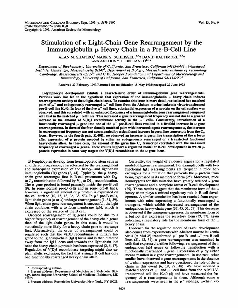
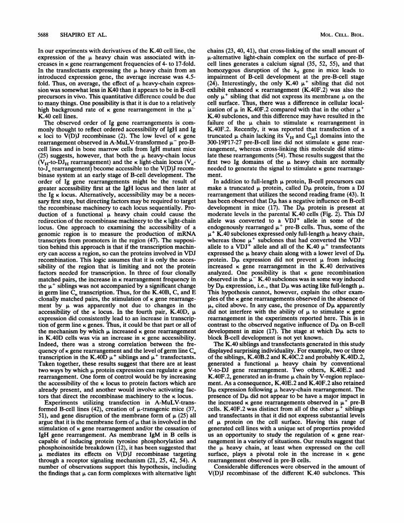
Isolation and characterization of matched p- and ,u+pre-B-cell lines. To examine the role that the Ig heavy chainplays in regulating K light-chain gene rearrangement duringB-lymphocyte development, we chose the K.40 pro-B-cellline, which exhibits a high rate of heavy-chain gene rear-rangement and a lower but detectable rate of light-chain generearrangement (5). K.40 cells have a nonfunctional rear-rangement (VDJ-) of one IgH allele and a partial rearrange-ment (DJ) of the other allele (5). First, five p- subclones ofK.40 were isolated by limiting dilution cloning. These K.40subclones were chosen because, like the parental cells, theywere mostly ,u- but did have a significant number of p. cellsin the population (-0.2%). The p,u cells in these populationsare likely to be the result of gene rearrangements thatbrought a VH gene to one of the two alleles after the cloningevent. In addition, the presence of such cells is indicative ofV(D)J recombinase activity being present. To isolate a ,uderivative of each K.40 subclone, sib selection based oncytoplasmic staining with fluoresceinated anti-p. antibodywas done to enrich for p,u cells. The cell populationsenriched for p.' cells were then subjected to limiting dilutioncloning, and p- and p,u pairs of cloned siblings for each ,u-K.40 subclone were isolated (Fig. 1). By this procedure, fivematched pairs of p- pro-B and p,u pre-B lines with func-tional rearrangements of endogenous IgH loci were ob-tained.To confirm that all of the p,u K.40 subclones did in fact
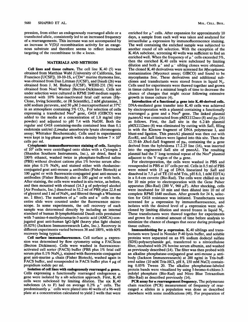
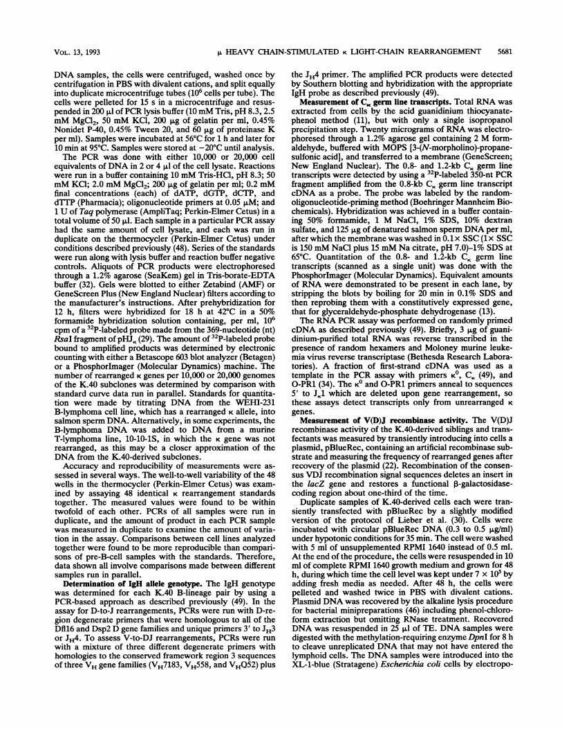
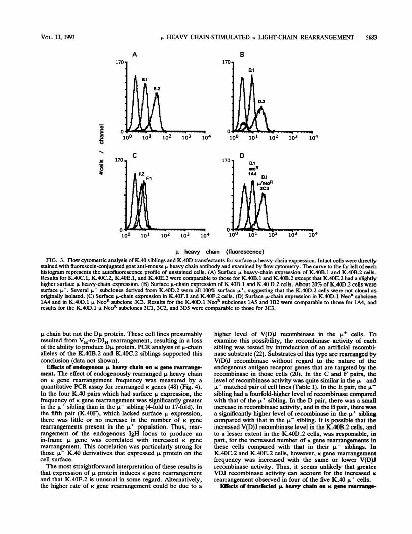
express a full-length p. chain, cell lysates were examined byimmunoblotting with anti-p antibody (Fig. 2). In cell linesexpressing full-length p., immunoblotting usually reveals twoadjacent p. protein bands which differ in their degree ofglycosylation (50). All of the ,u+ cells isolated by sib selec-tion except K.40F.2 expressed both p,-chain bands. K.40F.2expressed only the lower-molecular-weight band, indicatingless-extensive glycosylation, which could reflect a change inintracellular localization. Staining of cell surface heavy-chain p on K.40 subclones with fluoresceinated anti-p. anti-body followed by flow cytometery showed that, with theexception of K.40F.2, all of the K.40 p+ subclones ex-pressed p. on the cell surface at reasonably high levels (Fig.3A to C). These findings were unexpected, as most pro-B-cell lines express the p. protein primarily intracellularly andexpress only low levels on the cell surface. A few otherexamples of this phenotype have been reported previously(15, 18, 38, 53).
K.40 Cells (10% g+)
B C D E F Subclones
/111 ll |\ h~~~~~~~sb-selection
B.1 B.2 C.1 C.2 D.1 D.2 E.1 E.2 F.1 F.2- A+ - P+ ,- A+ g - A+ St- ,+FIG. 1. Generation of clonally matched B-lineage cell pairs.
Starting from a population of the pro-B-cell line K.40 (VDJ-/DJ) inwhich 10% of the cells expressed the p. chain, as assessed byimmunofluorescence, six p- subclones were isolated by limitingdilution cloning. These six subclones each exhibited ongoing rear-rangement of the IgH locus resulting in approximately 0.2% P. cellsin the populations soon after recloning. Sister clones that were ,u- or+ were isolated from five of the six K.40 subclones by sib
selection. For each clonally matched pair, the p- member wasreferred to as X.1 and the p,u member was referred to as X.2.
The immunoblotting experiment (Fig. 2) demonstrates thatall five of the ,u subclones expressed a shorter version of p.protein. This truncated p. protein is probably the D,u proteinthat is synthesized from most DJ alleles in which D and Jregions are linked in one particular reading frame (readingframe 2) (17, 43). Interestingly, three of the p,u derivatives ofK.40 (D.2, E.2, and F.2) continued to express the Dp,protein. PCR analysis of genomic p,-chain alleles of K.40E.2and K.40F.2 demonstrated that these Dp.+ K.40 derivativesretained their DJ alleles (data not shown). These resultssuggest that full-length p. protein in these cells was generatedby a V-region replacement event on the VDJ- chromosome,leaving the Dp.-expressing DJH allele on the other chromo-some intact. In contrast, flow cytometry suggested thatK.40D.2 cells were not clonal (Fig. 3B) and still containedabout 20% ,u- (and presumably Dp,+) cells in the population.Several subclones of K.40D.2 were isolated, and these celllines expressed p. on 100% of the cells and did not expressDp. The K.40B.2 and C.2 cells also expressed the full-length
v- C\j -r-- C\jcn CD CD
C\ V- Cm Cm0 D Ui UJ lL LL
^ .:>:.: .. .:
::* * w >:>> :: : .: : ->: :t ::. :; S.e ............. . : :.::
-4*---~t
-4---- Dla
FIG. 2. Heavy-chain pL and Dp expression in B-lineage pairs.Proteins from detergent-soluble lysates of K.40 subclones wereresolved by SDS-polyacrylamide gel electrophoresis, and the ex-pression of p and Dp proteins was assessed by immunoblotting withalkaline phosphatase-conjugated anti-mouse IgM heavy chain-spe-cific antibody. Colorimetric detection was used to visualize alkalinephosphatase-stained bands. The positions of the p and Dp, proteinsare indicated by arrows.
MOL. CELL. BIOL.
V1HEAVY CHAIN-STIMULATED K LIGHT-CHAIN REARRANGEMENT 5683
A
iicc
13*
4I
B
8.1
B.2
102 103 104
C
F.1
D.1
D.2
103 104
DD.1n,eoR
,u heavy chain (fluorescence)FIG. 3. Flow cytometric analysis of K.40 siblings and K.40D transfectants for surface p, heavy-chain expression. Intact cells were directly
stained with fluorescein-conjugated goat anti-mouse p heavy chain antibody and examined by flow cytometry. The curve to the far left of eachhistogram represents the autofluorescence profile of unstained cells. (A) Surface p. heavy-chain expression of K.40B.1 and K.40B.2 cells.Results for K.40C.1, K.40C.2, K.40E.1, and K.40E.2 were comparable to those for K.40B.1 and K.40B.2 except that K.40E.2 had a slightlyhigher surface p, heavy-chain expression. (B) Surface p.-chain expression of K.40D.1 and K.40 D.2 cells. About 20% of K.40D.2 cells weresurface p-. Several p,u subclones derived from K.40D.2 were all 100% surface p', suggesting that the K.40D.2 cells were not clonal asoriginally isolated. (C) Surface p-chain expression in K.40F.1 and K.40F.2 cells. (D) Surface ,u-chain expression in K.40D.1 NeoR subclone1A4 and in K.40D.1 p NeoR subclone 3C3. Results for the K.40D.1 NeoR subclones 1A5 and 1B2 were comparable to those for 1A4, andresults for the K.40D.1 p NeoR subclones 3C1, 3C2, and 3D5 were comparable to those for 3C3.
p chain but not the Dp, protein. These cell lines presumablyresulted from VH-to-DJH rearrangement, resulting in a lossof the ability to produce D,u protein. PCR analysis of p,-chainalleles of the K.40B.2 and K.40C.2 siblings supported thisconclusion (data not shown).
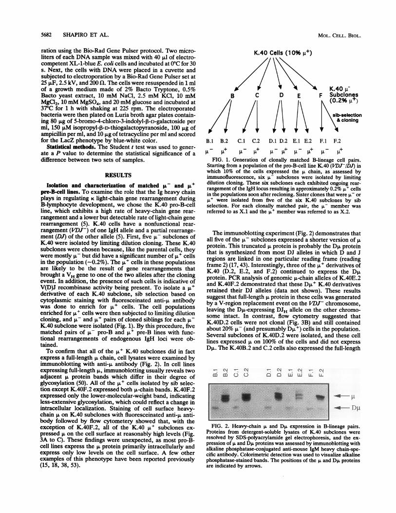
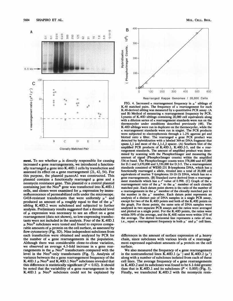
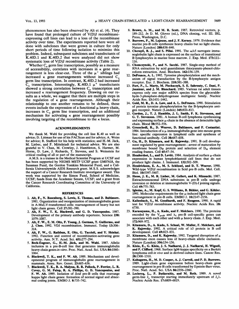
Effects of endogenous p heavy chain on K gene rearrange-ment. The effect of endogenously rearranged p heavy chainon K gene rearrangement frequency was measured by aquantitative PCR assay for rearranged K genes (48) (Fig. 4).In the four K.40 pairs which had surface p. expression, thefrequency of K gene rearrangement was significantly greaterin the ,u+ sibling than in the p- sibling (4-fold to 17-fold). Inthe fifth pair (K.40F), which lacked surface p. expression,there was little or no increase in the number of K generearrangements present in the p. population. Thus, rear-rangement of the endogenous IgH locus to produce anin-frame p. gene was correlated with increased K generearrangement. This correlation was particularly strong forthose ,u+ K.40 derivatives that expressed p. protein on thecell surface.The most straightforward interpretation of these results is
that expression of p. protein induces K gene rearrangementand that K.40F.2 is unusual in some regard. Alternatively,the higher rate of K gene rearrangement could be due to a
higher level of V(D)J recombinase in the p.' cells. Toexamine this possibility, the recombinase activity of eachsibling was tested by introduction of an artificial recombi-nase substrate (22). Substrates of this type are rearranged byV(D)J recombinase without regard to the nature of theendogenous antigen receptor genes that are targeted by therecombinase in those cells (20). In the C and F pairs, thelevel of recombinase activity was quite similar in the Au- andp.+ matched pair of cell lines (Table 1). In the E pair, the p-sibling had a fourfold-higher level of recombinase comparedwith that of the p,u sibling. In the D pair, there was a smallincrease in recombinase activity, and in the B pair, there wasa significantly higher level of recombinase in the ,u+ siblingcompared with that in the R- sibling. It is possible that theincreased V(D)J recombinase level in the K.40B.2 cells, andto a lesser extent in the K.40D.2 cells, was responsible, inpart, for the increased number of K gene rearrangements inthese cells compared with that in their p, siblings. InK.40C.2 and K.40E.2 cells, however, K gene rearrangementfrequency was increased with the same or lower V(D)Jrecombinase activity. Thus, it seems unlikely that greaterVDJ recombinase activity can account for the increased Krearrangement observed in four of the five K.40 ,u+ cells.
Effects of transfected ,I heavy chain on K gene rearrange-
VOL. 13, 1993
%0
100 101 102 103 104 104
5684 SHAPIRO ET AL.
A
0. 5 kb-r-
1(-
C
-
0coCE.E00
1(-
1.-
,1
0
09" 0 0I.- o " o 0 (l o6 6 0 N 0n D0 tcN_N o n
7-...-v
B C D E F
Clonally Matched Pair
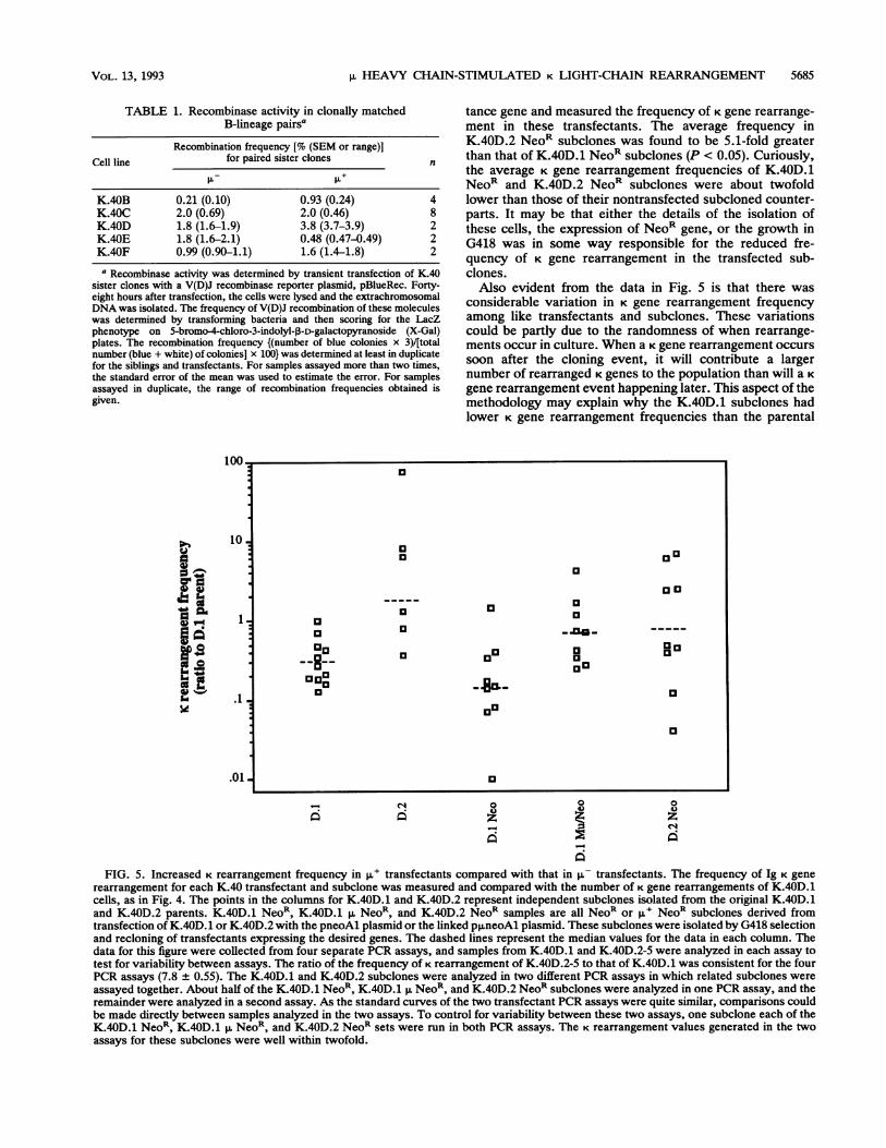
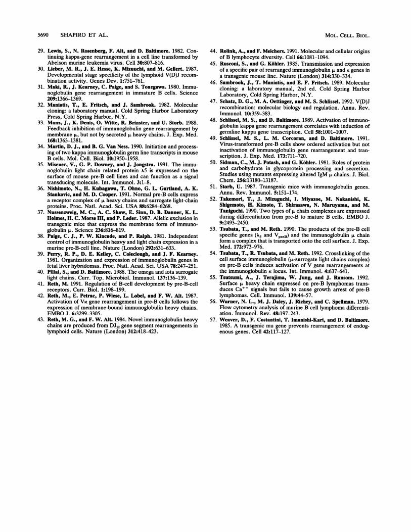
ment. To see whether IL is directly responsible for causingincreased K gene rearrangements, we introduced a function-ally rearranged gene into K.40D.1 cells by transfection andassessed its effect on K gene rearrangement (21, 42, 54). Forthis purpose, the plasmid p,uneoAl was constructed. Thisplasmid contains a functionally rearranged p. gene and aneomycin resistance gene. This plasmid or a control plasmidcontaining just the NeoR gene was transfected into K.40D.1cells, and clones were examined for p expression by immu-nofluorescence of permeabilized cells under the microscope.G418-resistant transfectants that were uniformly p,u andproduced an amount of roughly equal to that of the ,u+sibling K.40D.2 were subcloned and subjected to furtheranalysis. Preliminary results suggested that a threshold levelof expression was necessary to see an effect on K generearrangement (data not shown), so low-expressing transfec-tants were not included in the analysis. Four of the K.40D.1p. NeoR subclones were tested and found to express compa-rable amounts of protein on the cell surface, as assessed byflow cytometery (Fig. 3D). Nine independent subclones fromeach transfection were obtained and analyzed by PCR forthe number of K gene rearrangements in the population.Although there was considerable clone-to-clone variation,we observed an average 4.5-fold increase in K gene rear-rangements in the p. NeoR transfectants compared with thelevel in the NeoR_only transfectants (Fig. 5). Analysis ofvariance between the K gene rearrangement frequency of theK.40D.1 p. NeoR and K.40D.1 NeoR subclones revealed thatthis difference is statistically significant (P < 0.02). It shouldbe noted that the variability of K gene rearrangement in theK.40D.1 NeoR subclones could not be explained by
B 40)-
.N t 200-0
0. 100eI= /
_0
20-
0
0 100 200 300 400 500 600
Rearranged Kappa Genomes I 20,000 Cells
FIG. 4. Increased K rearrangement frequency in 1l siblings ofK.40 matched pairs. The frequency of K rearrangement for eachK.40-derived sibling was measured by a quantitative PCR assay. (Aand B) Method of measuring K rearrangement frequency by PCR.Lysates of K.40D siblings containing 20,000 cell equivalents alongwith a dilution series of K rearrangement standards were run on thethermocycler under conditions described previously (48). TheK.40D siblings were run in duplicate on the thermocycler, while theK rearrangement standards were run in singlet. The PCR productswere subjected to electrophoresis through a 1.2% agarose gel andblotted onto a filter. The rearranged K gene PCR product wasdetected by hybridization with a labeled 369-nt DNA fragment thatspans JK1 and most of the JK1-JK2 spacer. (A) Southern blot of theamplified PCR products of K.40D.1, K.40D.2-5, and the K rear-rangement standards. The amount of amplified product was deter-mined by scanning with the PhosphorImager and measuring theamount of signal (PhosphorImager counts) within the amplified536-nt band. The Phosphorlmager counts were 378,000 and 447,000for D.1 and 3,670,000 and 3,330,000 for D.2-5. The K rearrangementstandards consisted of WEHI-231 B-lymphoma DNA, which has afunctionally rearranged K allele, titrated into a total of 20,000 cellequivalents of murine T-lymphoma 10-10-iS DNA, which has no Kgene rearrangements. (B) Standard curve derived from K rearrange-ment standards which has a r2 value of 0.998. (C) Graph of the Krearrangement ratio of the p,u to the p- member of each clonallymatched pair. Each datum point shown is the ratio of the number ofK rearrangements in the ,u member of the clonally matched pair tothe number in the p- member. Each datum point represents ananalysis of a distinct pair of DNA samples in a single PCR assay,except for two of the K.40D points and both of the K.40E points onthe graph. For those points, the same sets of DNA samples wereanalyzed in two separate PCR assays and the ratios were averagedand plotted as a single point. For the K.40D points, the ratios werewithin 50% of the average, and the K.40E ratios were within 15% ofthe average. The dotted horizontal line represents a ratio of one,i.e., equal K rearrangement frequency in both p- and p,u cell lines.
differences in the amount of surface expression of p, heavychain, since subclones with various levels of K rearrange-ment expressed equivalent amounts of p. protein on the cellsurface.We also measured the frequency of K gene rearrangement
in the nontransfected lines K.40D.1 (p-) and K.40D.2 (,u+)along with a number of subclones isolated from each of thesecell lines. The average frequency of K gene rearrangementsin K.40D.2 and its subclones was found to be 6.1-fold greaterthan that in K.40D.1 and its subclones (P < 0.005) (Fig. 5).Finally, we transfected K.40D.2 with the neomycin resis-
D O
o 0
ao0 0 0
a
033
0~~~~~~~~~~~~~~~1-0
MOL. CELL. BIOL.
V1HEAVY CHAIN-STIMULATED K LIGHT-CHAIN REARRANGEMENT 5685
TABLE 1. Recombinase activity in clonally matchedB-lineage pairsa
Recombination frequency [% (SEM or range)]Cell line for paired sister clones n
~~~~~~~~ +
K.40B 0.21 (0.10) 0.93 (0.24) 4K.40C 2.0 (0.69) 2.0 (0.46) 8K.40D 1.8 (1.6-1.9) 3.8 (3.7-3.9) 2K.40E 1.8 (1.6-2.1) 0.48 (0.47-0.49) 2K.40F 0.99 (0.90-1.1) 1.6 (1.4-1.8) 2
a Recombinase activity was determined by transient transfection of K.4Asister clones with a V(D)J recombinase reporter plasmid, pBlueRec. Forty-eight hours after transfection, the cells were lysed and the extrachromosomalDNA was isolated. The frequency of V(D)J recombination of these moleculeswas determined by transforming bacteria and then scoring for the LacZphenotype on 5-bromo-4-chloro-3-indolyl-3-D-galactopyranoside (X-Gal)plates. The recombination frequency {(number of blue colonies x 3)/[totalnumber (blue + white) of colonies] x 100} was determined at least in duplicatefor the siblings and transfectants. For samples assayed more than two times,the standard error of the mean was used to estimate the error. For samplesassayed in duplicate, the range of recombination frequencies obtained isgiven.
IflA
10
-4
Ii
a
.C
1
.1
.01
e'i
tance gene and measured the frequency of K gene rearrange-ment in these transfectants. The average frequency inK.40D.2 NeoR subclones was found to be 5.1-fold greaterthan that of K.40D.1 NeoR subclones (P < 0.05). Curiously,the average K gene rearrangement frequencies of K.40D.1NeoR and K.40D.2 NeoR subclones were about twofoldlower than those of their nontransfected subcloned counter-parts. It may be that either the details of the isolation ofthese cells, the expression of NeoR gene, or the growth inG418 was in some way responsible for the reduced fre-quency of K gene rearrangement in the transfected sub-clones.Also evident from the data in Fig. 5 is that there was
considerable variation in K gene rearrangement frequencyamong like transfectants and subclones. These variationscould be partly due to the randomness of when rearrange-ments occur in culture. When a K gene rearrangement occurssoon after the cloning event, it will contribute a largernumber of rearranged K genes to the population than will a Kgene rearrangement event happening later. This aspect of themethodology may explain why the K.40D.1 subclones hadlower K gene rearrangement frequencies than the parental
a
0a)
zea
FIG. 5. Increased K rearrangement frequency in ,u+ transfectants compared with that in pL- transfectants. The frequency of Ig K gene
rearrangement for each K.40 transfectant and subclone was measured and compared with the number of K gene rearrangements of K.40D.1cells, as in Fig. 4. The points in the columns for K.40D.1 and K.40D.2 represent independent subclones isolated from the original K.40D.1and K.40D.2 parents. K.40D.1 NeoR, K.40D.1 ,u NeoR and K-40D.2 NeoR samples are all NeoR or p+ NeoR subclones derived fromtransfection of K.40D.1 or K.40D.2 with the pneoAl plasmid or the linked p,uneoAl plasmid. These subclones were isolated by G418 selectionand recloning of transfectants expressing the desired genes. The dashed lines represent the median values for the data in each column. Thedata for this figure were collected from four separate PCR assays, and samples from K.40D.1 and K.40D.2-5 were analyzed in each assay totest for variability between assays. The ratio of the frequency of K rearrangement of K.40D.2-5 to that of K.40D.1 was consistent for the fourPCR assays (7.8 0.55). The K.40D.1 and K.40D.2 subclones were analyzed in two different PCR assays in which related subclones wereassayed together. About half of the K.40D.1 NeoR, K.40D.1 p, NeoR, and K.40D.2 NeoR subclones were analyzed in one PCR assay, and theremainder were analyzed in a second assay. As the standard curves of the two transfectant PCR assays were quite similar, comparisons couldbe made directly between samples analyzed in the two assays. To control for variability between these two assays, one subclone each of theK.40D.1 NeoR, K.40D.1 NeoR, and K.40D.2 NeoR sets were run in both PCR assays. The K rearrangement values generated in the twoassays for these subclones were well within twofold.
a
a 0~~~~~o o0
13 0
a a~aa -flu-go
a 0~ag
an~~~~~~a~~~~~~
O o ,_____0
VOL. 13, 1993
5686 SHAPIRO ET AL.
TABLE 2. Comparison of the frequency of K rearrangement and recombinase activity in K.40D derivatives
Avg K rearrangement frequency Recombination activityType Cell line (no. of rearrangements/ [% (SEM or range)]'
20,000 genomes)a
K.40D siblings D.1 parent 36 1.8 (1.6-1.9)D.2-5 280 3.8 (3.7-3.9)
K.40D.1 transfected with D.1 Neo 1A3 2.3 0.52 (0.45-0.58)the NeoR gene D.1 Neo 1A4 0.32 1.5 (0.12)
D.1 Neo 1A5 6.1 1.5 (0.31)D.1 Neo lBl 51 2.5 (1.9-3.0)D.1 Neo 1B2 14 1.6 (1.3-1.8)
K.40D.1 transfected with D.1 p Neo 3C1 59 2.4 (1.9-2.9)the p. and NeoR genes D.1 p. Neo 3C2 41 4.4 (4.0-4.7)
D.1 p Neo 3C3 150 1.6 (1.4-1.7)D.1 p Neo 3D5 8.4 1.3 (1.2-1.3)D.1 p. Neo 5A4 9.7 1.7 (0.46)
K.40D.2 transfected with D.2 Neo 7A6 1.3 1.3 (0.75-1.8)the NeoR gene D.2 Neo 7B4 240 1.8 (1.7-1.9)
a The frequency of K rearrangement for each K.40D derivative was measured by using a quantitative PCR assay.bThe recombination activity was determined by transient transfection of the K.40D derivatives with a V(D)J recombination reporter plasmid, pBlueRec, as
described in Table 1, footnote a. For samples assayed more than twice, the standard error of the mean was used to estimate the error. For samples assayed induplicate, the range of measured recombination frequencies is given.
cell line: perhaps by chance the first K rearrangement in theparental cell line occurred relatively soon after cloning. Ofcourse, the higher the rate of rearrangement is, the greater isthe probability that a K gene rearrangement will occur earlyfollowing subcloning, so a general correlation is expectedbetween the rate of rearrangement and the number of rear-ranged K genes, the measured parameter. In addition, some ofthe variation observed may reflect true differences in Krearrangement rate among like subclones or transfectants.This phenomenon has been reported by others (4), althoughwe did not see a general loss of V(D)J recombinase activity inthe various derivatives of K.40D.1 (see below). For thesereasons, we believe that averaging of results from multiplesubclones, as we have done here, is currently the mostreliable way of assessing the K gene rearrangement rate.Various K.40D derivatives were assayed for V(D)J recom-
binase activity with the transiently transfected artificialsubstrate (Table 2). The recombinase activities of thesetransfectants were comparable to each other. No correlationwas found between the frequency of K gene rearrangementand the level of recombinase in the transfected cells. More-over, we did not observe a systematic loss in V(D)J recom-binase activity upon transfection or subcloning of thesecells. Taken together, these data strongly support the con-clusion that p. heavy-chain expression is responsible forpromoting K gene rearrangement. Moreover, this regulationdoes not require increased V(D)J recombinase activity asmeasured with an introduced recombinase substrate.
Effects of the i heavy chain on germ line C., transcription.To address the mechanism of how the ,u heavy chainincreases the frequency of K gene rearrangements, the effectof p. on the transcription of the unrearranged CK gene wasexamined. The CK locus has been observed to generate germline transcripts of 0.8 and 1.2 kb (the latter is also referred toas 1.1 kb [28]) (34). It has previously been observed thatincreases in germ line transcription correlate with increasedK gene rearrangement frequency (48). Whether these germline transcripts actually promote K gene rearrangement orare merely reflective of the increased accessibility of the Klocus to protein complexes such as RNA polymerase orV(D)J recombinase is not known (3, 47). RNA from each ofthe ,u- and ,u+ clonally matched siblings was isolated and
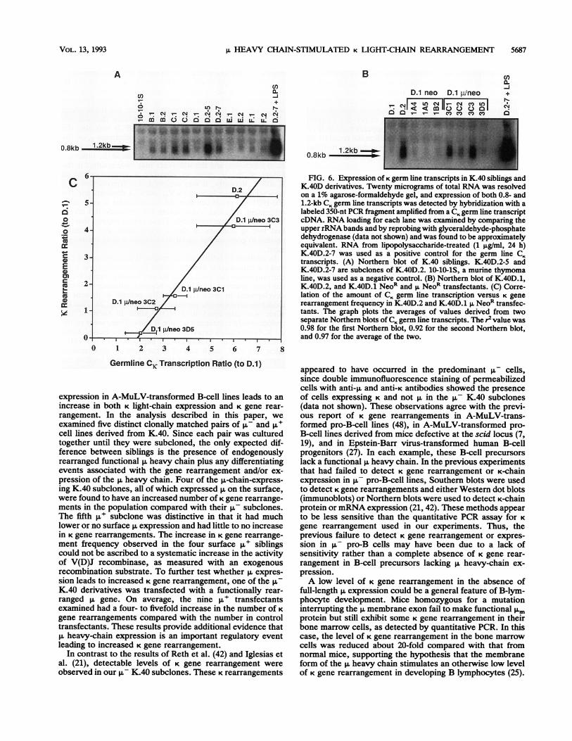
subjected to Northern (RNA) analysis using a KO probe thatdetects germ line CK transcripts (Fig. 6A). For the K.40F pair,which had no increase in K gene rearrangements, the level ofgerm line CK transcription was comparable for both the p-and ,u+ siblings. For three of the four K.40 pairs withincreased K gene rearrangements, the level of germ line CKtranscription was also quite similar for both the u- and ,u+siblings. Only in the case of the K.40D pair was there aconsiderable increase (approximately fivefold) in the amountof CK germ line transcription in the p,+ sibling compared withthat in the p- sibling. Very similar results were obtained witha quantitative RNA PCR assay for CK germ line transcription(48) (data not shown). Moreover, these PCR experimentsconfirmed that the 1.2-kb transcript is a germ line CK tran-script rather than a rearranged K transcript. Thus, with theexception of the K.40D pair, there was no correlation be-tween increases in CK germ line transcription and the in-creased K gene rearrangement frequency in the K.40 matchedpairs. Next, K germ line transcription was examined in thesome of the K.40D.1 NeoR-only and p. NeoR transfectants tosee whether the increase in germ line CK transcription inK.40D.2 was due to p. chain expression (Fig. 6B). All of theNeoR-only transfectants had a very low level of CK germ linetranscription like the parental K.40D.1 cells. In contrast, theK.40D.1 p. NeoR transfectants exhibited increased CK germline transcription. Increased CK germ line transcription in thetransfectants was confirmed by the quantitative RNA PCRassay for germ line CK transcription (data not shown). Inter-estingly, there was a linear relationship between the level ofgerm line transcription and the frequency of K gene rearrange-ment observed in K.40D.2 and K.40D.1 p. Neo transfectants(Fig. 6C). In contrast, the K.40D.1 NeoR (p-) clones failed toexhibit a correlation between these two parameters. Thus, forK.40D.1 there was a direct correlation between the amount ofCK germ line transcription induced by p.-chain expression andthe frequency of K gene rearrangements. K.40D.1 was uniquein this regard.
DISCUSSION
Previous work by Reth et al. (42), Iglesias et al. (21), andTsubata et al. (54) had demonstrated that p. heavy-chain
MOL. CELL. BIOL.
L1HEAVY CHAIN-STIMULATED K LIGHT-CHAIN REARRANGEMENT 5687
B
cn
v- I
_ m m 0 0 a a cz us X X X
cnCL-J
+-C,J
D.1 neo D.1 plneo.I eI Ln Cf11_rOUen I
0.8kb0.8kb 1.2kb
C
0-
0
0
cos
crE
cna:
0 1 2 3 4 5 6 7 8
Germline CK Transcription Ratio (to D.1)
expression in A-MuLV-transformed B-cell lines leads to anincrease in both K light-chain expression and K gene rear-rangement. In the analysis described in this paper, weexamined five distinct clonally matched pairs of ,- and R'cell lines derived from K.40. Since each pair was culturedtogether until they were subcloned, the only expected dif-ference between siblings is the presence of endogenouslyrearranged functional heavy chain plus any differentiatingevents associated with the gene rearrangement and/or ex-pression of the p. heavy chain. Four of the p,-chain-express-ing K.40 subclones, all of which expressed on the surface,were found to have an increased number of K gene rearrange-ments in the population compared with their p- subclones.The fifth p,u subclone was distinctive in that it had muchlower or no surface p. expression and had little to no increasein K gene rearrangements. The increase in K gene rearrange-ment frequency observed in the four surface ,u+ siblingscould not be ascribed to a systematic increase in the activityof V(D)J recombinase, as measured with an exogenousrecombination substrate. To further test whether expres-sion leads to increased K gene rearrangement, one of the p-K.40 derivatives was transfected with a functionally rear-ranged ,u gene. On average, the nine p+ transfectantsexamined had a four- to fivefold increase in the number of K
gene rearrangements compared with the number in controltransfectants. These results provide additional evidence thatp. heavy-chain expression is an important regulatory eventleading to increased K gene rearrangement.
In contrast to the results of Reth et al. (42) and Iglesias etal. (21), detectable levels of K gene rearrangement wereobserved in our ,u- K.40 subclones. These K rearrangements
FIG. 6. Expression of K germ line transcripts in K.40 siblings andK.40D derivatives. Twenty micrograms of total RNA was resolvedon a 1% agarose-formaldehyde gel, and expression of both 0.8- and1.2-kb CK germ line transcripts was detected by hybridization with alabeled 350-nt PCR fragment amplified from a CK germ line transcriptcDNA. RNA loading for each lane was examined by comparing theupper rRNA bands and by reprobing with glyceraldehyde-phosphatedehydrogenase (data not shown) and was found to be approximatelyequivalent. RNA from lipopolysaccharide-treated (1 pg/ml, 24 h)K.40D.2-7 was used as a positive control for the germ line CKtranscripts. (A) Northern blot of K.40 siblings. K.40D.2-5 andK.40D.2-7 are subclones of K.40D.2. 10-10-iS, a murine thymomaline, was used as a negative control. (B) Northern blot of K.40D.1,K.40D.2, and K.40D.1 NeoR and ,u NeoR transfectants. (C) Corre-lation of the amount of CK germ line transcription versus K generearrangement frequency in K.40D.2 and K.40D.1 p, NeoR transfec-tants. The graph plots the averages of values derived from twoseparate Northem blots of CK germ line transcripts. The r2 value was0.98 for the first Northern blot, 0.92 for the second Northern blot,and 0.97 for the average of the two.
appeared to have occurred in the predominant ,u- cells,since double immunofluorescence staining of permeabilizedcells with anti-,u and anti-K antibodies showed the presenceof cells expressing K and not p in the ,u- K.40 subclones(data not shown). These observations agree with the previ-ous report of K gene rearrangements in A-MuLV-trans-formed pro-B-cell lines (48), in A-MuLV-transformed pro-B-cell lines derived from mice defective at the scid locus (7,19), and in Epstein-Barr virus-transformed human B-cellprogenitors (27). In each example, these B-cell precursorslack a functional p heavy chain. In the previous experimentsthat had failed to detect K gene rearrangement or K-chainexpression in p- pro-B-cell lines, Southern blots were usedto detect K gene rearrangements and either Western dot blots(immunoblots) or Northern blots were used to detect K-chainprotein ormRNA expression (21, 42). These methods appearto be less sensitive than the quantitative PCR assay for Kgene rearrangement used in our experiments. Thus, theprevious failure to detect K gene rearrangement or expres-sion in p- pro-B cells may have been due to a lack ofsensitivity rather than a complete absence of K gene rear-rangement in B-cell precursors lacking p. heavy-chain ex-pression.A low level of K gene rearrangement in the absence of
full-length ,u expression could be a general feature of B-lym-phocyte development. Mice homozygous for a mutationinterrupting the membrane exon fail to make functional ,Umprotein but still exhibit some K gene rearrangement in theirbone marrow cells, as detected by quantitative PCR. In thiscase, the level of K gene rearrangement in the bone marrowcells was reduced about 20-fold compared with that fromnormal mice, supporting the hypothesis that the membraneform of the p. heavy chain stimulates an otherwise low levelof K gene rearrangement in developing B lymphocytes (25).
A cnCM-1
+-eCu
VOL. 13, 1993
5688 SHAPIRO ET AL.
In our experiments with derivatives of the K.40 cell line, theexpression of the p. heavy chain was associated with in-creases in K gene rearrangement frequencies of 4- to 17-fold.In the transfectants expressing the IL heavy chain from anintroduced expression gene, the average increase was 4.5-fold. Thus, on average, the effect of p. heavy-chain expres-sion was somewhat less in K40 than it appears to be in B-cellprecursors in vivo. This quantitative difference could be dueto many things. One possibility is that it is due to a relativelyhigh background rate of K gene rearrangement in the ,-K.40 cell lines.The observed order of Ig gene rearrangements is com-
monly thought to reflect ordered accessibility of IgH and IgK loci to V(D)J recombinase (2). The low level of K generearrangement observed in A-MuLV-transformed ,u- pro-B-cell lines and in bone marrow cells from IgH mutant mice(25) suggests, however, that both the p. heavy-chain locus(VH-to-DJH rearrangement) and the K light-chain locus (VK_to-JK rearrangement) become accessible to the V(D)J recom-binase system at an early stage of B-cell development. Theorder of Ig gene rearrangements might be the result ofgreater accessibility first at the IgH locus and then later atthe Ig K locus. Alternatively, accessibility may be a neces-sary first step, but directing factors may be required to targetthe recombinase machinery to each locus sequentially. Pro-duction of a functional p. heavy chain could cause theredirection of the recombinase machinery to the K light-chainlocus. One approach to examining the accessibility of agenomic region is to measure the production of mRNAtranscripts from promoters in the region (47). The supposi-tion behind this approach is that if the transcription machin-ery can access a region, so can the proteins involved in VDJrecombination. This logic assumes that it is only the acces-sibility of the region that is limiting and not the proteinfactors needed for transcription. In three of four clonallymatched pairs, the increase in K rearrangement frequency inthe ,u+ siblings was not accompanied by a significant changein germ line CK transcription. Thus, for the K.40B, C, and Eclonally matched pairs, the stimulation of K gene rearrange-ment by p. was apparently not due to changes in theaccessibility of the K locus. In the fourth pair, K.40D, p.expression did consistently lead to an increase in transcrip-tion of germ line K genes. Thus, it could be that part or all ofthe mechanism by which p. increased K gene rearrangementin K.40D cells was via an increase in K gene accessibility.Indeed, there was a strong correlation between the fre-quency of K gene rearrangement and the level of germ line CKtranscription in the K.40D p,u siblings and ,u+ transfectants.Taken together, these results suggest that there are at leasttwo ways by which p. protein expression can regulate K generearrangement. One form of control would be by increasingthe accessibility of the K locus to protein factors which arealready present, and another would involve activating fac-tors that direct the recombinase machinery to the K locus.Experiments utilizing transfection in A-MuLV-trans-
formed B-cell lines (42), creation of p.-transgenic mice (37,51), and gene disruption of the membrane form of p. (25) allargue that it is the membrane form of ,u that is involved in thestimulation of K gene rearrangement and/or the cessation ofIgH gene rearrangement. As membrane IgM in B cells iscapable of inducing protein tyrosine phosphorylation andphosphoinositide breakdown (12), it has been suggested thatp. mediates its effects on V(D)J recombinase targetingthrough a receptor signaling mechanism (21, 25, 42, 54). Anumber of observations support this hypothesis, includingthe findings that p. can form complexes with alternative light
chains (23, 40, 41), that cross-linking of the small amount ofp.-alternative light-chain complex on the surface of pre-B-cell lines generates a calcium signal (35, 52, 55), and thathomozygous disruption of the X5 gene in mice leads toimpairment of B-cell development at the pre-B-cell stage(24). Interestingly, the only K.40 ,u+ sibling that did notexhibit enhanced K rearrangement (K.40F.2) was also theonly p,u sibling that did not express its membrane p. on thecell surface. Thus, there was a difference in cellular local-ization of p. in K.40F.2 compared with that in the other p.+K.40 subclones, and this difference may have resulted in thefailure of the p. chain to stimulate K rearrangement inK.40F.2. Recently, it was reported that transfection of atruncated p. chain lacking its VH and CH1 domains into the300-19P17-27 pre-B-cell line did not stimulate K gene rear-rangement, whereas cross-linking this molecule did stimu-late these rearrangements (54). These results suggest that thefirst two Ig domains of the p. heavy chain are normallyneeded to generate the signal to stimulate K gene rearrange-ment.
In addition to full-length p. protein, B-cell precursors canmake a truncated p. protein, called Dp. protein, from a DJrearrangement that utilizes the second reading frame (43). Ithas been observed that Dp has a negative influence on B-celldevelopment in mice (17). The Dp. protein is present atmoderate levels in the parental K.40 cells (Fig. 2). This DJallele was converted to a VDJ+ allele in some of theendogenously rearranged ,u+ pre-B cells. Thus, some of the,u+ K.40 subclones expressed only full-length p. heavy chain,whereas those p,u subclones that had converted the VDJ-allele to a VDJ+ allele and all of the K.40 p,u transfectantsexpressed the p. heavy chain along with a lower level of Dp.protein. Dp. expression did not prevent p. from inducingincreased K gene rearrangement in the K.40 derivativesanalyzed. One possibility is that K gene recombinationobserved in the ,- K.40 subclones was in some way inducedby Dp. expression, i.e., that Dp. was acting like full-length P.This hypothesis cannot, however, explain the other exam-ples of the K gene rearrangements observed in the absence ofp., cited above. In any case, the presence of Dp. apparentlydid not interfere with the ability of p. to stimulate K generearrangement in the experiments reported here. This is incontrast to the observed negative influence of Dp. on B-celldevelopment in mice (17). The stage at which Dp. acts toblock B-cell development is not yet known.The K.40 siblings and transfectants generated in this study
displayed surprising individuality. For example, two or threeof the siblings, K.40B.2 and K.40C.2 and probably K.40D.2,generated a functional p. heavy chain by conventionalV-to-DJ gene rearrangement. Two others, K.40E.2 andK.40F.2, generated an in-frame p. chain by V-region replace-ment. As a consequence, K.40E.2 and K.40F.2 also retainedDp. expression following p. heavy-chain rearrangement. Thepresence of Dp. did not appear to be have a major impact inthe increased K gene rearrangements observed in ,u+ pre-Bcells. K.40F.2 was distinct from all of the other ,u+ siblingsand transfectants in that it did not express substantial levelsof p. protein on the cell surface. Having this range ofgenerated cell lines with a unique set of properties providedus an opportunity to study the regulation of K gene rear-rangement in a variety of situations. Our results suggest thatthe p. heavy chain, at least when expressed on the cellsurface, plays a pivotal role in the increase in K generearrangement observed in pre-B cells.
Considerable differences were observed in the amount ofV(D)J recombinase of the different K.40 subclones. This
MOL. CELL. BIOL.
V1HEAVY CHAIN-STIMULATED K LIGHT-CHAIN REARRANGEMENT 5689
phenomenon has also been observed by Alt et al. (4). Theyhave found that prolonged culture of V(D)J recombinase-expressing cell lines can lead to a loss of the recombinaseactivity over time. The experiments reported here were alldone with subclones that were grown in culture for onlyshort periods of time following isolation to minimize thisproblem. Indeed, subsequent subclones and transfectants ofK.40D.1 and K.40D.2 that were analyzed did not showsystematic loss of V(D)J recombinase activity (Table 2).Whether C,< germ line transcription, possibly as a measure
of accessibility, correlates with increases in K gene rear-rangement is less clear-cut. Three of the ,u+ siblings hadincreased K gene rearrangements without increased CKgerm line transcription. In contrast, K.40D.2 had increasedCK transcription. Interestingly, K.40D.1 ,u+ transfectantsshowed a strong correlation between CK transcription andincreased K rearrangement frequency. Drawing on our re-sults as a whole, we suggest that there are multiple steps inthe regulation of K gene rearrangement. Although theirrelationship to one another remains to be defined, theseevents include the expression of a functional ,u heavy chain,increases in CK germ line transcription, and an additionalmechanism for activating K gene rearrangement possiblyinvolving targeting of the recombinase to the K locus.
ACKNOWLEDGMENTS
We thank M. Wabl for providing the cell line K.40 as well asadvice; D. Littman for use of his FACScan and for advice; A. Weissfor advice; R. Stafford for his help with statistics; and H.-M. Jack,M. Lieber, and P. Mittelstadt for technical advice. We are alsograteful to V. Chan, M. Crowley, J. Hambleton, S. Harmer, M.Home, D. Law, J. Richards, T. Stevens, and S. Weinstein forcritical reading of all or part of the manuscript.A.M.S. is a trainee in the Medical Scientist Program at UCSF and
has been supported by NIGMS MSTP UCSF grant GM07618, theSussman Fund, the Gordon Tomkins Memorial Fund, and a Cali-fornia Universitywide AIDS Research grant. M.S.S. acknowledgesthe support of a Cancer Research Institute investigator award. Thiswork was supported by the Simon Fund, School of Medicine,UCSF; funds from the Academic Senate, UCSF; and a grant fromthe Cancer Research Coordinating Committee of the University ofCalifornia.
REFERENCES1. Alt, F., N. Rosenberg, S. Lewis, E. Thomas, and D. Baltimore.
1981. Organization and reorganization of immunoglobulin genesin A-MuLV-transformed cells: rearrangement of heavy but notlight chain genes. Cell 27:381-390.
2. Alt, F. W., T. K. Blackwell, and G. D. Yancopoulos. 1987.Development of the primary antibody repertoire. Science 238:1079-1087.
3. Alt, F. W., E. M. Oltz, F. Young, J. Gorman, T. Guillermo, andJ. Chen. 1992. VDJ recombination. Immunol. Today 13:306-314.
4. Alt, F. W., G. Rathbun, E. Oltz, G. Taccioli, and Y. Shinkai.1992. Function and control of recombination-activating geneactivity. Ann. N.Y. Acad. Sci. 651:277-294.
5. Beck-Engeser, G., H.-M. Jack, and M. Wabi. 1987. Allelicinclusion in a pre-B-cell line that generates immunoglobulinheavy chain genes in vitro. Proc. Natl. Acad. Sci. USA 84:1060-1064.
6. Blackwell, T. K., and F. W. Alt. 1989. Mechanism and devel-opmental program of immunoglobulin gene rearrangement inmammals. Annu. Rev. Genet. 23:605-636.
7. Blackwell, T. K., B. A. Malynn, R. R. Pollock, P. Ferrier, L. R.Covey, G. M. Fulop, R. A. Phillips, G. D. Yancopoulos, andF. W. Alt. 1989. Isolation of Scid pre-B cells that rearrangekappa light chain genes: formation of normal signal and abnor-mal coding joints. EMBO J. 8:735-742.
8. Brown, A. M., and M. R. Scott. 1987. Retroviral vectors, p.189-212. In D. M. Glover (ed.), DNA cloning, vol. III. IRLPress, Washington, D.C.
9. Burrows, P., M. Lejeune, and J. F. Kerney. 1979. Evidence thatmurine pre-B cells synthesize heavy chains but no light chains.Nature (London) 280:838-840.
10. Cherayil, B. J., and S. Pillai. 1991. The w/X5 surrogate immu-noglobulin light chain is expressed on the surface of transitionalB lymphocytes in murine bone marrow. J. Exp. Med. 173:111-116.
11. Chomczynski, P., and N. Sacchi. 1987. Single-step method ofRNA extraction by acid guanidinium thiocyanate-phenol-chlo-roform extraction. Anal. Biochem. 162:156-159.
12. DeFranco, A. L. 1992. Tyrosine phosphorylation and the mech-anism of signal transduction by the B-lymphocyte antigenreceptor. Eur. J. Biochem. 210:381-388.
13. Fort, P., L. Marty, M. Piechaczyk, S. E. Sabrouty, C. Dani, P.Jeanteur, and J. M. Blanchard. 1985. Various rat adult tissuesexpress only one major mRNA species from the glyceralde-hyde-3-phosphate-dehydrogenase multigenic family. NucleicAcids Res. 13:1431-1442.
14. Gold, M. R., D. A. Law, and A. L. DeFranco. 1990. Stimulationof protein tyrosine phosphorylation by the B-lymphocyte anti-gen receptor. Nature (London) 345:810-813.
15. Gordon, J., T. J. Hamblin, J. L. Smith, F. K. Stevenson, andG. T. Stevenson. 1981. A human B-cell lymphoma synthesizingand expressing surface ,u-chain in the absence of detectable lightchain. Blood 58:552-556.
16. Grosschedl, R., D. Weaver, D. Baltimore, and F. Costantini.1984. Introduction of a ,. immunoglobulin gene into mouse germline: specific expression in lymphoid cells and synthesis offunctional antibody. Cell 38:647-658.
17. Gu, H., D. Kitamura, and K. Rajewsky. 1991. B cell develop-ment regulated by gene rearrangement-arrest of maturation bymembrane bound DpL protein and selection of DH elementreading frames. Cell 65:47-54.
18. Hendershot, L., and D. Levitt. 1984. Analysis of surface ,u-chainexpression in human lymphoblastoid cell lines that do notproduce light chains. J. Immunol. 132:502-509.
19. Hendrickson, E. A., M. S. Schlissel, and D. T. Weaver. 1990.Wild-type V(D)J recombination in Scid pre-B cells. Mol. Cell.Biol. 10:5397-5407.
20. Hesse, J. E., M. R. Lieber, M. Gellert, and K. Mizuuchi. 1987.Extrachromosomal DNA substrates in pre-B cells undergoinversion or deletion at immunoglobulin V-(D)-J joining signals.Cell 49:775-783.
21. Iglesias, A., M. Kopf, G. S. Williams, B. Bfihler, and G. Kohler.1991. Molecular requirements for the p.-induced light chain generearrangement in pre-B cells. EMBO J. 10:2147-2156.
22. Kalienbach, S., M. Goodhardt, and F. Rougeon. 1990. A rapidtest for V(D)J recombinase activity. Nucleic Acids Res. 18:6730.
23. Karasuyama, H., A. Kudo, and F. Melchers. 1990. The proteinsencoded by the VpreB and X5 pre-B cell-specific genes canassociate with each other and with p. heavy chain. J. Exp. Med.172:969-972.
24. Kitamura, D., A. Kudo, S. Schaal, W. Muller, F. Melchers, andK. Rajewsky. 1992. A critical role of X5 protein in B celldevelopment. Cell 69:823-831.
25. Kitamura, D., and K. Rajewsky. 1992. Targeted disruption of Pmembrane exon causes loss of heavy-chain allelic exclusion.Nature (London) 356:154-156.
26. Klein, E., G. Klein, J. S. Nadkarni, J. J. Nadkarni, H. Wigzell,and P. Clifford. 1968. Surface IgM-kappa specificity on a Burkittlymphoma cell in vivo and in derived culture lines. Cancer Res.28:1300-1310.
27. Kubagawa, H., M. D. Cooper, A. J. Carroll, and P. D. Burrows.1989. Light-chain gene expression before heavy-chain generearrangement in pre-B cells transformed by Epstein-Barr virus.Proc. Natl. Acad. Sci. USA 86:2356-2360.
28. Leclercq, L., P. Butkeraitis, and M. Reth. 1989. A novelgerm-line JK transcript starting immediately upstream of JK1JNucleic Acids Res. 17:6809-6819.
VOL. 13, 1993
5690 SHAPIRO ET AL.
29. Lewis, S., N. Rosenberg, F. Alt, and D. Baltimore. 1982. Con-tinuing kappa-gene rearrangement in a cell line transformed byAbelson murine leukemia virus. Cell 30:807-816.
30. Lieber, M. R., J. E. Hesse, K. Mizuuchi, and M. Gellert. 1987.Developmental stage specificity of the lymphoid V(D)J recom-bination activity. Genes Dev. 1:751-761.
31. Maki, R., J. Kearney, C. Paige, and S. Tonegawa. 1980. Immu-noglobulin gene rearrangement in immature B cells. Science209:1366-1369.
32. Maniatis, T., E. Fritsch, and J. Sambrook. 1982. Molecularcloning: a laboratory manual. Cold Spring Harbor LaboratoryPress, Cold Spring Harbor, N.Y.
33. Manz, J., K. Denis, 0. Witte, R. Brinster, and U. Storb. 1988.Feedback inhibition of immunoglobulin gene rearrangement bymembrane ,u, but not by secreted ,u heavy chains. J. Exp. Med.168:1363-1381.
34. Martin, D. J., and B. G. Van Ness. 1990. Initiation and process-ing of two kappa immunoglobulin germ line transcripts in mouseB cells. Mol. Cell. Biol. 10:1950-1958.
35. Misener, V., G. P. Downey, and J. Jongstra. 1991. The immu-noglobulin light chain related protein X5 is expressed on thesurface of mouse pre-B cell lines and can function as a signaltransducing molecule. Int. Immunol. 3:1-8.
36. Nishimoto, N., H. Kubagawa, T. Ohno, G. L. Gartland, A. K.Stankovic, and M. D. Cooper. 1991. Normal pre-B cells expressa receptor complex of ,u heavy chains and surrogate light-chainproteins. Proc. Natl. Acad. Sci. USA 88:6284-6268.
37. Nussenzweig, M. C., A. C. Shaw, E. Sinn, D. B. Danner, K. L.Holmes, H. C. Morse III, and P. Leder. 1987. Allelic exclusion intransgenic mice that express the membrane form of immuno-globulin ,u. Science 236:816-819.
38. Paige, C. J., P. W. Kincade, and P. Ralph. 1981. Independentcontrol of immunoglobulin heavy and light chain expression in amurine pre-B-cell line. Nature (London) 292:631-633.
39. Perry, R. P., D. E. Kelley, C. Coleclough, and J. F. Kearney.1981. Organization and expression of immunoglobulin genes infetal liver hybridomas. Proc. Natl. Acad. Sci. USA 78:247-251.
40. Pillai, S., and D. Baltimore. 1988. The omega and iota surrogatelight chains. Curr. Top. Microbiol. Immunol. 137:136-139.
41. Reth, M. 1991. Regulation of B-cell development by pre-B-cellreceptors. Curr. Biol. 1:198-199.
42. Reth, M., E. Petrac, P. Wiese, L. Lobel, and F. W. Alt. 1987.Activation of VK gene rearrangement in pre-B cells follows theexpression of membrane-bound immunoglobulin heavy chains.EMBO J. 6:3299-3305.
43. Reth, M. G., and F. W. Alt. 1984. Novel immunoglobulin heavychains are produced from DJH gene segment rearrangements inlymphoid cells. Nature (London) 312:418-423.
44. Rolink, A., and F. Melchers. 1991. Molecular and cellular originsof B lymphocyte diversity. Cell 66:1081-1094.
45. Rusconi, S., and G. Kohler. 1985. Transmission and expressionof a specific pair of rearranged immunoglobulin p. and K genes ina transgenic mouse line. Nature (London) 314:330-334.
46. Sambrook, J., T. Maniatis, and E. F. Fritsch. 1989. Molecularcloning: a laboratory manual, 2nd ed. Cold Spring HarborLaboratory, Cold Spring Harbor, N.Y.
47. Schatz, D. G., M. A. Oettinger, and M. S. Schlissel. 1992. V(D)Jrecombination: molecular biology and regulation. Annu. Rev.Immunol. 10:359-383.
48. Schlissel, M. S., and D. Baltimore. 1989. Activation of immuno-globulin kappa gene rearrangement correlates with induction ofgermline kappa gene transcription. Cell 58:1001-1007.
49. Schlissel, M. S., L. M. Corcoran, and D. Baltimore. 1991.Virus-transformed pre-B cells show ordered activation but notinactivation of immunoglobulin gene rearrangement and tran-scription. J. Exp. Med. 173:711-720.
50. Sidman, C., M. J. Potash, and G. Kohler. 1981. Roles of proteinand carbohydrate in glycoprotein processing and secretion.Studies using mutants expressing altered IgM p. chains. J. Biol.Chem. 256:13180-13187.
51. Storb, U. 1987. Transgenic mice with immunoglobulin genes.Annu. Rev. Immunol. 5:151-174.
52. Takemori, T., J. Mizuguchi, I. Miyazoe, M. Nakanishi, K.Shigemoto, H. Kimoto, T. Shirasawa, N. Maruyama, and M.Taniguchi. 1990. Two types of p. chain complexes are expressedduring differentiation from pre-B to mature B cells. EMBO J.9:2493-2450.
53. Tsubata, T., and M. Reth. 1990. The products of the pre-B cellspecific genes (X5 and VpreB) and the immunoglobulin p. chainform a complex that is transported onto the cell surface. J. Exp.Med. 172:973-976.
54. Tsubata, T., R. Tsubata, and M. Reth. 1992. Crosslinking of thecell surface immunoglobulin (p,-surrogate light chains complex)on pre-B cells induces activation of V gene rearrangements atthe immunoglobulin K locus. Int. Immunol. 4:637-641.
55. Tsutsumi, A., J. Terajima, W. Jung, and J. Ransom. 1992.Surface p. heavy chain expressed on pre-B lymphomas trans-duces Ca"+ signals but fails to cause growth arrest of pre-Blymphomas. Cell. Immunol. 139:44-57.
56. Warner, N. L., M. J. Daley, J. Richey, and C. Spellman. 1979.Flow cytometry analysis of murine B cell lymphoma differenti-ation. Immunol. Rev. 48:197-243.
57. Weaver, D., F. Costantini, T. Imanishi-Kari, and D. Baltimore.1985. A transgenic mu gene prevents rearrangement of endog-enous genes. Cell 42:117-127.
MOL. CELL. BIOL.