stomatal development in arabidopsis - michigan technological...
TRANSCRIPT

The Arabidopsis Book ©2002 American Society of Plant Biologists
Stomata are not only central to plant productivity, but rep-resent an accessible model system for studying cell pat-terning and specification. Recent progress in Arabidopsisindicates that complex pathways regulate stomatal devel-opment and that intercellular communication may guidepatterning and stomatal initiation. First we review eventsthat form stomata such as the initiation and regulation ofprecursor cells, the generation of the spacing pattern, andmorphogenesis. Then we discuss the genes and mutationsknown to affect stomatal initiation, density, patterning, anddifferentiation.
STRUCTURE AND SIGNIFICANCE OF STOMATA
Stomata are specialized epidermal structures that act asturgor-operated valves for gas exchange (Schroeder et al.,2001). The term “stoma” denotes a mouth in Greek. Justas a mouth contains an opening surrounded by lips, thestoma consists of two guard cells that surround a pore(see list of definitions in appendix; Figure 1). Guard cellsare kidney-shaped in dicots.
The stomatal pore connects intercellular spaces insidethe plant to the atmosphere (Figure 2). This continuity iscritical for plant survival because it allows carbon dioxideto reach mesophyll chloroplasts for photosynthetic fixa-tion. Regulation of pore width restricts water loss.Stomatal opening and closing is controlled by environ-mental and plant parameters such as water stress and ismediated through complex signal transduction pathways(Schroeder et al., 2001). Several Arabidopsis stomatal
Stomatal Development in Arabidopsis
Jeanette A. Nadeau and Fred D. Sack1
Department of Plant Biology, Ohio State University, 1735 Neil Avenue, Columbus, Ohio 432101 Corresponding author: e-mail: [email protected]; fax 614-292-6345; telephone: 614-292-0896
ABSTRACT
Stomata consist of two guard cells around a pore and act as turgor-operated valves for gas exchange. Arabidopsis
stomata develop from one or more asymmetric divisions followed by the symmetric division of the guard mother
cell. Stomatal number is partly a function of the availability of smaller epidermal cells that are competent to divide
asymmetrically. Stomata are spaced apart from each other by at least one neighbor cell. Pattern generation may
involve cell-cell signaling that transmits spatial cues used to orient specific classes of asymmetric divisions. TOO
MANY MOUTHS may function in receiving or transducing these cues to orient asymmetric divisions. TMM also is a
negative or positive regulator of entry into the stomatal pathway, with the direction of the response dependent on
organ and location. STOMATAL DENSITY AND DISTRIBUTION1 is a negative regulator of stomatal formation
throughout the shoot and encodes a processing protease that may function in intercellular communication. FOUR
LIPS apparently controls the number symmetric divisions at the guard mother cell stage. In some organs, such as
the hypocotyl, the placement of stomata may be coordinated with internal features and involves genes that also reg-
ulate root hair and trichome formation. Other mutations affect guard cell morphogenesis, cytokinesis, and stom-
atal number in response to carbon dioxide concentration. The molecular analysis of stomatal development prom-
ises advances in understanding intercellular signaling, the control of the plane and polarity of asymmetric division,
the specification of cell fate, and the regulation of cell differentiation and shape.
INTRODUCTION
The Arabidopsis Book ©2002 American Society of Plant Biologists
First published on September 30, 2002; doi: 10.1199/tab.0066

The Arabidopsis Book 2 of 28
mutants have been isolated that affect these pathways.Stomatal opening occurs when ions from the surround-
ing apoplast are imported into guard cells. The resultingincrease in hydrostatic pressure reforms the guard cell inthree dimensions so that the pore widens (Franks andFarquhar, 2001). The cells that surround the stoma con-tribute indirectly to stomatal movement by ion exchange.Thus, neighbor cells – cells adjacent to guard cells - arefunctionally part of a stomatal complex.
The evolution of stomata was a central event in themovement of plants onto land because it allowed gasexchange while limiting desiccation. Stomata are criticalfor biosphere and crop productivity and for the food chain.The identification of the genes that specify stomatal for-mation and development should enable phylogeneticstudies and the manipulation of crop traits to improve pro-ductivity and stress tolerance.
Arabidopsis stomata are also valuable for studying celldevelopment. Their relatively accessible epidermal loca-tion has facilitated the isolation of mutants usingmicroscopy-based screens, as well as phenotypic analysisusing impression techniques that record the behavior ofthe same cells through time. Stomatal development dis-plays novel features compared to that of trichomes androot hairs, such as a progression of precursor cells andpatterning through asymmetric divisions (Larkin et al.,1997).
DISTRIBUTION AND PATTERNINGStomatal number varies in different organs of Arabidopsis.Stomatal distribution can also be confined to specificdomains such as the sides of the petiole or away from thevery edge of leaf. Despite these variations, stomata arepresent in the mature epidermis of all shoot organs exceptpetals and stamen filaments (Bowman, 1994; Sessions etal., 1997; Geisler et al., 1998).One-celled spacing pattern. A consistent feature of pat-terning is that stomata are separated from each other by atleast one cell (Sachs, 1991). In Arabidopsis, as well as inother species, the frequency of stomata in contact is muchlower than would be found in a random distribution(Figures 1 and 3; Korn, 1972; Sachs, 1978, 1991; Geisler etal., 2000). The stoma-free zone reduces the overlapbetween gaseous diffusion shells from nearby stomata.This increases the efficiency of each stoma, avoids unnec-essary evaporation, and establishes an optimal ratiobetween CO2 uptake and photosynthetic capacity. Theminimal one-celled spacing pattern also ensures the pres-ence of neighbor cells for ion exchange.Higher order spacing patterns. In some cases, differentstomatal densities result from higher order patterns. Forexample, more cells separate adaxial (upper epidermis)than abaxial (lower) stomata in Arabidopsis leaves andcotyledons (Serna and Fenoll, 2000a; Geisler et al., 2000;Geisler and Sack, 2002). Variations in spacing may opti-
Figure 1. Morphology and Distribution of Arabidopsis Stomata.(Left) Two kidney-shaped guard cells surround a pore. Transmission electron micrograph from Zhao and Sack (1999). (Right) Cryo-scanning electron micrograph of maturing epidermis from a cotyledon. The larger, non-stomatal cells are pavementcells that are shaped like pieces of a jigsaw puzzle. Bars = 2 µm (left) and 30 µm (right).

Stomatal Development in Arabidopsis 3 of 28
Figure 2. The Stomatal Pore Connects the Atmosphere and Air Spaces within the Leaf.(Top) Cross section through pore and substomatal cavity. Transmission electron micrograph from Zhao and Sack (1999).(Bottom) Cryo-scanning electron micrograph showing abaxial epidermis with one stoma at lower right, spongy mesophyll cells atcenter, and palisade mesophyll cells at upper left. Bars = 10 µm (top) and 40 µm (bottom).
The Arabidopsis Book 4 of 28
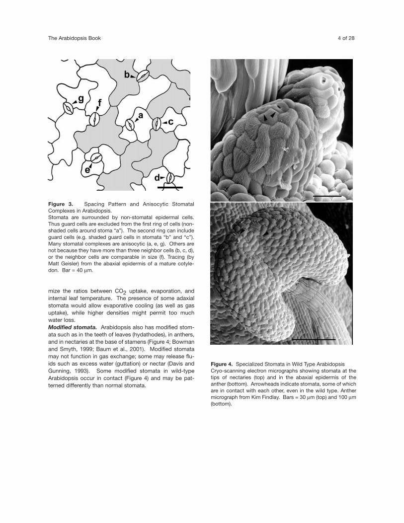
mize the ratios between CO2 uptake, evaporation, andinternal leaf temperature. The presence of some adaxialstomata would allow evaporative cooling (as well as gasuptake), while higher densities might permit too muchwater loss.Modified stomata. Arabidopsis also has modified stom-ata such as in the teeth of leaves (hydathodes), in anthers,and in nectaries at the base of stamens (Figure 4; Bowmanand Smyth, 1999; Baum et al., 2001). Modified stomatamay not function in gas exchange; some may release flu-ids such as excess water (guttation) or nectar (Davis andGunning, 1993). Some modified stomata in wild-typeArabidopsis occur in contact (Figure 4) and may be pat-terned differently than normal stomata.
Figure 3. Spacing Pattern and Anisocytic StomatalComplexes in Arabidopsis.Stomata are surrounded by non-stomatal epidermal cells.Thus guard cells are excluded from the first ring of cells (non-shaded cells around stoma “a”). The second ring can includeguard cells (e.g. shaded guard cells in stomata “b” and “c”).Many stomatal complexes are anisocytic (a, e, g). Others arenot because they have more than three neighbor cells (b, c, d),or the neighbor cells are comparable in size (f). Tracing (byMatt Geisler) from the abaxial epidermis of a mature cotyle-don. Bar = 40 µm.
Figure 4. Specialized Stomata in Wild Type ArabidopsisCryo-scanning electron micrographs showing stomata at thetips of nectaries (top) and in the abaxial epidermis of theanther (bottom). Arrowheads indicate stomata, some of whichare in contact with each other, even in the wild type. Anthermicrograph from Kim Findlay. Bars = 30 µm (top) and 100 µm(bottom).

Stomatal Development in Arabidopsis 5 of 28
STOMATAL PRECURSORS
Understanding the cell types and stages of stomatal devel-opment provides a foundation for explaining the mecha-nisms of patterning, the regulation of stomatal density, andthe functions of proteins involved in stomatal develop-ment. Because dicot stomatal development is variable insome features, it has proven helpful to analyze develop-ment by studying the behavior of cells through time usingvarious impression methods (Sachs, 1991; Sachs andKagan, 1993). The following description of precursor cellsand of stomatal development in Arabidopsis is based uponthe dental resin impression method, as well as cytologicalstudies (Yang and Sack, 1995; Zhao and Sack, 1999;Berger and Altmann, 2000; Geisler et al., 2000).
Stomatal development in Arabidopsis invariably requiresthree different precursor cells (Figures 5-9), the meriste-moid mother cell (MMC), the meristemoid, and the guardmother cell (GMC).MMCs. The first precursor cell is the first cell in the stom-atal pathway to divide asymmetrically. This division pro-duces a smaller, usually triangular meristemoid and a larg-er sister cell (Figure 5). As far as is known, all Arabidopsisstomata are produced by at least one asymmetric division,that of the MMC. MMCs that are committed to this divi-sion can be recognized by their polarized cytoplasm withthe nucleus and preprophase band of microtubules locat-ed at one pole, and a vacuole at the other (Zhao and Sack,1999). MMCs typically originate from relatively small epi-dermal cells that do not have very sinuous cell walls.Meristemoids. Some newly formed meristemoids convertdirectly into guard mother cells, the third and final precur-sor. Other meristemoids divide one to three times asym-metrically before conversion. As in the asymmetric divi-sions of MMCs, meristemoids that are about to divide canbe recognized by their polarized cytoplasm (Figure 9;Galatis and Mitrakos, 1979). Each time a meristemoiddivides, it regenerates a meristemoid and a larger sistercell. In this way, a meristemoid can be considered a“stem” cell. We were not able to predict the number oftimes a meristemoid will divide by its size, position, or anyother parameter that can be detected by the dental resinmethod. Thus, the simplest assumption is that all meriste-moids have the same potential for divisions, and that thefactors that regulate the number of divisions need to bedetermined. It is, however, useful to separate out oneclass of meristemoids, satellite meristemoids, based onwhere they form. Satellite meristemoids originate fromMMCs that are located next to a pre-existing stoma or pre-cursor and, as will be discussed, are central to the gener-ation of the spacing pattern. GMCs. Guard mother cells divide symmetrically to pro-
duce two guard cells. The GMC division site, the locationof cell plate fusion with the parental cell wall, is marked bya preprophase band of microtubules, a marker found inalmost all types of cells (Zhao and Sack, 1999 Smith,2001). In Arabidopsis GMCs, the division site is alsomarked by wall thickenings at opposite ends of the cell(Figure 9; Zhao and Sack, 1999). While similar thickeningshave been found in GMCs in the Fabaceae (Galatis andMitrakos, 1979; Galatis et al., 1982), they have not beenfound in any other cell type. It is not clear whether thesethickenings function in guiding cytokinesis and/or whetherthey help the GMC attain an oval shape.
STOMATAL MORPHOGENESIS
GMC and stoma. The final stage of stomatal develop-ment involves the differentiation of the stoma itself.Differentiation in Arabidopsis is comparable to thatdescribed for other dicots (Galatis and Mitrakos, 1980;Sack, 1987; Zhao and Sack, 1999). Stomatal morphogen-esis actually begins in the GMC. The oval outline of thestoma is first established in the GMC, perhaps by the endwall thickenings that may restrict local elongation of thecell wall (Figure 9). Other features of mature stomata, suchas starch accumulation and vacuolar enlargement also ini-tiate in the GMC, and then continue until the stomamatures.Stomatal pore. Pore formation starts with a lens-shapedthickening located at the middle of the ventral wall (thenew wall formed in the GMC; Figure 9). This thickeningmay be organized by radial arrays of microtubules thathave gamma-tubulin at their focus (Marc, 1997). It is strik-ing that each guard cell develops with a mirror-like sym-metry with respect to the other. For example, the radialmicrotubule arrays and the wall thickenings in each guardcell are precisely opposite each other (Wasteneys et al.,1997). After the pore thickening reaches a critical stage,the anticlinal walls separate in the region of the thickening.This creates the stomatal pore. Obviously, the location ofwall separation is regulated spatially, a trait under strongselection pressure because a separation elsewhere couldresult in desiccation.Wall specialization. Stomata have an elaborate architec-ture. In Arabidopsis, there are distinct differences in thethickness of the different cell walls in the mature guard cell(Zhao and Sack, 1999). In addition, cellulose is depositedin arrays that radiate out from the pore, arrays that pre-sumably result from microtubule-directed deposition. Thecollective distributions of cellulose, wall thickenings, andhinges presumably translate changes in hydrostatic pres-

The Arabidopsis Book 6 of 28
sure into pore opening and closing. The elimination ofplasmodesmata during stomatal development helps main-tain turgor by making each guard cell an island of sym-plasm in the epidermis.
STOMATAL PATHWAY
This overview shows that each precursor cell is capable ofdivision, and that changes in fate are progressive.Although meristemoids sometimes do not divide, MMCsand GMCs invariably do so. Each type of precursor cellhas a different fate, shape, identity and behavior. However,each step in the pathway advances towards the terminalevents of stomatal specification and differentiation.
As stated, the pathway begins with the selection andasymmetric division of the MMC. This also initiates thestomatal cell lineage, which is a series of clonally relatedcells derived from the MMC.
Although differentiation terminates the stomatal path-
way from the meristemoid, the larger daughter cells pro-duced by asymmetric divisions may follow three differentfates (Figure 10). First, they may differentiate into pave-ment cells, large jigsaw puzzle-shaped cells that undergoendoreduplication (Melaragno et al., 1993). Second, theymay become MMCs and start a new stomatal lineage.Third, they may divide symmetrically and produce twocells that can independently follow one of these threefates. One consequence of asymmetric divisions in thestomatal pathway is that these divisions produce abouthalf of all pavement cells and perhaps up to three-quartersof all epidermal cells in leaves.
GENERATION AND TYPES OF STOMATALCOMPLEXES
A stomatal complex can be defined as the stoma plusadjacent epidermal cells. In many species, a set numberand arrangement of cells surround each stoma. Stomatal
Figure 5. Diagram Showing Key Cell Types and Divisions in Arabidopsis Stomatal Development.The first asymmetric division (in a MMC) produces a meristemoid. Meristemoids can divide before converting into a guardmother cell (GMC). GMCs divide symmetrically producing two guard cells. Note that the process can reiterate; neighborcells can re-enter the pathway by becoming MMCs and dividing asymmetrically to produce satellite meristemoids. Theplane of division is oriented so that the new meristemoid is placed away from the pre-existing stoma or precursor cell.Yellow, meristemoid mother cells (MMCs); red, meristemoids.

Stomatal Development in Arabidopsis 7 of 28
complexes can be distinct taxonomically and develop-mentally (Esau, 1977). Here we address whether theArabidopsis stomatal complex is predictable in the posi-tion, number, and origin of neighbor cells. This analysisalso relates to patterning in the stomatal pathway.Anisocytic complexes. Arabidopsis and other membersof the Brassicaceae exhibit anisocytic complexes wherethe stoma is surrounded by three cells, one of which issmaller (Figures 5 and 9B; Metcalfe and Chalk, 1950; Pantand Kidwai, 1967). This arrangement can result from threeconsecutive asymmetric divisions, one of the MMC andtwo of the meristemoid (Berger and Altmann, 2000; Geisleret al., 2000). Because the divisions are arranged in aninward spiral, the larger daughter cells become located tothe outside; as a result they completely surround thestoma and their relative size correlates with their age. Herethe complex is monoclonal because all five cells (threeneighbor cells plus two guard cells) originated from thesame MMC. Anisocytic complexes make up 40-66% of allcomplexes in leaves and cotyledons (Geisler et al., 2000;Serna and Fenoll, 2000a).
The inward spiral of asymmetric divisions can occur in aclockwise (Figure 5) or in a counterclockwise direction(Serna et al., 2002). Moreover, the angle of the symmetric
division of the GMC is often roughly parallel to that of thelast asymmetric division of the meristemoid (Galatis andMitrakos, 1979; Serna et al., 2002). Nothing is known inthis system about how division planes are positioned withrespect to each other over four sequential divisions. Complexes other than anisocytic. Other complexes inArabidopsis are not anisocytic either because the stomataare surrounded by 2, 4 or 5 neighbor cells, or because thethree cells are of equal size, or because two complexesshare one neighbor cell (Figure 3; Geisler et al., 2000;Serna and Fenoll, 2000a). Using the dental resin impres-sion method, we found that this variability results in partfrom how many times meristemoids divide (Figure 5;Geisler et al., 2000). We estimated that about a third of allmeristemoids in a lineage divide once or not at all beforeconverting to a GMC. The resulting complexes would con-tain at least one non-clonal neighbor cell and would thusbe polyclonal. Other estimates of the number of polyclon-al complexes are lower (13 to 23%) and were derived fromthe analysis of transposon-induced sectors marked byGUS-staining (Larkin et al., 1996; Serna and Fenoll, 2000a;Serna et al., 2002). Thus, about 13-34% of Arabidopsisstomatal complexes are polyclonal, and 33-60% are notanisocytic.
These findings show that unlike the one-celled stomatalspacing pattern, the anisocytic pattern is not obligate.Because not all cells adjacent to stomata are distinct mor-phologically, a requirement for the term “subsidiary cell”(Esau, 1977), we refer to these cells generally as neighborcells. More importantly, the existence of polyclonal com-plexes has implications for how the stomatal spacing pat-tern is generated (see below).
GENERATION OF SPACING PATTERN
Hypotheses for spacing. As mentioned the central fea-ture of patterning is that stomata are separated by at leastone intervening cell. Several hypotheses have been pro-posed to explain how this pattern is generated in dicotyle-dons (Figure 11; Sachs, 1979, 1991; Sylvester et al., 1996;Larkin et al., 1997; Croxdale, 2000).
The classical lateral inhibition hypothesis invokes thepresence of an inhibitory field around developing stomatathat prevents new stomata from forming in neighbor cells(Bünning, 1953; Korn, 1993, 1994). This implies that aneighbor cell should be prohibited from functioning as anMMC.
In the cell lineage hypothesis, a series of stereotypedasymmetric divisions creates a monoclonal boundary ofneighbor cells (Sachs, 1991; Larkin et al., 1997; Serna and
Figure 6. Living Abaxial Epidermis from Developing LeafShowing Asynchrony in Stomatal Development and DifferentCell Types.Confocal scanning laser micrograph of green fluorescent pro-tein line that labels cell membrane (“Q8” from Cutler et al.,2000). Because the cell walls are thin, the two cell mem-branes appear as one line at this magnification. GMCs (whiteasterisks) tend to have convex walls, whereas meristemoids(red asterisks and arrowheads) are more triangular. The stom-atal pore is not visible because of the sharp curvature of thepore wall or the plane of the optical section. Bar = 20 µm.

The Arabidopsis Book 8 of 28
Figure 7. Example of Dental Resin Data.Bright-field light micrographs of nail polish replicas of dental impressions showing the same field of cells over a 24-hour period (6-7 days after germination). Abaxial epidermis of cotyledon. The MMC (left) divided asymmetrically producing a satellite meriste-moid (right). Another neighbor cell divided symmetrically. Figure from Matt Geisler. Bar = 25 µm.
Figure 8. Key Events in Stomatal Development Shown in Dental Impression Series. The abaxial epidermis of a single cotyledon is shown through time. Cells “c” (day 7) and “f” (day 8) are MMCs because they divid-ed asymmetrically to produce meristemoids. The initial asymmetric division of MMC “f” took place next to a pre-existing stomaand produced a satellite meristemoid. Both MMCs arose from smaller, less sinuous cells. One smaller cell (“e”, day 7) divided sym-metrically by day 8. Meristemoid “a” (day 6) divided twice asymmetrically in an inward spiral. Two apparent meristemoids are adja-cent (day 6, lower right); the upper one formed a stoma but the lower meristemoid did not progress in development (“b” and “d”).From Geisler et al. (2000).Yellow, meristemoid mother cells (MMCs); red, meristemoids; blue, guard mother cells; green, stomata.

Stomatal Development in Arabidopsis 9 of 28
Figure 9. Asymmetric and Symmetric Divisions.(A) A newly formed meristemoid is not polarized cytologically and is much smaller than its sister cell.(B) The meristemoid shown was produced by two previous asymmetric divisions (1 & 2). It is now polarized prior to a likely thirdasymmetric division (arrows). The series of divisions occurs in an inward spiral.(C) A higher magnification of a polarized meristemoid with an asymmetrically placed nucleus. This nucleus is close to the future siteof division as marked by a preprophase band of microtubules (shown in Zhao and Sack, 1999).(D) The division site in GMCs is marked by wall thickenings before symmetric division. (E) The GMC division site wall thickenings are still visible in a developing stoma (arrowheads).Adapted from Zhao and Sack (1999). Bars = 3 µm (A), 5 µm (B), 1 µm (C-E).

Fenoll, 2000a; Serna et al., 2002). According to thishypothesis, the placement of neighbor cells is generatedwithin the cell lineage and does not require communicationwith surrounding cells. This is thought to prevent the for-mation of stomata in contact by generating interveningcells.
A third view, based on study of the same cells throughtime, is that stomata are mostly randomly placed, but ori-ented divisions correct occasional patterning mistakes(Sachs, 1991; Sachs and Kagan, 1993; Sachs andNovoplansky, 1993). For example, Sachs and co-workersfound that when meristemoids formed in contact, onemeristemoid divided away, behavior thought to result fromcell signaling.Stomata are spaced by the placement of satellitemeristemoids. We studied the development of manyArabidopsis stomata using the dental resin impressionmethod (Geisler et al., 2000). This made it possible to ana-lyze how Arabidopsis stomata are patterned. We foundthat many neighbor cells become MMCs, including cellsadjacent to a stoma, a GMC or a meristemoid. The asym-metric division of the neighbor cell MMC is invariably ori-ented so that the new meristemoid, the satellite meriste-moid, is placed away from the pre-existing stoma/precur-sor (Figure 12).
This is a central event in stomatal patterning inArabidopsis in the abaxial leaf epidermis. The majority ofstomata in these tissues originate from satellite meriste-moids. Satellite meristemoid placement creates the one-
cell spacing around this precursor and maintains thespace around the pre-existing stoma or precursor so thatboth become patterned.
It is likely that cell signaling influences the orientation ofthe asymmetric divisions that produce satellite meriste-moids. Satellite meristemoids were correctly placedregardless of whether the new MMC was or was not clon-ally related to the adjacent stoma/precursor. This arguesagainst the idea that division orientation requires the inher-itance of mitosis allocated factors which organize the siteof subsequent divisions as is found in yeast budding (Chenet al., 2000). The simplest explanation is that the neighborcell receives signals from the pre-existing stoma or pre-cursor, positional information that is used to select thelocation of the division site.Relationship to other patterning hypotheses. Ourresults support Sachs’ (1991) concept that the positions ofthe first generation of stomata are generally not regulatedwith respect to each other. We found that MMCs formedin contact with each other with a frequency predicted bychance (Figures 10 and 13; Geisler et al., 2000). This sug-gests that the first precursor cells are randomly placed andthat MMCs do not laterally inhibit each other.
As noted, Sachs (1991) suggested that oriented divi-sions could correct patterning mistakes. We also foundthat when meristemoids were in contact, that either orboth divided away (Geisler et al., 2000). While this con-tributes to stomatal patterning, we found it occurs less fre-quently in Arabidopsis than satellite meristemoid forma-tion, and thus it is a secondary mechanism of patterning.
Bünning’s (1953) formulation of the lateral inhibitionhypothesis (Figure 11) implies that the presence of a stomaprevents adjacent cells from becoming stomata. Lateral
The Arabidopsis Book 10 of 28
Figure 10. Fates of Sister Cell to Meristemoid.The same asymmetric division that produces a meristemoid(red) also produces a larger sister cell (*). This cell is plastic infate. It can divide asymmetrically to produce a satellite meris-temoid. It can divide symmetrically to produce two newneighbor cells that in turn have the same plasticity in cell fate.It may stay relatively small and not divide, or it may enlargeand differentiate into a pavement cell (latter not shown).
Figure 11. Hypotheses for the Generation of the StomatalOne-Celled Spacing Pattern.Hypotheses tested by analysis of dental resin impressionseries (Geisler et al., 2000).

Stomatal Development in Arabidopsis 11 of 28
inhibition usually means that one cell prevents an adjacentcell from assuming a particular fate (Appel et al., 2001).But the only way that a neighbor cell can form a stoma isby functioning as an MMC, a frequent event inArabidopsis. Thus, in this sense, patterning does notinvolve lateral inhibition. However, the presence of astoma or precursor does seem to inhibit the formation of ameristemoid nearby. But the mechanism is the control ofthe placement of the MMC division site, not the fate of theneighbor cell itself.
Our data suggest that cell lineage does not generate theone-celled spacing pattern in the leaf epidermis. First, thelineage hypothesis doesn’t provide a mechanism for howstomata in polyclonal complexes are spaced. Up to a thirdof all complexes are polyclonal in the abaxial epidermis ofthe first leaf and cotyledon (Geisler et al., 2000). The celllineage hypothesis as formulated fails to explain why thenon-clonal cells in polyclonal complexes are not stomata.
Second, even when a monoclonal complex is produced,
neighbor cells do not provide a boundary or buffer,because many divide and produce stomata. It is not theexistence of the neighbor cell itself, but the correct place-ment of the satellite meristemoid that establishes the min-imal one-celled spacing pattern.
Third, even when adjacent complexes are each mono-clonal (Serna and Fenoll, 2000a; Serna et al., 2002), itremains to be demonstrated that a series of stereotypeddivisions within a cell lineage actually establishes the one-cell spacing pattern. The key question is when in devel-opment the separation between meristemoids is first gen-erated. A single asymmetric division can be sufficient tocreate a one-celled spacing such as in the placement ofthe satellite meristemoid or in the division of meristemoidsin contact. In addition, many meristemoids are correctlyspaced by chance (Geisler et al., 2000). In all three cases,subsequent asymmetric divisions in a lineage wouldincrease the number of cells between stomata but wouldnot affect the already established minimal one-celledspacing. Although cell lineage may not generate the one-celled spacing pattern, divisions within a cell lineage arecrucial for establishing the position, arrangement andhigher order spacing of stomata and other epidermal cells.Cell position and events in the stomatal pathway.Some events in the stomatal pathway seem independentof cell position. For example, MMCs can form in contact,and the divisions of these MMCs appear to be randomlyoriented (Figure 13A-E). It is unclear whether other eventsrequire positional cues communicated via intercellular sig-naling. For example, the divisions of meristemoids in aninward spiral could result from an endogenous patterningprogram such as through the allocation of spatial cuesthrough mitosis.
Other events seem to require the communication ofinformation about adjacent cell position. Examples men-tioned include the orientation of asymmetric divisions inneighbor cells and in adjacent meristemoids (Figure 13).Cell signaling also appears to regulate the fate of cells intwo specific positions in the stomatal pathway inArabidopsis (Figure 13 H and I; Geisler et al., 2000). First,when meristemoids accidentally form in contact, some-times one meristemoid arrests and does not divide orbecome a GMC. Second, cells next to any combination oftwo stomata, meristemoids, and GMCs seem to be pro-hibited from dividing asymmetrically. Neither event seemsto play a major role in stomatal patterning (Geisler et al.,2000).
In domains that have fewer stomata and satellite meris-temoids, such as the adaxial epidermis, spacing may relymore upon chance or minor spacing mechanisms. Buteven in the adaxial epidermis, we estimate that the major-ity of all stomata are patterned by satellite meristemoidplacement (Geisler and Sack, 2002).
In summary, some stomata are placed by chance. The
Figure 12. Satellite Meristemoid Formation.(Top) Dental resin series showing asymmetric division of anMMC (yellow). (Bottom) Cryo-scanning electron micrograph. Asterisksshow a satellite meristemoid (red), or GMCs produced bysatellite meristemoids (blue). Successive stages of poredevelopment are shown at the upper left and lower center.Note that five of the six existing and future stomata shown arepatterned by satellite meristemoid placement. Cells locatednext to two stomata and/or precursors (such as the cell to theright of the red asterisk) usually do not divide. Adapted fromGeisler et al. (2000). Bar (bottom) = 10 µm.

The Arabidopsis Book 12 of 28
remainder is spaced largely by oriented asymmetric divi-sions of neighbor cells. Other position-dependent eventsplay either a secondary role or are not directly involved ingenerating the minimal one-celled spacing pattern.
REGULATION OF STOMATAL NUMBER
Many factors influence stomatal number such as environ-mental and developmental signals. For example, light and
CO2 concentration regulate the stomatal index which isdefined as the ratio of stomata to all epidermal cells(Woodward and Kelly, 1995; Tang and Liang, 2000;Brownlee, 2001). Signals that influence stomatal numbermust ultimately regulate the specification of the first pre-cursor cell in the stomatal pathway, the MMC. Genesinvolved in this regulation are reviewed later. Here theimportance of cell position, ploidy and size are discussed.Cell competence and selection as MMCs. Stomatal for-mation appears prohibited in several positions. In somecases, such as around trichomes, this prohibition occursbefore asymmetric divisions start in the developing leaf
Figure 13. Position Independent and Dependent Events in the Stomatal Pathway(A) and (B) The placement of the first precursor cell, the MMC, appears to be random so that MMCs sometimes form in contact(B).(C) The plane of asymmetric division in separated MMCs also seems to be random.(D) and (E) Divisions of MMCs in contact are randomly oriented and can produce meristemoids in contact.(F) The orientation of asymmetric divisions next to a single stoma or precursor is position-sensitive and patterns most stomata.(G) Asymmetric divisions of adjacent meristemoids can be oriented and thus space stomata.(H) Cells next to two stomata or precursors (asterisks) usually do not divide asymmetrically.(I) The arrest of an adjacent meristemoid (asterisk) spaces some stomata.Adapted from Geisler et al. (2000).Yellow, meristemoid mother cells (MMCs); red, meristemoids; blue, guard mother cell (GMCs); tan, cells whose fate is affected bytheir position.

Stomatal Development in Arabidopsis 13 of 28
(Larkin et al., 1997; Martin and Glover, 1998; Glover andMartin, 2000). In other cases, specific cells become pro-hibited even in regions actively forming stomata. Forexample, we found that cells next to two stomata and/orprecursors in any combination usually do not divide asym-metrically (such as the cell to the right of red asterisk inFigure 12 bottom; Geisler et al. 2000).
The differentiation of pavement cells also removes cellsfrom the pool of those competent to form MMCs. Large,sinuous cells that are known to undergo endoreduplicationhave not been observed to divide asymmetrically(Melaragno et al., 1993; Geisler et al., 2000; Glover, 2000).Conversely, smaller less wavy cells with 2C levels of DNAare most likely to be division competent and form MMCs(Donnelly et al., 1999; Geisler et al., 2000). Such cells areabundant in the young, developing epidermis. Later in leafdevelopment, smaller cells are mostly located next tostomata (Figure 14). Thus the generalized capacity of theprotoderm to initiate a stomatal lineage later becomes pro-gressively restricted to small, less sinuous cells. Multiplegenerations of stomata can develop over the course of leafdevelopment when sister cells to satellite meristemoidsthemselves divide asymmetrically and so on (Landré,1972; Berger and Altmann, 2000).
Other small cells arise from symmetric divisions.Symmetric divisions produce cells of comparable size anddevelopmental potential (Geisler et al., 2000). Symmetricdivisions occur in neighbor cells (Figures 7 and 10) or incells removed from stomata or their precursors. The poolof cells competent to form stomata is filled by asymmetricand symmetric divisions, and drained by cell enlargementand differentiation. Stomatal number is therefore partly afunction of the balance between cell proliferation and celldifferentiation.
In summary, stomatal number is regulated by the avail-ability of division competent cells and by how many ofthose cells are induced to divide asymmetrically. Many ofthe cells capable of forming MMCs originate from priorasymmetric divisions in the stomatal pathway.Stomatal index. This ratio depends upon how manystomata and other cells arise during leaf development.MMC divisions directly increase the number of both stom-ata and epidermal cells. Symmetric divisions of epidermalcells and asymmetric divisions of meristemoids directlyincrease only the number of epidermal cells. Thus, thestomatal index may be modulated by the differential regu-lation of these three types of divisions, a possibility thathas not been studied. Such an analysis could identify themechanisms by which environmental parameters influenceplant productivity. Longitudinal gradients in organs. In developingArabidopsis leaves most stomata form and mature in aroughly tip to base gradient (Pyke et al., 1991; Donnelly etal., 1999). Successive waves of stomatal initiation, satel-
lite meristemoid formation, and generalized cell enlarge-ment start at the tip and proceed basipetally.
Grasses also display a tip-to-base gradient of stomatalformation. But the gradient in stomatal development is pre-dictable in time and space within cell files. Meristemoidsappear to be absent in grasses (Larkin et al., 1997).Stomatal number in monocots is controlled partly by theproduction of new regions of the leaf blade. In Arabidopsisleaves the initial longitudinal gradient in stomatal formationis much less strictly defined and is disrupted by mosaicgrowth. Stomatal formation is asynchronous and variable inthe number of asymmetric divisions. Stomata are producediteratively and progressively including in regions that areotherwise mature (Figures 12 and 14).
Relatively little is known about stomatal development inArabidopsis stems and other cylindrical organs such aspetioles, carpels and pedicels. In hypocotyls, a series oflongitudinal and transverse divisions seems to create apool of cells competent to form MMCs (Berger et al.,1998b). In stems and hypocotyls stomata appear tomature acropetally (Nadeau and Sack, unpublished).These observations imply that longitudinal waves of com-petence to form MMCs pass through developing leavesand stems.
COORDINATION WITH INTERNAL FEATURES
Because the stomatal pore regulates gas exchangebetween the atmosphere and subjacent tissues, it mightbe expected that position-dependent signals would coor-dinate the placement of stomata and underlying features.This section reviews two lines of evidence relating to sucha position-dependent coordination.Leaves. There seems to be no obligate relationshipbetween the placement of stomata and substomatal cavi-ties in dicot leaves. In one study, 13% of mature stomatawere located directly over a mesophyll cell rather than asubstomatal cavity (Sachs, 1979). Stomatal precursorcells also can be positioned over mesophyll cells insteadof cell junctions (Figure 15; reviewed in Sachs, 1978).
Serna and Fenoll (2000a) analyzed whether stomatalplacement is coordinated with internal tissues inArabidopsis. Because substomatal cavities are small orabsent in the adaxial side of the leaf, they used junctionsrather than cavities between mesophyll cells for scoring.Over 90% of stomata and meristemoids were located overcell junctions. This percentage differed significantly fromtheir estimate of a randomly generated placement.However, meristemoids only form at cell edges, a featurethat would differ from a randomly generated control. Also,
The Arabidopsis Book 14 of 28
epidermal cell outlines may be positionally out of registerwith the cells below (Figure 15). Using these parameters,it would be valuable to confirm that the distribution overcell junctions is non-random. If so, then radial signalingmay take place between these two cell layers. One sce-nario could be that a signal from mesophyll cells couldhelp orient asymmetric divisions so that meristemoids tendto form over cell junctions (Serna and Fenoll, 2000a).Hypocotyls. Hypocotyl stomata are present in files of
short, flattened cells and excluded from files of cells thatare elongated and bulging (Gendreau et al., 1997). Theprotruding epidermal cells are located over the periclinalwall of a single underlying cortical cell. The files that pro-duce stomata are above anticlinal walls (Schiefelbein2002; Berger et al., 1998b; Hung et al., 1998). Thus, thecompetence to form MMCs is restricted in a position-dependent manner to hypocotyl cells that are radiallyaligned with subjacent landmarks.
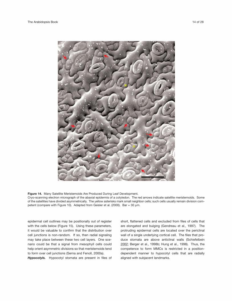
Figure 14. Many Satellite Meristemoids Are Produced During Leaf Development.Cryo-scanning electron micrograph of the abaxial epidermis of a cotyledon. The red arrows indicate satellite meristemoids. Someof the satellites have divided asymmetrically. The yellow asterisks mark small neighbor cells; such cells usually remain division com-petent (compare with Figure 10). Adapted from Geisler et al. (2000). Bar = 30 µm.

Stomatal Development in Arabidopsis 15 of 28
A similar mechanism controls root hair formation andconsiderably more is known about root hair patterning.Root hairs form in cells (H cells) located over anticlinal cor-tical cell walls rather than in cells (N cells) over periclinalwalls (Schiefelbein, 2000, 2002). The signal that coordi-nately establishes root hairs over anticlinal walls isunknown. The cellular pattern is set up in the embryo andis maintained in derivatives of the root apical meristem(Berger et al., 1998a). These authors killed subjacent rootcortical cells using laser ablation before the fate of recentepidermal derivatives was fixed so that the derivatives nolonger contacted living cortical cells. The isolated deriva-tives continued to express markers appropriate to theirposition relative to underlying cells. These data argueagainst the need for continuous communication betweencell layers, and are consistent with the possibility that theouter cell walls of dead cortical cells retain some factorthat maintains the fate of the overlying epidermal cells(Berger et al., 1998a; Benfey, 1999).
The formation of stomata and root hairs over anticlinalcortical walls is a striking example of the conservation of aradial patterning mechanism. It places stomata over futureintercellular spaces and root hairs over the shortest routeto the xylem. This conservation involves common molec-ular pathways, the subject of the next section.
“ROOT HAIR” GENES AND MMC FORMATION INHYPOCOTYLS
It is appropriate that this discussion of the genes regulat-ing stomatal development begins with genes that ultimate-ly control the first step in the pathway, the creation of smallcells that are capable of forming MMCs. Loci known toaffect both root hair and stomatal patterning include GL2,TTG, and WER (see model in Schiefelbein 2002; Berger etal., 1998b; Hung et al., 1998; Schiefelbein, 2000).
In roots, these three gene products block the formationof root hairs in N cells. Mutations in all three result inectopic root hairs in N cells. GL2, a homeodomain protein,is preferentially expressed in non-hair files. The expres-sion of GL2 is positively regulated by WER, a myb protein,and by TTG, a WD-40 protein. Both maintain the higherrelative expression levels of GL2 in N cells (Schiefelbein,2000). These data are consistent with a model wherebythe expression of GL2 in N cells negatively regulates roothair specification (Lee and Schiefelbein, 1999;Schiefelbein, 2000, 2002). Conversely, reduced GL2expression in H cells allows hairs to form.
In addition, TTG may bind to a basic Helix-Loop-Helixtranscriptional activator. When this complex is bound toWER, it may promote GL2 expression in N cells. In H cells,
Figure 15. Stomatal Precursor Cell Placement with Respect to the Mesophyll.Tracing from a pea stipule showing epidermal cells (broken lines), stomata and their precursors (parallel hatching), and the subja-cent mesophyll (solid lines). Figure from Sachs (1978) who did not find a consistent spatial relationship between stomatal precur-sor cell placement and the anticlinal walls of the mesophyll. Bar = 30 µm.
The Arabidopsis Book 16 of 28
TTG-bHLH may bind to CPC, a myb protein. This complexis hypothesized to repress GL2 expression because CPClacks an activation domain. This model is supported bythe findings that a 35S::R transgene (a maize bHLH) in attg mutant background restores a non-hair fate to N cellsand represses hair formation in H cells (Galway et al.,1994; Schiefelbein, 2000, 2002).
In hypocotyls, GL2 and WER are preferentiallyexpressed in protruding cells. Mutations in GL2, TTG, andWER exhibit ectopic stomata in GL2-expressing cells(Hung et al., 1998; Lee and Schiefelbein, 1999). In wild-type hypocotyls, 2-5% of all stomata were found to beectopic. In contrast 12-27% were ectopic in gl2, ttg andwer mutants or when the R transgene was introducedunder the control of the 35S promoter (Berger et al.,1998b; Hung et al., 1998; Lee and Schiefelbein, 1999). Thegl2 and ttg mutations had no effect on stomatal density oron relative cell length in the normal stomatal files, but35S::R reduced the total number of stomata by 80%.
GL2, WER, TTG, and the 35S::R transgene all negative-ly regulate stomatal formation in cells that normallyexpress GL2. A model for how these genes function in thehypocotyl is comparable to their position-dependenteffects in roots (Berger et al., 1998b; Hung et al., 1998).Thus, ectopic stomata may form because (1) GL2 is notfunctional in gl2 mutants, (2) GL2 is not expressed wheneither TTG and WER, which positively regulate GL2expression, are non-functional, or (3) the 35S::R transgenedisrupts the differential expression of GL2 (Hung et al.,1998; Lee and Schiefelbein, 1999; Schiefelbein 2002).
The effects of the same ttg and gl2 alleles appear to bemore severe in roots than in hypocotyls. These mutationscause almost all non-hair cells to form ectopic root hairs(Galway et al., 1994; Masucci and Schiefelbein, 1996). Incontrast, relative few ectopic stomata are formed inmutant hypocotyls. This difference in apparent severitymight be related to the findings that wild-type hypocotylshave relatively few stomata and that the protruding cellsfiles can be disrupted by intervening smaller cells (Bergeret al., 1998b; Gendreau et al., 1997).
GL2 may negatively regulate stomatal formation byspecifying a protruding cell fate. These cells are larger,and divide considerably less than the non-GL2 expressingcells (Gendreau et al., 1997; Hung et al., 1998; Berger etal., 1998b). Protruding cells probably undergo endoredu-plication because half of all hypocotyl cells do so andbecause endoreduplication can be proportional to cell size(Gendreau et al., 1998). These cells are probably removedfrom the pool of cells competent to divide. Thus, GL2 mayblock MMC formation by promoting an alternate cell fate ina position-dependent manner. Whereas in roots, almost allnon-GL2 expressing cells form hairs, in hypocotyls only afew stomata form per file. This suggests that the absenceof GL2 expression only results in MMC and stomatal for-
mation in a subset of cells.The finding that genes that affect root hairs also affect
stomatal patterning provides a new context for studyingthe functions of these genes. It will be interesting to seewhether additional loci that affect root hair patterning,such as CPC, show a stomatal phenotype, and whetherany Arabidopsis bHLH homologues of the maize R gene(Payne et al., 2000) is expressed in stomatal-forming files.Also, many of the root hair mutations seem to affect later-al roots, not just the primary, embryonic root (Schiefelbein,personal communication). This invites a determination ofwhether these genes influence stomatal patterning in theshoot, not just in embryonic tissues such as the hypocotyl.
TOO MANY MOUTHS (TMM)
While GL2, TTG, and WER affect the distribution andpatterning of stomata in the hypocotyl, the minimal one-celled spacing pattern is unaffected. In contrast, muta-tions in several loci have been shown to disrupt the funda-mental spacing pattern by allowing stomata to form incontact. The best described of these loci are TOO MANYMOUTHS, STOMATAL DENSITY AND DISTRIBUTION1,and FOUR LIPS.Clusters result from disruption of the number, planeand polarity of asymmetric divisions. TMM is essentialfor the establishment of stomatal patterning (Yang andSack, 1995; Larkin et al., 1997). A major tmm phenotypeis the presence of clusters of adjacent stomata in leavesand cotyledons (Figure 16). Stomata in clusters arearranged at different angles with respect to each other, andthe stomata vary in size and stage of development. Thesetraits suggest that the clusters develop progressively anditeratively.
Analysis of cluster development using both dental resinimpressions and static images shows that tmm disruptsseveral spacing mechanisms (Figure 17; Geisler et al.,2000).
First, instead of satellite meristemoids being placedaway from pre-existing stomata, some tmm meristemoidsform in contact with the stoma (Figure 17; Yang and Sack,1995). These ectopic satellite meristemoids result from themisorientation of the plane of asymmetric divisions inneighbor cells. Analysis of division angles shows that tmmrandomizes the division plane. This appears to be themajor defect resulting in cluster formation.
Second, tmm disrupts a secondary stomatal spacingmechanism, the oriented divisions of meristemoids in con-tact (Figures 11 and 13; Geisler et al., 2000). Unlike thewild type, tmm meristemoids fail to divide away from each

Stomatal Development in Arabidopsis 17 of 28
other.Third, stomatal clusters also develop from alterations in
the frequencies of cell divisions. Satellite meristemoids,like all meristemoids, can divide asymmetrically, divisionsthat often add to the separation between the pre-existingstoma and the new one. However, tmm meristemoidsdivide fewer times compared to the wild type resulting inan acceleration of when meristemoids convert into GMCs.This reduces the likelihood that meristemoid divisions willcorrect patterning mistakes.
Fourth, clusters form partly from an overproduction ofsatellite meristemoids. While the number of asymmetricdivisions of meristemoids is reduced in tmm, the numberof neighbor cells that divide asymmetrically increases.Thus, TMM acts as a negative regulator of asymmetricdivisions in neighbor cells, and a positive regulator of divi-sions in meristemoids. The consequence of disrupting thisregulation is an overproduction of satellite meristemoids,
many of which are misplaced and which may not divide.Fifth, cells that do not divide in the wild type do so in
tmm. Cells located between two stomata or precursorsappear to be prohibited from dividing asymmetrically in thewild type. The asymmetric divisions of these cells in tmmcombined with randomized division planes produces morestomata in contact (Figure 17). Thus, cluster formation alsoresults from the disruption of the position-dependent con-trol of cell fate.
Sixth, tmm also occasionally alters the fates of daughtercells of divisions that normally would be asymmetric.Usually tmm asymmetric divisions are clearly polar mean-ing that they produce daughter cells of different sizes andfates. However, sometimes both products of a division ina neighbor cell develop into stomata. This shows thatTMM is also necessary for the polarity of the asymmetricdivision, and that tmm may disrupt the segregation of dif-ferent fate factors.
Figure 16. Stomatal Clusters in too many mouthsCryo-scanning electron micrograph of tmm abaxial cotyledon epidermis. Bar = 15 µm.
The Arabidopsis Book 18 of 28
Thus, stomatal clusters result from alterations in the ori-entation and polarity of asymmetric divisions, from disrup-tions in cell fate, and from defects in the regulation of divi-sion frequency. Many of these events probably requirecell-cell signaling to communicate spatial cues. Both tmmalleles are recessive and are presumed to be loss-of-func-tion mutations. We hypothesize that TMM functions in anintercellular communication pathway that receives andconveys information about the identity and location ofnearby cells. Positional cues may play several roles.These may orient and fix the plane of asymmetric divisionsin neighbor cells. They could be required for the correctdistribution of cell fate determinants that result in the
asymmetric fates of daughter cells. Also spatial signalsmay guide the divisions of meristemoids in contact, andensure that cells adjacent to two stomata or precursorcells do not divide. Thus, TMM may help interpret spatialsignals so as to regulate the number, plane and polarity ofasymmetric divisions. Further study of TMM may provideaccess to genes and events that regulate the placement ofasymmetric divisions in plants.TMM is a negative or positive regulator of entry intothe stomatal pathway. In addition to a role in stomatalpatterning, TMM is also a key regulator of stomatal pro-duction. tmm stems have virtually no stomata (Yang andSack, 1995; Geisler et al., 1998). Thus tmm has two com-pletely opposite stomatal phenotypes, clusters and theelimination of stomata.
Sometimes these phenotypes are found in separateorgans. As mentioned, leaves have clusters and stemslack stomata (Figure 18). Other organs display both phe-notypes. The abaxial sepal epidermis has clusters, but nostomata are present in the adaxial epidermis (Figure 19;Geisler et al., 1998). The most striking case is in the flowerstalk (pedicel) where clusters are present at the floral end,
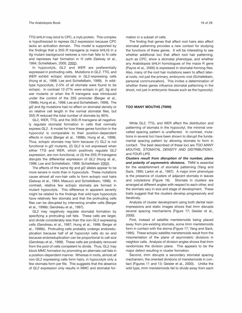
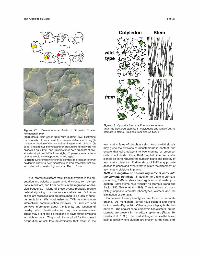
Figure 17. Developmental Basis of Stomatal ClusterFormation in tmm(Top) Dental resin series from tmm (bottom row) illustratingthat stomatal clusters result from several defects including (1)the randomization of the orientation of asymmetric division, (2)cells (*) next to two stomata and/or precursors normally do notdivide but do in tmm, and (3) sometimes both products of divi-sion develop into GMCs (lower right). Top row shows cartoonof what would have happened in wild type. (Bottom) Differential interference contrast micrograph of tmmepidermis showing new meristemoids (red asterisks) that arein contact with developing stomata. Bar = 10 µm.
Figure 18. Opposite Stomatal Phenotypes in tmmtmm has clustered stomata in cotyledons and leaves but nostomata in stems. Tracings from cleared tissue.

Stomatal Development in Arabidopsis 19 of 28
stomata are absent at the stem end, and there is a gradi-ent in between (Figure 20).
TMM is thus essential for stomatal formation in somecontexts, and a negative regulator in others (Figure 21).TMM appears to control how many cells are induced todivide asymmetrically (to function as MMCs). The direc-tion of that regulation depends upon organ and domain.Thus, TMM is a major determinant of stomatal formationas well as patterning. It remains to be seen whether theseare separate functions of the same protein, or whether theyresult from the same key function such as regulation of thefrequency and execution of asymmetric divisions.
The diversity of tmm phenotypes suggests that TMMinteracts with partners in a molecular complex or pathway.In addition, cues conveying tissue identity and positionmay signal the direction of the interaction thereby increas-ing or decreasing the number of MMCs as appropriate tolocation.
STOMATAL DENSITY AND DISTRIBUTION1 (SDD1)
The sdd1 mutation dramatically increases stomatal num-ber, index, and density throughout the shoot (Figure 22top; Berger and Altmann, 2000; Von Groll and Altmann,2001). Three defects contribute to this increase (Figure 22bottom; Berger and Altmann, 2000). First, more protoder-mal cells enter the stomatal pathway (function as MMCs)in sdd1-1. Second, more satellite stomata are produced insuccession (multiple generations of satellite meriste-moids). Third, meristemoids divide fewer times; the stom-atal index increases when fewer daughter cells are avail-able to form pavement cells.
sdd1 also disrupts the primary mechanism of stomatalpatterning in that it disorients asymmetric divisions inneighbor cells; this produces ectopic satellite meriste-moids and stomatal clusters (Figure 22 bottom). No dataare available on whether SDD1 is also required for a sec-ondary patterning mechanism, the ability of adjacentmeristemoids to divide away from each other.
TMM and SDD1 seem to regulate many of the sameevents in leaves (Figure 21). Both affect the orientation ofasymmetric divisions, both function as positive regulatorsof meristemoid division, and both are negative regulatorsof MMC/stomatal formation (Berger and Altmann, 2000;Geisler et al., 2000). Also, both prevent cells adjacent totwo stomata from dividing (Figure 22 bottom; Geisler et al.,2000). But it is not clear whether TMM and SDD1 restrictstomatal number by the same mechanisms. For example,data are not yet available as to whether tmm, like sdd1,increases MMC formation from protodermal cells and per-mits extra generations of satellite meristemoids (Bergerand Altmann, 2000). Moreover, the phenotypes of sdd1and tmm are quite distinct (Figure 22 top). Leaves of bothmutants have many more stomata than the wild type. Butsdd1 has many more correctly patterned stomata thantmm and many fewer stomatal clusters. Where present,sdd1 clusters tend to be smaller than in tmm. These datasuggest that SDD1 is less important for the one-celledspacing pattern than TMM, but more important for deter-mining stomatal density.
sdd1 displays roughly the same phenotype throughoutthe plant whereas tmm has opposite stomatal phenotypesin different organs. Thus, SDD1 is negative regulator ofentry into the stomatal pathway throughout the shoot,whereas TMM can be a negative or positive regulatordepending upon context.
SDD1 encodes a subtilisin-like serine protease that mayfunction as a processing protease in developmental sig-naling (Berger and Altmann, 2000). SDD1 is expressed inmeristemoids and GMCs, and SDD1 has a signal peptide(Von Groll and Altmann, 2001). Thus SDD1 may process
Figure 19. tmm Alters Stomatal Production in an Organ-Specific MannerDiagram showing the distribution of stomata in the reproduc-tive shoot of wild type (top) and tmm (bottom). The stomataldensity is more or less uniform throughout the stem and floralorgans in the wild type. In tmm, stomata are absent from thestem, the base of the flower stalk, the ends of the silique andthe adaxial sepal. The abaxial tmm sepal has more stomatathan the wild type. A stomatal unit is defined as a singlestoma or a cluster of stomata in contact. The density of stom-atal units is shown in color coding at the bottom left. Adaptedfrom Geisler et al. (1998).

The Arabidopsis Book 20 of 28
some signal either in the endomembrane system of GMCsand meristemoids or outside these cells. This signal mayinhibit some neighbor cells from dividing asymmetrically. IfSDD1 also negatively regulates stomatal initiation in fieldsof cells devoid of any stomatal precursor, then this com-munication may take place over larger distances. In con-
trast, SDD1 may positively regulate the divisions of meris-temoids, the same cells in which it is expressed.
The phenotypes of tmm and sdd1 and the molecularidentity of SDD1 support the idea that there is a balancebetween cell proliferation and differentiation. One balancepoint might be between the induction of asymmetric divi-sions that cause entry into the stomatal pathway and thespecification of a pavement cell fate for neighbor cells.Another might be between the induction of meristemoiddivisions and the conversion of the meristemoid into aGMC. It is likely that these set points are responsive tointercellular communication.
This balance might also function in patterning. The dis-ruption of signaling in cells selected to divide could causeabnormal division planes. The analysis of SDD1 and TMMshould help define cell signaling pathways required for bal-ancing cell proliferation and differentiation, for orientingasymmetric divisions, and for specifying cell fate.
FOUR LIPS (FLP)
four lips mutants have clusters of laterally-aligned stomatain direct contact (Yang and Sack, 1995; Larkin et al., 1997).In some alleles, clusters consist primarily of 4 guard cells.Larger clusters are present in other alleles. All allelesexhibit unpaired guard cells that either form in isolation, orin contact with pairs of guard cells. Most cell walls instomatal clusters are parallel but other angles are found aswell. Large clusters contain mature and developing stom-ata, but also other cells of uncertain identity. Only a sub-set of stomata is clustered (Figure 23). The proportion ofclusters to normal stomata is roughly similar in differentplants of the same allele.
Stomatal clusters are found throughout the plant in allplaces where stomata are found in the wild type (Geisler etal., 1998). In flp-1, clusters are less frequent in cylindricalorgans such as stems, flower stalks, and siliques, than indorsi-ventral ones such as rosette leaves.
No other defects have been found in flp. The flp-1 alleledoes not appear to affect the total number of meristemoidsor pavement cells that are produced (Yang and Sack,1995; Geisler et al., 1998). Although guard cell develop-ment can be arrested in clusters, stomatal morphogenesisis not seriously affected. flp plants are healthy, and tri-chome and root hair development appear normal.
The arrangement of cell walls in clusters is consistentwith the hypothesis that most or all cells derive from thesame parent cell. Dental resin impressions of developingclusters show that clusters originate from a single guard
Figure 20. Gradient in Stomatal Phenotypes in tmm FlowerStalksCleared and stained flower stalks of wild type (left) and tmm(right). Dark blue ovals are stomata. Note the even distribu-tion of stomata in the wild type. In contrast, tmm displays agradient in phenotype from excessive stomata, many in stom-atal clusters at the floral end (top) to the elimination of stoma-ta at the base (bottom). Reprinted from Geisler et al. (1998).Bar = 200 µm

Stomatal Development in Arabidopsis 21 of 28
mother cell. As in the wild type, flp GMCs usually dividesymmetrically. In some flp stomata, the two cell productsdifferentiate into guard cells as indicated by the develop-ment of a lens-shaped pore thickening. This scenarioleads to the development of normal unclustered stomata.But in other cases, the two daughter cells fail to developpore thickenings and they divide again indicating thatthese cells do not differentiate normally as guard cells.
These data suggest that the four lips patterning defectresults from a reiteration of the guard mother cell programin the stomatal cell lineage. The longer the GMC programpersists, the larger the cluster. GMC persistence might becaused by a failure to positively regulate guard cell identi-ty or to negatively regulate guard mother cell identity inprogeny cells, or by a defect in exiting from the cell cycle.FLP is therefore required to limit the number of GMC divi-sions to one and may act at the intersection of the regula-tion of the cell cycle and cell specification.
Whereas stomatal clusters in tmm result partly fromexcess asymmetric divisions, flp clusters develop mostlyfrom excess symmetric divisions. Based on cluster phe-notypes, it is likely that FLP acts later in the stomatal path-way than TMM (Figure 21). It is not clear whether there isany direct interaction between TMM and FLP. tmm isepistatic to flp-1 in many organs. For example, stems ofthe double mutant lack stomata as in tmm (Geisler et al.,1998). Stomatal clusters are smaller in the double mutantthan would be predicted if the phenotypes of each mutant
were simply additive. But this apparent interactionbetween the mutations might instead be due to packingconstraints. tmm clusters are tightly grouped. Perhapsthis density restricts the numbers of extra GMCs that formin the double mutant.
Finally, while flp clearly affects the one celled spacingpattern, data to date suggest that it does not disrupt thispattern by misplacing precursor cells. Instead, patternviolations appear to result from cell duplications late in thepathway. Thus, FLP may function as a negative regulatorof cell division at the GMC to guard cell transition.
HIGH CARBON DIOXIDE (HIC)
The many influences of the environment on stomatal den-sity indicate that these signals operate through complexmolecular pathways that modulate stomatal number. Onesuch locus is HIGH CARBON DIOXIDE (HIC). Wild typeArabidopsis plants show a decrease in stomatal numberwhen exposed to abnormally high concentrations of car-bon dioxide (Woodward and Kelly, 1995; Gray et al., 2000).This decrease depends on the CO2 concentration aroundmature rather than developing leaves suggesting that olderleaves send a signal that inhibits stomatal formation in
Figure 21. Model for Where Gene Products Act During Stomatal DevelopmentSummary of major events disrupted by different mutations. Yellow represents the selection of an MMC fate. Meristemoids are red.Negative regulation is indicated by “T”-shaped lines, positive regulation is indicated when just the gene abbreviation is shown.

The Arabidopsis Book 22 of 28
developing leaves (Brownlee, 2001; Lake et al., 2001; Lakeet al., 2002). In contrast to the wild type, the hic mutantexhibits an increase in stomata in elevated CO2 (Gray etal., 2000). High CO2 increases both the stomatal densityand the stomatal index. Stomatal patterning and develop-ment are unaffected regardless of the CO2 level. Besides
the conditional response to high CO2 no other phenotypehas been reported for hic.
hic was isolated as a promoter trap line that exhibits -glucuronidase (GUS) staining in stomata. The GUS geneinserted in the 3’ untranslated region of the HIC gene thatis also disrupted in the open reading frame. HIC encodes
Figure 22. sdd1 has a Higher Stomatal Density but Fewer Clusters than tmm(Top) Micrographs from leaves of wild type (left), tmm (middle), and sdd1-2 (right). sdd1-2 has a higher stomatal density but fewerstomata in contact than tmm. Bar at left = 50 µm for all three micrographs.(Bottom) Developmental basis of sdd1-1 phenotype. Adapted from dental resin series from Berger and Altmann (2000). Wild type(C24 ecotype) at top, sdd1-1 at bottom. sdd1 produces extra generations of meristemoids. One stomatal cluster develops froman ectopic satellite meristemoid (arrow, day 6). However, most extra stomata are correctly spaced.Yellow, meristemoid mother cells (MMCs); red, meristemoids; green, stomata.

Stomatal Development in Arabidopsis 23 of 28
a putative 3-keto acyl coenzyme A synthase, a fatty acidelongase that may be involved in the synthesis of very longchain fatty acids such as waxes and cutin. HIC appears tofunction in a signal transduction pathway that negativelyregulates stomatal number in response to an increase inCO2. It is hypothesized to affect the movement of arepressor through guard cell walls, perhaps by altering wallpermeability via very long chain lipids (Gray et al. 2000).Mutations in other loci, such as those affecting jasmonate,ethylene and abscisic acid signaling, also show defects inregulating stomatal number in response to high CO2 (Lakeet al., 2002).
This initial work with HIC raises several intriguing issuesrelated to CO2 concentration. Because no abnormal hicphenotype was found at ambient CO2 levels, HIC may only
function when CO2 levels are high. One alternative is thatredundancy may prevent the detection of a hic phenotypeand function at ambient CO2. Perhaps HIC-related path-ways evolved in response to severe fluctuations in atmos-pheric CO2 concentrations during the evolution of landplants.
Other issues concern development and signaling. It willbe important to determine whether HIC is expressed instomatal precursors as well as in guard cells. As discussedMMCs arise next to precursor cells, not just stomata. Arepressor that acted only around stomata would regulateonly a subset of MMCs. Also, HIC would be expected to actin developing leaves because this is a strategic stage tocontrol stomatal density and because young leaves areapparently receive long-distance CO2 signals. But HIC
Figure 24. Phenotypes of mus and cyd1(Top) Differential interference contrast optics micrographs ofwild type (left) and mustaches (right) stomata. In mus, thepore wall and the shape of the guard cells are skewed withrespect to each other. Wild type guard cells are bilaterallysymmetric.(Bottom) Transmission electron micrograph of cyd1 stomawith incomplete cytokinesis as shown by wall stubs at bothend. The left wall stub has a small stomatal pore that is lack-ing in the stub on the right side (arrowhead). From Yang et al.(1999). Both bars = 5 µm.
Figure 23. Paired Stomata in four lips(Top) flp-1 displays both unclustered and clustered stomata.Stomata visualized using KAT::GUS staining (promoter fromRebecca Hirsch and Michael Sussman, methods as in Larkinet al. 1997).(Bottom) Cryo-scanning electron micrograph showing pairedstomata at right. Bars = 200 µm (top) and 10 µm (bottom).

The Arabidopsis Book 24 of 28
might also function in perceiving the CO2 concentration inolder leaves and in transmitting that signal to youngerleaves. If so, then it will be critical to work out how a fattyacid elongase influences long-distance signaling.
OTHER MUTANTS
Mutants known to affect stomatal development to varyingdegrees are shown in Table 1. Like the mutants discussedpreviously (flp, hic, sdd1, and tmm), mustaches appears toonly affect stomatal development. mus has abnormally-shaped guard cells, and absent or skewed stomatal poresand thus seems to regulate stomatal morphogenesis(Figure 24 top; Nadeau and Sack, 1997).
Other mutations shown in Table 1 are pleiotropic. Some,such as ectopic root hair3 (Schneider et al., 1997), gl2,wer, and ttg affect the patterning of several different celltypes. Others affect basic signaling processes that alsocontrol stomatal development. For example, mutations atsome loci that cause constitutive photomorphogenesis inthe dark such as cpd, and cop10 also display stomatalclusters (Szekeres et al., 1996; Serna and Fenoll, 2000b).axr2, which is auxin resistant, affects the stomatal index instems (Timpte et al., 1994).
cyd1 exhibits cytokinesis defects throughout the shoot,but these defects are particularly noticeable in GMCsbecause their divisions are so stereotyped (Figure 24 bot-
tom; Yang et al., 1999). Some mature stomata lack anydividing wall. Others have wall stubs with or withoutpores. The resulting cells display cytological traits typicalof stomata and express appropriate markers indicatingthat aspects of stomatal differentiation do not requirecytokinesis of the GMC. The incomplete walls are cor-rectly placed opposite the division site wall thickenings ofthe GMC indicating that it is the execution, not the place-ment of cytokinesis that is defective.
OVERVIEW AND FUTURE DIRECTIONS
Although the events and genes involved in stomatal devel-opment are just beginning to be described, what we havelearned reveals a pathway that is fascinating and complex.The minimal one-celled spacing apparently results fromcell communication that orients specific asymmetric divi-sions (Figure 21). TMM is required for the orientation ofthese divisions and for regulating the fate of various cellsin a position-dependent manner. TMM seems to functionas a negative or positive regulator of entry into the path-way depending upon the organ and domain. SDD1 is anegative regulator of stomatal formation throughout theplant. Although tmm and sdd1 disrupt some of the sameevents, their phenotypes are distinct. FLP may limit GMCdivisions and MUS affects stomatal differentiation; thusboth probably act downstream of TMM and SDD1 in the

Stomatal Development in Arabidopsis 25 of 28
developmental pathway.Much remains to be clarified such as the molecular and
cell biological mechanisms that control the position andtype of division, the specification of different precursors,and the progressive morphogenesis of the guard cell.Little is known about the roles of environmental cues suchas the photoreceptors and downstream targets that influ-ence stomatal number and how environmental stressessuch as high relative humidity and mechanical perturbationdisrupt stomatal patterning (Serna and Fenoll, 1997). Incontrast to stomatal movement where the participation ofgrowth regulators is well established, the roles of hor-mones in controlling stomatal density are not yet defined.
Of course, characterization of molecular identities andexpression patterns of existing loci will allow pathwaysand additional players to be determined. Of key interest iswhether cell communication in the pathway relies upon apeptide signal, e.g. one released by the action of SDD1, aprocessing protease. Similarly, the nature and extent ofcommunication between the epidermis and subjacent lay-ers in stomatal patterning deserves careful analysis.
Many more loci must function in stomatal developmentthan have been found to date. There are at least about fivetimes as many genes known to affect the development ofroot hairs or trichomes than stomata (Schiefelbein 2002;Marks, 2002). The detection of stomatal mutants is rela-tively labor intensive, and thus the screen is probably farfrom saturated. For example, TMM probably interacts withother factors that regulate whether it functions positively ornegatively in stomatal formation. And the elaborate andunique architecture of the guard cell presumably requiresmore than MUS for morphogenesis. It remains to be seenwhether stomatal development requires novel types ofgenes and molecular pathways and/or whether it usesmembers of known gene families in new contexts.Regardless, given the complexity and importance of stom-atal development, this field is likely to inform and flourishfor years to come.
ACKNOWLEDGMENTS
Thanks to Tsuyoshi Nakagawa, Matt Geisler, Liming Zhao,Kim Findlay, and Lien Lai for sharing ideas and figures.Supported by grants from NSF and USDA.
APPENDIX: STOMATAL TERMINOOGY RELEVANT TO
ARABIDOPSIS
Stoma (pl. stomata; also stomate and stomates): an epi-dermal structure consisting of two guard cells that sur-round a pore whose width is actively regulated.
Guard cell: A kidney-shaped cell that changes shape withalterations in turgor thus affecting pore width. Wild typeguard cells are paired and face each other.
Stomatal complex: The stoma plus adjacent, surroundingepidermal cells.
Neighbor cell: an epidermal cell in contact with a guard cellor precursor.
Subsidiary cell: a neighbor cell that is morphologically dis-tinct from other epidermal cells.
Anisocytic complex: a stomatal complex common to theBrassicaceae which includes Arabidopsis; the stoma issurrounded by three subsidiary cells, one of which ismuch smaller.
Sub-stomatal cavity: the airspace in the mesophyll underthe stomatal pore.
Stomatal lineage: starts with the selection and division ofan MMC. The formation of an MMC from a cell (largersister cell to a meristemoid) that was produced by anearlier lineage can either be considered the start of anew lineage, or a continuation of the older one, depend-ing on context and author.
Clonal vs. non-clonal neighbor cell: A neighbor cell thatdoes or does not share a common cell lineage with theadjacent stoma, meristemoid or GMC.
Monoclonal vs. polyclonal: Complexes in which all neigh-bor cells derive from the same cell lineage as the centralstoma are considered to be monoclonal.
Asymmetric division: each daughter cell has a differentfate; the asymmetric divisions of MMCs and meriste-moids are also asymmetric in cell size.
Symmetric division: The two daughter cells have the samefate and usually size. The symmetric division of smallerepidermal cells produces two cells of equal develop-mental potential.
Meristemoid mother cell (MMC): a relatively small epider-mal cell that has entered the stomatal pathway bybecoming committed to divide asymmetrically. Divisionproduces a smaller cell that becomes a meristemoid.The larger daughter cell can become a pavement cell oran MMC.
Meristemoid: A small often triangular cell that can divideasymmetrically. Asymmetric division regenerates themeristemoid and produces a larger daughter cell. Somemeristemoids do not divide. Meristemoids eventuallyconvert into guard mother cells.
The Arabidopsis Book 26 of 28
Satellite meristemoid: Similar to other meristemoidsexcept that they form in neighbor cells i.e. the neighborcell becomes an MMC. The orientation of the MMC divi-sion is regulated so that the satellite meristemoid islocated away from the pre-existing stoma/precursor.This is a major mechanism of stomatal patterning.
Guard mother cell (GMC): The terminal precursor thatdivides symmetrically and produces two guard cells.
Pavement cells: Generic, relatively large, jigsaw puzzle-shaped epidermal cells including some neighbor cells.
Small less sinuous epidermal cells: These cells can dividesymmetrically or asymmetrically or differentiate as apavement cell. They are found next to stomata and inthe protoderm.
Stomatal density: Number of stomata per unit area.Stomatal index: Number of stomata divided by total num-
ber of epidermal cells including stomata.
REFERENCES
Appel, B., Givan, L.A., and Eisen, J.S. (2001). Delta-Notch sig-naling and lateral inhibition in zebrafish spinal cord develop-ment. BMC Devel. Biol. 1, 13.
Baum, S.F., Eshed, Y., and Bowman, J.L. (2001). The Arabidopsisnectary is an ABC-independent floral structure. Development128, 4657-4667.
Benfey, P.N. (1999). Is the shoot a root with a view? Curr. Opin.Plant Biol. 2, 39-43.
Berger, D., and Altmann, T. (2000). A subtilisin-like serine pro-tease involved in the regulation of stomatal density and distri-bution in Arabidopsis thaliana. Genes Devel. 14, 1119-1131.
Berger, F., Haseloff, J., Schiefelbein, J., and Dolan, L. (1998a).Positional information in root epidermis is defined duringembryogenesis and acts in domains with strict boundaries.Curr. Biol. 8, 421-430.
Berger, F., Linstead, P., Dolan, L., and Haseloff, J. (1998b).Stomata patterning on the hypocotyl of Arabidopsis thaliana iscontrolled by genes involved in the control of root epidermispatterning. Devel. Biol. 194, 226-234.
Bowman, J.L. (1994). Arabidopsis: an atlas of morphology anddevelopment (NY: Springer-Verlag).
Bowman, J.L., and Smyth, D.R. (1999). CRABS CLAW, a genethat regulates carpel and nectary development in Arabidopsis,encodes a novel protein with zinc finger and helix-loop- helixdomains. Development 126, 2387-2396.
Brownlee, C. (2001). The long and the short of stomatal densitysignals. Trends Pl. Sci. 6, 441-442.
Bünning, E. (1953). Entwicklungs- und Bewegungsphysiologie derPflanze (Berlin: Springer-Verlag).
Chen, T., Hiroko, T., Chaudhuri, A., Inose, F., Lord, M., Tanaka,S., Chant, J., and Fujita, A. (2000). Multigenerational corticalinheritance of the Rax2 protein in orienting polarity and divisionin yeast. Science 290, 1975-1978.
Croxdale, J.L. (2000). Stomatal patterning in angiosperms. Am. J.Bot. 87, 1069-1080.
Cutler, S.R., Ehrhardt, D.W., Griffitts, J.S., and Somerville, C.R.(2000). Random GFP::cDNA fusions enable visualization of sub-cellular structures in cells of Arabidopsis at a high frequency.Proc. Natl. Acad. Sci. 97, 3718-3723.
Davis, A.R., and Gunning, B.E.S. (1993). The modified stomata ofthe floral nectary of Vicia faba L. 3. Physiological aspects,including comparisons with foliar stomata. Bot. Acta 106, 241-253.
Donnelly, P.M., Bonetta, D., Tsukaya, H., Dengler, R.E., andDengler, N.G. (1999). Cell cycling and cell enlargement indeveloping leaves of Arabidopsis. Devel. Biol. 215, 407-419.
Esau, K. (1977). Anatomy of Seed Plants (New York: John Wiley).Franks, P.J., and Farquhar, G.D. (2001). The effect of exogenous
abscisic acid on stomatal development, stomatal mechanics,and leaf gas exchange in Tradescantia virginiana. Plant Physiol.125, 935-942.
Galatis, B., and Mitrakos, K. (1979). On the differential divisionsand preprophase microtubule bands involved in the develop-ment of stomata of Vigna sinensis L. J Cell Sci 37, 11-37.
Galatis, B., and Mitrakos, K. (1980). The ultrastructural cytologyof the differentiating guard cells of Vigna sinensis. Am. J. Bot.67, 1243-1261.
Galatis, B., Apostolakos, P., Katsaros, C., and Loukari, H.(1982). Pre-prophase microtubule band and local wall thicken-ing in guard cell mother cells of some Leguminosae. Ann. Bot.50, 779-791.
Galway, M.E., Masucci, J.D., Lloyd, A.M., Walbot, V., Davis,R.W., and Schiefelbein, J.W. (1994). The TTG gene is requiredto specify epidermal cell fate and cell patterning in theArabidopsis root. Devel. Biol. 166, 740-754.
Geisler, M., Yang, M., and Sack, F.D. (1998). Divergent regulationof stomatal initiation and patterning in organ and suborganregions of the Arabidopsis mutants too many mouths and fourlips. Planta 205, 522-530.
Geisler, M., Nadeau, J., and Sack, F.D. (2000). Oriented asym-metric divisions that generate the stomatal spacing pattern inArabidopsis are disrupted by the too many mouths mutation.Plant Cell 12, 2075-2086.
Geisler, M., and Sack, F.D. (2002). Variable timing of the develop-mental progression in the stomatal pathway in Arabidopsiscotyledons. New Phytol. 153, 469-476.
Gendreau, E., Traas, J., Desnos, T., Grandjean, O., Caboche,M., and Hofte, H. (1997). Cellular basis of hypocotyl growth inArabidopsis thaliana. Plant Physiol. 114, 295-305.
Gendreau, E., Hofte, H., Grandjean, O., Brown, S., and Traas, J.(1998). Phytochrome controls the number of endoreduplicationcycles in the Arabidopsis thaliana hypocotyl. Plant J. 13, 221-230.
Glover, B.J. (2000). Differentiation in plant epidermal cells. J. Exp.Bot. 51, 497-505.
Glover, B.J., and Martin, C. (2000). Specification of epidermal cellmorphology. Adv. Bot. Res. 31, 193-217.
Gray, J.E., Holroyd, G.H., van der Lee, F.M., Bahrami, A.R.,Sijmons, P.C., Woodward, F.I., Schuch, W., andHetherington, A.M. (2000). The HIC signalling pathway linksCO2 perception to stomatal development. Nature 408, 713-716.

Stomatal Development in Arabidopsis 27 of 28
Hung, C.Y., Lin, Y., Zhang, M., Pollock, S., Marks, M.D., andSchiefelbein, J. (1998). A common position-dependent mech-anism controls cell-type patterning and GLABRA2 regulation inthe root and hypocotyl epidermis of Arabidopsis. Plant Physiol.117, 73-84.
Korn, R.W. (1972). Arrangement of stomata on the leaves ofPelargonium zonale and Sedum stahlii. Ann. Bot. 36, 325-333.
Korn, R.W. (1993). Evidence for stomatal patterning by inhibition.Int. J. Plant Sci. 154, 367-377.
Korn, R.W. (1994). Pattern formation in the epidermis through inhi-bition of immediately adjacent cells by pattern elements.Protoplasma 180, 145-152.
Lake, J.A., Quick, W.P., Beerling, D.J., and Woodward, F.I.(2001). Plant development. Signals from mature to new leaves.Nature 411, 154.
Lake, J.A., Woodward, F.I., and Quick, W.P. (2002). Long-dis-tance CO2 signalling in plants. J. Exp. Bot. 53, 183-193.
Landré, P. (1972). Origine et développemente des épidermescotylédonaires et foliares de la moutarde (Sinapis alba L.).Différenciation ultrastructurale des stomates. Ann. Sci. Nat.Bot. Biol. Veg. 12, 247-322.
Larkin, J.C., Young, N., Prigge, M., and Marks, M.D. (1996). Thecontrol of trichome spacing and number in Arabidopsis.Development 122, 997-1005.
Larkin, J.C., Marks, M.D., Nadeau, J., and Sack, F. (1997).Epidermal cell fate and patterning in leaves. Plant Cell 9, 1109-1120.
Lee, M.M., and Schiefelbein, J. (1999). WEREWOLF, a MYB-related protein in Arabidopsis, is a position-dependent regulatorof epidermal cell patterning. Cell 99, 473-483.
Marc, J. (1997). Microtubule-organizing centres in plants. TrendsPl. Sci. 2, 223-230.
Martin, C., and Glover, B.J. (1998). Cellular differentiation in theshoot epidermis. Curr. Opin. Plant Biol. 1, 511-519.
Masucci, J.D., and Schiefelbein, J.W. (1996). Hormones actdownstream of TTG and GL2 to promote root hair outgrowthduring epidermis development in the Arabidopsis root. PlantCell 8, 1505-1517.
Melaragno, J.E., Mehrotra, B.M., and Coleman, A.W. (1993).Relationship between endopolyploidy and cell size in epidermaltissue of Arabidopsis. Plant Cell 5, 1661-1668.
Metcalfe, C.R., and Chalk, L. (1950). Anatomy of Dicotyledons.(Oxford: Clarendon Press).
Nadeau, J.A., and Sack, F.D. (1997). The Arabidopsis mutantmustaches is defective in guard cell shape and pore formation.International Arabidopsis Meeting, Abstract 3:49.
Nagpal, P., Walker, L.M., Young, J.C., Sonawala, A., Timpte, C.,Estelle, M., and Reed, J.W. (2000). AXR2 encodes a memberof the Aux/IAA protein family. Plant Physiol 123, 563-574.
Pant, D.D., and Kidwai, P.F. (1967). Development of stomata insome Cruciferae. Ann. Bot. 31, 513-521.
Payne, C.T., Zhang, F., and Lloyd, A.M. (2000). GL3 encodes abHLH protein that regulates trichome development inArabidopsis through interaction with GL1 and TTG1. Genetics156, 1349-1362.
Pyke, K., Marrison, J.L., and Leech, R.M. (1991). Temporal andspatial development of the cells of the expanding first leaf ofArabidopsis thaliana (L.) Heynh. J. Exp. Bot. 42, 1407-1416.
Sachs, T. (1978). The development of spacing patterns in the leafepidermis. Pp., 161-183 In: The Clonal Basis of Development,ed. S. Subtelny and I. M. Sussex (New York. Academic Press).
Sachs, T. (1979). Cellular interactions in the development of stom-atal patterns in Vinca major L. Ann. Bot 43, 693-700.
Sachs, T. (1991). Pattern Formation in Plant Tissues (New York:Cambridge University Press).
Sachs, T., and Kagan, M.L. (1993). Variable development and cel-lular patterning in the epidermis of Ruscus hypoglossum. Ann.Bot. 71, 237-243.
Sachs, T., and Novoplansky, N. (1993). The development andpatterning of stomata and glands in the epidermis ofPeperomia. New Phytol. 123, 567-574.
Sack, F.D. (1987). The development and structure of stomata. Pp.,59-89 In: Stomatal Function, ed. E. Zeiger, G. D. Farquhar andI. R. Cowan (Stanford. Stanford University Press).
Schiefelbein, J.W. (2000). Constructing a plant cell. The geneticcontrol of root hair development. Plant Physiol. 124, 1525-1531.
Schneider, K., Wells, B., Dolan, L., and Roberts, K. (1997).Structural and genetic analysis of epidermal cell differentiationin Arabidopsis primary roots. Development 124, 1789-1798.
Schroeder, J.I., Allen, G.J., Hugouvieux, V., Kwak, J.M., andWaner, D. (2001). Guard cell signal transduction. Annu. Rev.Plant Physiol. Plant Molec. Biol. 52, 627-658.
Schwechheimer, C., and Deng, X.W. (2000). The COP/DET/FUSproteins-regulators of eukaryotic growth and development.Sem. Cell Devel. Biol. 11, 495-503.
Serna, L., and Fenoll, C. (1997). Tracing the ontogeny of stomatalclusters in Arabidopsis with molecular markers. Plant J. 12,747-755.
Serna, L., and Fenoll, C. (2000a). Stomatal development and pat-terning in Arabidopsis leaves. Physiol. Plant. 109, 351-358.
Serna, L., and Fenoll, C. (2000b). Stomatal development inArabidopsis: How to make a functional pattern. Trends Pl. Sci.5, 458-460.
Serna, L., Torres-Contreras, J., and Fenoll, C. (2002). Clonalanalysis of stomatal development and patterning in Arabidopsisleaves. Devel. Biol. 241, 24-33.
Sessions, A., Nemhauser, J.L., McColl, A., Roe, J.L., Feldmann,K.A., and Zambryski, P.C. (1997). ETTIN patterns theArabidopsis floral meristem and reproductive organs.Development 124, 4481-4491.
Smith, L.G. (2001). Plant cell division: Building walls in the rightplaces. Nat. Rev. Mol. Cell Biol. 2, 33-39.
Sylvester, A.W., Smith, L., and Freeling, M. (1996). Acquisition ofidentity in the developing leaf. Annu. Rev. Cell Devel. Biol. 12,257-304.
Szekeres, M., Nemeth, K., Koncz-Kalman, Z., Mathur, J.,Kauschmann, A., Altmann, T., Redei, G.P., Nagy, F., Schell,J., and Koncz, C. (1996). Brassinosteroids rescue the deficien-cy of CYP90, a cytochrome P450, controlling cell elongationand de-etiolation in Arabidopsis. Cell 85, 171-182.
Tang, Y., and Liang, N. (2000). Characterization of the photosyn-thetic induction response in a Populus species with stomatabarely responding to light changes. Tree Physiol. 20, 969-976.
Timpte, C., Wilson, A.K., and Estelle, M. (1994). The axr2-1mutation of Arabidopsis thaliana is a gain-of-function mutationthat disrupts an early step in auxin response. Genetics 138,1239-1249.

The Arabidopsis Book 28 of 28
von Groll, U., and Altmann, T. (2001). Stomatal cell biology. Curr.Opin. Plant Biol. 4, 555-560.
Wasteneys, G.O., WillingaleTheune, J., and Menzel, D. (1997).Freeze shattering: a simple and effective method for permeabi-lizing higher plant cell walls. J. Microscopy 188, 51-61.
Wei, N., Kwok, S.F., von Arnim, A.G., Lee, A., McNellis, T.W.,Piekos, B., and Deng, X.-W. (1994). Arabidopsis COP8,COP10, and COP11 genes are involved in repression of photo-morphogenic development in darkness. Plant Cell 6, 629-643.
Woodward, F.I., and Kelly, C.K. (1995). The influence of CO2 con-centration on stomatal density. New Phytol. 131, 311-327.
Yang, M., and Sack, F.D. (1995). The too many mouths and fourlips mutations affect stomatal production in Arabidopsis. PlantCell 7, 2227-2239.
Yang, M., Nadeau, J.A., Zhao, L.M., and Sack, F.D. (1999).Characterization of a cytokinesis defective (cyd1) mutant ofArabidopsis. J. Exp. Bot. 50, 1437-1446.
Zhao, L., and Sack, F.D. (1999). Ultrastructure of stomatal devel-opment in Arabidopsis (Brassicaceae) leaves. Am. J. Bot. 86,929-939.