stopping the circadian pacemaker with inhibitors of protein synthesis
TRANSCRIPT

Proc. Natl. Acad. Sci. USAVol. 89, pp. 10862-10866, November 1992Neurobiology
Stopping the circadian pacemaker with inhibitors ofprotein synthesis
(rhythm/Biula/moflusc)
SAT BIR S. KHALSA, DAVID WHITMORE, AND GENE D. BLOCK*National Science Foundation Center for Biological Timing, Department of Biology, University of Virginia, Charlottesville, VA 22901
Communicated by Colin S. Pittendrigh, June 15, 1992
ABSTRACT The requirement for protein synthesis in themechanism of a circadian pacemaker was investigated by usinginhibitors of protein synthesis. Continuous treatment of theocular circadian pacemaker ofthe molluscBudagouddiana withanisomycin or cycloheximide substantially lengthened (up to 39and 52 hr, respectively) the free-running period of the rhythm.To determine whether high concentrations of inhibitor couldstop the pacemaker, long pulse treatments ofvarious durations(up to 44 hr) were applied and the subsequent phase of therhythm was assayed. The observed phases of the rhythm afterthe treatments were a function of the time of the end of thetreatment pulse, but only for treatments which spanned sub-jective dawn. The results provide evidence that protein syn-thesis is required in a phase-dependent manner for motion ofthe circadian pacemaker to continue.
While there is considerable experimental interest in biolog-ical timing, little is known about the cellular and subcellularevents which are responsible for generating circadian peri-odicities. Given the ubiquitous role of proteins and enzymesin intracellular processes and systems, it is reasonable tohypothesize that intracellular clocks are built by using pro-teins-that the biochemical processes responsible for gener-ating circadian timing intimately involve transcription and/ortranslation and/or proteins themselves (1).
This hypothesis has received support from numerous anddiverse studies. For example, mutations have been examinedwhich show deficits or alterations in the physiology of thecircadian pacemaker (reviewed in refs. 1-3). Furthermore,there are studies demonstrating proteins which oscillate in acircadian cycle representing putative "clock proteins" (4-6)(reviewed in refs. 7 and 8). Induction of proteins afterphase-shifting perturbations of the circadian pacemaker hasalso been reported (9-13), and the role of protein synthesis asa process in phase shifting has been discussed (14). Otherstudies with transcription inhibitors have suggested a role forRNA synthesis in the pacemaker mechanism (4, 15-20), andmodels for a pacemaker mechanism incorporating transcrip-tion have been described and discussed (8, 21, 22).Perhaps the largest body of evidence implicating proteins
in the circadian pacemaker mechanism is from studies em-ploying translation inhibitors (reviewed in refs. 7, 8, and23-26). Continuous treatment of circadian pacemakers withtranslation inhibitors lengthens the period of the rhythms(27-30), whereas pulse treatments generate phase-dependentphase shifts of the circadian pacemaker (27, 28, 31-49) in avariety of organisms. On the basis of this type of experimentit has been suggested that translation is a requirement for thecircadian pacemaker (17, 27, 28, 30-32, 35, 36, 40, 43, 44, 47,49, 50), or more specifically, that translation is a phase-dependent requirement for the circadian pacemaker (24, 28,
32, 35, 36, 40, 43, 44, 49). A prediction drawn from thissuggestion is that inhibition of protein synthesis should becapable of stopping the motion of the circadian pacemaker(14, 32); without protein synthesis, the pacemaker should notcomplete a full cycle of circadian oscillation.Using an experimental paradigm that allows us to test the
prediction that the circadian pacemaker can be stopped, wehave conducted experiments with protein synthesis inhibi-tors on a molluscan preparation. The eyes of the molluscsBulla gouldiana and Aplysia californica contain populationsof electrically coupled retinal cells; recordings of optic nerveimpulse activity from isolated eyes reveal a precise andpersistent circadian rhythm (reviewed in refs. 23, 25, and 51).We now report evidence for a phase-specific requirement forprotein synthesis in maintaining pacemaker motion: disrup-tion of protein synthesis during a critical phase "stops theclock," which resumes its motion only upon removal of theprotein synthesis inhibitor.
MATERIALS AND METHODSB. gouldiana were obtained from Marinus (Long Beach, CA)and maintained in a temperature-controlled seawater tank at15TC. Animals were exposed to a light cycle of 12 hr of lightand 12 hr of darkness (L:D 12:12) for at least 1 week prior toexperimental set-up. Two hours before the onset of darkness,animals were immobilized with an injection of 10 ml ofisotonic MgCl2 and then placed on ice for dissection.For extracellular recordings, both eyes were removed from
each animal and placed in separate dishes of artificial sea-water (ASW, 20 ml per dish). The composition of ASW was395 mM NaCl, 10 mM KCI, 10 mM CaCl2, 50 mM MgCl2, 28mM Na2SO4, 30 mM Hepes buffer, 100,000 units of penicillinper liter, and 100,000 ,ug of streptomycin per liter. The pHwas adjusted to 7.8. The optic nerve from each eye was pulledup into an ASW-filled micropipette suction electrodemounted on a recording dish, which was then placed in alight-tight recording chamber and maintained at 15°C.
Optic nerve compound action potential activity was re-corded on a Grass polygraph and also computer counted into15-min bins. The phase reference point for circadian cycles ofactivity was determined by calculating the time of occurrenceof the half-maximal spike frequency on the rising phase ofeach cycle. An acceptable cycle was defined as one whichhad at least one bin of eight events (a minimum frequency of16 impulses per 0.5 hr), except for experiments measuringperiod lengthening with anisomycin, where a minimum bin offive events was considered acceptable.
Clock time or Zeitgeber time (ZT) based on the previouslight cycle was used in the measurement of the phases ofactivity cycles and the timing oftreatments. Therefore, phasesand times reported in ZT in this study are slightly later than
Abbreviations: CHX, cycloheximide; ASW, artificial seawater; ZT,Zeitgeber time.*To whom reprint requests should be addressed.
10862
The publication costs of this article were defrayed in part by page chargepayment. This article must therefore be hereby marked "advertisement"in accordance with 18 U.S.C. §1734 solely to indicate this fact.

Proc. Natl. Acad. Sci. USA 89 (1992) 10863
true circadian time (CT), since the period of the Bulla ocularrhythm in vitro is on the average slightly less than 24 hr (51).Two types of experiments were performed: (i) Continuous
treatment experiments to evaluate changes in period. Eyeswere set up in vitro in ASW at ZT 12 (subjective dusk). Onehour later, at ZT 13, the experimental solution was appliedand the preparations were then left undisturbed. (ii) Long-duration pulse treatments of inhibitor, in which up to fivesubsequent cycles of activity were recorded. Eyes were setup in vitro in ASW at ZT 12. Pulses were begun at ZT 13, ZT18, or ZT 6 before or during the first cycle of activity, andpulse lengths ranged from 5 to 44 hr. At least two eyes wereevaluated for each pulse length, and eyes from the sameanimal were never subjected to the same pulse length.For each solution exchange, at least 95% of the volume of
the dish was first removed and 20 ml of the exchangingsolution (maintained at 15'C) was infused. For each pulse onecomplete exchange of the inhibitor solution was applied at thestart of the pulse and five complete exchanges ofASW wereapplied at the end. Cycloheximide (CHX), anisomycin, anddeacetylanisomycin were obtained from Sigma.
Protein synthesis inhibition was quantified by determiningthe [35S]methionine incorporated into newly synthesizedproteins, using a standard trichloroacetic acid precipitationprotocol. Eight whole eyes were pulsed with 10 mM CHX asdescribed above; the contralateral eyes served as controls.Eyes were transferred to an Eppendorf tube with 345.6 L4 ofASW and 14.4 ILI of [35S]methionine (40 ,ACi/ml; 1 Ci = 37GBq) and incubated in darkness at 15'C for 1 hr. Eyes werewashed five times in 40C ASW and homogenized in a grindingbuffer of 75 Al of Tris-HCl, pH 7.4, and 30 /4 of Anderson'sbuffer, which is 2% each of SDS, glycerol, Ampholine(LKB), 3-[3-cholamidopropyl)dimethylammonio]-1-pro-panesulfonate (CHAPS), and dithiothreitol. Samples wereheated to 950C for 5 min and frozen at -800C.To determine the amount of label incorporated, 10 Al of
each sample was added to 300 Al of a bovine serum albuminsolution (0.2 mg/ml) and 100 Al of 50% (wt/vol) trichloro-acetic acid. This was mixed, placed on ice for 20 min, andcentrifuged, and the supernatant was aspirated and the re-maining pellet was washed three times in 5% trichloroaceticacid. The pellet was solubilized with scintillation fluid and theradioactivity was measured in a scintillation counter. Theabove procedure was repeated three times with additional10-Al aliquots of every sample and the four resultant countswere averaged. The radioactivity of incubating solution wasmeasured as a control to ensure that equal amounts of isotopewere added to all eyes. Counts on four empty vessels servedas background values. Inhibition of protein synthesis wascalculated as 100%o minus the percent of the average exper-imental counts relative to control counts.
RESULTSContinuous application of both translation inhibitors CHXand anisomycin (52) lengthened the free-running period ofthecircadian rhythm in a concentration-dependent manner (Fig.1). Where three or more cycles of activity were expressed,the period length was stable over the course of the treatmentfor both inhibitors. Period lengthening to 52 hr with 5 mMCHX and to 38 hr with 1 ,uM anisomycin was observed; theinhibition of spontaneous impulse activity at these highinhibitor concentrations precluded the observation ofgreaterlengthening. The data in Fig. 1 are fit well with an exponentialfunction, suggesting that the circadian pacemaker may beheld "motionless" at concentrations above those whichwould drive the period to extreme values.To test the hypothesis that the motion of the pacemaker
can be stopped, we adopted an experimental paradigm inwhich the phase of the circadian rhythm subsequent to atreatment of various durations is used as an indication of
(I)
0,-
40~c
ID
48
42
36
30
24
10-7 I0-i 103Inhibitor Concentration (M)
10-2
FIG. 1. Period length increases as a function of inhibitor concen-tration. Various concentrations of anisomycin (o) or CHX (0) wereapplied continuously. Period length was calculated as the averagedifference in time between successive cycles of activity for each eye.With increasing concentrations of the inhibitors the activity rhythmswere progressively reduced in amplitude, with fewer cycles ex-pressed.
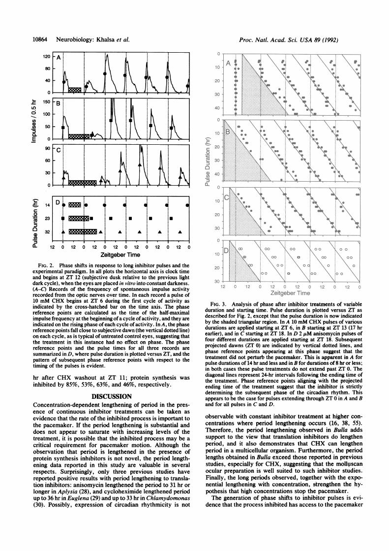
pacemaker motion during the treatment (53, 54). Concentra-tions of CHX and anisomycin above those observed to causethe longest period lengthening were used as the treatment (10mM and 2 AuM, respectively). Fig. 2 summarizes the exper-imental protocol and shows examples of impulse activityrecords from three eyes subjected to different durations ofCHX. In the summary plot in Fig. 2D, the shortest pulse ofCHX (14 hr) fails to perturb the phase of the rhythm from thatexpected of an untreated control rhythm, near subjectivedawn (ZT 0). However, the two longer pulses (23 and 32 hr)shift the phase of the rhythm with a magnitude which is afunction of the end of the inhibitor pulse.Summary plots of data from four complete experiments are
shown in Fig. 3. For CHX pulses terminating before ZT 0 inFig. 3A, the phase of the circadian rhythm appears unper-turbed by the inhibitor treatment, since the phase referencepoints on subsequent cycles are near ZT 0, as expected foruntreated eyes. This is also true in Fig. 3B, although only twopulses end before ZT 0 and show this effect. However, afterlonger-duration pulses that extend through ZT 0 in Fig. 3 A andB a distinctive relationship is established. The phase subse-quent to the treatment is a strict function of the ending time ofthe treatment: the phase ofthe subsequent rhythm is delayedby the number ofhours that the inhibitorpulse extends pastZT0. In Fig. 3C, this relationship is confirmed; all of the phasereference points are determined by the ending time ofthe CHXtreatment, since all pulses extend through ZT 0.Long pulses of 2 1M anisomycin generated data similar to
those for CHX. In Fig. 3D phase reference points afteranisomycin pulses are a function of the ending time of thetreatment, although the location ofthe phase reference pointsappears to be delayed relative to the same points obtainedafter CHX pulses (compare Fig. 3C). Pulses of the inactiveanalogue deacetylanisomycin (50) (2 ,uM) from ZT 18 to 12over the first cycle did not inhibit spontaneous impulseactivity during the treatment, nor did they generate signifi-cant phase shifts relative to untreated controls as measuredon the fourth cycle (mean = -0.1 hr, SD = 1.0 hr, n = 4).
Protein synthesis inhibition was quantified at different timepoints over the course of a 17-hr pulse of 10 mM CHXdelivered from ZT 18 to ZT 11, a treatment yielding a phaseshift of about 12 hr (see Fig. 2C). Over the 1-hr period of ZT20 to ZT 21 (beginning 2 hr after CHX application), proteinsynthesis inhibition was 85%. At the end of the inhibitorpulse, over the 1-hr period of ZT 10 to ZT 11 (16 hr afterCHX), inhibition increased to 93%. Recovery from inhibitionwas measured over the 1-hr periods beginning 2, 4, 6, and 12
Neurobiology: Khalsa et al.
10864 Neurobiology: Khalsa et al. Proc. Natl. Acad. Sci. USA 89 (1992)
120
80
40
-
c)0CE0-E
9
a
3
61
IC,.5
0~
00
10
12 0 12 0 1 0 1 0 1 0
12 0 12 0 12 0 12 0 12 0 12 0 12 0Zeitgeber Time
FIG. 2. Phase shifts in response to long inhibitor pulses and theexperimental paradigm. In all plots the horizontal axis is clock timeand begins at ZT 12 (subjective dusk relative to the previous lightdark cycle), when the eyes are placed in vitro into constant darkness.(A-C) Records of the frequency of spontaneous impulse activityrecorded from the optic nerves over time. In each record a pulse of10 mM CHX begins at ZT 6 during the first cycle of activity asindicated by the cross-hatched bar on the time axis. The phasereference points are calculated as the time of the half-maximalimpulse frequency at the beginning ofa cycle of activity, and they areindicated on the rising phase ofeach cycle of activity. In A, the phasereference points fall close to subjective dawn (the vertical dotted line)on each cycle, as is typical of untreated control eyes, suggesting thatthe treatment in this instance had no effect on phase. The phasereference points and the pulse times for all three records aresummarized in D, where pulse duration is plotted versus ZT, and thepattern of subsequent phase reference points with respect to thetiming of the pulses is evident.
hr after CHX washout at ZT 11; protein synthesis wasinhibited by 85%, 53%, 63%, and 46%, respectively.
DISCUSSIONConcentration-dependent lengthening of period in the pres-ence of continuous inhibitor treatments can be taken asevidence that the rate of the inhibited process is important tothe pacemaker. If the period lengthening is substantial anddoes not appear to saturate with increasing levels of thetreatment, it is possible that the inhibited process may be acritical requirement for pacemaker motion. Although theobservation that period is lengthened in the presence ofprotein synthesis inhibitors is not novel, the period length-ening data reported in this study are valuable in severalrespects. Surprisingly, only three previous studies havereported positive results with period lengthening to transla-tion inhibitors: anisomycin lengthened the period to 31 hr orlonger in Aplysia (28), and cycloheximide lengthened periodup to 36 hr in Euglena (29) and up to 33 hr in Chlamydomonas(30). Possibly, expression of circadian rhythmicity is not
4-0 ......0
....
-t L: :. -*all. a.
l..X \
2 ',.
2O-C
6a)-5-
,00
.,a
\.aI .H
Ir t . . . \ .b r . . : >w
-Xo. iE \ ats . A;. I,. .. \
!,:t::: V:::. ... .:: :\An..-......:......:: :, \
TIC i[:: :' ....1: :-: -. .: ;, "
0 0 0.,e .s soNo ...i'* S..
90
S. S
.'S
0
Cr ,,
T.(. ., !- . 1:, :\;, 'em '.,:~q. , 1: \|Ne Y *j~~~N *... ..... .
Ze'tgeber,IFr.e
FIG. 3. Analysis of phase after inhibitor treatments of variableduration and starting time. Pulse duration is plotted versus ZT asdescribed for Fig. 2, except that the pulse duration is now indicatedby the shaded triangular region. In A 10 mM CHX pulses of variousdurations are applied starting at ZT 6, in B starting at ZT 13 (17 hrearlier), and in C starting at ZT 18. In D 2 1.tM anisomycin pulses offour different durations are applied starting at ZT 18. Subsequentprojected dawns (ZT 0) are indicated by vertical dotted lines, andphase reference points appearing at this phase suggest that thetreatment did not perturb the pacemaker. This is apparent in A forpulse durations of 14 hr and less and in B for durations of 8 hr or less;in both cases these pulse treatments do not extend past ZT 0. Thediagonal lines represent 24-hr intervals following the ending time ofthe treatment. Phase reference points aligning with the projectedending time of the treatment suggest that the inhibitor is strictlydetermining the subsequent phase of the circadian rhythm. Thisappears to be the case for pulses extending through ZT 0 in A and Band for all pulses in C and D.
observable with constant inhibitor treatment at higher con-centrations where period lengthening occurs (16, 38, 55).Therefore, the period lengthening observed in Bulla addssupport to the view that translation inhibitors do lengthenperiod, and it also demonstrates that CHX can lengthenperiod in a multicellular' organism. Furthermore, the periodlengths obtained in Bulla exceed those reported in previousstudies, especially for CHX, suggesting that the molluscanocular preparation is well suited to such inhibitor studies.Finally, the long periods observed, together with the expo-nential lengthening with concentration, strengthen the hy-pothesis that high concentrations stop the pacemaker.The generation of phase shifts to inhibitor pulses is evi-
dence that the process inhibited has access to the pacemaker
KT

Proc. Natl. Acad. Sci. USA 89 (1992) 10865
mechanism, and it may be evidence that the process isinvolved in the pacemaker mechanism. A large body of datahas been generated by using translation inhibitors applied inpulses to generate phase shifts. Phase shifts and/or full phaseresponse curves have been obtained for cycloheximidepulses in Acetabularia (35, 43), Neurospora (36, 40, 43),Gonyaulax (38, 42, 45-47), Phaseolus (39), Aplysia (31, 34),and hamster (33, 41) and for anisomycin pulses in Gonyaulax(37, 38, 42, 45, 47), Aplysia (27, 28, 32, 34), chick pineal (49),and hamster (33, 48). On the basis of these data, it has beensuggested that translation is a requirement for the circadianpacemaker mechanism (24, 27, 28, 30-32, 35, 36, 40, 43, 44,47, 49, 50), or more specifically, that translation is a phase-dependent requirement (24, 28, 32, 35, 36, 40, 43, 44, 49).Furthermore, models describing the circadian pacemakermechanism which include translation have been extensivelydescribed and discussed (14, 23-26, 28, 39, 40, 43).The experimental paradigm in this study, a set oflong pulse
durations, is similar to traditional phase shifting experimentsin that it uses a high inhibitor concentration in a restrictedpulse. However, it extends the traditional phase-shiftingparadigm to effectively demonstrate a nonsaturating "phaseshift" to pulse duration. The subsequent phase of the pace-maker is therefore determined solely by pulse duration andthe pacemaker does not continue its motion until the inhibitorpulse is terminated. Previously, Lotshaw and Jacklet (32),using 3-, 6-, and 9-hr pulses of anisomycin and puromycin inAplysia starting at CT 21, observed an increase in phase shiftwhich was linear with pulse duration and suggested that thepacemaker was being stopped. However, they did not extendpulse duration beyond 9 hr and throughout a circadian cycle,conditions that are necessary to demonstrate that the phaseshift is truly not saturating with pulse duration.The paradigm used in this study can also be thought of as
an extension of the period-lengthening experiment. How-ever, the period during the treatment is calculated retrospec-tively, and the concentration is chosen to yield effectively"infinite period" or a stopping of pacemaker motion. Previ-ously, Feldman (29) used this concept in demonstrating thereversibility of CHX period lengthening. In this study amoderate period-lengthening concentration was applied fordurations from 24 to 72 hr, and the subsequent phase after thepulse was consistent with the expected period lengtheningoccurring during the pulse.The data in this study also address the issue of phase
dependence of protein synthesis. Given that subsequentphase was not perturbed by inhibitor pulses until the pulsespanned subjective dawn (Fig. 3 A and B), and that thesubsequent phase was determined solely by the terminationtime of the pulse for all CHX and anisomycin pulses whichbegan as late as ZT 18 (Fig. 3 C and D), the critical phase mustlie in a region within the late subjective night. The differencein starting times for different pulses in this study (3 hr apart)is too large to permit sufficient resolution for an exactdetermination of the time of the critical phase for proteinsynthesis. However, phase-shifting studies in Acetabularia(35), Neurospora (36), Phaseolus (39), Aplysia (27, 28, 31, 32,34), chick pineal (49), and hamster (33, 41, 48) have alsosuggested that the critical phase is in the late subjective nightsince phase delays appear for pulses of inhibitor beginning inthe mid to late subjective night. In Aplysia, critical proteinsynthesis occurs from CT 20 to CT 8 based on phase shifts toCHX pulses (34) and from CT 18 to CT 2 based on phase shiftsto anisomycin and puromycin pulses (32).The prediction that high concentrations of protein synthe-
sis inhibitor should be capable of stopping the pacemaker ata unique phase derives naturally from the hypothesis sug-gested by numerous investigators, based on phase-shiftingand period-lengthening data, that protein synthesis is a phase-dependent requirement for the circadian pacemaker. This
study demonstrates this phase-dependent requirement andtherefore serves as a confirmation of this widely held con-cept. Without protein synthesis at a critical phase of thecircadian oscillation near subjective dawn, pacemaker mo-tion is stopped. This confirmation is directly attributable tothe application ofthe experimental paradigm used; however,it has been possible only because of the surprising toleranceofthe molluscan eye preparation to the long durations of highinhibitor concentration applied.To address the question as to whether the data with long
CHX pulses may be a result ofsome action ofCHX other thanthe inhibition ofprotein synthesis (33, 39-41, 44, 46), we haveapplied anisomycin, another translation inhibitor. Anisomy-cin was also capable of stopping the pacemaker for pulsesbeginning at ZT 18; however, the resultant phases followingtreatment were more delayed compared with the same pulsetreatments ofCHX (Fig. 3 C and D). The delays observed arepossibly a result of the slow recovery time reported for thewash-out of anisomycin (32, 34). Eskin (34) found in acomparative study that CHX recovery time in Aplysia wasmuch faster than that for anisomycin. Therefore, after longpulses of anisomycin, one would expect that the anisomycinremaining after wash-out would lengthen the period andresult in a delayed subsequent phase of the rhythm ascompared with the same duration pulse of CHX. The as-sumption that protein synthesis inhibition accounts for theobservations is further supported by the result that theinactive analogue of anisomycin, deacetylanisomycin (38,50), was ineffective at perturbing the phase of the rhythm.Finally, labeled methionine incorporation experiments in thisstudy, which are a measure of protein synthesis inhibition,have indicated that CHX is effective at inhibiting proteinsynthesis over the time course of the long pulses used in thisstudy. No appreciable degradation ofinhibition was apparentover 17 hr of inhibitor application. Incorporation studies inAplysia eye, using CHX and/or anisomycin, are also con-sistent with the effective inhibition of protein synthesis withthese compounds (31, 32, 34, 50). The recovery of inhibitionafter 17 hr of CHX was slow, as compared with recoveryreported in Aplysia eye after short pulses ofCHX (34), sinceprotein synthesis inhibition was still inhibited by 46% as longas 12 hr after the wash-out of the inhibitor. This observationmay account for the low amplitude and distorted waveformoften observed on the first cycle of compound action poten-tial activity following long pulses (Fig. 2 A-C).
Application of constant light has also been reported as atreatment which appears to stop pacemaker motion (53, 54).However, recent studies have raised the alternative interpre-tation that the underlying pacemaker actually continues itsmotion in constant light and that the transition from constantlight to dark instantaneously "resets" the pacemaker to aspecific phase (56-59). This interpretation is plausible forDrosophila, where the free-running period can be lengthenedmodestly (although exponentially with intensity), to about 26hr, in constant light. Resetting is clearly the only interpreta-tion for the effects of dim light pulses. The Drosophilapacemaker continues its motion during constant dim lightwith a circadian period of less than 25 hr, and yet a pulse oflight at this intensity is capable of fully resetting the pace-maker (57). The resetting interpretation is not compelling forthe Bulla eye, however, where long noncircadian periodici-ties are observed in the presence of lower concentrations ofprotein synthesis inhibitors. Applying the resetting interpre-tation in this case would suggest the implausible result thatcircadian periodicities are somehow present at high proteinsynthesis inhibitor concentrations, even though lower con-centrations yield substantial period lengthening.Although the action of CHX and anisomycin may be by
their inhibition of protein synthesis, "whether the effects ofinhibitors of protein synthesis on the circadian oscillator are
Neurobiology: Khalsa et al.

Proc. Natl. Acad. Sci. USA 89 (1992)
direct or indirect is not clear" (49). For example, proteinsynthesis inhibitors may act via an "input pathway" to thepacemaker (33, 34), or they may affect one or more processesin the cell which are more directly involved with the pace-maker mechanism (29, 30, 39). Another limitation of allstudies based solely on application of protein synthesisinhibitors is that it is not possible to distinguish betweenprotein synthesis as a central pacemaker variable and as anonrhythmic supportive process (7, 8, 35, 40, 43). As aptlystated by Dunlap and Feldman (40):
There are two general hypotheses. In one. . ., the periodicsynthesis of one or more proteins is a required step in thecycle. Here, the process of synthesizing a protein or proteinsis an indispensable part of the clock, and drug-induced reset-ting is due to interference with this process.... the kineticsof the clock reflect in some way the short-term rate oftranslation. A second type ofmodel . . . suggests that proteinsare needed to carry out an enzymatic reaction or transportprocess required for the clock, but synthesis per se of theseproteins is not a step in the oscillation. Here, the short-termrate of protein synthesis is unimportant so long as a sufficientamount of the critical enzymatic activity has been made by thetime it is needed. The critical proteins here are viewed ashaving a high turnover rate and, therefore, must be resynthe-sized each day during (but not necessarily only during) thetime corresponding to the sensitive phase of the cycle.
Studies examining phase shifts to translation inhibitors incombination with temperature changes (42, 43, 45, 47), or toinhibitor-insensitive mutants (40), have provided evidencefavoring the argument that translation is not an integral partof the clock mechanism but simply a requirement.The experimental paradigm employed in this study, al-
though not a novel one, is particularly suited for pacemakeranalysis. In circumstances where the effects of a treatmentabolish or obscure the overt circadian rhythm, the long-pulseparadigm can potentially serve as a useful technique forassessing the behavior ofthe underlying circadian pacemakerduring the treatment. Previously, Pittendrigh (60) has dem-onstrated that the Drosophila circadian pacemaker appearsto stop in a phase-dependent manner during exposure tooxygen-free nitrogen atmosphere. In Bulla, the circadianpacemaker appears to stop in a phase-dependent manner atlow pH (61) and in the presence oftranscription inhibitor (19).The results of period-changing and phase-shifting experi-ments can lead to the suggestion that a process plays animportant role in the pacemaker system. The experimentalparadigm used in the present study is a valuable adjunct tothese techniques in providing direct evidence that a processnot only influences free-running period or pacemaker phasebut also is required for the pacemaker to complete a fullcycle. Without the contributing process, the pacemakerstops, possibly in a phase-dependent manner, and motionresumes only when the process is restored.
We thank Dr. Barbara Bogart for extensive efforts on, and Dr.Arnold Eskin for assistance with, the trichloroacetic acid precipita-tion procedure. This work was supported by National Institutes ofHealth Grant NS15264 to G.D.B.
1. Dunlap, J. C. (1990) Trends Genet. 6, 159-165.2. Rosbash, M. & Hall, J. C. (1989) Neuron 3, 387-398.3. Hall, J. C. & Rosbash, M. (1988) Annu. Rev. Neurosci. 11, 373-393.4. Strumwasser, F. (1988) J. Physiol. (Paris) 83, 246-254.5. Hartwig, R., Schweiger, M., Schweiger, R. & Schweiger, H. G. (1985)
Proc. Natl. Acad. Sci. USA 82, 6899-6902.6. Siwicki, K. K., Eastman, C., Petersen, G., Rosbash, M. & Hall, J. C.
(1988) Neuron 1, 141-150.7. Rensing, L. & Hardeland, R. (1990) Chronobiol. Int. 7, 353-370.8. Edmunds, L. N. (1988) Cellular and Molecular Bases of Biological
Clocks (Springer, New York).
9. Rusak, B., Robertson, H. A., Wisden, W. & Hunt, S. P. (1990) Science248, 1237-1240.
10. Raju, U., Yeung, S. J. & Eskin, A. (1990) Am. J. Physiol. 258, R256-R262.
11. Yeung, S. J. & Eskin, A. (1987) Proc. Natd. Acad. Sci. USA 84,279-283.12. Aronin, N., Sagar, S. M., Sharp, F. R. & Schwartz, W. J. (1990) Proc.
Natl. Acad. Sci. USA 87, 5959-5962.13. Kornhauser, J. M., Nelson, D. E., Mayo, K. E. & Takahashi, J. S.
(1990) Neuron 5, 127-134.14. Cornelius, G. & Rensing, L. (1982) Biosystems 15, 35-47.15. Rothman, B. S. & Strumwasser, F. (1977) Fed. Proc. 36, 2050-2055.16. Karakasian, M. W. & Hastings, J. W. (1962) Proc. Natl. Acad. Sci. USA
48, 2130-2137.17. MacDowall, F. D. H. (1964) Can. J. Bot. 42, 115-122.18. Raju, U., Koumenis, C., Nunez-Regueiro, M. & Eskin, A. (1991) Science
253, 673-675.19. Khalsa, S. B. S. & Block, G. D. (1990) Soc. Neurosci. Abstr. 16, 640.20. Ohi, K. & Takahashi, J. S. (1991) Soc. Neurosci. Abstr. 17, 675.21. Ehret, C. F. & Trucco, E. (1967) J. Theor. Biol. 15, 240-262.22. Hardin, P. E., Hall, J. C. & Rosbash, M. (1990) Nature (London) 343,
536-540.23. Jacklet, J. W. (1989) in Neuronal and Cellular Oscillators, ed. Jacklet,
J. W. (Dekker, New York), pp. 483-527.24. Takahashi, J. S. (1990) Curr. Opin. Neurobiol. 1, 556-561.25. Jacklet, J. W. (1981) Biol. Bull. 160, 199-227.26. Schweiger, H. G., Hartwig, R. & Schweiger, M. (1986) J. Cell Sci. Suppl.
4, 181-200.27. Jacklet, J. W. (1977) Science 198, 69-71.28. Jacklet, J. W. (1980) J. Exp. Biol. 84, 1-15.29. Feldman, J. F. (1967) Proc. Natd. Acad. Sci. USA 57, 1080-1087.30. Goodenough, J. E., Bruce, V. G. & Carter, A. (1981) Biol. Bull. 161,
371-381.31. Rothman, B. S. & Strumwasser, F. (1976) J. Gen. Physiol. 68, 359-384.32. Lotshaw, D. P. & Jacklet, J. W. (1986) Am. J. Physiol. 250, R5-R17.33. Takahashi, J. S. & Turek, F. W. (1987) Brain Res. 405, 199-203.34. Yeung, S. J. & Eskin, A. (1988) J. Biol. Rhythms 3, 225-236.35. Karakashian, M. W. & Schweiger, H. G. (1976) Exp. Cell Res. 98,
303-312.36. Nakashima, H., Perlman, J. & Feldman, J. F. (1981) Am. J. Physiol. 241,
R31-R35.37. Taylor, W. R., Krasnow, R., Dunlap, J. C., Broda, H. & Hastings, J. W.
(1982) J. Comp. Physiol. 148, 11-25.38. Taylor, W. R., Dunlap, J. C. & Hastings, J. W. (1982) J. Exp. Biol. 97,
121-136.39. Mayer, W. E. & Knoll, U. (1981) Z. Pflanzenphysiol. 103, 413-425.40. Dunlap, J. C. & Feldman, J. F. (1988) Proc. Natl. Acad. Sci. USA 85,
1096-1100.41. Wollnik, F., Turek, F. W., Majewski, P. & Takahashi, J. S. (1989) Brain
Res. 49, 82-88.42. Broda, H., Johnson, C. H., Taylor, W. R. & Hastings, J. W. (1989) J.
Biol. Rhythms 4, 327-333.43. Karakasian, M. W. & Schweiger, H. G. (1976) Proc. Natl. Acad. Sci.
USA 73, 3216-3219.44. Nakashima, H., Perlman, J. & Feldman, J. F. (1980) Science 212,
361-362.45. Thorey, I., Rode, I., Harnau, G. & Hardeland, R. (1987) J. Comp.
Physiol. B 157, 85-89.46. Dunlap, J. C., Taylor, W. & Hastings, J. W. (1980) J. Comp. Physiol.
138, 1-8.47. Olesiak, W., Ungar, A., Johnson, C. H. & Hastings, J. W. (1987)J. Biol.
Rhythms 2, 121-138.48. Inouye, S. T., Takahashi, J. S., Wollnik, F. & Turek, F. W. (1988) Am.
J. Physiol. 255, R1OSS-R1058.49. Takahashi, J. S., Murakami, N., Nikaido, S. S., Pratt, B. L. & Robert-
son, L. M. (1989) Recent Prog. Horm. Res. 45, 279-352.50. Jacklet, J. W. (1980) J. Exp. Biol. 85, 33-42.51. Block, G. D., Khalsa, S. B. S., McMahon, D. G., Michel, S. & Geusz,
M. Int. Rev. Cytol., in press.52. Vazquez, D. (1979) Mol. Biol. Biochem. Biophys. 30, 1-312.53. Pittendrigh, C. S. (1960) Cold Spring Harbor Symp. Quant. Biol. 25,
159-184.54. Pittendrigh, C. S. (1966) Z. Pflanzenphysiol. S4, 275-307.55. Mergenhagen, D. & Schweiger, H. G. (1975) Exp. Cell. Res. 94, 321-326.56. Pittendrigh, C. S. (1976) in The Molecular Basis of Circadian Rhythms,
eds. Hastings, J. W. & Schweiger, H. (Abakon, Berlin), pp. 11-48.57. Pittendrigh, C. S. (1981) in Handbook of Behavioral Biology, ed.
Aschoff, J. (Plenum, New York), Vol. 4, pp. 95-124.58. Saunders, D. S. (1976) J. Comp. Physiol. 110, 111-133.59. Prichard, R. G. & Lickey, M. E. (1981) J. Neurosci. 1, 835-839.60. Pittendrigh, C. S. (1974) in The Neurosciences 3rd Study Program, eds.
Schmitt, F. 0. & Worden, F. G. (MIT Press, Boston), pp. 437-458.61. Khalsa, S. B. S., Ralph, M. R. & Block, G. D. (1991) J. Neurosci. 11,
2672-2679.
10866 Neurobiology: Khalsa et al.