strategies for optimising musculoskeletal health in the ...epubs.surrey.ac.uk/851790/1/strategies...
TRANSCRIPT
REVIEW Open Access
Strategies for optimising musculoskeletalhealth in the 21st centuryRebecca Lewis1†, Constanza B. Gómez Álvarez1†, Margaret Rayman2, Susan Lanham-New2, Anthony Woolf7
and Ali Mobasheri1,3,4,5,6*
Abstract
We live in a world with an ever-increasing ageing population. Studying healthy ageing and reducing thesocioeconomic impact of age-related diseases is a key research priority for the industrialised and developingcountries, along with a better mechanistic understanding of the physiology and pathophysiology of ageing thatoccurs in a number of age-related musculoskeletal disorders. Arthritis and musculoskeletal disorders constitute amajor cause of disability and morbidity globally and result in enormous costs for our health and social-care systems.By gaining a better understanding of healthy musculoskeletal ageing and the risk factors associated with prematureageing and senescence, we can provide better care and develop new and better-targeted therapies for commonmusculoskeletal disorders. This review is the outcome of a two-day multidisciplinary, international workshopsponsored by the Institute of Advanced Studies entitled “Musculoskeletal Health in the 21st Century” and held atthe University of Surrey from 30th June-1st July 2015.The aim of this narrative review is to summarise current knowledge of musculoskeletal health, ageing and diseaseand highlight strategies for prevention and reducing the impact of common musculoskeletal diseases.
Keywords: Ageing, Musculoskeletal health, Musculoskeletal disorders, Global burden, Joint diseases, Osteoarthritis(OA), Rheumatoid arthritis (RA), Low back pain (LBP), Osteoporosis (OP), Sarcopenia, Obesity, Type II diabetes,Metabolic disease
BackgroundThe current rise in life-expectancy, the subsequent in-crease in numbers of older people and increasing pen-sion costs have prompted the UK government to changepolicy, extend working years and delay pensionable age.By 2034 the number of people in the UK aged 85 andover is projected to be 2.5-times larger than in 2009,reaching 3.5 million and accounting for 5 % of the popu-lation. The number of people aged 65+ is expected torise to over 16 million in the UK by the same time point(according to Age UK and the Office for National Statis-tics [1]). However, working for an extended period of
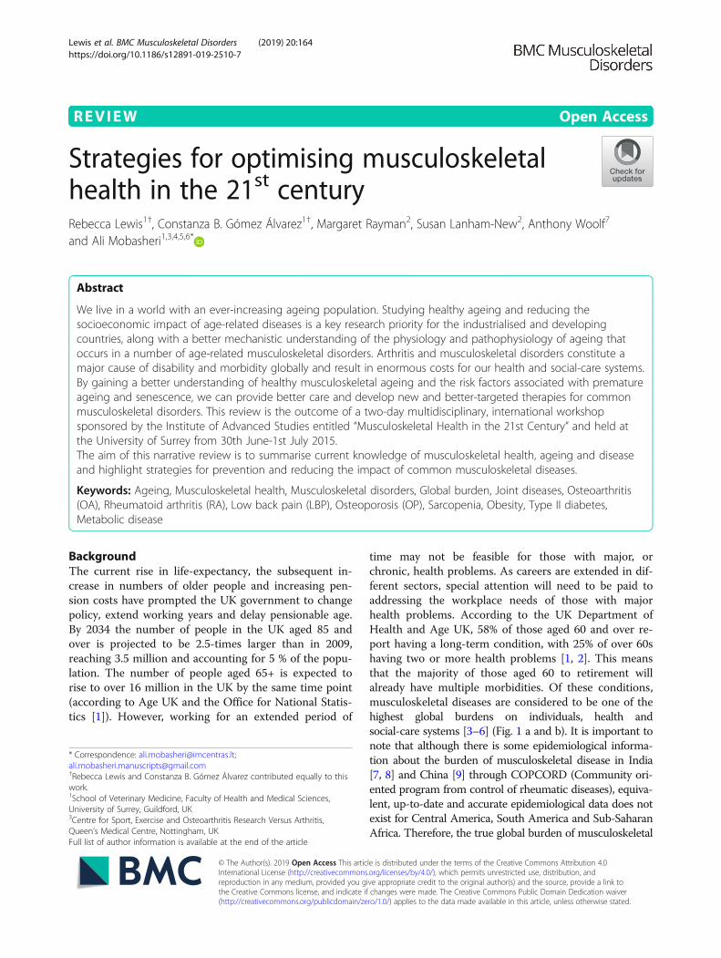
time may not be feasible for those with major, orchronic, health problems. As careers are extended in dif-ferent sectors, special attention will need to be paid toaddressing the workplace needs of those with majorhealth problems. According to the UK Department ofHealth and Age UK, 58% of those aged 60 and over re-port having a long-term condition, with 25% of over 60shaving two or more health problems [1, 2]. This meansthat the majority of those aged 60 to retirement willalready have multiple morbidities. Of these conditions,musculoskeletal diseases are considered to be one of thehighest global burdens on individuals, health andsocial-care systems [3–6] (Fig. 1 a and b). It is important tonote that although there is some epidemiological informa-tion about the burden of musculoskeletal disease in India[7, 8] and China [9] through COPCORD (Community ori-ented program from control of rheumatic diseases), equiva-lent, up-to-date and accurate epidemiological data does notexist for Central America, South America and Sub-SaharanAfrica. Therefore, the true global burden of musculoskeletal
© The Author(s). 2019 Open Access This article is distributed under the terms of the Creative Commons Attribution 4.0International License (http://creativecommons.org/licenses/by/4.0/), which permits unrestricted use, distribution, andreproduction in any medium, provided you give appropriate credit to the original author(s) and the source, provide a link tothe Creative Commons license, and indicate if changes were made. The Creative Commons Public Domain Dedication waiver(http://creativecommons.org/publicdomain/zero/1.0/) applies to the data made available in this article, unless otherwise stated.
* Correspondence: [email protected];[email protected]†Rebecca Lewis and Constanza B. Gómez Álvarez contributed equally to thiswork.1School of Veterinary Medicine, Faculty of Health and Medical Sciences,University of Surrey, Guildford, UK3Centre for Sport, Exercise and Osteoarthritis Research Versus Arthritis,Queen’s Medical Centre, Nottingham, UKFull list of author information is available at the end of the article
Lewis et al. BMC Musculoskeletal Disorders (2019) 20:164 https://doi.org/10.1186/s12891-019-2510-7
disease is likely to be grossly underestimated. Com-mon musculoskeletal conditions include osteoarthritis(OA), rheumatoid arthritis (RA), psoriatic arthritis(PsA), gout, lower back pain (LBP) and osteoporosis(OP). Between the fifth and ninth decade of life, OAand LBP are major contributors to musculoskeletalimpairment (Fig. 1 c). Arthritis is of particular con-cern for the UK population as some 10 millionpeople are now estimated to be suffering from the
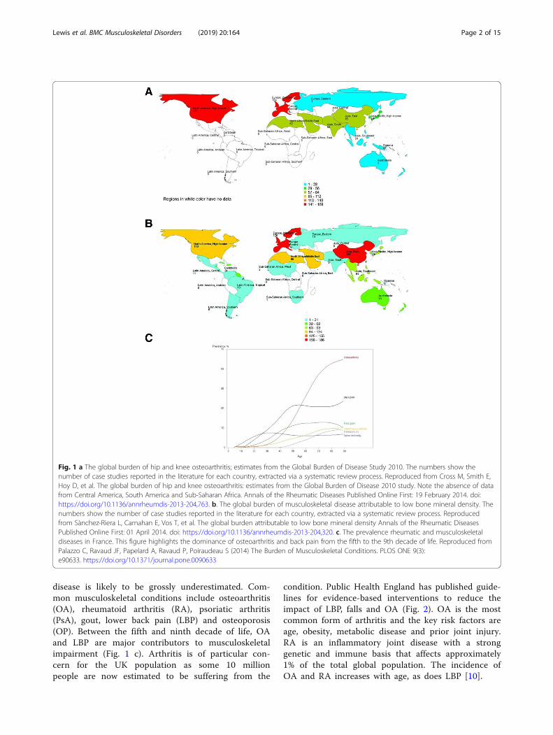
condition. Public Health England has published guide-lines for evidence-based interventions to reduce theimpact of LBP, falls and OA (Fig. 2). OA is the mostcommon form of arthritis and the key risk factors areage, obesity, metabolic disease and prior joint injury.RA is an inflammatory joint disease with a stronggenetic and immune basis that affects approximately1% of the total global population. The incidence ofOA and RA increases with age, as does LBP [10].
Fig. 1 a The global burden of hip and knee osteoarthritis; estimates from the Global Burden of Disease Study 2010. The numbers show thenumber of case studies reported in the literature for each country, extracted via a systematic review process. Reproduced from Cross M, Smith E,Hoy D, et al. The global burden of hip and knee osteoarthritis: estimates from the Global Burden of Disease 2010 study. Note the absence of datafrom Central America, South America and Sub-Saharan Africa. Annals of the Rheumatic Diseases Published Online First: 19 February 2014. doi:https://doi.org/10.1136/annrheumdis-2013-204,763. b. The global burden of musculoskeletal disease attributable to low bone mineral density. Thenumbers show the number of case studies reported in the literature for each country, extracted via a systematic review process. Reproducedfrom Sànchez-Riera L, Carnahan E, Vos T, et al. The global burden attributable to low bone mineral density Annals of the Rheumatic DiseasesPublished Online First: 01 April 2014. doi: https://doi.org/10.1136/annrheumdis-2013-204,320. c. The prevalence rheumatic and musculoskeletaldiseases in France. This figure highlights the dominance of osteoarthritis and back pain from the fifth to the 9th decade of life. Reproduced fromPalazzo C, Ravaud JF, Papelard A, Ravaud P, Poiraudeau S (2014) The Burden of Musculoskeletal Conditions. PLOS ONE 9(3):e90633. https://doi.org/10.1371/journal.pone.0090633
Lewis et al. BMC Musculoskeletal Disorders (2019) 20:164 Page 2 of 15
This narrative review is the outcome of a multidiscip-linary, international workshop sponsored by the Instituteof Advanced Studies (IAS) entitled: “MusculoskeletalHealth in the 21st Century” and held at the University ofSurrey, UK, from 30th June-1st July 2015. The abstractsfrom this workshop were published in a special supple-ment of BMC Musculoskeletal Disorders in 2015. [11].Diverse topics were discussed and debated, ranging fromthe global burden of OA to the link between diabetesand joint disease, diet and nutrition in arthritis, OneHealth [12], ageing, advances in imaging and musculo-skeletal health and disease in military personnel andcompanion animals. One of the key deliverables of theinternational workshop was a paper that summarisedthe topics that were discussed. In this paper, we sum-marise and disseminate some of the key outcomes fromthe workshop in the context of currently available infor-mation, discuss the current challenges faced by societyin relation to the rising burden of musculoskeletal disor-ders and review existing strategies and recommendationsfor prevention and mitigation of the impact of thesediseases.The workshop reached an important consensus state-
ment, namely that development of more precise diag-nostic and prognostic tools and targeted treatments isnecessary, along with better disseminated preventativemeasures, which is in complete alignment with thoseoutlined by the Arthritis Research UK (now rebranded asVersus Arthritis) approach plan (2014). In order toachieve this, greater transfer and translation of know-ledge between the veterinary and human fields isneeded as well as more funding for musculoskeletalresearch and investigation of populations with rare
diseases and those with higher incidences of joint in-juries, such as youth sport and the military.
Current societal challengesPhysical inactivity and sedentary behaviourSedentary behaviour and the rapid growth of legions of videogame, social media and movie-streaming addicted couch po-tatoes is a consequence of sustained and systemic urbanisa-tion in developed countries, including the United States,where nearly 50% of the population do not undertakeeven the bare minimum levels of aerobic activity rec-ommended by Physical Activity Guidelines [13]. Thisrise in sedentary behaviour is seen as a major risk factorfor a number of chronic diseases recognised by the Na-tional Health Service (NHS), costing around £1billion ayear in the UK, and is recognised as a substantial globaleconomic burden [14]. Statistics recently provided bythe Centre for Economics and Business Research showthe cost of “doing nothing”: half a million Europeansdie every year as a result of being physically inactiveand this costs the economy over €80bn annually [15].This global challenge requires urgent and feasible solu-tions. Increasing physical activity and optimising exer-cise (as recommended by Arthritis Research UK/VersusArthritis and World Health Organisation (WHO)) isseen as an optimal way to improve musculoskeletalhealth. Only 36% of the adult population in the UK takepart in moderate intensity physical activity of about 30min at least once a week [16]. An increasing body ofevidence is showing that even the effects of a sedentarylifestyle (for example, of those with a desk job) can bemitigated by a small amount of activity every day. A re-cent meta-analysis of the data from studies involving
Fig. 2 Evidence-based interventions for the musculoskeletal conditions that cause the most DALYs (Disability-adjusted life years) in England,including low back and neck pain, falls andosteoarthritis. https://publichealthmatters.blog.gov.uk/2016/01/11/preventing-musculoskeletal-disorders-has-wider-impacts-for-public-health/
Lewis et al. BMC Musculoskeletal Disorders (2019) 20:164 Page 3 of 15
over 1 million individuals concluded that an hour ofmoderate level activity per day eliminated the in-creased risk of death associated with 8 h of sitting[17]. Interestingly, the study also found that theselevels of activity did not have an effect on the in-creased risk of death associated with high levels ofleisure-time sedentary behaviour such as watchingtelevision. Whilst this level of activity is far morethan those recommended by WHO, it is clear thatkeeping a moderate level of physical activity is a keyrequirement for healthy ageing and maintaining mus-culoskeletal health [17]. WHO recommendations sug-gest that healthy individuals should take around twohours a week of moderate physical activity or ap-proximately 20 min a day of doing any kind of phys-ical activity like brisk walking. This level of exercise,which involves elevation of heart-rate, has been asso-ciated with lower lifetime risk of cardiovascular dis-ease in a 25 year longitudinal study of approximately13,000 adults [18]. In addition to benefits for muscu-loskeletal health, improvements in cardiorespiratoryfitness can be achieved by changing sedentary behav-iour to achieve a low-intensity physical activity suchas walking [19]. Furthermore, in patients with kneeOA, improvement of locomotor function, includingbalance and strength, and a reduction in pain wasseen following supplementation of home exercise withan eight-week class-based programme [20]. Patientssuffering with chronic LBP who were given an exer-cise programme combining muscle strength, flexibilityand aerobic fitness also reported a reduction in stiff-ness, which can result in back pain [21]. The idea ofexercise for rehabilitation of musculoskeletal injurieshas been widely accepted for many years now, andthe idea of prescribing exercise as a preventativehealth measure is also more widely investigated, withguidelines around the type, frequency and duration ofactivity being considered [22].Sedentary behaviour is also contributing to the rise
in obesity and type-2 diabetes. Obesity is a majorcontributor to the development and progression ofOA [23] and numerous epidemiological studies haveconfirmed the link between adiposity and joint degen-eration. The prevalence of diabetes mellitus is be-tween 7 and 14% globally [24]. Diabetes is animportant predictor for severe forms of arthritis [25]and has recently been shown to be an independentrisk factor for the progression of OA in men [26].This shows that measures to increase levels of phys-ical activity will not only increase musculoskeletal(MSK) health but also decrease the risk of sufferingfrom obesity-related diseases such as diabetes. Thiswill reduce the numbers in the population susceptibleto MSK disorders and ill health.
Healthy ageing and physical exerciseWhilst ageing is inevitable, the benefits of exercise onthe ageing body are numerous and, in some circum-stances, can reduce the manifestations of ageing, particu-larly the "ageing phenotype" of the elderly. A recentsystematic review looked at evidence supporting nutritionand physical activity in the prevention and treatment ofsarcopenia [27]. Sarcopenia results in loss of musclestrength and mass and this can lead to weakening of mus-culoskeletal structures and impair tendon, ligament, boneand cartilage function, which will destabilise the joint andincrease the risk of arthritis and other musculoskeletal dis-orders. The authors identified a total of 37 randomisedcontrol trials to explore the effect of combined exerciseand nutritional intervention for overcoming muscle sarco-penia. They concluded that physical exercise has a positiveimpact on muscle mass and function in healthy subjectsaged 60 and above, however, there were huge variations inoutcomes connected with dietary supplementation,highlighting the difficulties associated with cohort studiesof well-nourished human beings, where the interactive ef-fects of dietary supplementation may be masked or com-pletely limited [27]. Physical exercise improves muscleperformance by increasing the ratio of type I to type IImuscle fibres and increasing the cross-sectional area oftype II muscle fibres [28].A European Society for Clinical and Economic Aspects
of Osteoporosis, Osteoarthritis and Musculoskeletal Dis-eases (ESCEO) taskforce looked at dietary protein andvitamin D and calcium supplementation and recom-mended higher protein intake in combination with phys-ical exercise particularly in post-menopausal women atrisk of developing menopause-associated musculoskeletaldisease, such as OP [29]. Physical exercise programmesimprove strength and balance in ageing women with OP[30]. Fragility fracture risk, associated with OP, can be de-creased by following an exercise programme, as exerciseincreases bone density and reduces inflammatory markers[31]. However, the incidence of OP is usually highest inelderly females who are most unlikely to perform the dy-namic exercises needed for bone modelling/remodelling[32]. This highlights the challenge of preventing OP andOP-related fractures in elderly females that cannot per-form ballistic exercise. Some medications are availablefor these frail patients but having an active lifestylefrom an early age and following recommendations forexercise could be more beneficial. The same principleapplies to frailty in ageing companion animals, wherelife-long spontaneous exercise significantly slowsdown the progression of frailty [33].Physical activity is also known to increase insulin me-
diated glucose uptake and, in individuals without type-2diabetes, exercise positively impacts on systemic glucosehomeostasis. However, in patients with type-2 diabetes
Lewis et al. BMC Musculoskeletal Disorders (2019) 20:164 Page 4 of 15
where beta-cell impairment is significant, physical train-ing does not decrease insulin secretion [34]. A recentstudy found that a 30 min daily increment in moderateto vigorous intensity physical activity also significantlyreduced glycated haemoglobin, a measure of type-2 dia-betes risk [19]. Physical exercise reduces the risk of car-diovascular and metabolic comorbidities associated withjoint diseases.In addition to the positive effects of exercise on phys-
ical and mental well-being, there is also ample evidenceto suggest that exercise and mechanical loading have apositive impact at the molecular, cellular and tissuelevels. For example, in tendons, where ageing decreasesthe potential for cell proliferation and number of stem/progenitor-like cells, it has been shown that exercise/loading can induce an increase in tendon collagen syn-thesis [35], increasing tendon strength. In an animalmodel of ageing, it was found that moderate exercisecould also enhance the quality of tissue produced duringhealing of injured tendons [36]. There is plenty of pub-lished evidence to support a positive role for physical ex-ercise and mechanical loading for cartilage [37] andbone health [38].
ObesityThe rise in sedentary behaviour and unhealthy diets con-tributes to the global obesity epidemic and the sharp risein the incidence of type-2 diabetes. When combinedthese are powerful risk factors for cardiovascular andneurodegenerative diseases, which further complicatesthe management of musculoskeletal diseases. As withphysical inactivity, the NHS now recognises obesity asan independent major risk factor for populationill-health, costing the UK over £5billion. Obesity isthought to be a key co-morbidity of many musculoskel-etal conditions and is closely related to the developmentof OA, one of the commonest musculoskeletal health is-sues. Reducing levels of obesity in the adult populationmay lead to reduced occurrence of OA and can alleviatesome of the pain of the condition. Obesity also happensto be one of the most modifiable risk factors for OA[39]. Exercise and weight loss has been successfullytrialled in overweight and obese adults with knee OA[40]. There are ongoing trials assessing the effects ofcombined physical activity and weight loss and it will beinteresting to see how combining exercise and weightloss may act synergistically to improve musculoskeletalfunction. It is now clear that combining modest weightloss with moderate exercise can provide the best overallimprovement in symptoms of pain and joint function.Furthermore, improved diet, moderate exercise andweight loss are probably the best short-term solutionsfor the ineffective surgical interventions for OA patients.
Childhood obesity and physical inactivityReducing obesity in children may also reduce the risk ofdeveloping musculoskeletal pain later in life [41], al-though the full impact of obesity on development of thechild’s musculoskeletal system is still poorly understood[42]. A major contributor to the obesity epidemic is thelack of physical exercise in the population, with manypeople leading an increasingly sedentary lifestyle. Simi-larly, unhealthy diets are a common problem, due to theready availability of a bewildering variety and quantity offast foods, ready meals and the relentless advertising ofthese products. The low nutritional value and high cal-orie content of these foods further contributes to theobesity epidemic. They may also play a role in the highprevalence of type-2 diabetes and cardiovascular diseaseswhich are exacerbated by a lack of fitness and byinactivity.
The companion animal linkThe health issues that impact on society are not just lim-ited to humans. We co-exist with a variety of companionanimals and share the same diet and environment. In astudy of approximately 700 dogs, 40% were overweightand 20% obese [43]. In addition, there is a strong correl-ation between the canine obesity and the BMI of theirowners, indicating that the lifestyle and diet of theowners is having a direct impact on their pets as theyshare the same environment, and probably with similarimpacts on their families [44]. This highlights an oppor-tunity for vets and medics to collaborate to tackle obes-ity, diabetes and cardiovascular co-morbidities thatimpact on humans and their companion animals.
Dietary factorsIt is generally accepted that maintaining a healthy weightcan help to improve musculoskeletal health and preventdegenerative diseases, but research also focuses onwhether dietary factors can influence disease progres-sion. Eating a varied diet high in fresh fruit and vegeta-bles is recommended by many health organisations. Acombined regime of exercise and increased intake offruit and vegetables increased life expectancy in women;those in their 70s with the highest level of activity andvegetable consumption were eight-times more likely tosurvive a five-year follow up period [45]. However, fewdirect links between fruit and vegetable intake and im-proved musculoskeletal health have been shown but onethree year follow-up study of nearly 400 adults showedthat diets high in potassium (from fruit and vegetable in-take) reduced the amount of muscle loss in adults > 65years [46]. Dietary flavonoid intake, the compoundfound in many fruit and vegetables, was positively corre-lated with good bone health (measured by bone mineral
Lewis et al. BMC Musculoskeletal Disorders (2019) 20:164 Page 5 of 15
density and bone resorption) in a population ofperi-menopausal women [47].As current treatment options in OA are very limited,
OA patients may benefit from the ability to self-managetheir condition through improving their diet [48]. Vita-min D, calcium and protein (particularly protein) opti-mise muscle, bone and functional outcomes in olderpeople reducing falls and fractures [49]. Calcium andprotein intake work together to optimise bone health[50]. Previously, it was thought that, in older patients,diets too high in protein should be avoided due to thedetrimental effect on the kidneys. However, increasingevidence now shows that protein levels should not bedecreased, as the effects of metabolic acidosis on thekidneys can be offset by increased fruit and vegetable in-take (as these foods decrease renal acid load) [51, 52].Several studies recommend the benefits of supple-
menting the diet with “nutraceuticals”. A recent system-atic review found promising but nevertheless limitedresearch evidence to support the oral use of severalherbal supplements including Boswellia serrata extractand pycnogenol, curcumin and methylsulfonylmethanein people with OA despite the poor quality of the pub-lished studies [53]. Dietary strategies for improving mus-culoskeletal health can include consumption oflong-chain fatty acids and vitamins D and K [54, 55] aswell as decreasing blood cholesterol [56]. OA patientsshould ensure that they meet the recommended intakesfor micronutrients such as vitamin K, which has a rolein bone and cartilage mineralization. However, the cur-rently available evidence for a role of vitamin D supple-mentation in OA is unconvincing [48]. Research hasfocussed on a number of dietary supplements to modu-late progression of the disease or ease joint stiffness (andtherefore pain). A diet rich in antioxidants may providea useful therapeutic tool for athletes, improving tissuerepair, although the optimum dosage is unknown [57].Combining exercise with a dietary supplement of wheyprotein fortified with vitamin D is effective at increasingmuscle mass and strength in elderly people affected withsarcopenia [58].Both human and veterinary research has shown prom-
ise for a number of other natural products or com-pounds derived from natural sources for alleviation ofarthritic symptoms. Green-lipped mussel extract hasbeen shown to be an effective chondroprotective agent[59], reducing OA symptoms in dogs with OA [60].More recently, fish oil and krill oil have been found tohave even greater protective effects against proteoglycanand collagen degradation in an in vitro model of caninearthritis [61]. These substances are all known to be richin long-chain omega-3 polyunsaturated fatty acids. Aseparate study found that supplementing the diet of dogswith eicosapentaenoic acid and docosahexaenoic acid
(both long-chain omega-3 polyunsaturated fatty acids)significantly reduced the clinical signs of OA [62]. Inhumans, these substances have also been demonstratedto provide an alternative to anti-inflammatory pain med-ications for the relief of chronic neck and back pain aswell as joint pain in rheumatoid arthritis [63, 64]. Whilstthe molecular mechanism of action of these fatty acids isnot entirely understood, it has been shown that thesecompounds reduce the expression of cartilage-degradingproteases and inflammatory cytokines [65].Curcumin is a well-known plant-derived compound
with anti-oxidant and anti-inflammatory properties. Itseffects have been described in numerous chronic ill-nesses in humans, including diabetes, allergies and arth-ritis. It modulates growth factors, transcription factorsand inflammatory cytokines [66]. Interestingly, a recentsystematic review, has investigated the efficacy and safetyof dietary supplements for OA human patients andfound that lesser known supplements such as curcuminfrom Curcuma longa and Boswellic acid from Boswelliaserrata were more effective than well-known nutraceuti-cals, such as glucosamine and chondroitin [67]. Glucosa-mine and chondroitin are popular supplements thathave been suggested to promote healthy joint function.However, there is little evidence of their benefit [67].Glucosamine was recommended in early guidelines is-sued by the European League Against Rheumatism(EULAR) and the Osteoarthritis Research Society Inter-national (OARSI) for the management of knee OA [68,69] but it was not recommended in the National Insti-tute for Health and Care Excellence (NICE) guidelinesand in the most recent set of OARSI recommendationsit was identified as being “uncertain” [70]. More recentguidelines published by EULAR, OARSI and ACR donot recommend glucosamine.In vitro evidence for glucosamine is generally good. At
relatively high concentrations, glucosamine has beenshown to have a protective effect on cartilage and syn-ovial fluid; however many clinical trials have shown thatthis substance is unable to reach the tissue that it ismeant to affect in appropriate and sufficient doses [68].Structural heterogeneity of chondroitin sulphate, anothersupplement commonly taken alongside glucosamine,means that it is difficult to see consistent effects of thissupplement, and the supplements available on the mar-ket are unlikely to be pure due to contamination withother glycosaminoglycans during the manufacturingprocess [71]. There is some evidence that glucosamineand chondroitin sulphate together provide superior ef-fects on the inhibition of nitric oxide and prostaglandinsynthesis and on protection of cartilage structure, thanwhen applied alone [72, 73]. This combination may alsobe effective with the addition of manganese ascorbate, asshown in patients with knee OA [74]. However, this is
Lewis et al. BMC Musculoskeletal Disorders (2019) 20:164 Page 6 of 15
again in an in vitro model, so it is doubtful that thesesubstances would reach the required tissues at an appro-priate dose. A small number of studies have found sideeffects of glucosamine and chondroitin sulphate treat-ment, which include inducing insulin resistance and glu-cose metabolism disorders [75]. There is also concernover the formulation of glucosamine, as it is commonlygiven as a salt (glucosamine sulphate potassium/sodiumchloride) which could affect renal function, particularlyin elderly patients, who are arguably more likely to betaking the supplement [68].The gut microbiome is also an area of increasing focus
for health research. There is an association between TheWestern Ontario and McMaster Universities Osteoarth-ritis Index (WOMAC) pain scores of hip and knee OAand the gut microbiome of individuals [76], so possiblefuture dietary interventions for OA could include main-taining a healthy gut flora. Since dysbiosis of the intes-tinal microbiota is strongly associated with thepathogenesis of several metabolic and inflammatory dis-eases, it is conceivable that also the pathogenesis of OAmight be related to it. However, the mechanisms and thecontribution of intestinal microbiota metabolites to OApathogenesis are still not clear [77]. Other foods andfood supplements, such as blueberry leaves and milkthistle, have been found to have an anti-inflammatory oranti-oxidant effect in other body systems, however theeffect of their function on the musculoskeletal system isyet to be realised [78, 79].
Other co-morbidities and musculoskeletal disordersThere is increasing awareness of the effects of the painfrom musculoskeletal disorders on mental health. Astudy on the effects of musculoskeletal chronic pain of5900 individuals (including fibromyalgia or chronic backor neck pain) indicated that they are at increased risk ofpoor mental health and diminished quality of life com-pared to those who did not report suffering from theseconditions [80]. Whilst the links between mental healthand musculoskeletal disorders are complex, it is thoughtthat living with the pain of OA can lead to depressionand anxiety; conversely, psychological distress and de-pression worsen pain [81–83]. This can develop into avicious cycle with worsening pain and low mood. AnAustralian study has found a strong association betweenmusculoskeletal health and mental health; 470,000 moreAustralians had both a musculoskeletal condition and amental disorder than would be expected if occurrencesof the two conditions were independent of each other[84]. Chronic insomnia can also indicate musculoskeletalpain, as the two commonly co-occur, and doctors shouldenquire about sleep patterns in patients consulting withpain conditions [85].
A significant number of people suffering from OA alsohave OP, which affects approximately 3 million people inthe UK. However, it was found that unless bone mineraldensity measurements were taken from sites other thanthe OA affected joints, there was a high likelihood of anosteoporosis diagnosis being missed [86]. Another mus-culoskeletal disorder, fibromyalgia, is often associatedwith chronic fatigue, sleep disorders, irritable bowel syn-drome and other psychological disorders, as well as car-diovascular dysregulation [87].Chronic musculoskeletal disorders can also aggravate
other disease conditions, due to their activity-limiting ef-fects. Where patients are diagnosed with a musculoskel-etal condition, it often means that they will have limitedmobility or that activity is painful to them. This restric-tion of movement can then cause other ill health, suchas obesity or diabetes, or contribute to the effects of re-spiratory disease [88]. For example, in a population ofwar veterans, arthritis was shown to be associated withdiabetes, obesity and cardiovascular comorbidities [89].Obesity has also been shown to reduce the efficacy ofanti-TNF treatment in rheumatoid arthritis [90].
Diagnostic and prognostic tools forunderstanding mechanisms of diseaseBiomarkers are routinely being used for diagnosis ofvarious diseases. These can include imaging techniques,as well as detection and measurement of biochemicalmarkers found in the blood and urine. Currently mostOA diagnoses are made by radiography, once the patienthas presented with severe joint pain, by which pointthere is little the clinician can do except manage thatpain at some level. Biomarkers for early diagnosis of dis-eases could help to detect at-risk individuals and theycould then be put onto treatment plans to help preventfurther development of disease. Currently diagnosis ofOA, for example, is generally confirmed using imagingtechniques such as radiography [91]. Ultrasound is com-monly used for diagnosis of soft tissue disorders such asmyopathies [92]. Magnetic Resonance Imaging (MRI)and Computed Tomography (CT) can also be useful im-aging techniques, however these more expensivemethods are less frequently used. MRI is the only one ofthese techniques that is capable of assessing all thestructures of the joint, including cartilage and ligaments,in 3D [91]. Radiography and CT techniques can be lim-ited, however, as the degree of joint damage seen doesnot necessarily correlate with the pain the individual isexperiencing, or the actual degree of cartilage damage,for example. Radiographic diagnosis of arthritis can bedetermined by joint space width measurements andosteophyte development [93]. Ultrasound can also pro-vide a useful view of the different tissues within a joint,as it can provide a view on early inflammatory features
Lewis et al. BMC Musculoskeletal Disorders (2019) 20:164 Page 7 of 15
and allows for detailed measurements of the jointstructures [94]. Development of more sophisticatedimaging techniques and image analysis tools is neces-sary to correctly diagnose stages of musculoskeletaldiseases. While some techniques can distinguish smallchanges in joint tissues during disease, it is unclearhow these correlate to specific grades of musculoskel-etal conditions [95]. A technique that is starting to re-ceive more attention is the use of non-invasive probes formonitoring joint damage and inflammation. Radiotracers,or contrast agents, are used for detecting inflammation toprovide a method for detection of specific subtypes of in-flammatory activity in musculoskeletal conditions, by pro-viding earlier and more reliable assessments of tissueinflammation [96].Gait analysis can be used as a biofeedback marker of
musculoskeletal health as it provides physical functionaloutcome measures to quantify improvement, monitortreatment and can provide early diagnosis of mechanicalcompensation due to patient pain or discomfort. Diversegait-analysis technologies have been developed and usedin humans and sport horses over the past 40 years, andare constantly being refined to provide reliable measuresof improvement from disease [97]. Current technologiesinclude wearables, inertial measurement units or acceler-ometers which are lightweight, wireless devices to investi-gate activity levels, gait patterns and fitness parameters forhumans and other animals [98–100]. Real-time ordelayed-time parameters that can be analysed includeground reaction forces and foot-pressure distribution, thekinematics of joints and segments, along with dynamicelectromyograms [101]. The information gained fromthese analyses enables clinicians to quantify, andtherefore monitor and evaluate, gait and posture pa-rameters such as asymmetry and other abnormalmovement patterns, possibly indicating pain or dis-comfort in the musculoskeletal system. Increasing evi-dence indicates that cytokines and mediators togetherwith mechanical stress are key to the development ofcartilage damage; this mechanical stress due to abnor-mal movement patterns can also be quantified by gaitanalyses [37]. Gait analysis has great clinical value asa test for patients with neurological and orthopaedicdisorders as it provides quantifiable, objective, data toaid the clinician in selection of any surgical procedureneeded and then to monitor outcomes and follow uppost-surgery. This is a valuable addition to the use oftraditional clinical examination. Nowadays, more ac-curate and user-friendly technology for gait analysisallows investigation of musculoskeletal diseases in hu-man patients [101] and other species, with the goal ofobtaining a better definition of specific clinical hall-marks of diseases such as rheumatoid arthritis [102]and OA [103–105].
Imaging tools, diagnostic biomarkers and gait ana-lysis should be combined for a more integrated ap-proach to diagnosis of musculoskeletal disorders toensure that the clinician not only has a clear andquantifiable overview of the patient’s clinical signs,but is also able to consider what is happening at amolecular and tissue level.
Management of musculoskeletal diseasesCommon musculoskeletal disorders include LBP, fibro-myalgia, gout, OA, tendinitis and RA. These result inpain and disability, affecting quality of life and product-ivity. To address this, Arthritis Research UK, recentlyrebranded as Versus Arthritis in the UK, has developedseveral recommendations to improve musculoskeletalhealth, including advice on diet and lifestyle as preventa-tive measures [41]. However, none of these recommen-dations is related to actual treatment of disease orimprovement of diagnostic tools. Treatment of OA, forexample, currently only consists of pain management,which is often insufficient, whilst diagnosis is generallylimited to clinical examination by a general practitioneror, in some cases, diagnostic imaging.Management of musculoskeletal diseases should start
with proper and complete pain management, includingaccurate diagnosis and grading of pain [106]. As discussedpreviously, pain from musculoskeletal conditions severelyaffects patients’ quality of life. In humans, paracetamoland non-steroidal anti-inflammatory drugs (NSAIDs) aremost often prescribed to treat OA pain, with some clini-cians preferring paracetamol/opioid combinations or anopioid alone, depending on patient age and other comor-bidities (for example, renal disease, diabetes, hypertension,gastrointestinal, etc.) [106]. These medications may be ap-propriate for the majority of patients, however, there aresome barriers to optimal pain management, including pa-tient compliance, self-medication and lack of monitoringby the clinician [106]. It is recognised that opioid misusein the U.S. has reached epidemic proportions [107], forwhich the U.S. Department for Health and Human Ser-vices has announced a new combative strategy [108].Therefore, other treatment options may need to be con-sidered if the pain cannot be appropriately controlled or ifthe prescription of particular medications is problematicin that country.
One health, OA and learning from studies in thecanine speciesOA is also a commonly presented condition in dogs.The recommended conservative management in thisspecies usually includes nutritional management andweight control (with considered exercise options), along-side pain management/disease modifying agents andphysical rehabilitation [109]. The usual approach when a
Lewis et al. BMC Musculoskeletal Disorders (2019) 20:164 Page 8 of 15
canine presents with OA is to prioritise weight loss andexercise, as the canine patients are often overweight orobese, thus weight management is seen as a priority inthese cases [110]. Similarly, it is becoming increasinglyrecognised that obesity is a risk factor for OA inhumans, with increasing prevalence in the last few de-cades [111, 112]. Where canine pain is managed, theusual treatments are similar to human medicine and in-clude opioids and NSAIDs. Systemic and intraarticularcorticosteroids have been shown to be effective for painmanagement in dogs and horse, despite being less com-monly used in these species, and may provide a protect-ive articular effect [113, 114]. In contrast, intraarticularcorticosteroid injections for human knee osteoarthritishave been shown to give similar reductions in pain asthe placebo injection [115]. The latter could be ex-plained by a mechanical effect of the fluid volume beinginjected. Research into musculoskeletal conditions inveterinary species is lacking in quantitative markers ofevaluation, such as objective gait analysis. Despite lame-ness scoring by a veterinarian (a semi-quantitative tool),owner perception is often relied upon to determine pa-tient improvement. The “caregiver placebo effect” is re-ported on in veterinary literature, affecting both owner’sand veterinary practitioner’s judgement when they areassessing an animal’s lameness against an objectivemeasure [116]. Further research into quantitative bio-mechanical markers in veterinary species is essential,along with increased use of objective measures in clinicalpractice.In humans and veterinary species, the treatment regi-
mens are very similar with corticosteroids. The usualdosing pattern would be one intraarticular injectionevery six weeks, with no more than 3–4 per year.Long-term steroid use (i.e. more often than four timesper year) is recommended to be avoided in all speciesdue to systemic side-effects [117]. NSAIDs are alsoknown for having side effects in the gastrointestinal sys-tem, with potentially severe effects in the lower gastro-intestinal tract, as well as in other systems [118].Therefore, finding alternatives to these therapies forhigh-risk patients seems to be the logical option. Somepossibilities may be topical NSAIDs, which have fewerside effects [119] or other modalities such as photobio-modulation (low level laser therapy; LLLT) [120]. Animalstudies suggest that at the right dose, laser therapy canbe more effective at modulating the inflammatoryprocess, than topical NSAIDs which treat pain alone[120]. Treatment of the inflammatory processes may alsodecrease the pain that is reported with many musculo-skeletal disorders or injuries. Traditionally, people mayuse cryotherapy or topical NSAIDs such as diclofenacfor treatment of acute musculoskeletal injuries. However,a recent study showed that LLLT was more effective at
decreasing pro-inflammatory cytokine release in an ani-mal model than topical diclofenac and cryotherapy[121]. Other treatments such as acupuncture, ultrasoundand transcutaneous electrical nerve stimulation (TENS)are known to be effective for pain relief in both humanand veterinary medicine, however, the methodologicalquality of some studies has been questioned and com-parisons between the studies show large heterogeneityand significant publication bias [122]. For example, acu-puncture has been shown to be effective for the shortterm relief of pain [123]; TENS was shown to be inef-fective for musculoskeletal pain [124, 125]; ultrasoundand shockwave therapy also do not appear to improvepain significantly [126, 127]. However, there do seem tobe patient-reported beneficial effects of these treatmentsand reduced lameness in animals, which may mean thatwhilst there is no evidence for clinical mechanisms ofaction of these treatments, some of them may be work-ing to improve patient health. Further, NSAIDs, opioidsand topical steroids were found to be beneficial in theshort term for pain relief, but not over a longer timeperiod. In contrast, exercise therapy and psychosocial in-terventions not only relieved pain but improved functionin a human primary care setting [122]. Psychosocial in-terventions may include self-management, behaviouraland/or cognitive changes. These are longer-term therap-ies, which may improve patient outcomes as patients be-come empowered to manage their own conditions.Physical rehabilitation will usually include several dif-
ferent techniques and modalities, in order to slow pro-gression of the disease, improve patients’ activity levelsand, in turn, decrease their level of disability. Some tech-niques used in rehabilitation include passive ranges ofmotion, where the therapist will move or manipulate thejoint for the patient and thus increase the metabolism ofthe tissues. Rehabilitation can also include therapeuticexercises, which are controlled movements where thepatient will perform active ranges of motion (i.e. the pa-tient is self-motivated to move), achieving the same tis-sue metabolic and physical effects [128]. Rehabilitationin this manner aims to ensure muscle tone is improvedand that joints are utilised effectively. Specifically, im-proving muscle tone should decrease the rate of progres-sion of the disease (as the joint becomes stabilised) andhopefully decrease a patient’s pain levels. Whilst thesetherapies will not cure arthritis, improving the jointcharacteristics in this manner will enable the patient tokeep active for longer.Patients can be referred to a specialist for physical
therapy and orthotics to improve posture and gait imbal-ances, which may not only prevent but improve muscu-loskeletal ailments that may ultimately result in injuriesto cartilage and soft tissues. Identifying the needs of apatient before a musculoskeletal condition becomes an
Lewis et al. BMC Musculoskeletal Disorders (2019) 20:164 Page 9 of 15
issue, for example, utilising workstation assessments(usually implemented by an Occupational Health profes-sional, under employer guidance) in order to improvethe ergonomics of items the patient uses every day andthen training the patient in the best use of these items,will improve a patients’ posture and could lead to pre-vention of musculoskeletal injuries. Incorporatingreal-time visual feedback into the assessment of patientbehaviour has been shown to be an effective tool for im-proving posture [129]. Increases in patient numbers re-ferred for this preventative kind of therapy could reducepatient presentations at a later stage for more seriousmusculoskeletal complaints. This referral will only workif the general practitioner is able to identify these imbal-ances at an early stage.It is clear that a holistic and individualised approach to
managing treatment in musculoskeletal conditions is ne-cessary, with the input of multidisciplinary health profes-sionals, including general practitioners, dieticians, physicaltherapists and fitness specialists. Thus, the problem wouldneed to be assessed from multiple viewpoints and at amuch earlier stage with greater input from the patientsand their commitment if behaviour change is required.Equally, this issue would also be more likely to fulfil pa-tients’ needs and, most importantly, place greater em-phasis on their medical background, lifestyle, anyco-morbidities and their family history and susceptibilityto developing musculoskeletal disease.
Current research approaches for musculoskeletaldiseasesArthritis Research UK, recently rebranded as Versus Arth-ritis, has presented a new approach plan towards arthritisand related musculoskeletal conditions by providing awealth of information to the public, funding and undertak-ing research, improving data collection and influencing re-lated policies [41]. Whilst data on the levels of fundingreceived for different conditions can be difficult to obtainfor European countries, in the United States, musculoskel-etal conditions received the least funding from the NationalInstitutes of Health Research compared to other conditionssuch as cardiovascular disease or cancers (Fig. 3 a). Of thosemusculoskeletal conditions funded, the largest proportionwent to investigating conditions resulting from injuries oraccidents (Fig. 3 b).Understanding common diseases such as OA and RA
would help to develop better and more precise diagnosticand prognostic tools, improve treatment management andlead to the recommendation of effective preventive mea-sures. There are currently few funding streams that enableresearchers to join together and work on all these aspectsof musculoskeletal health. A multidisciplinary approachcombining these factors could not only improve patientdiagnosis and outcomes but also help further our
understanding of disease progression. Current research onmusculoskeletal diseases is mainly focused on understand-ing the mechanisms of disease to identify a better andearlier diagnosis. At a cellular level this includes studieson chondrocytes and synovial tissue as well as osteoblasts[130–132]. Studies with animal models to investigate OAare being widely used to understand the disease and itssymptoms such as pain [133–135]. Veterinary research isimportant, not only on established animal models butalso on companion animals because species, such as rab-bits, dogs and horses, are also commonly affected by simi-lar musculoskeletal diseases with similar clinical signs tohumans [136, 137]. As these animals have a shorter lifespan, this allows for observation of the complete processof disease over a shorter time period.There is also the opportunity when studying rare mus-
culoskeletal diseases, such as alkaptonuria (AKU), for re-searchers to determine whether treatments developedspecifically for these can facilitate the development ofnew therapies for the more common disorders. Theremay also be common pathways in disease progression,which may help understanding of how and why individ-uals develop musculoskeletal problems [138].In addition to the general population, there is also an in-
creasing need for more research in musculoskeletal condi-tions affecting the military; in 2006 there were nearly threequarters of a million reported musculoskeletal injuries orconditions in the US military [139]. This sector of the popu-lation is an interesting target for research studies as it has ahigh incidence of musculoskeletal injuries. For example, ithas been shown that conservative physical therapy is a valu-able first approach for musculoskeletal conditions in a USnaval department [140]. On the other hand, the vast major-ity of US army active soldiers are being prescribed NSAIDs,where more investigation is needed to stablish the pros andcons of this practice in that population [141]. These contra-dictions could be tackled by increasing the number ofhigh-quality research studies and thus establish new guide-lines and recommendations for the improved managementof musculoskeletal health in the military.Until the underlying mechanisms of these diseases are
revealed and important details that give us more infor-mation are clarified, little can be done in terms of devel-oping new treatment options. Although it is importantto work on a preventative approach, treatment stillneeds to be optimised to reduce pain and disability, es-pecially given the rising numbers of elderly people aswell as an unfit, sedentary and overweight population.
ConclusionThe burden of musculoskeletal diseases will only increasewith an increasing ageing population and an increasednumber of people not taking diet, lifestyle, health andphysical activity seriously. A pragmatic multidisciplinary
Lewis et al. BMC Musculoskeletal Disorders (2019) 20:164 Page 10 of 15
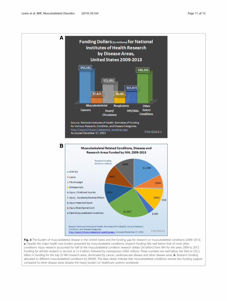
Fig. 3 The burden of musculoskeletal disease in the United States and the funding gap for research on musculoskeletal conditions (2009–2013).a. Despite the major health care burden presented by musculoskeletal conditions, research funding falls well below that of most otherconditions. Injury research accounted for half of the musculoskeletal condition research dollars ($4 billion) from NIH for the years 2009 to 2013.Funding for arthritis research is second, at $1.4 billion, followed by osteoporosis ($965 million). These numbers are well below the $8.6 to $55.2billion in funding for the top 25 NIH research areas, dominated by cancer, cardiovascular disease and other disease areas. b. Research fundingallocated to different musculoskeletal conditions by NIAMS. The data clearly indicate that musculoskeletal conditions receive less funding supportcompared to other disease areas despite the heavy burden on healthcare systems worldwide
Lewis et al. BMC Musculoskeletal Disorders (2019) 20:164 Page 11 of 15
approach to treatment and prevention is needed to tacklethese problems; including raising public awareness of riskfactors and improving the understanding of these ap-proaches within the medical and scientific community.Behaviour change and patient participation is absolutelycrucial for success. We must make use of efficient diag-nostic tools available and develop new ones that enableearlier identification of MSK disease; and we need to in-vestigate the mechanisms of disease progression that willlead to preventive measures and individually tailored andmore holistic treatment programmes.
AbbreviationsACR: American College of Rheumatology; AKU: Alkaptonuria;COPCORD: Community oriented program for control of rheumatic diseases;CT: Computed Tomography; ESCEO: European Society for Clinical andEconomic Aspects of Osteoporosis, Osteoarthritis and MusculoskeletalDiseases ; EULAR: European League Against Rheumatism; IAS: Institute ofAdvanced Studies; LBP: Low back pain; LLLT: Low level laser therapy;MRI: Magnetic Resonance Imaging; NHS: National Health Service;NIAMS: National Institute of Arthritis and Musculoskeletal and Skin Diseases;NICE: National Institute for Health and Care Excellence; NIH: NationalInstitutes of Health; NSAIDs: Non-steroidal anti-inflammatory drugs;OA: Osteoarthritis; OARSI: Osteoarthritis Research Society International;OP: Osteoporosis; PsA: Psoriatic arthritis; RA: Rheumatoid arthritis;TENS: Transcutaneous electrical nerve stimulation; WHO: World HealthOrganisation; WOMAC: The Western Ontario and McMaster UniversitiesOsteoarthritis Index
AcknowledgementsA.M. is the co-ordinator of the D-BOARD Consortium funded by the EuropeanCommission Framework 7 programme (EU FP7; HEALTH.2012.2.4.5-2, projectnumber 305815; Novel Diagnostics and Biomarkers for Early Identification ofChronic Inflammatory Joint Diseases). A.M. has also received support from theInnovative Medicines Initiative Joint Undertaking under grant agreement No.115770, resources of which are composed of financial contribution from theEuropean Union’s Seventh Framework programme (FP7/2007-2013) and EFPIAcompanies’ in-kind contribution. A.M. is also a member of the Centre for Sport,Exercise, and Osteoarthritis Research Versus Arthritis, funded by Versus Arthritis(Grant Reference: 20194) and has also received funding from the Deanship ofScientific Research (DSR), King AbdulAziz University (grant no. 1-141/1434 HiCi).A.M. is currently funded by the European Social Fund according to the activity‘Improvement of researchers’ qualification by implementing world-class R&Dprojects’ of Measure No. 09.3.3-LMT-K-712 (grant application code: 09.3.3-LMT-K-712-01-0157, agreement No. DOTSUT-215).
FundingThe authors were recipients of a workshop grant conceived, co-ordinatedand submitted by A.M. from the Institute of Advanced Studies (IAS) at theUniversity of Surrey (http://www.ias.surrey.ac.uk). The funders had no role instudy design, data collection and analysis, decision to publish, or preparationof the manuscript.
Availability of data and materialsNot applicable.
Authors’ contributionsRL and CGA contributed equally in manuscript preparation. AM conceivedthe idea for the workshop and was granted with funding. RL, CGA, MR, SLNand AM organised the workshop, including the topics for the programmeand subsequent publication of proceedings. The authors researched,discussed and approved the concept, drafted and submitted thecommissioned paper. All co-authors made a significant intellectualcontribution to the concept of the manuscript.
Ethics approval and consent to participateNot applicable.
Consent for publicationNot applicable.
Competing interestsThe authors wrote this paper within the scope of their academic andaffiliated research positions. The authors declare no conflict of interests.
Publisher’s NoteSpringer Nature remains neutral with regard to jurisdictional claims inpublished maps and institutional affiliations.
Author details1School of Veterinary Medicine, Faculty of Health and Medical Sciences,University of Surrey, Guildford, UK. 2School of Biosciences and Medicine,Faculty of Health and Medical Sciences, University of Surrey, Guildford, UK.3Centre for Sport, Exercise and Osteoarthritis Research Versus Arthritis,Queen’s Medical Centre, Nottingham, UK. 4Department of RegenerativeMedicine, State Research Institute Centre for Innovative Medicine, Vilnius,Lithuania. 5The D-BOARD FP7 Consortiumhttp://www.d-board.eu. 6TheAPPROACH IMI Consortiumhttps://www.approachproject.eu. 7Department ofRheumatology, Royal Cornwall Hospital, Truro, UK.
Received: 16 February 2018 Accepted: 17 March 2019
References1. Age UK. Later Life in the United Kingdom. 2018. http://www.ageuk.org.uk/
Documents/EN-GB/Factsheets/Later_Life_UK_factsheet.pdf.2. Policy paper. Long term conditions compendium of information: Third
Edition. 2012. https://assets.publishing.service.gov.uk/government/uploads/system/uploads/attachment_data/file/216528/dh_134486.pdf.
3. Cross M, Smith E, Hoy D, Nolte S, Ackerman I, Fransen M, Bridgett L,Williams S, Guillemin F, Hill CL, et al. The global burden of hip and kneeosteoarthritis: estimates from the global burden of disease 2010 study. AnnRheum Dis. 2014;73(7):1323–30.
4. Hoy DG, Smith E, Cross M, Sanchez-Riera L, Buchbinder R, Blyth FM, BrooksP, Woolf AD, Osborne RH, Fransen M, et al. The global burden ofmusculoskeletal conditions for 2010: an overview of methods. Ann RheumDis. 2014;73(6):982–9.
5. Woolf AD. The bone and joint decade. Strategies to reduce the burden ofdisease: the bone and joint monitor project. J Rheumatol Suppl.2003;67:6–9.
6. Woolf AD, Pfleger B. Burden of major musculoskeletal conditions. Bull WorldHealth Organ. 2003;81(9):646–56.
7. Chopra A, Saluja M, Patil J, Tandale HS. Pain and disability, perceptions andbeliefs of a rural Indian population: a WHO-ILAR COPCORD study. WHO-international league of associations for rheumatology. Community orientedprogram for control of rheumatic diseases. J Rheumatol. 2002;29(3):614–21.
8. Chopra A. The WHO-ILAR COPCORD Bhigwan (India) model: foundation fora future COPCORD design and data repository. Clin Rheumatol. 2006;25(4):443–7.
9. Zeng QY, Chen R, Darmawan J, Xiao ZY, Chen SB, Wigley R, Le Chen S,Zhang NZ. Rheumatic diseases in China. Arthritis Res Ther. 2008;10(1):R17.
10. Hoy D, Brooks P, Blyth F, Buchbinder R. The epidemiology of low back pain.Best Pract Res Clin Rheumatol. 2010;24(6):769–81.
11. Proceedings of the Musculoskeletal Health in the 21st Century Workshop.BMC Musculoskelet Disord 2015, 16 Suppl 1:I1-S20.
12. World Health Organisation (WHO). One health. 2017. https://www.who.int/features/qa/one-health/en/.
13. Centers for Disease Control and Prevention (CDC), U.S. Department ofHealth & Human Services: Behavioural risk factors surveillance system(BRFSS) data. 2017. https://www.cdc.gov/brfss/index.html.
14. Ding D, Lawson KD, Kolbe-Alexander TL, Finkelstein EA, Katzmarzyk PT, vanMechelen W, Pratt M. Lancet physical activity series 2 executive C: theeconomic burden of physical inactivity: a global analysis of major non-communicable diseases. Lancet. 2016.
15. The Economic Cost of Physical Inactivity in Europe. In: Centre for Economicsand Business Research. 2015. https://inactivity-time-bomb.nowwemove.com/report/.
16. Measuring national well-being in the UK: international comparisons, 2019.In: Office for National Statistics. 2019. https://www.ons.gov.uk/
Lewis et al. BMC Musculoskeletal Disorders (2019) 20:164 Page 12 of 15
peoplepopulationandcommunity/wellbeing/articles/measuringnationalwellbeing/internationalcomparisons2019.
17. Ekelund U, Steene-Johannessen J, Brown WJ, Fagerland MW, Owen N,Powell KE, Bauman A, Lee IM. Lancet physical activity series 2 executive C,lancet sedentary behaviour working G: does physical activity attenuate, oreven eliminate, the detrimental association of sitting time with mortality? Aharmonised meta-analysis of data from more than 1 million men andwomen. Lancet. 2016.
18. Kubota Y, Evenson KR, MacLehose RF, Roetker NS, Joshu CE, Folsom AR.Physical Activity and Lifetime Risk of Cardiovascular Disease and Cancer.Med Sci Sports Exerc. 2017.
19. van der Velde JH, Koster A, van der Berg JD, Sep SJ, van der Kallen CJ,Dagnelie PC, Schram MT, Henry RM, Eussen SJ, van Dongen MC, et al.Sedentary behavior, physical activity, and fitness-the Maastricht study. MedSci Sports Exerc. 2017.
20. McCarthy CJ, Mills PM, Pullen R, Richardson G, Hawkins N, Roberts CR,Silman AJ, Oldham JA. Supplementation of a home-based exerciseprogramme with a class-based programme for people with osteoarthritis ofthe knees: a randomised controlled trial and health economic analysis.Health Technol Assess. 2004;8(46):iii–v 1-61.
21. Gordon R, Bloxham S. A Systematic Review of the Effects of Exercise andPhysical Activity on Non-Specific Chronic Low Back Pain. Healthcare (Basel).2016;4(2).
22. Warburton DE, Nicol CW, Bredin SS. Prescribing exercise as preventivetherapy. CMAJ. 2006;174(7):961–74.
23. Felson DT, Anderson JJ, Naimark A, Walker AM, Meenan RF. Obesity and kneeosteoarthritis The Framingham Study. Ann Intern Med. 1988;109(1):18–24.
24. Zhang P, Zhang X, Brown J, Vistisen D, Sicree R, Shaw J, Nichols G. Globalhealthcare expenditure on diabetes for 2010 and 2030. Diabetes Res ClinPract. 2010;87(3):293–301.
25. Schett G, Kleyer A, Perricone C, Sahinbegovic E, Iagnocco A, Zwerina J,Lorenzini R, Aschenbrenner F, Berenbaum F, D'Agostino MA, et al. Diabetesis an independent predictor for severe osteoarthritis: results from alongitudinal cohort study. Diabetes Care. 2013;36(2):403–9.
26. Eymard F, Parsons C, Edwards MH, Petit-Dop F, Reginster JY, Bruyere O,Richette P, Cooper C, Chevalier X. Diabetes is a risk factor for kneeosteoarthritis progression. Osteoarthr Cartil. 2015;23(6):851–9.
27. Beaudart C, Dawson A, Shaw SC, Harvey NC, Kanis JA, Binkley N, ReginsterJY, Chapurlat R, Chan DC, Bruyere O, et al. Nutrition and physical activity inthe prevention and treatment of sarcopenia: systematic review. OsteoporosInt. 2017.
28. Lundberg IE, Nader GA. Molecular effects of exercise in patients withinflammatory rheumatic disease. Nat Clin Pract Rheumatol. 2008;4(11):597–604.
29. Rizzoli R, Stevenson JC, Bauer JM, van Loon LJ, Walrand S, Kanis JA, CooperC, Brandi ML, Diez-Perez A, Reginster JY, et al. The role of dietary proteinand vitamin D in maintaining musculoskeletal health in postmenopausalwomen: a consensus statement from the European Society for Clinical andEconomic Aspects of osteoporosis and osteoarthritis (ESCEO). Maturitas.2014;79(1):122–32.
30. Otero M, Esain I, Gonzalez-Suarez AM, Gil SM. The effectiveness of a basicexercise intervention to improve strength and balance in women withosteoporosis. Clin Interv Aging. 2017;12:505–13.
31. Ahn N, Kim K. Effects of 12-week exercise training on osteocalcin, high-sensitivity C-reactive protein concentrations, and insulin resistance in elderlyfemales with osteoporosis. J Phys Ther Sci. 2016;28(8):2227–31.
32. Sugiyama T, Oda H. Osteoporosis therapy: bone modeling during growthand aging. Front Endocrinol (Lausanne). 2017;8:46.
33. Gomez-Cabrera MC, Garcia-Valles R, Rodriguez-Manas L, Garcia-Garcia FJ,Olaso-Gonzalez G, Salvador-Pascual A, Tarazona-Santabalbina FJ, Vina J. Anew frailty score for experimental animals based on the clinical phenotype:inactivity as a model of frailty. J Gerontol A Biol Sci Med Sci. 2017.
34. Dela F, Prats C, Helge JW. Exercise interventions to prevent and managetype 2 diabetes: physiological mechanisms. Med Sport Sci. 2014;60:36–47.
35. Svensson RB, Heinemeier KM, Couppe C, Kjaer M, Magnusson SP. Effect ofaging and exercise on the tendon. J Appl Physiol (1985). 2016;121(6):1353–62.
36. Zhang JY, Yuan T, Wang JHC. Moderate treadmill running exercise prior totendon injury enhances wound healing in aging rats. Oncotarget. 2016;7(8):8498–512.
37. Guilak F. Biomechanical factors in osteoarthritis. Best Pract Res ClinRheumatol. 2011;25(6):815–23.
38. Grabowski P. Physiology of bone. Endocr Dev. 2009;16:32–48.
39. Mobasheri A, Batt M. An update on the pathophysiology of osteoarthritis.Ann Phys Rehabil Med. 2016;59(5–6):333–9.
40. Messier SP, Loeser RF, Miller GD, Morgan TM, Rejeski WJ, Sevick MA,Ettinger WH Jr, Pahor M, Williamson JD. Exercise and dietary weightloss in overweight and obese older adults with knee osteoarthritis: thearthritis, diet, and activity promotion trial. Arthritis Rheum. 2004;50(5):1501–10.
41. Arthritis Research UK/Versus Arthritis. In: Musculoskeletal health – a publichealth approach. 2014. https://www.versusarthritis.org/policy/policy-reports/musculoskeletal-health.
42. Chan G, Chen CT. Musculoskeletal effects of obesity. Curr Opin Pediatr.2009;21(1):65–70.
43. Courcier EA, Thomson RM, Mellor DJ, Yam PS. An epidemiological study ofenvironmental factors associated with canine obesity. J Small Anim Pract.2010;51(7):362–7.
44. Nijland ML, Stam F, Seidell JC. Overweight in dogs, but not in cats, isrelated to overweight in their owners. Public Health Nutr. 2010;13(1):102–6.
45. Nicklett EJ, Semba RD, Xue QL, Tian J, Sun K, Cappola AR, Simonsick EM,Ferrucci L, Fried LP. Fruit and vegetable intake, physical activity, andmortality in older community-dwelling women. J Am Geriatr Soc. 2012;60(5):862–8.
46. Dawson-Hughes B, Harris SS, Ceglia L. Alkaline diets favor lean tissue massin older adults. Am J Clin Nutr. 2008;87(3):662–5.
47. Hardcastle AC, Aucott L, Reid DM, Macdonald HM. Associations betweendietary flavonoid intakes and bone health in a Scottish population. J BoneMiner Res. 2011;26(5):941–7.
48. Thomas S, Browne H, Mobasheri A, Rayman MP. What is the evidence for arole for diet and nutrition in osteoarthritis? Rheumatology (Oxford). 2018;57(suppl_4):iv61–74.
49. Daly RM. Exercise and nutritional approaches to prevent frail bones, fallsand fractures: an update. Climacteric. 2017;20(2):119–24.
50. Meyer HE, Pedersen JI, Loken EB, Tverdal A. Dietary factors and theincidence of hip fracture in middle-aged Norwegians. A prospective study.Am J Epidemiol. 1997;145(2):117–23.
51. Heaney RP, Layman DK. Amount and type of protein influences bonehealth. Am J Clin Nutr. 2008;87(5):1567S–70S.
52. Remer T, Manz F. Potential renal acid load of foods and its influence onurine pH. J Am Diet Assoc. 1995;95(7):791–7.
53. Liu X, Eyles J, McLachlan AJ, Mobasheri A. Which supplements can I recommendto my osteoarthritis patients? Rheumatology (Oxford). 2018;57(suppl_4):iv75–87.
54. Cao Y, Winzenberg T, Nguo K, Lin J, Jones G, Ding C. Association betweenserum levels of 25-hydroxyvitamin D and osteoarthritis: a systematic review.Rheumatology (Oxford). 2013;52(7):1323–34.
55. Misra D, Booth SL, Tolstykh I, Felson DT, Nevitt MC, Lewis CE, Torner J,Neogi T. Vitamin K deficiency is associated with incident knee osteoarthritis.Am J Med. 2013;126(3):243–8.
56. Sturmer T, Sun Y, Sauerland S, Zeissig I, Gunther KP, Puhl W, Brenner H.Serum cholesterol and osteoarthritis. The baseline examination of the Ulmosteoarthritis study. J Rheumatol. 1998;25(9):1827–32.
57. Sen CK. Antioxidants in exercise nutrition. Sports Med. 2001;31(13):891–908.58. Molnar A, Jonasne Sztruhar I, Csontos AA, Ferencz C, Varbiro S, Szekacs B.
Special nutrition intervention is required for muscle protective efficacy ofphysical exercise in elderly people at highest risk of sarcopenia. Physiol Int.2016;103(3):368–76.
59. Pearson W, Orth MW, Karrow NA, Maclusky NJ, Lindinger MI. Anti-inflammatory and chondroprotective effects of nutraceuticals from Sasha'sblend in a cartilage explant model of inflammation. Mol Nutr Food Res.2007;51(8):1020–30.
60. Rialland P, Bichot S, Lussier B, Moreau M, Beaudry F, del Castillo JR, GauvinD, Troncy E. Effect of a diet enriched with green-lipped mussel on painbehavior and functioning in dogs with clinical osteoarthritis. Can J Vet Res.2013;77(1):66–74.
61. Buddhachat K, Siengdee P, Chomdej S, Soontornvipart K, Nganvongpanit K.Effects of different omega-3 sources, fish oil, krill oil, and green-lippedmussel against cytokine-mediated canine cartilage degradation. In Vitro CellDev Biol Anim. 2017.
62. Mehler SJ, May LR, King C, Harris WS, Shah Z. A prospective, randomized,double blind, placebo-controlled evaluation of the effects ofeicosapentaenoic acid and docosahexaenoic acid on the clinical signs anderythrocyte membrane polyunsaturated fatty acid concentrations in dogswith osteoarthritis. Prostag Leukotr Ess. 2016;109:1–7.
Lewis et al. BMC Musculoskeletal Disorders (2019) 20:164 Page 13 of 15
63. Maroon JC, Bost JW. Omega-3 fatty acids (fish oil) as an anti-inflammatory:an alternative to nonsteroidal anti-inflammatory drugs for discogenic pain.Surg Neurol. 2006;65(4):326–31.
64. Goldberg RJ, Katz J. A meta-analysis of the analgesic effects of omega-3polyunsaturated fatty acid supplementation for inflammatory joint pain.Pain. 2007;129(1–2):210–23.
65. Hurst S, Zainal Z, Caterson B, Hughes CE, Harwood JL. Dietary fatty acidsand arthritis. Prostaglandins Leukot Essent Fatty Acids. 2010;82(4–6):315–8.
66. Stanic Z. Curcumin, a compound from natural sources, a true scientificchallenge - a review. Plant Foods Hum Nutr. 2017;72(1):1–12.
67. Liu X, Machado GC, Eyles JP, Ravi V, Hunter DJ. Dietary supplements fortreating osteoarthritis: a systematic review and meta-analysis. Br J SportsMed. 2017.
68. Henrotin Y, Mobasheri A, Marty M. Is there any scientific evidence for theuse of glucosamine in the management of human osteoarthritis? ArthritisRes Ther. 2012;14(1):201.
69. Henrotin Y, Marty M, Mobasheri A. What is the current status of chondroitinsulfate and glucosamine for the treatment of knee osteoarthritis? Maturitas.2014;78(3):184–7.
70. McAlindon TE, Bannuru RR, Sullivan MC, Arden NK, Berenbaum F, Bierma-Zeinstra SM, Hawker GA, Henrotin Y, Hunter DJ, Kawaguchi H, et al. OARSIguidelines for the non-surgical management of knee osteoarthritis.Osteoarthr Cartil. 2014;22(3):363–88.
71. Santos GR, Piquet AA, Glauser BF, Tovar AM, Pereira MS, Vilanova E,Mourao PA. Systematic Analysis of Pharmaceutical Preparations ofChondroitin Sulfate Combined with Glucosamine. Pharmaceuticals(Basel). 2017;10(2).
72. Chan PS, Caron JP, Rosa GJ, Orth MW. Glucosamine and chondroitinsulfate regulate gene expression and synthesis of nitric oxide andprostaglandin E(2) in articular cartilage explants. Osteoarthr Cartil. 2005;13(5):387–94.
73. Silva FS Jr, Yoshinari NH, Castro RR, Girao VC, Pompeu MM, Feitosa JP,Rocha FA. Combined glucosamine and chondroitin sulfate providesfunctional and structural benefit in the anterior cruciate ligamenttransection model. Clin Rheumatol. 2009;28(2):109–17.
74. Das A Jr, Hammad TA. Efficacy of a combination of FCHG49 glucosaminehydrochloride, TRH122 low molecular weight sodium chondroitin sulfateand manganese ascorbate in the management of knee osteoarthritis.Osteoarthr Cartil. 2000;8(5):343–50.
75. Pham T, Cornea A, Blick KE, Jenkins A, Scofield RH. Oral glucosamine indoses used to treat osteoarthritis worsens insulin resistance. Am J Med Sci.2007;333(6):333–9.
76. Boer CG, Radjabzadeh D, Uitterlinden AG, Kraaij R, van Meurs JB. The role ofthe gut microbiome in osteoarthritis and joint pain. Osteoarthr Cartil. 2017;25:S10.
77. Szychlinska MA, Di Rosa M, Castorina A, Mobasheri A, Musumeci G. Acorrelation between intestinal microbiota dysbiosis and osteoarthritis.Heliyon. 2019;5(1):e01134.
78. Huang WY, Fu L, Li CY, Xu LP, Zhang LX, Zhang WM. Quercetin, Hyperin,and Chlorogenic Acid Improve Endothelial Function by Antioxidant,Antiinflammatory, and ACE Inhibitory Effects. J Food Sci. 2017.
79. Trappoliere M, Caligiuri A, Schmid M, Bertolani C, Failli P, Vizzutti F, Novo E,di Manzano C, Marra F, Loguercio C, et al. Silybin, a component of sylimarin,exerts anti-inflammatory and anti-fibrogenic effects on human hepaticstellate cells. J Hepatol. 2009;50(6):1102–11.
80. Bjornsdottir SV, Jonsson SH, Valdimarsdottir UA. Mental health indicatorsand quality of life among individuals with musculoskeletal chronic pain: anationwide study in Iceland. Scand J Rheumatol. 2014;43(5):419–23.
81. Dickens C, McGowan L, Clark-Carter D, Creed F. Depression in rheumatoidarthritis: a systematic review of the literature with meta-analysis. PsychosomMed. 2002;64(1):52–60.
82. Krishnan KR, Delong M, Kraemer H, Carney R, Spiegel D, Gordon C, McDonaldW, Dew M, Alexopoulos G, Buckwalter K, et al. Comorbidity of depression withother medical diseases in the elderly. Biol Psychiatry. 2002;52(6):559–88.
83. Sareen J, Jacobi F, Cox BJ, Belik SL, Clara I, Stein MB. Disability and poorquality of life associated with comorbid anxiety disorders and physicalconditions. Arch Intern Med. 2006;166(19):2109–16.
84. Australian Government - Australian Institute of Health and Welfare. Whenmusculoskeletal conditions and mental disorders occur together. 2010.https://www.aihw.gov.au/reports/arthritis-other-musculoskeletal-conditions/musculoskeletal-conditions-mental-disorder/formats.
85. Baker S, McBeth J, Chew-Graham CA, Wilkie R. Musculoskeletal pain and co-morbid insomnia in adults; a population study of the prevalence andimpact on restricted social participation. BMC Fam Pract. 2017;18(1):17.
86. Lingard EA, Mitchell SY, Francis RM, Rawlings D, Peaston R, Birrell FN,McCaskie AW. The prevalence of osteoporosis in patients with severe hipand knee osteoarthritis awaiting joint arthroplasty. Age Ageing. 2010;39(2):234–9.
87. Romeyke T, Noehammer E, Scheuer HC, Stummer H. Severe forms offibromyalgia with acute exacerbation of pain: costs, comorbidities, andlength of stay in inpatient care. Clinicoecon Outcomes Res. 2017;9:317–25.
88. Slater M, Perruccio AV, Badley EM. Musculoskeletal comorbidities incardiovascular disease, diabetes and respiratory disease: the impact onactivity limitations; a representative population-based study. BMC PublicHealth. 2011;11:77.
89. Rivera JC, Amuan ME, Morris RM, Johnson AE, Pugh MJ. Arthritis,comorbidities, and care utilization in veterans of operations enduring andIraqi freedom. J Orthop Res. 2017;35(3):682–7.
90. Shan J, Zhang J. Impact of obesity on the efficacy of different biologicagents in inflammatory diseases: a systematic review and meta-analysis.Joint Bone Spine. 2018.
91. Hayashi D, Roemer FW, Jarraya M, Guermazi A. Imaging in osteoarthritis.Radiol Clin N Am. 2017;55(5):1085–102.
92. Takamatsu N, Mori A. Nodera H: [sonographic evaluation of myopathy].Brain Nerve. 2014;66(3):247–57.
93. Braun HJ, Gold GE. Diagnosis of osteoarthritis: imaging. Bone. 2012;51(2):278–88.
94. Sudol-Szopinska I, Schueller-Weidekamm C, Plagou A, Teh J. Ultrasound inarthritis. Radiol Clin N Am. 2017;55(5):985–96.
95. Sudol-Szopinska I, Jans L, Teh J. Rheumatoid arthritis: what do MRI andultrasound show. J Ultrason. 2017;17(68):5–16.
96. Zeman MN, Scott PJ. Current imaging strategies in rheumatoid arthritis. AmJ Nucl Med Mol Imaging. 2012;2(2):174–220.
97. Back, Clayton: Equine Locomotion, 2nd edn: Saunders Ltd; 2013.98. Rhodin M, Bergh A, Gustas P, Gomez Alvarez CB. Inertial sensor-based
system for lameness detection in trotting dogs with induced lameness. VetJ. 2017;222:54–9.
99. Bertuletti S, Cereatti A, Comotti D, Caldara M, Della Croce U. Static andDynamic Accuracy of an Innovative Miniaturized Wearable Platform forShort Range Distance Measurements for Human Movement Applications.Sensors (Basel). 2017;17(7).
100. Keegan KG, Wilson DA, Kramer J, Reed SK, Yonezawa Y, Maki H, Pai PF,Lopes MA. Comparison of a body-mounted inertial sensor system-basedmethod with subjective evaluation for detection of lameness in horses. AmJ Vet Res. 2013;74(1):17–24.
101. Simon SR. Quantification of human motion: gait analysis-benefits and limitationsto its application to clinical problems. J Biomech. 2004;37(12):1869–80.
102. Barn R, Brandon M, Rafferty D, Sturrock RD, Steultjens M, Turner DE,Woodburn J. Kinematic, kinetic and electromyographic response tocustomized foot orthoses in patients with tibialis posterior tenosynovitis,pes Plano valgus and rheumatoid arthritis. Rheumatology (Oxford). 2014;53(1):123–30.
103. Meyer CA, Corten K, Fieuws S, Deschamps K, Monari D, Wesseling M, SimonJP, Desloovere K. Biomechanical gait features associated with hiposteoarthritis: towards a better definition of clinical hallmarks. J Orthop Res.2015;33(10):1498–507.
104. Tas S, Guneri S, Kaymak B, Erden Z. A comparison of results of 3-dimensional gait analysis and observational gait analysis in patients withknee osteoarthritis. Acta Orthop Traumatol Turc. 2015;49(2):151–9.
105. Carroll M, Parmar P, Dalbeth N, Boocock M, Rome K. Gait characteristicsassociated with the foot and ankle in inflammatory arthritis: a systematicreview and meta-analysis. BMC Musculoskelet Disord. 2015;16:134.
106. Austine J, Nair S, Mirza K. Perspective of orthopedists on pain Managementin Osteoarthritis: a qualitative study. Indian J Palliat Care. 2016;22(4):410–5.
107. Substance Abuse and Mental Health Services Administration, U.S.Department of Health and Human Services: The National Survey on DrugUse and Health. 2017. https://www.samhsa.gov/data/sites/default/files/nsduh-ppt-09-2018.pdf.
108. National Institutes of Health, U.S. Department of Health and HumanServices: NIH HEAL Initiative: Helping to end addiction long-term. 2018.https://www.nih.gov/research-training/medical-research-initiatives/heal-initiative.
Lewis et al. BMC Musculoskeletal Disorders (2019) 20:164 Page 14 of 15
109. Harper TAM. Conservative Management of hip Dysplasia. Vet Clin North AmSmall Anim Pract. 2017;47(4):807–21.
110. Frye CW, Shmalberg JW, Wakshlag JJ. Obesity, exercise and orthopedicdisease. Vet Clin North Am Small Anim Pract. 2016;46(5):831–41.
111. Flegal KM, Carroll MD, Ogden CL, Johnson CL. Prevalence and trends inobesity among US adults, 1999-2000. JAMA. 2002;288(14):1723–7.
112. Ogden CL, Carroll MD, Curtin LR, McDowell MA, Tabak CJ, Flegal KM.Prevalence of overweight and obesity in the United States, 1999-2004.JAMA. 2006;295(13):1549–55.
113. Murphy DJ, Todhunter RJ, Fubini SL, Vernier-Singer M, Straubinger RK, LustG. The effects of methylprednisolone on normal and monocyte-conditionedmedium-treated articular cartilage from dogs and horses. Vet Surg. 2000;29(6):546–57.
114. Pelletier JP, Martel-Pelletier J. Protective effects of corticosteroids oncartilage lesions and osteophyte formation in the pond-Nuki dog model ofosteoarthritis. Arthritis Rheum. 1989;32(2):181–93.
115. Juni P, Hari R, Rutjes AW, Fischer R, Silletta MG, Reichenbach S, da Costa BR.Intra-articular corticosteroid for knee osteoarthritis. The Cochrane databaseof systematic reviews. 2015;(10) Cd005328.
116. Conzemius MG, Evans RB. Caregiver placebo effect for dogs with lamenessfrom osteoarthritis. J Am Vet Med Assoc. 2012;241(10):1314–9.
117. Innes J: Arthritis. In: Veterinary Surgery Small Animal. Volume 1, edn. Editedby Tobias KM, Johnston SA: Elsevier Saunders; 2011: 1078–1111.
118. Mandegaran R, Conway C, Elton C. Lower gastrointestinal adverse effects ofNSAIDS: an extreme example of a common problem. BMJ Case Rep. 2013;2013.
119. Loveless MS, Fry AL. Pharmacologic therapies in musculoskeletal conditions.Med Clin North Am. 2016;100(4):869–90.
120. Tomazoni SS, Frigo L, Dos Reis Ferreira TC, Casalechi HL, Teixeira S, deAlmeida P, Muscara MN, Marcos RL, Serra AJ, de Carvalho PTC, et al. Effectsof photobiomodulation therapy and topical non-steroidal anti-inflammatorydrug on skeletal muscle injury induced by contusion in rats-part 2:biochemical aspects. Lasers Med Sci. 2017.
121. de Almeida P, Tomazoni SS, Frigo L, de Carvalho PT, Vanin AA, Santos LA,Albuquerque-Pontes GM, De Marchi T, Tairova O, Marcos RL, et al. What isthe best treatment to decrease pro-inflammatory cytokine release in acuteskeletal muscle injury induced by trauma in rats: low-level laser therapy,diclofenac, or cryotherapy? Lasers Med Sci. 2014;29(2):653–8.
122. Babatunde OO, Jordan JL, Van der Windt DA, Hill JC, Foster NE, Protheroe J.Effective treatment options for musculoskeletal pain in primary care: asystematic overview of current evidence. PLoS One. 2017;12(6):e0178621.
123. Vickers AJ, Cronin AM, Maschino AC, Lewith G, MacPherson H, Foster NE,Sherman KJ, Witt CM, Linde K, Acupuncture Trialists C. Acupuncture forchronic pain: individual patient data meta-analysis. Arch Intern Med. 2012;172(19):1444–53.
124. Kroeling P, Gross A, Graham N, Burnie SJ, Szeto G, Goldsmith CH, Haines T,Forget M. Electrotherapy for neck pain. Cochrane Database Syst Rev. 2013;8:CD004251.
125. Rutjes AW, Nuesch E, Sterchi R, Kalichman L, Hendriks E, Osiri M, Brosseau L,Reichenbach S, Juni P. Transcutaneous electrostimulation for osteoarthritisof the knee. Cochrane Database Syst Rev. 2009;4:CD002823.
126. Seco J, Kovacs FM, Urrutia G. The efficacy, safety, effectiveness, and cost-effectiveness of ultrasound and shock wave therapies for low back pain: asystematic review. Spine J. 2011;11(10):966–77.
127. Robertson VJ, Baker KG. A review of therapeutic ultrasound: effectivenessstudies. Phys Ther. 2001;81(7):1339–50.
128. Dycus DL, Levine D, Marcellin-Little DJ. Physical rehabilitation for theManagement of Canine hip Dysplasia. Vet Clin North Am Small Anim Pract.2017;47(4):823–50.
129. Sigurdsson SO, Austin J. Using real-time visual feedback to improve postureat computer workstations. J Appl Behav Anal. 2008;41(3):365–75.
130. Lewis R, May H, Mobasheri A, Barrett-Jolley R. Chondrocyte channeltranscriptomics: do microarray data fit with expression and functional data?Channels. 2013;7(6):459–67.
131. Harre U, Schett G. Cellular and molecular pathways of structural damage inrheumatoid arthritis. Semin Immunopathol. 2017;39(4):355–63.
132. Corrado A, Maruotti N, Cantatore FP. Osteoblast Role in Rheumatic Diseases.Int J Mol Sci. 2017;18(6).
133. Little CB, Zaki S. What constitutes an "animal model of osteoarthritis"--theneed for consensus? Osteoarthr Cartil. 2012;20(4):261–7.
134. Holland C, Vollrath F, Gill HS. Horses and cows might teach us abouthuman knees. Naturwissenschaften. 2014;101(4):351–4.
135. Piel MJ, Kroin JS, van Wijnen AJ, Kc R, Im HJ. Pain assessment in animalmodels of osteoarthritis. Gene. 2014;537(2):184–8.
136. McIlwraith CW, Frisbie DD, Kawcak CE. The horse as a model of naturallyoccurring osteoarthritis. Bone Joint Res. 2012;1(11):297–309.
137. Bergknut N, Rutges JP, Kranenburg HJ, Smolders LA, Hagman R, Smidt HJ,Lagerstedt AS, Penning LC, Voorhout G, Hazewinkel HA, et al. The dog as ananimal model for intervertebral disc degeneration? Spine (Phila Pa 1976).2012;37(5):351–8.
138. Gallagher JA, Ranganath LR, Boyde A. Lessons from rare diseases of cartilageand bone. Curr Opin Pharmacol. 2015;22:107–14.
139. Hauret KG, Jones BH, Bullock SH, Canham-Chervak M, Canada S.Musculoskeletal injuries description of an under-recognized injury problemamong military personnel. Am J Prev Med. 2010;38(1 Suppl):S61–70.
140. McGee JC, Bise CG, Stevens JM. An orthopedic performance improvementinitiative at a small military treatment facility. Mil Med. 2017;182(S1):18–25.
141. Walker LA, Zambraski EJ, Williams RF. Widespread use of prescriptionnonsteroidal anti-inflammatory drugs among U.S. Army active duty soldiers.Mil Med. 2017;182(3):e1709–12.
Lewis et al. BMC Musculoskeletal Disorders (2019) 20:164 Page 15 of 15