stroke - intracranial hemorrhagedramiteshaggarwal.yolasite.com/resources/cerebral hemorrhage.pdf ·...
TRANSCRIPT

Stroke - Intracranial
hemorrhage
Dr. Amitesh Aggarwal
Associate Professor
Department of Medicine

Etiology and pathogenesis
ICH accounts for ~10% of all strokes
30 day mortality - 35–45%
Incidence rates higher in Asians
MAJOR CAUSES
Hypertension
Coagulopathy
Sympathomimetic drugs (cocaine, methamphetamine)
Cerebral amyloid angiopathy

Other Etiologies
Advanced age
Heavy alcohol consumption
Atherosclerosis
Bleeding tendency (hemophilia, etc)
Congenital angiomatous malformation
Amyloid
Trauma
Aneurysm
Tumor
Vasculitis (PAN / SLE)

Hypertensive ICH
ICH usually results from spontaneous rupture of a small
penetrating artery deep in the brain.
The most common sites are
Basal ganglia (50%)
Lobar regions (20-50%)
Thalamus (10-15%)
Pons (5-12%)
Cerebellum (1-5%)

Clinical features
Abrupt onset of focal neurologic deficit
Seizures are uncommon
Clinical symptoms may be maximal at onset
Diminishing level of consciousness
Signs of increased ICP such as headache and vomiting
Focal neurological deficits depending on the location
of bleed

• Penetrating cortical branches
of ACA, MCA, & PCA
• Major neurologic deficit
• Occipital hemorrhage -
hemianopia
• Left temporal hemorrhage -
aphasia and delirium
• Parietal hemorrhage -
hemisensory loss
• Frontal hemorrhage - arm
weakness
• Large hemorrhages - stupor
or coma
Lobar hemorrhage

• Ascending lenticulostriate
branches of MCA
• Wide spectrum of severity
extending to coma and
decerebrate rigidity
• Contralateral hemiplegia,
hemianesthesia, and hemianopia
• Eyes are frequently deviated
toward the side of the affected
hemisphere
• Aphasia if dominant hemisphere
is affected
• Ventricular extension carries very
poor prognosis
Basal ganglia

•Ascending thalamogeniculate
branches of PCA
• Contralateral hemiplegia,
hemianesthesia, and hemianopia
• Deep sensation disturbance
• Ocular signs
• Disturbance of consciousness
• Patients may later develop a
chronic, contralateral pain
syndrome (Déjérine-Roussy
syndrome)
•Abrupt hydrocephalus from
aqueductal obstruction from
intraventricular clot
Thalamus
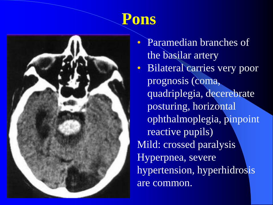
• Paramedian branches of
the basilar artery
• Bilateral carries very poor
prognosis (coma,
quadriplegia, decerebrate
posturing, horizontal
ophthalmoplegia, pinpoint
reactive pupils)
Mild: crossed paralysis
Hyperpnea, severe
hypertension, hyperhidrosis
are common.
Pons

• Penetrating branches of the
PICA, AICA, SCA
• Abrupt onset vertigo, inability
to walk in absence of weakness
• Ipsilateral ataxia, horizontal
gaze palsy, peripheral facial
palsy
• Unpredictable deterioration to
coma
• Occipital headache, nystagmus
• Severe cerebellar hemorrhage :
coma, compression of brain
stem, tonsillar herniation
Cerebellum

Other causes
Anticoagulant related - may continue over 24–48 h
Hematologic disorders (leukemia, aplastic anemia,
thrombocytopenic purpura) - any site, multiple
Metastatic tumors - Choriocarcinoma, malignant
melanoma, renal cell carcinoma, bronchogenic
carcinoma
Cerebral amyloid angiopathy – MC cause in elderly,
multiple hemorrhages (& infarcts) over several months
or years

Investigation
1. CT
First choice
High density blood
Mass effect and edema
2. MRI
Brain stem hemorrhage
<24h, not distinguishable with thrombosis
3. DSA
Young and with normal blood pressure
4. Platelet count and PT//INR – coagulopathy
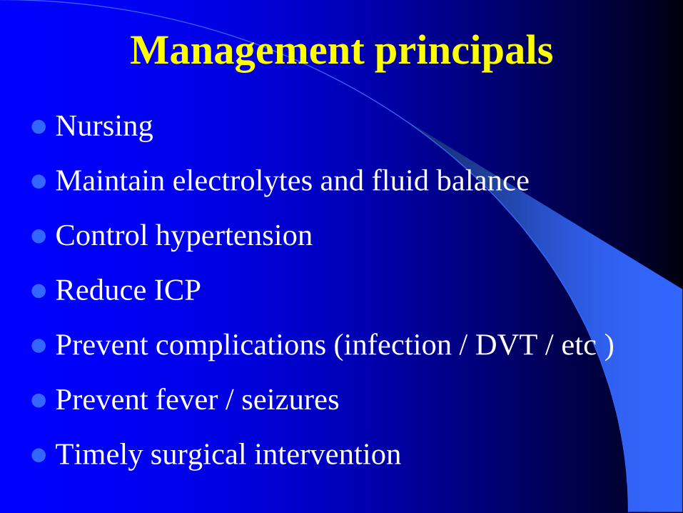
Management principals
Nursing
Maintain electrolytes and fluid balance
Control hypertension
Reduce ICP
Prevent complications (infection / DVT / etc )
Prevent fever / seizures
Timely surgical intervention

BP Management
BP is elevated on admission in over 2/3 of patients
Tends to return to baseline 7-10 days post ICH
How fast should BP be lowered?
– Rapidly lowering MAP by 15% does not lower
CBF
– Current guidelines suggest a reduction of ≤ 20% in
the first 24 hrs
Which agents should be used?
– Short and rapidly acting IV antihypertensive
– Labetalol, hydralazine, esmolol, nicardipine,
enalapril

Raised ICP Management
CSF volume
Mannitol or hypertonic solution
External CSF drainage
Ventricular catheter
Ventriculo -peritoneal or atrial shunt
Lumbar drain
Serial lumbar punctures
Brain volume
Mannitol or hypertonic saline
Decompressive craniotomy
Resection of tumor or other mass lesion
Blood volume
Mannitol or
hypertonic saline
Hyperventilation
Hypothermia
Head elevation,
neutral neck
position
Deep propofol or
barbiturate sedation
± paralysis Seizure Control

Coagulopathy Management
Goal of treatment: fully reverse INR to normal range
High dose Vitamin K 10-20 mg IV slow infusion
– Effect takes 12-24hrs
– Helps achieving sustained reversal of INR
Fresh frozen plasma 15cc/kg 4U
– Volume overload, insufficient factor IX
– ABO compatibility, thawing, infusion time (30hrs)
Prothrombin Complex Concentrates (PCC)
– Combination of II, VII, IX, X, variable protein C and S
– Dosage dependant on initial INR
– Smaller volume, correct INR as fast as 30 min
When ICH is associated with thrombocytopenia (platelet
count <50,000/μL), transfusion of fresh platelets is indicated

Existing data do not support routine surgical evacuation
of supratentorial hemorrhages in stable patients
Putamen, lobar > 50 ml, deteriorating/ brainstem
compression/ hydrocephalus
Cerebellum > 30 ml, diameter > 3cm
Thalamus - obstructive hydrocephalus →ventricular
drainage
Surgical indications

18
http://dramiteshaggarwal.yolasite.com