stroke, tia, and other central focal conditions
TRANSCRIPT

STROKE, TIA, AND STROKE, TIA, AND OTHER CENTRAL OTHER CENTRAL
FOCAL CONDITIONSFOCAL CONDITIONS
Dee Mortensen PGY2Dee Mortensen PGY2
November 10, 2005November 10, 2005
Tintinalli Ch. 228Tintinalli Ch. 228
BackgroundBackground
Third leading cause of death in USThird leading cause of death in US Leading cause of adult disabilityLeading cause of adult disability 700,000 patients/year700,000 patients/year 1/3 of patients younger than 651/3 of patients younger than 65

Definition of StrokeDefinition of Stroke
Any disease process that disrupts blood flow Any disease process that disrupts blood flow to a focal region of the brain. to a focal region of the brain.
Stroke TypesStroke Types
80% ischemic80% ischemic ThrombosisThrombosis EmbolismEmbolism HypoperfusionHypoperfusion
20% hemorrhagic20% hemorrhagic IntracerebralIntracerebral SubarachnoidSubarachnoid
Ischemic StrokesIschemic Strokes
ThrombosisThrombosis-most common cause-most common cause Etiology Etiology
Atherosclerotic disease-most commonAtherosclerotic disease-most common VasculitisVasculitis DissectionDissection PolycythemiaPolycythemia Hypercoagulable statesHypercoagulable states Infectious Diseases-HIV, TB, syphilisInfectious Diseases-HIV, TB, syphilis

Ischemic StrokesIschemic Strokes
1/51/5 thth due to due to EmbolismEmbolism EtiologyEtiology
CardiacCardiac Valvular VegetationsValvular Vegetations Mural thrombi- caused by A-fib, MI, or dysrhythmiasMural thrombi- caused by A-fib, MI, or dysrhythmias Paradoxical emboli-from ASD, VSDParadoxical emboli-from ASD, VSD Cardiac tumors-myxomaCardiac tumors-myxoma
Fat emboliFat emboli Particulate emboli – IV drug injectionsParticulate emboli – IV drug injections Septic EmboliSeptic Emboli

Ischemic StrokesIschemic Strokes
HypoperfusionHypoperfusion- less common mechanism- less common mechanism Typically caused by cardiac failureTypically caused by cardiac failure More diffuse injury pattern vs thrombosis or More diffuse injury pattern vs thrombosis or
embolismembolism Usually occur in watershed regions of brainUsually occur in watershed regions of brain
Hemorrhagic StrokesHemorrhagic Strokes
Intracerebral hemorrhage (ICH)Intracerebral hemorrhage (ICH)- approx. 10% of all strokes- approx. 10% of all strokes Risk FactorsRisk Factors
HTNHTN Increasing AgeIncreasing Age Race: Asians and Blacks Race: Asians and Blacks Amyloidosis- esp. in the elderlyAmyloidosis- esp. in the elderly AVMs or tumorsAVMs or tumors Anticoagulants/Thrombolitic useAnticoagulants/Thrombolitic use History of previous strokeHistory of previous stroke Tobacco, ETOH, and cocaine useTobacco, ETOH, and cocaine use
Subarachnoid hemorrhage (SAH)Subarachnoid hemorrhage (SAH) Result from rupture of berry aneurysm or rupture Result from rupture of berry aneurysm or rupture
of AVMsof AVMs
Hemorrhagic StrokeHemorrhagic Stroke

Cerebral Anatomy Cerebral Anatomy
Vascular circulation: Anterior and Posterior Vascular circulation: Anterior and Posterior Anterior circulationAnterior circulation
Origin: carotid systemOrigin: carotid system supplies 80% brain- optic nerve, retina, supplies 80% brain- optic nerve, retina,
frontoparietal and anterotemporal lobes of brainfrontoparietal and anterotemporal lobes of brain
Anterior Circulation AnatomyAnterior Circulation Anatomy
Common carotid arteryCommon carotid artery Divides into Internal and External carotids at angle Divides into Internal and External carotids at angle
of mandibleof mandible Internal carotid artery – terminates at anterior and Internal carotid artery – terminates at anterior and
middle cerebral artery at the circle of Willismiddle cerebral artery at the circle of Willis Ophthalmic artery – 1Ophthalmic artery – 1stst branch off internal carotid branch off internal carotid
-supplies optic nerve and retina-supplies optic nerve and retina

Posterior circulation: supplies 20% of brainPosterior circulation: supplies 20% of brain Derived from vertebral arteriesDerived from vertebral arteries Posterior circulation supplies brainstem, Posterior circulation supplies brainstem,
cerebellum, thalamus, auditory centers and cerebellum, thalamus, auditory centers and visual cortexvisual cortex
Posterior Circulation AnatomyPosterior Circulation Anatomy

Neurons are very sensitive to cerebral blood Neurons are very sensitive to cerebral blood flow and die within minutes of complete flow and die within minutes of complete cessationcessation
Extent of injury depends on vessel involved Extent of injury depends on vessel involved and presence or absence of collateral blood and presence or absence of collateral blood flowflow
Penumbra Penumbra Reversibly injured neurons surrounding the Reversibly injured neurons surrounding the
primary injury with collateral circulation, which primary injury with collateral circulation, which can be preserved with proper timely intervention can be preserved with proper timely intervention
Ischemic PathophysiologyIschemic Pathophysiology

Hemorrhagic PathophysiologyHemorrhagic Pathophysiology
In ICH and SAH, intracranial pressure rises In ICH and SAH, intracranial pressure rises following vascular rupture with resulting following vascular rupture with resulting global hypoperfusionglobal hypoperfusion
Marked reduction in perfusion occurs near the Marked reduction in perfusion occurs near the hematoma as a result of local compressionhematoma as a result of local compression
Clinical FeaturesClinical Features
Stroke presentation often subtle and variedStroke presentation often subtle and varied Key aspects in determining the underlying Key aspects in determining the underlying
cause and location of the lesion include:cause and location of the lesion include: HistoryHistory Physical ExamPhysical Exam Neurologic ExamNeurologic Exam

HistoryHistory
History of:History of: HTNHTN CADCAD DMDM Previous TIA in same vascular distributionPrevious TIA in same vascular distribution Symptomatic deficits that wax and wan Symptomatic deficits that wax and wan Gradual onsetGradual onset
Suggests: atherosclerotic disease and thrombosisSuggests: atherosclerotic disease and thrombosis

HistoryHistory
History of History of A-FibA-Fib Valvular replacementValvular replacement Recent MIRecent MI Multiple TIAs involving different vascular Multiple TIAs involving different vascular
distributionsdistributions Sudden onset of symptomsSudden onset of symptoms
Suggests: EmbolismSuggests: Embolism

History of :History of : Recent neck injury-MVA, Sports injuryRecent neck injury-MVA, Sports injury Chiropractic manipulationChiropractic manipulation
Suggests: Carotid dissectionSuggests: Carotid dissection
HistoryHistory

History of:History of: Straining or coughing immediately preceding Straining or coughing immediately preceding
symptomssymptoms
Suggests: ruptured aneurysmSuggests: ruptured aneurysm
HistoryHistory

HistoryHistory
History of:History of: Sudden onset of symptomsSudden onset of symptoms Headache (minority of patients with ischemic Headache (minority of patients with ischemic
stroke)stroke)
Suggests: Hemorrhagic strokeSuggests: Hemorrhagic stroke

Physical ExamPhysical Exam
Not inclusive, but some pointers:Not inclusive, but some pointers: Signs of emboli- Janeway lesions, Osler nodesSigns of emboli- Janeway lesions, Osler nodes Bleeding dyscrasia- ecchymosis, petechiaeBleeding dyscrasia- ecchymosis, petechiae Papilledema- mass lesion, HTN crisis, cerebral Papilledema- mass lesion, HTN crisis, cerebral
vein thrombosisvein thrombosis Carotid bruit or murmurs- vascular or cardiac etiol. Carotid bruit or murmurs- vascular or cardiac etiol.

Neurologic Exam (see Ch 226)Neurologic Exam (see Ch 226)
National Institutes of Health (NIH) Stroke National Institutes of Health (NIH) Stroke Scale- correlates to infarct volumeScale- correlates to infarct volume
Six major areas:Six major areas:1.1. LOCLOC2.2. Visual AssessmentVisual Assessment3.3. Motor FunctionMotor Function4.4. Cerebellar FunctionCerebellar Function5.5. Sensation and NeglectSensation and Neglect6.6. Cranial NervesCranial Nerves
Stroke SyndromesStroke Syndromes
Classic physical exam findings that assist in Classic physical exam findings that assist in localizing the lesion.localizing the lesion.

Transient Ischemic Attack (TIA)Transient Ischemic Attack (TIA) Neurologic deficit that resolves within 24 hoursNeurologic deficit that resolves within 24 hours
Most TIAs resolve < 30 minutesMost TIAs resolve < 30 minutes Approx. 10% of patients will have a stroke in 90 daysApprox. 10% of patients will have a stroke in 90 days
Half of these in just 2 daysHalf of these in just 2 days
Ischemic Stroke SyndromeIschemic Stroke Syndrome

Anterior Cerebral Artery InfarctionAnterior Cerebral Artery Infarction Contralateral weakness/numbness greater in leg than armContralateral weakness/numbness greater in leg than arm DyspraxiaDyspraxia Speech perseverationSpeech perseveration Slow responsesSlow responses
Ischemic Stroke SyndromesIschemic Stroke Syndromes

Middle cerebral artery occlusionMiddle cerebral artery occlusion Dominant Hemisphere (usually the left)Dominant Hemisphere (usually the left)
Contralateral weakness/numbness in arm and face Contralateral weakness/numbness in arm and face greater than leggreater than leg
Contralateral hemianopsiaContralateral hemianopsia Gaze preference toward side of infarct Gaze preference toward side of infarct Aphasia (Wernicke’s -receptive, Broca’s -expressive or Aphasia (Wernicke’s -receptive, Broca’s -expressive or
may have both)may have both) DysarthriaDysarthria
Ischemic Stroke SyndromesIschemic Stroke Syndromes

Ischemic Stroke SyndromesIschemic Stroke Syndromes
Middle cerebral artery occlusionMiddle cerebral artery occlusion Nondominant hemisphereNondominant hemisphere
Contralateral weakness/numbness in arm and face Contralateral weakness/numbness in arm and face greater than in the leggreater than in the leg
Constructional ApraxiaConstructional Apraxia DysarthriaDysarthria Inattention, neglect, or extinctionInattention, neglect, or extinction

Posterior Cerebral Artery InfarctPosterior Cerebral Artery Infarct Often unrecognized by patient- minimal motor Often unrecognized by patient- minimal motor
involvementinvolvement Light-touch/pinprick may be significantly reducedLight-touch/pinprick may be significantly reduced Visual cortex abnormalities also minimalVisual cortex abnormalities also minimal
Ischemic Stroke SyndromesIschemic Stroke Syndromes
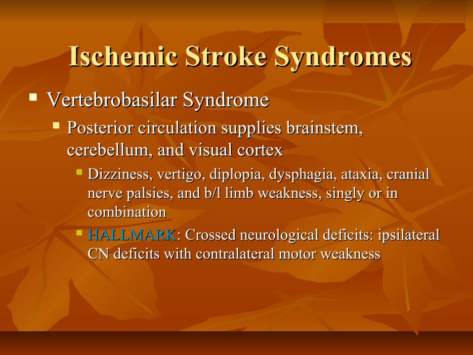
Vertebrobasilar SyndromeVertebrobasilar Syndrome Posterior circulation supplies brainstem, Posterior circulation supplies brainstem,
cerebellum, and visual cortexcerebellum, and visual cortex Dizziness, vertigo, diplopia, dysphagia, ataxia, cranial Dizziness, vertigo, diplopia, dysphagia, ataxia, cranial
nerve palsies, and b/l limb weakness, singly or in nerve palsies, and b/l limb weakness, singly or in combinationcombination
HALLMARKHALLMARK: Crossed neurological deficits: ipsilateral : Crossed neurological deficits: ipsilateral CN deficits with contralateral motor weaknessCN deficits with contralateral motor weakness
Ischemic Stroke SyndromesIschemic Stroke Syndromes

Ischemic Stroke SyndromesIschemic Stroke Syndromes
Lateral Medullary (Wallenburg) SyndromeLateral Medullary (Wallenburg) Syndrome Specific post. Circulation infarct involving Specific post. Circulation infarct involving
vertebrobasilar and/or post inferior cerebellar Art.vertebrobasilar and/or post inferior cerebellar Art. Signs:Signs:
Ipsilateral loss of facial pain and temperature with Ipsilateral loss of facial pain and temperature with contralateral loss of these senses over the bodycontralateral loss of these senses over the body
Gait and limb ataxiaGait and limb ataxia Partial ipsilateral loss of CN V, IX, X, and XIPartial ipsilateral loss of CN V, IX, X, and XI Ipsilateral Horner Syndrome may be presentIpsilateral Horner Syndrome may be present
Ischemic Stroke SyndromesIschemic Stroke Syndromes
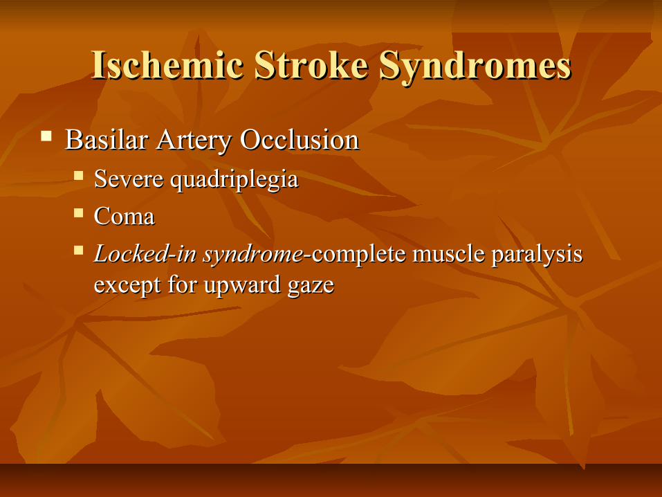
Basilar Artery OcclusionBasilar Artery Occlusion Severe quadriplegiaSevere quadriplegia ComaComa Locked-in syndrome-Locked-in syndrome-complete muscle paralysis complete muscle paralysis
except for upward gazeexcept for upward gaze

Ischemic Stroke SyndromesIschemic Stroke Syndromes
Cerebellar Infarction-subset of post. circ. infarctsCerebellar Infarction-subset of post. circ. infarcts Symptoms: “drop attack” with sudden inability to walk or Symptoms: “drop attack” with sudden inability to walk or
stand, often a/w vertigo, HA, nausea/vomiting, neck painstand, often a/w vertigo, HA, nausea/vomiting, neck pain
Diagnosis: MRI, MRA as bone artifact obscures CTDiagnosis: MRI, MRA as bone artifact obscures CT Cerebral edema develops w/in 6-12 hrs Cerebral edema develops w/in 6-12 hrs →→ increased increased
brainstem pressure and decreased LOCbrainstem pressure and decreased LOC Treatment: decrease ICP and emergent surgical Treatment: decrease ICP and emergent surgical
decompressiondecompression
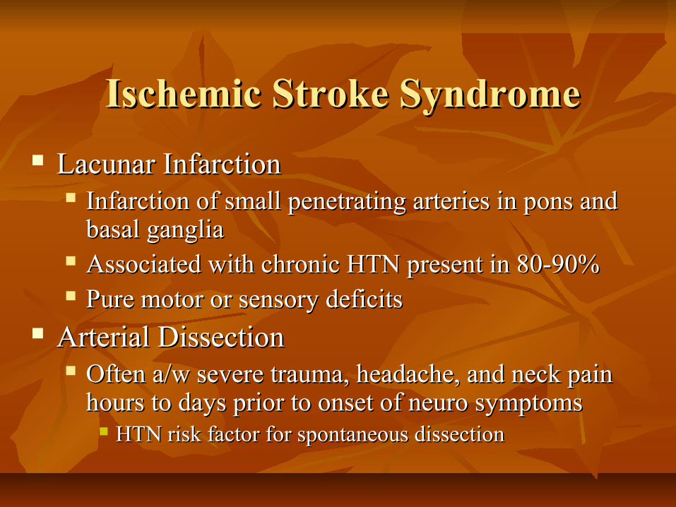
Lacunar InfarctionLacunar Infarction Infarction of small penetrating arteries in pons and Infarction of small penetrating arteries in pons and
basal gangliabasal ganglia Associated with chronic HTN present in 80-90%Associated with chronic HTN present in 80-90% Pure motor or sensory deficitsPure motor or sensory deficits
Arterial DissectionArterial Dissection Often a/w severe trauma, headache, and neck pain Often a/w severe trauma, headache, and neck pain
hours to days prior to onset of neuro symptomshours to days prior to onset of neuro symptoms HTN risk factor for spontaneous dissectionHTN risk factor for spontaneous dissection
Ischemic Stroke SyndromeIschemic Stroke Syndrome

Intracerebral HemorrhageIntracerebral Hemorrhage ICH – sudden onset HA, N/V, elevated BPICH – sudden onset HA, N/V, elevated BP Progressive focal neurologic deficits over minutesProgressive focal neurologic deficits over minutes Patients may rapidly deterioratePatients may rapidly deteriorate Exertion commonly triggers symptomsExertion commonly triggers symptoms Bleeding localized to putamen, thalamus, Bleeding localized to putamen, thalamus,
pons-pinpoint pupils, and cerebellumpons-pinpoint pupils, and cerebellum
Hemorrhagic SyndromesHemorrhagic Syndromes

Hemorrhagic SyndromesHemorrhagic Syndromes
Cerebellar HemorrhageCerebellar Hemorrhage Sudden onset dizziness, vomiting, truncal ataxia, Sudden onset dizziness, vomiting, truncal ataxia,
inability to walkinability to walk Possible gaze palsies and increasing stuporPossible gaze palsies and increasing stupor Treatment: urgent surgical decompression or Treatment: urgent surgical decompression or
hematoma evacuationhematoma evacuation

Hemorrhagic SyndromeHemorrhagic Syndrome
Subarachnoid hemorrhageSubarachnoid hemorrhage Severe HA, vomiting, decreasing LOCSevere HA, vomiting, decreasing LOC HA- often occipital or nuchal in locationHA- often occipital or nuchal in location Sudden onset of symptoms– history may reveal Sudden onset of symptoms– history may reveal
activities a/w activities a/w ↑ HTN such as defecation, coughing ↑ HTN such as defecation, coughing or intercourseor intercourse
Diagnosis-Critical PathwayDiagnosis-Critical Pathway
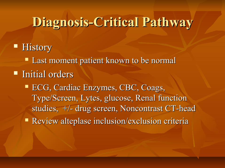
HistoryHistory Last moment patient known to be normalLast moment patient known to be normal
Initial ordersInitial orders ECG, Cardiac Enzymes, CBC, Coags, ECG, Cardiac Enzymes, CBC, Coags,
Type/Screen, Lytes, glucose, Renal function Type/Screen, Lytes, glucose, Renal function studies, +/- drug screen, Noncontrast CT-headstudies, +/- drug screen, Noncontrast CT-head
Review alteplase inclusion/exclusion criteriaReview alteplase inclusion/exclusion criteria
Diagnostic TestsDiagnostic Tests
Emergent noncontrast CT of headEmergent noncontrast CT of head Differentiate hemorrhage vs ischemiaDifferentiate hemorrhage vs ischemia
MOST ischemic strokes (-) by CT for at least 6 hrsMOST ischemic strokes (-) by CT for at least 6 hrs Hypodensity indicating infarct seen 24-48 hrsHypodensity indicating infarct seen 24-48 hrs
Can identify hemorrhage greater than 1cm, and 95% of Can identify hemorrhage greater than 1cm, and 95% of SAHSAH
If CT (-) but still considering SAH may do L.P.If CT (-) but still considering SAH may do L.P.

Depending on circumstances, other helpful testsDepending on circumstances, other helpful tests Echocardiogram – identifies mural thrombus, tumor, Echocardiogram – identifies mural thrombus, tumor,
valvular vegetations in suspected cardioembolic strokevalvular vegetations in suspected cardioembolic stroke Carotid duplex -for known/suspected high grade stenosisCarotid duplex -for known/suspected high grade stenosis Angiography – “gold standard” identifies occlusion or Angiography – “gold standard” identifies occlusion or
stenosis of large and small vessels of head/neck, stenosis of large and small vessels of head/neck, dissections and aneurysmsdissections and aneurysms
MRI scan – identifies posterior circulation strokes better MRI scan – identifies posterior circulation strokes better and ischemic strokes earlier than CTand ischemic strokes earlier than CT
Emergent MRI- considered for suspected brainstem lesion or dural Emergent MRI- considered for suspected brainstem lesion or dural sinus thrombosissinus thrombosis
MRA scan – identifies large vessel occlusions – may MRA scan – identifies large vessel occlusions – may replace angiography in the futurereplace angiography in the future
Diagnostic TestsDiagnostic Tests

Differential DiagnosisDifferential Diagnosis
Ddx of Acute Stroke (not inclusive)Ddx of Acute Stroke (not inclusive) Epidural/subdural hematomaEpidural/subdural hematoma HyponatremiaHyponatremia Brain tumor/abscessBrain tumor/abscess Postictal paralysis (Todd paralysis)Postictal paralysis (Todd paralysis) Hypertensive encephalopathyHypertensive encephalopathy Meningitis/encephalitisMeningitis/encephalitis Hyperosmotic comaHyperosmotic coma

Wernicke EncephalopathyWernicke Encephalopathy Drug toxicity (lithium, phenytoin, Drug toxicity (lithium, phenytoin,
carbamazepine)carbamazepine) Complicated MigraineComplicated Migraine Bells palsyBells palsy Multiple sclerosisMultiple sclerosis Meniere’s diseaseMeniere’s disease LabyrinthitisLabyrinthitis
Differential Diagnosis Cont.Differential Diagnosis Cont.

Special Populations In StrokeSpecial Populations In Stroke
Sickle Cell Disease (SCD)Sickle Cell Disease (SCD) Most common cause of ischemic stroke in childrenMost common cause of ischemic stroke in children 10% of patients with Sickle Cell Disease have stroke by 10% of patients with Sickle Cell Disease have stroke by
age 20age 20 SCD-SCD-↑↑ frequency of cerebral aneurysm—think SAH frequency of cerebral aneurysm—think SAH Treatment: emergent simple or exchange transfusion to Treatment: emergent simple or exchange transfusion to
decrease HbS to < 30%, thus improving blood flow and decrease HbS to < 30%, thus improving blood flow and oxygen delivery to infarct zoneoxygen delivery to infarct zone

Young Adults (age 15 to 50)Young Adults (age 15 to 50) 20% of ischemic strokes due to arterial dissection20% of ischemic strokes due to arterial dissection
Often preceded by minor traumaOften preceded by minor trauma Cardioembolic etiologies- MVP, rheumatic heart disease, Cardioembolic etiologies- MVP, rheumatic heart disease,
or paradoxical embolismor paradoxical embolism Migrainous stroke- infarction a/w typical attackMigrainous stroke- infarction a/w typical attack Air embolism-scuba diving or recent invasive procedureAir embolism-scuba diving or recent invasive procedure Drugs: heroin, cocaine, amphetaminesDrugs: heroin, cocaine, amphetamines
Special Populations In StrokeSpecial Populations In Stroke
PregnancyPregnancy ↑↑risk during peripartum and up to 6 weeks risk during peripartum and up to 6 weeks
postpartumpostpartum Contributors to risk-preeclampsia/eclampsia, decrease Contributors to risk-preeclampsia/eclampsia, decrease
in blood vol. and hormonal status following birthin blood vol. and hormonal status following birth
Special Populations In StrokeSpecial Populations In Stroke

Ischemic Stroke ManagementIschemic Stroke Management
General ManagementGeneral Management A, B, CsA, B, Cs IV, oxygen, monitor, elevate head of bed slightlyIV, oxygen, monitor, elevate head of bed slightly E.D. protocols/Notify stroke teamE.D. protocols/Notify stroke team Treat dehydration and hypotension Treat dehydration and hypotension Avoid overhydration – cerebral edemaAvoid overhydration – cerebral edema Avoid IVF with glucose – except if hypoglycemicAvoid IVF with glucose – except if hypoglycemic Fever – worsens neurologic deficitsFever – worsens neurologic deficits

Ischemic Stroke ManagementIschemic Stroke Management
HypertensionHypertension Treatment indicated for SBP Treatment indicated for SBP > 220 mm Hg or > 220 mm Hg or
mean arterial pressure > 130 mm Hgmean arterial pressure > 130 mm Hg Lowering BP too much reduces perfusion to penumbra Lowering BP too much reduces perfusion to penumbra
converting reversible injury to infarctionconverting reversible injury to infarction Use easily titratable Rx (labetalol or enalaprilat)Use easily titratable Rx (labetalol or enalaprilat) SL Ca-channel blockers should be avoidedSL Ca-channel blockers should be avoided

Management of HTN cont.Management of HTN cont.
Thrombolytic candidates- use NTG paste or Thrombolytic candidates- use NTG paste or Labetalol to reduce BP Labetalol to reduce BP << 185/115 to allow tx 185/115 to allow tx
Requirements for more aggressive treatment Requirements for more aggressive treatment exclude the use of tissue plasminogen exclude the use of tissue plasminogen activator.activator.

Thrombolysis BackgroundThrombolysis Background
NIH/NINDS studyNIH/NINDS study 624 patients, RDBPC trial IV tPA vs placebo624 patients, RDBPC trial IV tPA vs placebo
Treatment w/in 3 hrs of onsetTreatment w/in 3 hrs of onset At 3 months pts tx’d with tPA were at least 30% more At 3 months pts tx’d with tPA were at least 30% more
likely to have minimal/no disability…absolute favorable likely to have minimal/no disability…absolute favorable outcome in 11-13 percentoutcome in 11-13 percent
6.4% of patients treated with tPA developed symptomatic 6.4% of patients treated with tPA developed symptomatic ICH compared with 0.6% in placebo groupICH compared with 0.6% in placebo group
Mortality rate at 3 months not significantly differentMortality rate at 3 months not significantly different tPA group had significantly less disabilitytPA group had significantly less disability FDA approved in 1996FDA approved in 1996

tPA Dose and ComplicationstPA Dose and Complications
IV tPA –Total dose 0.9 mg/kg, max. 90mgIV tPA –Total dose 0.9 mg/kg, max. 90mg 10% as bolus, remaining infusion over 60 min.10% as bolus, remaining infusion over 60 min. BP and Neuro checks q 15 min x 2 hrs initiallyBP and Neuro checks q 15 min x 2 hrs initially
Treatment must begin w/in 3 hrs of symptoms Treatment must begin w/in 3 hrs of symptoms and meet inclusion and exclusion criteriaand meet inclusion and exclusion criteria
No ASA or heparin given x 24 hrs after txNo ASA or heparin given x 24 hrs after tx

Emergent Mngt of HTN Emergent Mngt of HTN during/following rtPA in Acute Strokeduring/following rtPA in Acute Stroke
Monitor BP closelyMonitor BP closely q 15 min x 2 hrs, then q 30 min x 6 hrs, then q 60 min for 24 hr Totalq 15 min x 2 hrs, then q 30 min x 6 hrs, then q 60 min for 24 hr Total
If SBP 180-230 or DBP 105-120 mmHgIf SBP 180-230 or DBP 105-120 mmHg 10 mg labetalol IVP q 10-20 min, max 150 mg10 mg labetalol IVP q 10-20 min, max 150 mg
If SBP > 230 or DBP 121-140 mmHgIf SBP > 230 or DBP 121-140 mmHg 10 mg labetalol may repeat q 10-20 min, max 150 mg 10 mg labetalol may repeat q 10-20 min, max 150 mg If BP not controlled by labetalol then consider nitroprusside (0.5-If BP not controlled by labetalol then consider nitroprusside (0.5-
1.0mcg/kg/min), continuous arterial monitoring advised1.0mcg/kg/min), continuous arterial monitoring advised
If DBP > 140 mmHgIf DBP > 140 mmHg Infuse sodium nitroprusside (0.5-1.0mcg/kg/min), continuous Infuse sodium nitroprusside (0.5-1.0mcg/kg/min), continuous
arterial monitoring advisedarterial monitoring advised

IV Thrombolysis Criteria in Ischemic StrokeIV Thrombolysis Criteria in Ischemic Stroke
Inclusion criteriaInclusion criteria Age 18 years or olderAge 18 years or older Time since onset well established to be Time since onset well established to be < 3 hrs< 3 hrs Clinical diagnosis of ischemic strokeClinical diagnosis of ischemic stroke

Exclusion criteriaExclusion criteria Minor/rapidly improving neurologic signsMinor/rapidly improving neurologic signs Evidence of intracranial hemorrhage on Evidence of intracranial hemorrhage on
pretreatment noncontrast head CTpretreatment noncontrast head CT History of intracranial hemorrhageHistory of intracranial hemorrhage High suspicion of SAH despite normal CTHigh suspicion of SAH despite normal CT GI or GU bleeding within last 21 daysGI or GU bleeding within last 21 days
Criteria for IV Thrombolysis cont.Criteria for IV Thrombolysis cont.

Exclusion criteriaExclusion criteria Known bleeding diathesisKnown bleeding diathesis
Platelet count < 100,000 /mmPlatelet count < 100,000 /mm33
Heparin within 48 hours and has an elevated PTTHeparin within 48 hours and has an elevated PTT Current use of anticoagulation or PT > 15 seconds or Current use of anticoagulation or PT > 15 seconds or
INR > 1.7INR > 1.7
Criteria for IV Thrombolysis cont.Criteria for IV Thrombolysis cont.

Exclusion criteriaExclusion criteria Intracranial surgery, serious head trauma or Intracranial surgery, serious head trauma or
previous stroke within 3 monthsprevious stroke within 3 months Major surgery within 14 daysMajor surgery within 14 days Recent arterial puncture at non compressible siteRecent arterial puncture at non compressible site Lumbar puncture within 7 daysLumbar puncture within 7 days Seizure at onset of strokeSeizure at onset of stroke
Criteria for IV Thrombolysis cont.Criteria for IV Thrombolysis cont.

Exclusion criteriaExclusion criteria History of ICH, AVM or aneurysmHistory of ICH, AVM or aneurysm Recent MIRecent MI Sustained pretreatment systolic pressure > 185 mmHg or Sustained pretreatment systolic pressure > 185 mmHg or
diastolic pressure > 110 mmHg despite aggressive diastolic pressure > 110 mmHg despite aggressive treatment to reduce BP to within these limitstreatment to reduce BP to within these limits
Blood glucose < 50 or > 400 mg/dLBlood glucose < 50 or > 400 mg/dL
Criteria for IV Thrombolysis cont.Criteria for IV Thrombolysis cont.

Drug Therapy in Ischemic StrokeDrug Therapy in Ischemic Stroke
Majority of pts not thrombolytic candidatesMajority of pts not thrombolytic candidates Antiplatelet agents-cornerstone for 2Antiplatelet agents-cornerstone for 2°° prevention prevention
Antiplatelet agentsAntiplatelet agents ASA: ASA: ↓ risk 20-25% vs placebo↓ risk 20-25% vs placebo
50-300 mg dose and will not interfere with tPA therapy50-300 mg dose and will not interfere with tPA therapy
Dipyridamole: alone (200mg BID) Dipyridamole: alone (200mg BID) ↓ risk 15%↓ risk 15% Plavix: (75 mg qd) 0.5% absolute Plavix: (75 mg qd) 0.5% absolute annual risk annual risk
reduction when compared to ASAreduction when compared to ASA Good Rx for pts who cannot tolerate or fail ASA Good Rx for pts who cannot tolerate or fail ASA

Heparin: unprovenHeparin: unproven Pts may expect fewer strokes but benefit is offset Pts may expect fewer strokes but benefit is offset
by increased ICHby increased ICH Similar results with LMWHSimilar results with LMWH Use of UFH, LMWH, or heparinoids to tx a Use of UFH, LMWH, or heparinoids to tx a
specific stroke subtype or TIA cannot be specific stroke subtype or TIA cannot be recommended based on available evidence.recommended based on available evidence.
AnticoagulantsAnticoagulants

TIA ManagementTIA Management
Admit-Evaluate for cardiac sources of emboli Admit-Evaluate for cardiac sources of emboli or high grade stenosis of carotid arteriesor high grade stenosis of carotid arteries
Rx: ASARx: ASA UFH-for high risk of recurrence UFH-for high risk of recurrence
Known high grade stenosis in appropriate distribution Known high grade stenosis in appropriate distribution of symptoms, cardioembolic source, Crescendo TIAs, of symptoms, cardioembolic source, Crescendo TIAs, TIAs despite antiplatelet therapyTIAs despite antiplatelet therapy
Urgent CEA for TIAs that resolve in Urgent CEA for TIAs that resolve in < 6 hrs < 6 hrs and a/w > 70% stenosis of carotid arteryand a/w > 70% stenosis of carotid artery

Treat HTN Treat HTN >220 mm Hg systolic or > 120 mm Hg >220 mm Hg systolic or > 120 mm Hg diastolic using labetalol or nitroprussidediastolic using labetalol or nitroprusside Reduce gradually to prehemorrhage levelsReduce gradually to prehemorrhage levels
Elevate HOB to 30°Elevate HOB to 30° HHyperventilation-target PaCO2 30-35 mm Hg yperventilation-target PaCO2 30-35 mm Hg Osmotherapy Osmotherapy
Mannitol (0.25-1.0 g/kg IV), and lasix (10 mg IV)– target Mannitol (0.25-1.0 g/kg IV), and lasix (10 mg IV)– target serum osmolality serum osmolality ≤ 310 mOsm/kg≤ 310 mOsm/kg
Hyperventilation/osmotherapy used Hyperventilation/osmotherapy used for signs of progressive for signs of progressive ↑↑ ICP ICP i.e. mass effect, midline shift or herniationi.e. mass effect, midline shift or herniation
Steroids – not recommendedSteroids – not recommended
ICH ManagementICH Management

ICH Management cont.ICH Management cont.
ICP Monitoring considered if GCS ICP Monitoring considered if GCS < 9< 9 Consider seizure prophylaxis with phenytoinConsider seizure prophylaxis with phenytoin Surgery – controversialSurgery – controversial
Depends on neuro status of pt, size and location of Depends on neuro status of pt, size and location of hemorrhagehemorrhage
Best benefit in cerebellar hemorrhage Best benefit in cerebellar hemorrhage

SAH ManagementSAH Management
Major complications w/in 1Major complications w/in 1stst 24 hrs 24 hrs Rebleeding and vasospasmRebleeding and vasospasm
To To ↓ rebleed risk: reduce SBP to 160 mm Hg and/or maintain MAP ↓ rebleed risk: reduce SBP to 160 mm Hg and/or maintain MAP of 110 mm Hgof 110 mm Hg
Cerebral ischemia 2° to vasospasm occurs 2-21 days after Cerebral ischemia 2° to vasospasm occurs 2-21 days after aneurysm ruptureaneurysm rupture
Nimodipine 60 mg PO q 6 hr-↓ incidence and severity of Nimodipine 60 mg PO q 6 hr-↓ incidence and severity of vasospasmsvasospasms
Prophylactic treatment of pain, N/V and seizuresProphylactic treatment of pain, N/V and seizures Obtain Neurosurgical consultationObtain Neurosurgical consultation

Summary of Summary of Emergency Department RoleEmergency Department Role
Stabilization- A,B,Cs Stabilization- A,B,Cs Quick accurate diagnosis-hx, PE/neuro examQuick accurate diagnosis-hx, PE/neuro exam Determine appropriateness of fibrinolyticsDetermine appropriateness of fibrinolytics
NIH stroke scaleNIH stroke scale
Early neurology/neurosurgery consultEarly neurology/neurosurgery consult Manage blood pressure appropriatelyManage blood pressure appropriately

Free HANDi Stroke Rx for PDAFree HANDi Stroke Rx for PDA
program includes:program includes: An NIHSS calculatorAn NIHSS calculator Indications and Contraindications for t-PA useIndications and Contraindications for t-PA use A t-PA dosing calculatorA t-PA dosing calculator Sample orders for patients receiving t-PASample orders for patients receiving t-PA ReferencesReferences

HANDi Stroke Rx HANDi Stroke Rx
To download and install HANDi Stroke Rx, follow these simple instructions: To download and install HANDi Stroke Rx, follow these simple instructions: 1. On your computer, open your web browsing program such as Netscape 1. On your computer, open your web browsing program such as Netscape
Navigator or Microsoft Internet Explorer. Navigator or Microsoft Internet Explorer. 2. Go to the FERNE website, 2. Go to the FERNE website, http://www.FERNE.org http://www.FERNE.org 3. Click on the “Download the Stroke Management Program for Handhelds” 3. Click on the “Download the Stroke Management Program for Handhelds”
button. button. 4. From the “Software” page, select the “NIHSS, Stroke Management, and t-4. From the “Software” page, select the “NIHSS, Stroke Management, and t-
PA Administration” link. PA Administration” link. 5. Select the type of computer system you are running (IBM/PC or Macintosh) 5. Select the type of computer system you are running (IBM/PC or Macintosh)
from the Handheld Computer Stroke Program screen. from the Handheld Computer Stroke Program screen. 6. Input your name and email address on the “Handheld Computer Stroke 6. Input your name and email address on the “Handheld Computer Stroke
Program Download” page, then click the “Submit” button. Program Download” page, then click the “Submit” button. Follow the operating system-specific instructions for the remainder of the Follow the operating system-specific instructions for the remainder of the
installation process. installation process.

QuestionsQuestions
1. T/F -Seizure at onset of stroke wouldn’t preclude use of 1. T/F -Seizure at onset of stroke wouldn’t preclude use of tPA.tPA.
2. T/F -Maximum dose of tPA is 100 mg2. T/F -Maximum dose of tPA is 100 mg 3. T/F –The use of heparin after tPA is prohibited for 24 3. T/F –The use of heparin after tPA is prohibited for 24
hours.hours. 4. T/F –Middle Cerebral artery occlusion in the dominant 4. T/F –Middle Cerebral artery occlusion in the dominant
hemisphere may be associated with receptive or expressive hemisphere may be associated with receptive or expressive aphasia.aphasia.
5. T/F –Asians, Blacks and Caucasians are at increased risk 5. T/F –Asians, Blacks and Caucasians are at increased risk for intracerebral hemorrhage.for intracerebral hemorrhage.

AnswersAnswers
1. F- seizures at onset is a contraindication to 1. F- seizures at onset is a contraindication to tPAtPA
2. F – max dose is 90 mg2. F – max dose is 90 mg 3. T3. T 4. T4. T 5. F – Only Asians and Blacks5. F – Only Asians and Blacks