structural and functional characterisation of three novel
TRANSCRIPT

This is a repository copy of Structural and functional characterisation of three novel fungal amylases with enhanced stability and pH tolerance.
White Rose Research Online URL for this paper:https://eprints.whiterose.ac.uk/151337/
Version: Accepted Version
Article:
Roth, Christian orcid.org/0000-0001-5806-0987, Moroz, Olga V., Turkenburg, Johan P. et al. (8 more authors) (2019) Structural and functional characterisation of three novel fungal amylases with enhanced stability and pH tolerance. International Journal of Molecular Sciences. 4902. ISSN 1422-0067
https://doi.org/10.3390/ijms20194902
[email protected]://eprints.whiterose.ac.uk/
Reuse
Items deposited in White Rose Research Online are protected by copyright, with all rights reserved unless indicated otherwise. They may be downloaded and/or printed for private study, or other acts as permitted by national copyright laws. The publisher or other rights holders may allow further reproduction and re-use of the full text version. This is indicated by the licence information on the White Rose Research Online record for the item.
Takedown
If you consider content in White Rose Research Online to be in breach of UK law, please notify us by emailing [email protected] including the URL of the record and the reason for the withdrawal request.

Int. J. Mol. Sci. 2019, 20, x; doi: FOR PEER REVIEW www.mdpi.com/journal/ijms
Article 1
Structural and functional characterisation of three 2
novel fungal amylases with enhanced stability and 3
pH tolerance 4
Christian Roth 1,4# , Olga V Moroz 1#, Johan P. Turkenburg1, Elena Blagova 1, Jitka Waterman 1,5, 5
Antonio Ariza1,6 , Li Ming 2, Sun Tianqi 2, Carsten Andersen3, Gideon J Davies 1 and Keith S 6 Wilson 1* 7
1 York Structural Biology Laboratory, Department of Chemistry, University of York, Heslington, York, YO10 8 5DD, UK 9 2 Novozymes (China) Investment Co. Ltd, 14 Xinli Road, Haidian District, Beijing 100085, People’s Republic 10 of China 11 3 Novozymes (Denmark), Krogshojvej 36, DK-2880 Bagsvaerd, Denmark 12 4 Present address: Carbohydrates: Structure and Function, Biomolecular Systems, Max Planck Institute of 13 Colloids and Interfaces,14195 Berlin, Germany 14 5 Present address: Diamond Light Source, Diamond House, Harwell Science and Innovation Campus, Fermi 15 Ave, Didcot OX11 0DE 16 6 Present address: Sir William Dunn School of Pathology, University of Oxford, Oxford, OX1 3RE, UK 17 # The first two authors contributed equally to this work. 18 * Correspondence: [email protected] +44 1904 328262 19
Received: date; Accepted: date; Published: date 20
Abstract: Amylases are probably the best studied glycoside hydrolases and have a huge 21 biotechnological value for industrial processes on starch. Multiple amylases from fungi and 22 microbes are currently in use. Whereas bacterial amylases are well suited for many industrial 23 processes due to their high stability, fungal amylases are recognized as safe and are preferred in the 24
food industry, although they lack the pH tolerance and stability of their bacterial counterparts. Here, 25 we describe three amylases, two of which have a broad pH spectrum extending to pH 8 and higher 26 stability well suited for a broad set of industrial applications. These enzymes have the characteristic 27 GH13 α-amylase fold with a central (β/α)8-domain, an insertion domain with the canonical calcium 28
binding site and a C-terminal β-sandwich domain. The active site was identified based on the 29 binding of the inhibitor acarbose in form of a transglycosylation product, in the amylases from 30 Thamnidium elegans and Cordyceps farinosa. The three amylases have shortened loops flanking the 31 nonreducing end of the substrate binding cleft, creating a more open crevice. Moreover, a potential 32 novel binding site in the C-terminal domain of the Cordyceps enzyme was identified, which might 33
be part of a starch interaction site. In addition, Cordyceps farinosa amylase presented a successful 34 example of using the microseed matrix screening technique to significantly speed-up crystallization. 35
Keywords: α-amylase; starch degradation; biotechnology; structure 36 37
1. Introduction 38
The use of enzymes in industrial processes is a multi-billion-dollar market. One of the first 39 enzymes discovered in 1833 was diastase, an enzyme able to hydrolyze starch [1]. Nowadays, 40 amylases, also able to hydrolyze starch, constitute up to 25% of the market for enzymes and have 41
virtually replaced chemical methods for degrading starch in the industrial sector (reviewed in [2]). 42 Amylases are the most important class of enzymes for degrading starch and can be subdivided into 43 three subclasses: α-, β-, and gluco-amylases based on their reaction specificity and product profiles. 44
Int. J. Mol. Sci. 2019, 20, x FOR PEER REVIEW 2 of 15
α-amylases degrade the α- 1,4 linkage between adjacent glucose units and are extensively used for 45
example in bioethanol production or in washing powder and detergents [3] (and reviewed in [4]). 46 One of the most widely used α-amylases is that from Bacillus licheniformis, known under the 47 tradename “Termamyl”. Microbial amylases are generally used in detergent applications and other 48 industrial processes, including bioethanol production, with new amylases, in particular those from 49
hyperthermophilic organisms, offering further improvement in the production process (reviewed in 50 [5]). 51
a-amylases belong to glycoside hydrolase family 13 (GH13) in the CAZy database classification 52 [6]. They have a (β/α)8 barrel domain harbouring the active site, a subdomain which includes the 53 canonical calcium binding site inserted between the third β-strand and the third α-helix and a C-54
terminal β-sandwich domain, thought to be important for the interaction with raw starch (reviewed 55 in [7]), [8, 9]. Amylases follow a retaining mechanism with an aspartate as nucleophile and one 56 glutamate as general acid/base [10, 11]. Up to ten consecutive sugar subsites forming the active site 57 cleft, have been identified in bacterial amylases, [12]. 58
To date, recombinant fungal amylases have been isolated from mesophilic hosts such as 59
Aspergillus oryzae and are of particular interest to the food industry as they match the temperature 60 and pH range used in typical applications in the baking process, where they are active in the dough 61 but inactivated during baking. Due to the widespread use of fungal enzymes for the production of 62 food and food ingredients (such as citric acid), they are classified as GRAS (generally recognized as 63
safe) organisms by organizations including the FDA (US Food and Drug Administration) [13]. 64 Up till now, fungal enzymes with a higher pH-tolerance and thermostability have not been 65
reported. Here we describe the structure and function of three novel α-amylases from Cordyceps 66 farinosa (CfAM), Rhizomucor pusillus (RpAM) and Thamnidium elegans (TeAM) with a higher stability 67 and pH-tolerance with the potential to act as novel biocatalysts for various industrial processes. The 68
sequence of all three enzymes groups them in the GH13 sub-family 1 along with, for example, the 69 amylase from Aspergillus oryzae (also known as TAKA amylase). However, unlike other fungal 70 amylases, the enzymes in this study have been shown to have a broad pH profile with an optimum 71 around pH 5 while retaining activity at pH 8. Furthermore, their more open crevice leads to the 72 production of longer oligomers compared to TAKA amylase. 73
The native RpAM and TeAM have a four-domain fold with a carbohydrate binding domain 74 (CBM20) at the C-terminus and a short serine-rich linker in between, while native CfAM lacks this 75 CBM20 domain. In this study, only the core of the amylases including the A, B and C domains was 76 cloned and expressed. In addition, crystallisation of Cordyceps farinosa amylase again demonstrates 77
the power of the microseed matrix screening technique [14]. 78
2. Results 79
2.1. Biochemical characterization 80
The pH-, temperature- and product profiles were characterized for all three amylases. Of great 81 desire are amylases with a broader pH-tolerance compared to TAKA amylase. Our analysis showed 82
that all three amylases have a pH optimum around 5. Whereas TeAM has no significant activity above 83 pH 7, RpAM and CfAM retain significant activity at pH 7 extending up to a pH of 9 (Figure 1a). In 84 particular CfAM shows the highest pH-tolerance, retaining 70 % of its activity at pH 8. RpAM and 85 TeAM both show a pronounced shoulder suggesting the involvement of more titratable residues in 86 the substrate recognition and catalysis process. The temperature profiles reveal that RpAM and 87
CfAM also have a considerably higher thermotolerance compared to TAKA and TeAM (Figure 1b). 88 In particular, RpAM retains full activity even at 80°C, making it an attractive enzyme for industrial 89 high temperature starch saccharification processes. Compared to TAKA amylase, all three amylases 90 show a tendency to produce higher amounts of oligomers with a degree of polymerization (dp) of 91 three, with trace amounts of oligomers with a dp of up to seven for TeAM (Figure 1c). 92

Int. J. Mol. Sci. 2019, 20, x FOR PEER REVIEW 3 of 15
93
Figure 1. Biochemical characterization of RpAM, CfAM. TeAM and TAKA. (a) pH-profile of all three 94 amylases in comparison with TAKA amylase. (b) Temperature profile of all three amylases in 95 comparison with TAKA amylase. (c) Product profile of all three amylases and the abundance of 96 oligomers with a degree of polymerization (dp) of 1 to 7 after hydrolysis of starch. 97
2.2. Overall fold 98
The structures were solved using molecular replacement starting from the A. oryzae amylase as 99
template (pdb-ID: 7taa and 3vx0) to a resolution of 1.4 Å for RpAM, 1.2 Å for TeAM and 1.35 Å for 100 CfAM respectively. The final model of RpAM includes two monomers in the asymmetric unit 101 comprising residues 1 to 438 in both chains, which superpose on each other with an r.m.s.d. of 0.54 102 Å. The model of TeAM contains one monomer in the asymmetric unit including residues 1 to 438. 103
Int. J. Mol. Sci. 2019, 20, x FOR PEER REVIEW 4 of 15
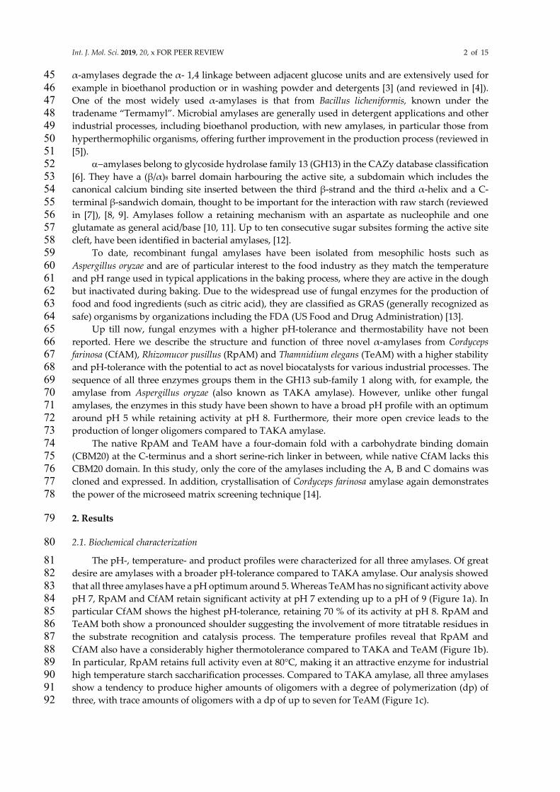
For CfAM, there are two monomers in the asymmetric unit comprising residues 19 to 459 for chain 104
A and 19 to 460 for chain B, which superpose with an r.m.s.d. of 0.3 Å. All three amylases have the 105 classical domain structure with a central (β/α)8-barrel with the active site located on its C-terminal 106 face, together with a small subdomain, inserted between the third strand and helix and a C-terminal 107 β-sandwich (Figure 2a). All three superpose with each other (Figure 2b) and with TAKA-amylase 108
with an r.m.s.d. between 0.6 to 0.9 Å for up to 423 residues. Two conserved disulphide bridges 109 stabilize flexible loops in subdomains A and B. There is an additional disulphide bridge in CfAM, 110 located in the C-terminal domain. All three α-amylases have the conserved canonical calcium binding 111 site located between the (β/α)8-barrel and the insertion domain B. 112
113
Figure 2. Structural overviews. (a) Ribbon representation of the structure of CfAM amylase in ribbon 114 representation. The domains are coloured separately with the central barrel in purple. subdomain B 115 in yellow and the C-terminal b-sandwich in green. The bound ligands acarbose transglycosylation 116 product (ATgp) and maltose are shown as spheres. (b) Structural superposition of CfAM (purple) 117 TeAM (orange) and RpAM (green). 118
2.3. Ligand binding site: 119
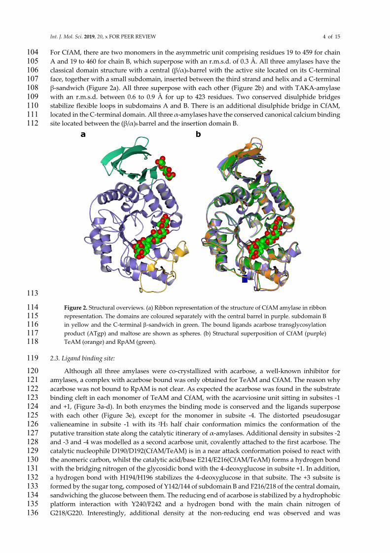
Although all three amylases were co-crystallized with acarbose, a well-known inhibitor for 120 amylases, a complex with acarbose bound was only obtained for TeAM and CfAM. The reason why 121
acarbose was not bound to RpAM is not clear. As expected the acarbose was found in the substrate 122 binding cleft in each monomer of TeAM and CfAM, with the acarviosine unit sitting in subsites -1 123 and +1, (Figure 3a-d). In both enzymes the binding mode is conserved and the ligands superpose 124 with each other (Figure 3e), except for the monomer in subsite -4. The distorted pseudosugar 125 valieneamine in subsite -1 with its 2H3 half chair conformation mimics the conformation of the 126
putative transition state along the catalytic itinerary of α-amylases. Additional density in subsites -2 127 and -3 and -4 was modelled as a second acarbose unit, covalently attached to the first acarbose. The 128 catalytic nucleophile D190/D192(CfAM/TeAM) is in a near attack conformation poised to react with 129 the anomeric carbon, whilst the catalytic acid/base E214/E216(CfAM/TeAM) forms a hydrogen bond 130 with the bridging nitrogen of the glycosidic bond with the 4-deoxyglucose in subsite +1. In addition, 131
a hydrogen bond with H194/H196 stabilizes the 4-deoxyglucose in that subsite. The +3 subsite is 132 formed by the sugar tong, composed of Y142/144 of subdomain B and F216/218 of the central domain, 133 sandwiching the glucose between them. The reducing end of acarbose is stabilized by a hydrophobic 134 platform interaction with Y240/F242 and a hydrogen bond with the main chain nitrogen of 135
G218/G220. Interestingly, additional density at the non-reducing end was observed and was 136
Int. J. Mol. Sci. 2019, 20, x FOR PEER REVIEW 5 of 15
modelled as an additional acarbose unit in subsites -2 and -3 and -4. The glucose in subsite -2 is 137
stabilized by multiple hydrogen bonds with D323/325, R327/329 and W375/377. The glucose in subsite 138 -3 is held in place by only one hydrogen bond with D323/325. The last visible part of the acarbose 139 molecule is the acarviosine unit in subsite -4, which is not stabilized by direct interactions with the 140 protein. Furthermore, the acarviosine unit is in two different positions in the two structures, reflecting 141
the lack of strong stabilizing interactions between the ligand and the protein beyond subsite -3 (Figure 142 3e). 143
144
Figure 3. Acarbose transglycosylation product binding in CfAM and TeAM. (a) and (b) Stick 145 representation of the acarbose derived transglycosylation product in the substrate binding crevice of 146 CfAM and TeAM respectively. The 2Fo-Fc electron density around the ligands is contoured at 147 0.3 e/Å3. The interacting residues are shown as cylinders. (c) and (d) Hydrogen bonding pattern 148 between ATgp and CfAM and TeAM in the active site. (e) Stereo view of the overlay of the binding 149 crevice of CfAM (purple) and TeAM (orange). The residues and the ligands overlap very closely with 150 the only major difference being the orientation of the acarviosine subunit in subsite -4. 151
2.4. Secondary glucose binding site 152
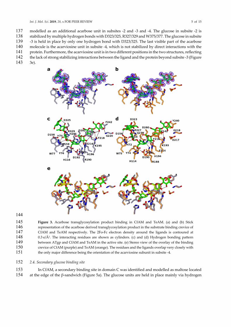
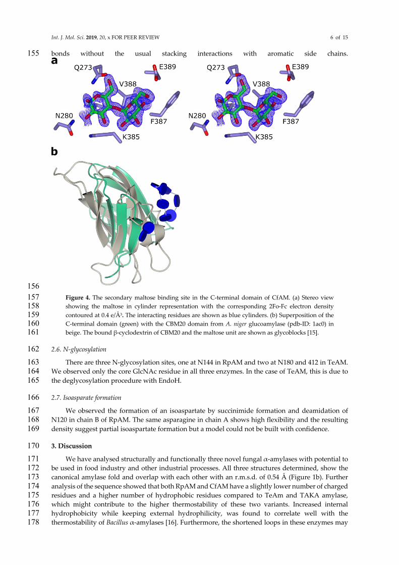
In CfAM, a secondary binding site in domain C was identified and modelled as maltose located 153
at the edge of the β-sandwich (Figure 5a). The glucose units are held in place mainly via hydrogen 154
Int. J. Mol. Sci. 2019, 20, x FOR PEER REVIEW 6 of 15
bonds without the usual stacking interactions with aromatic side chains. 155
156
Figure 4. The secondary maltose binding site in the C-terminal domain of CfAM. (a) Stereo view 157 showing the maltose in cylinder representation with the corresponding 2Fo-Fc electron density 158 contoured at 0.4 e/Å3. The interacting residues are shown as blue cylinders. (b) Superposition of the 159 C-terminal domain (green) with the CBM20 domain from A. niger glucoamylase (pdb-ID: 1ac0) in 160 beige. The bound β-cyclodextrin of CBM20 and the maltose unit are shown as glycoblocks [15]. 161
2.6. N-glycosylation 162
There are three N-glycosylation sites, one at N144 in RpAM and two at N180 and 412 in TeAM. 163 We observed only the core GlcNAc residue in all three enzymes. In the case of TeAM, this is due to 164
the deglycosylation procedure with EndoH. 165
2.7. Isoasparate formation 166
We observed the formation of an isoaspartate by succinimide formation and deamidation of 167 N120 in chain B of RpAM. The same asparagine in chain A shows high flexibility and the resulting 168
density suggest partial isoaspartate formation but a model could not be built with confidence. 169
3. Discussion 170
We have analysed structurally and functionally three novel fungal α-amylases with potential to 171 be used in food industry and other industrial processes. All three structures determined, show the 172 canonical amylase fold and overlap with each other with an r.m.s.d. of 0.54 Å (Figure 1b). Further 173
analysis of the sequence showed that both RpAM and CfAM have a slightly lower number of charged 174 residues and a higher number of hydrophobic residues compared to TeAm and TAKA amylase, 175 which might contribute to the higher thermostability of these two variants. Increased internal 176 hydrophobicity while keeping external hydrophilicity, was found to correlate well with the 177 thermostability of Bacillus α-amylases [16]. Furthermore, the shortened loops in these enzymes may 178

Int. J. Mol. Sci. 2019, 20, x FOR PEER REVIEW 7 of 15
also contribute to the overall rigidity of the enzymes and therefore the thermostability as observed 179
for other enzymes as well [17, 18]. 180 The substrate crevice in all three amylases, if defined on the basis of protein carbohydrate 181
interactions, spans from subsite -3 to +3. Having only three defined subsites for the non-reducing end 182 is common for amylases and is in line with the number of donor subsites described for TAKA-183
amylase. Potentially, there could be more subsites for additional carbohydrate units at the reducing 184 end, which might connect the active site crevice with the observed second binding site (see below). 185
The observed complexes are most likely the result of limited transglycosylation, an unusual side 186 reaction previously reported in crystallo for several amylases, for example TAKA-amylase [19]. 187 Though this reaction is common in the closely related CGTases (GH13_2) and amylomaltases (GH77), 188
it was not observed in solution for α-amylases. However, in crystals, transglycosylation products 189 with 10 or more units have been reported as a result of multiple transglycosylation events. 190 Interestingly, the final complex always has the pseudosaccharide unit, thought to mimic the 191 transition state, in the -1 subsite, rendering the enzyme inactive. Other binding modes are clearly 192 possible as evidenced by the final product and a pre-Michaelis complex observed for GH77 Thermus 193
aquaticus amylomaltase with acarbose [20]. 194 All three amylases have as hallmark a shortened loop between β2/α3 and two shorter loops in 195
subdomain B located between β3 and α4 of the central (β/α)8-barrel, compared to structures of other 196 fungal amylases, e.g. TAKA-amylase (Figure 5a). The importance of subdomain B for the 197
physicochemical properties, for example pH-stability, as well as substrate and product specificity, is 198 well known [21-24]. Indeed, the shorter loops open up the substrate crevice on the non-reducing end 199 (Figure 5b), which might explain the shift in the product profile for all three amylases towards 200 oligomers with a higher dp compared to TAKA amylase (Figure 1c, Figure 5c). 201
202
Figure 5. (a) Stereo view of all three amylases compared to TAKA-amylase with the three shortened 203 loops in the front marked with arrows. The ligand in CfAM is shown as sticks to identify the active 204 site. (b) Surface representation of CfAM with the bound ligand. The substrate is more open on the 205 donor subsite. (c) Surface representation of TAKA-amylase. The elongated loops create a more 206

Int. J. Mol. Sci. 2019, 20, x FOR PEER REVIEW 8 of 15
restricted active site crevice precluding the binding mode observed in CfAM and TeAM due to steric 207 clashes. . 208
The C-terminal domain in α-amylases is implicated in starch binding and shows structural 209 similarity to classic CBM domains, based on an analysis using PDBeFOLD [25]. The additional 210 binding site in this domain in CfAM strengthen the role of this domain in substrate binding. 211 Additional carbohydrate binding sites have been observed as well for example in barley α-amylase 212
1 [26]. While none of these sites overlap with the binding site seen in CfAM, a structure of a CBM20 213 in complex with β-cyclodextrin revealed two binding sites, with the site termed SB1 in close 214 proximity to the binding site in CfAM (Figure 4b) [27]. This was confirmed to be the primary binding 215 site for the interaction with raw starch and it is likely that the observed binding site in CfAM is a 216
genuine carbohydrate binding site. Furthermore, it is intriguing to speculate about a potential path 217 from the primary substrate crevice to the secondary glucose binding site, which could be rather easily 218 thought as a simple extension of the acarbose from the reducing end. 219
Only limited information about the influence of glycosylation on amylase activity is available. It 220 was shown that for α-amylase Amy1 from the yeast Cryptococcus flavus N-glycosylation enhances 221
thermostability and resistance to proteolytic degradation [28]. The same effect is observed for 222 Trichoderma reesei Cel7a [29]. Indeed N144 is located in an extended loop and N-glycosylation might 223 help to shield the loop against proteolytic attack. The other two glycosylation sites are located in or 224 at the beginning of secondary structure elements, with N412 being located in the C- domain. 225
The observed isoaspartate formation is thought usually to be an age related side effect of protein 226
decomposition but a functional role cannot be ruled out [30]. Indeed, it was shown in GH77 enzymes 227 that such unusual posttranslational rearrangement might play a functional role in glycoside 228 hydrolases [31, 32]. The observed isoaspartate is located in one of the shortened loops in subdomain 229 B close to the substrate binding cleft, suggesting a functional role in CfAM as well. 230
4. Materials and Methods 231
4.1. Macromolecule production 232
The coding sequence of CfAM for the A, B and C domains was amplified from Cordyceps farinosa 233 gDNA by the polymerase chain reaction (PCR). The PCR fragment was obtained using primer pairs: 234 5’-ACACAACTGGGGATCCACCATGAAGCTTACTGCGTCCCTC-3’ and 5’-235
GATGGTGATGGGATCCTTACTGCGCAACAAAAACAATGGG-3’. The fragment was then ligated 236 in the expression vector pSUN515 using BamHI and XhoI restriction sites. The ligation protocol was 237 performed according to the IN-FUSION™ Cloning Kit instructions. A transformation of TOP10 238 competent E. coli cells (Tiangen, China) with the plasmid, containing the CfAM gene, was performed 239 and positive clones confirmed by sequencing. The transformation of Aspergillus oryzae (strain 240
MT3568) with the expression vector comprising CfAM gene was performed according to patent 241 application WO95/002043 [33]. After incubation for 4-7 days at 37°C spores of four transformants 242 were inoculated into 3 ml of YPM medium. After 3-day cultivation at 30°C, the culture broths were 243 analysed by SDS-PAGE to identify the transformant producing the largest amount of recombinant 244 mature amylase with an estimated size of 48 kDa. Spores from the best expressing transformant were 245
cultivated in YPM medium in shake flasks for 4 days at a temperature of 30°C. The culture broth was 246 harvested by filtration using a 0.2 µm filter device, and the filtered fermentation broth was used for 247 purification and further assays. 248
RpAM was cloned and expressed in a similar manner as CfAM while TeAM was expressed in 249
Pichia pastoris with a similar protocol to that described for the lipase from Gibberella zeae [34].The entire 250 coding sequence of TeAM was amplified from cDNA by the polymerase chain reaction and 251 transformation into ElectroMax DH10B competent cells (Invitrogen) by electroporation. Transformed 252 cells were plated on LB plates containing 100 mM ampicillin. After overnight incubation at 27°C, a 253 positive clone was selected by colony PCR and confirmed by sequencing. The plasmid DNA of the 254
positive clone was linearized with PmeI (NEB) and transformed into Pichia pastoris KM71 (Invitrogen) 255
Int. J. Mol. Sci. 2019, 20, x FOR PEER REVIEW 9 of 15
following the manufacturer’s instructions. An amylase positive clone was inoculated into 3 ml BMSY 256
and incubated at 28°C for 3 days until the OD600 reached 20. Methanol was added to the culture 257 daily to a final concentration of 0.5% for the following 4 days. On day 4 of induction, the culture 258 supernatant was separated from the cells by centrifugation and the pH of the supernatant was 259 adjusted to 7.0. 260
The CfAM culture broth was precipitated with ammonium sulphate (80% saturation), then 261 dialyzed with 20 mM Na-acetate at pH 5.0. The solution was loaded on to a Q Sepharose Fast Flow 262 column (GE Healthcare) equilibrated with 20 mM Na Acetate at pH 5.0. Protein was eluted with a 263 salt gradient from zero to 1 M NaCl Fractions were analysed for amylase activity and pooled 264 accordingly. The flow-through fraction, containing the bulk of amylase activity was supplemented 265
with ammonium sulphate to a final concentration of 1.2 M and then loaded on to Phenyl Sepharose 266 6 Fast Flow column (GE Healthcare). The activity was eluted by a linear gradient of decreasing salt 267 concentration. The fractions with activity were analysed by SDS-PAGE and then concentrated for 268 further use. 269
Amylase activity was detected by AZCL-HE-amylose (Megazyme International Ireland Ltd.) as 270
substrate. 10µl enzyme sample and 120 µl 0.1% substrate at pH 7 were mixed in a Microtiter plate 271 and incubated at 50°C for 30 min. Then 70 µl supernatant was transferred to a new microtiter plate 272 and the absorption at 595 nm determined. All reactions were done as duplicates. 273
4.2. Biochemical characterisation 274
pH-Optimum: 275
To determine the pH-Optimum each enzyme (3 µl of a 0.5 mg/ml solution) was incubated with 276 40 µl 1% substrate (AZCL-HE-amylose (Megazyme International Ireland Ltd.)). The pH between 2 277 and 11 was adjusted using 100 µl of B&R buffer (Britton-Robinson buffer: 0.1 M boric acid, 0.1 M 278 acetic acid, and 0.1 M phosphoric acid, adjusted to pH-values 3.0, 4.0, 5.0, 6.0, 7.0, 8.0, 9.0, 10.0 and 279
11.0 with HCl or NaOH) [35]. The reactions were incubated at 30°C for 30 minutes and afterwards 280 60µl were transferred in a new microtiter plate and the absorption was measured at 595 nm. 281
Temperature-Optimum: 282
To determine the Temperature-Optimum each enzyme was incubated with 100 µl 0.1 % 283 substrate (AZCL-HE-amylose (Megazyme International Ireland Ltd.)) in 50 mM Na Acetate pH 4.3. 284
The substrate solution was preincubated at 20-90 °C for 5 minutes and the reaction was started by 285 addition of 3 µl of enzyme solution (0.5 mg/ml). The reaction mixture was further incubated at the 286 respective temperature for 30 minutes at 950 rpm. The reaction was stopped by rapid cooling on ice. 287 Afterwards 60 µl of each reaction was transferred in a microtiterplate and the absorption was 288
measured at 595 nm. Each reaction was performed in triplicate. 289
Product profile: 290
For product profile determination each enzyme (15 µl) was incubated with 120 µl 0.1% substrate 291 (AZCL-HE-amylose (Megazyme International Ireland Ltd.)) at pH 5 and 62 °C for 14 hours. 70 µl of 292 each reaction was mixed with equal amounts of Acetonitril. The mixture was centrifuged for 30 min 293
at 16.000xg and the supernatant was analyzed using HPAEC with pulsed amperometric detection. 294
4.3. Crystallisation 295
RpAM: 296
The concentrated protein was mixed with acarbose in a molar ration of 4:1 before the initial 297 screening in 96 well format using commercially available screens. An initial hit (0.2 M NaCl, 0.1 M 298
Na-acetate pH 4.6, 30 %MPD), was further refined in 24 well format using the initial crystals as seeds. 299

Int. J. Mol. Sci. 2019, 20, x FOR PEER REVIEW 10 of 15
Crystals suitable for data collection were cryoprotected using 25% glycerol and flash frozen in liquid 300
nitrogen prior data collection. 301
TeAM: 302
Prior to providing the sample to York, the protein was deglycosylated using Endo-H treatment. 303 The protein was concentrated using Amicon filter units and stored at -80°C for later use. For the 304
crystallisation the protein was mixed with 5 mM acarbose prior to setting up the screen. Initial screens 305 were set up in 96 well sitting drop format using commercially available screens. Initial hits were 306 further refined in 24 well hanging drop format. The best crystals grew in 0.1 M di-hydrogen 307 phosphate, 1.8 M ammonium sulphate. Crystals were cryoprotected by addition of ethylene glycol to 308 a final concentration of 15 %. The crystals were flash frozen in liquid nitrogen prior to data collection. 309
CfAM: 310
Prior to crystallisation, the protein was concentrated to 22.5 mg/ml by ultrafiltration in an 311 Amicon centrifugation filter unit (Millipore), aliquoted to 50 µl; aliquots that were not immediately 312 set up for crystallisation were flash frozen in liquid nitrogen and stored at -80°C to use later in 313 optimizations. Initial crystallisation experiments were carried out in the presence or absence of 4 mM 314
CaCl2 and 40 mM acarbose. An initial hit was obtained for an acarbose complex, in just one condition 315 (H3, Bis-tris 5.5, 25% w/v PEG3350) of JCSG screen (Figure 6a), out of total 192 conditions in two 316 initial screens set up – JCSG (ref) and PACT premier™ HT-96 (Molecular Dimensions). The crystals 317 were imperfect and were used to make the seeding stock. The seeding stock was prepared and 318
microseed matrix screening (MMS, recent review in [14]) carried out using an Oryx robot (Douglas 319 instruments) according to the published protocols [36, 37]. Briefly, crystals were crushed, and diluted 320 with ~50µl of mother liquor. The solution was transferred into a seed bead containing reaction tube 321 and vortexed for three minutes. The seeding stock was used straightaway, and the remaining seeds 322 were frozen and kept at -20°C. MMS was carried out in the PACT screen, giving a significant number 323
of hits (Figure 6b). Crystals from condition A11 were used to make a seeding stock for the next 324 seeding round. This time it was not a “classical” MMS – seeding into a random screen, but rather 325 seeding into an optimisation screen based on the initial conditions, but with different pH, salts and 326 PEGs/PEG concentrations. The crystallisation drops contained 150 nl protein + 50 nl seeding stock + 327 100 nl mother liquor from a new random screen. The final, good quality crystal was obtained in 12% 328
PEG 3350 0.2 M NaNO3, CAPS pH 11.0 (Figure 6c). 329
330
Int. J. Mol. Sci. 2019, 20, x FOR PEER REVIEW 11 of 15
Figure 6. Crystal optimization using microseed matrix screening. 331
4.4. Data collection and processing 332
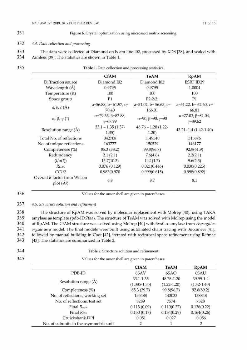
The data were collected at Diamond on beam line I02, processed by XDS [38], and scaled with 333 Aimless [39]. The statistics are shown in Table 1. 334
Table 1. Data collection and processing statistics. 335
CfAM TeAM RpAM
Diffraction source Diamond I02 Diamond I02 ESRF ID29 Wavelength (Å) 0.9795 0.9795 1.0004 Temperature (K) 100 100 100
Space group P1 P212121 P1
a, b, c (Å) a=56.88, b= 61.97, c=
70.40 a=51.02, b= 56.63, c=
166.01 a=51.22, b= 62.60, c=
66.81
α, β, γ (°) a=79.33, b=82.88,
g=67.99 a=90, b=90, g=90
a=77.03, b=81.04, g=89.62
Resolution range (Å) 33.1 – 1.35 (1.37-
1.35) 48.76 – 1.20 (1.22-
1.20) 43.21- 1.4 (1.42-1.40)
Total No. of reflections 342708 1149540 315876 No. of unique reflections 163777 150529 146177
Completeness (%) 85.3 (38.2) 99.8(96.7) 92.9(61.9) Redundancy 2.1 (2.1) 7.6(4.6) 2.2(2.1)
⟨I/σ(I)⟩ 13.7(10.3) 14.1(1.7) 9.6(2.3) Rr.i.m. 0.076 (0.129) 0.021(0.446) 0.030(0.225)
CC1/2 0.983(0.970 0.999(0.615) 0.998(0.892) Overall B factor from Wilson
plot (Å2) 6.8 8.7 8.1
Values for the outer shell are given in parentheses. 336
4.5. Structure solution and refinement 337
The structure of RpAM was solved by molecular replacement with Molrep [40], using TAKA 338
amylase as template (pdb-ID:7taa). The structure of TeAM was solved with Molrep using the model 339 of RpAM. The CfAM structure was solved using Molrep [40] with 3vx0 α-amylase from Aspergillus 340 oryzae as a model. The final models were built using automated chain tracing with Buccaneer [41], 341 followed by manual building in Coot [42], iterated with reciprocal space refinement using Refmac 342 [43]. The statistics are summarized in Table 2. 343
Table 2. Structure solution and refinement. 344
Values for the outer shell are given in parentheses. 345
CfAM TeAM RpAM
PDB-ID 6SAV 6SAO 6SAU
Resolution range (Å) 33.1-1.35
(1.385-1.35) 48.76-1.20 (1.22-1.20)
39.99-1.4 (1.42-1.40)
Completeness (%) 85.3 (39.7) 99.8(96.7) 92.8(89.2) No. of reflections, working set 155488 143033 138848
No. of reflections, test set 8289 7574 7328 Final Rcryst 0.113 (0.09) 0.110(0.27) 0.136(0.22) Final Rfree 0.150 (0.17) 0.134(0.29) 0.164(0.26)
Cruickshank DPI 0.051 0.027 0.056 No. of subunits in the asymmetric unit 2 1 2
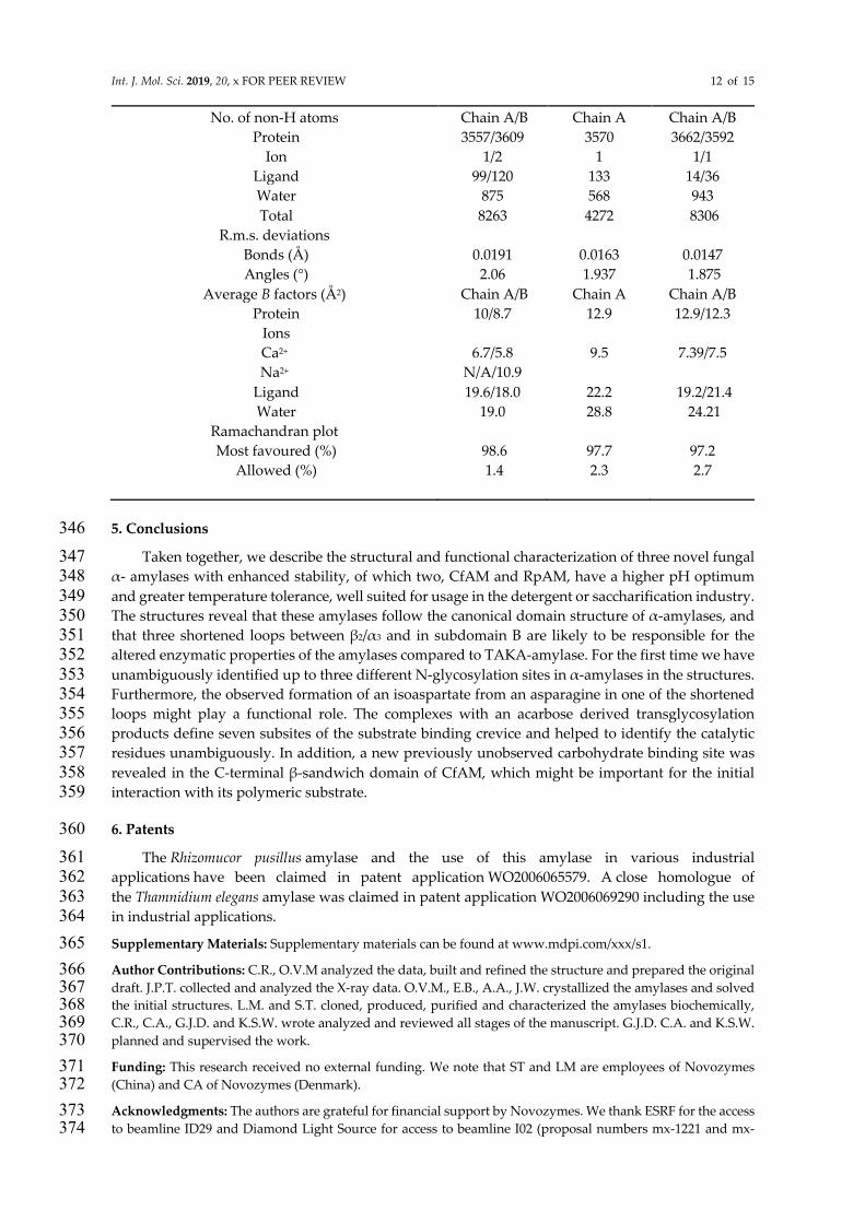
Int. J. Mol. Sci. 2019, 20, x FOR PEER REVIEW 12 of 15
No. of non-H atoms Chain A/B Chain A Chain A/B Protein 3557/3609 3570 3662/3592
Ion 1/2 1 1/1 Ligand 99/120 133 14/36 Water 875 568 943 Total 8263 4272 8306
R.m.s. deviations
Bonds (Å) 0.0191 0.0163 0.0147 Angles (°) 2.06 1.937 1.875
Average B factors (Å2) Chain A/B Chain A Chain A/B Protein 10/8.7 12.9 12.9/12.3
Ions
Ca2+ 6.7/5.8 9.5 7.39/7.5 Na2+ N/A/10.9
Ligand 19.6/18.0 22.2 19.2/21.4 Water 19.0 28.8 24.21
Ramachandran plot
Most favoured (%) 98.6 97.7 97.2 Allowed (%) 1.4 2.3 2.7
5. Conclusions 346
Taken together, we describe the structural and functional characterization of three novel fungal 347 α- amylases with enhanced stability, of which two, CfAM and RpAM, have a higher pH optimum 348
and greater temperature tolerance, well suited for usage in the detergent or saccharification industry. 349 The structures reveal that these amylases follow the canonical domain structure of α-amylases, and 350 that three shortened loops between β2/α3 and in subdomain B are likely to be responsible for the 351 altered enzymatic properties of the amylases compared to TAKA-amylase. For the first time we have 352
unambiguously identified up to three different N-glycosylation sites in α-amylases in the structures. 353 Furthermore, the observed formation of an isoaspartate from an asparagine in one of the shortened 354 loops might play a functional role. The complexes with an acarbose derived transglycosylation 355 products define seven subsites of the substrate binding crevice and helped to identify the catalytic 356 residues unambiguously. In addition, a new previously unobserved carbohydrate binding site was 357
revealed in the C-terminal β-sandwich domain of CfAM, which might be important for the initial 358 interaction with its polymeric substrate. 359
6. Patents 360
The Rhizomucor pusillus amylase and the use of this amylase in various industrial 361 applications have been claimed in patent application WO2006065579. A close homologue of 362
the Thamnidium elegans amylase was claimed in patent application WO2006069290 including the use 363 in industrial applications. 364
Supplementary Materials: Supplementary materials can be found at www.mdpi.com/xxx/s1. 365
Author Contributions: C.R., O.V.M analyzed the data, built and refined the structure and prepared the original 366 draft. J.P.T. collected and analyzed the X-ray data. O.V.M., E.B., A.A., J.W. crystallized the amylases and solved 367 the initial structures. L.M. and S.T. cloned, produced, purified and characterized the amylases biochemically, 368 C.R., C.A., G.J.D. and K.S.W. wrote analyzed and reviewed all stages of the manuscript. G.J.D. C.A. and K.S.W. 369 planned and supervised the work. 370
Funding: This research received no external funding. We note that ST and LM are employees of Novozymes 371 (China) and CA of Novozymes (Denmark). 372
Acknowledgments: The authors are grateful for financial support by Novozymes. We thank ESRF for the access 373 to beamline ID29 and Diamond Light Source for access to beamline I02 (proposal numbers mx-1221 and mx-374
Int. J. Mol. Sci. 2019, 20, x FOR PEER REVIEW 13 of 15
9948) that contributed to the results presented here. The authors also thank Sam Hart for assistance during data 375 collection. 376
Conflicts of Interest: The authors declare no conflict of interest, but we note that the Novozymes authors declare 377 the following competing financial interest(s): Novozymes are a commercial enzyme supplier. 378
Abbreviations 379
CfAM. Cordyceps farinosa amylase RpAM Rhizomucor pusillus amylase TeAM Thamnidium elegans amylase TAKA dp
Aspergillus oryzae amylase Degree of polymerisation
References 380
1. Payen, A.P.J.F., Memoire sur la diastase, les principaux produits de ses réactions et leurs applications aux arts 381
industriels" (Memoir on diastase, the principal products of its reactions, and their applications to the industrial 382
arts). Annales de Chimie et de Physique, 1833. 2: p. 73-92. 383
2. Gurung, N., et al., A broader view: microbial enzymes and their relevance in industries, medicine, and beyond. 384
Biomed Res Int, 2013. 2013: p. 329121. 385
3. Roy, J.K., et al., Cloning and extracellular expression of a raw starch digesting alpha-amylase (Blamy-I) and its 386
application in bioethanol production from a non-conventional source of starch. J Basic Microbiol, 2015. 55(11): 387
p. 1287-98. 388
4. Gupta, R., et al., Microbial α-amylases: a biotechnological perspective. Process Biochemistry, 2003. 38(11): p. 389
1599-1616. 390
5. Niehaus, F., et al., Extremophiles as a source of novel enzymes for industrial application. Appl Microbiol 391
Biotechnol, 1999. 51(6): p. 711-29. 392
6. Lombard, V., et al., The carbohydrate-active enzymes database (CAZy) in 2013. Nucleic Acids Res, 2014. 393
42(Database issue): p. D490-5. 394
7. Janecek, S., B. Svensson, and E.A. MacGregor, Structural and evolutionary aspects of two families of non-395
catalytic domains present in starch and glycogen binding proteins from microbes, plants and animals. Enzyme 396
Microb Technol, 2011. 49(5): p. 429-40. 397
8. Liu, Y., et al., Crystal structure of a raw-starch-degrading bacterial alpha-amylase belonging to subfamily 37 of 398
the glycoside hydrolase family GH13. Sci Rep, 2017. 7: p. 44067. 399
9. Mehta, D. and T. Satyanarayana, Domain C of thermostable alpha-amylase of Geobacillus thermoleovorans 400
mediates raw starch adsorption. Appl Microbiol Biotechnol, 2014. 98(10): p. 4503-19. 401
10. Sogaard, M., et al., Site-directed mutagenesis of histidine 93, aspartic acid 180, glutamic acid 205, histidine 290, 402
and aspartic acid 291 at the active site and tryptophan 279 at the raw starch binding site in barley alpha-amylase 403
1. J Biol Chem, 1993. 268(30): p. 22480-4. 404
11. Kadziola, A., et al., Molecular structure of a barley alpha-amylase-inhibitor complex: implications for starch 405
binding and catalysis. J Mol Biol, 1998. 278(1): p. 205-17. 406
12. Brzozowski, A.M., et al., Structural analysis of a chimeric bacterial alpha-amylase. High-resolution analysis of 407
native and ligand complexes. Biochemistry, 2000. 39(31): p. 9099-107. 408
13. Pritchard, P.E., Studies on the bread-improving mechanism of fungal alpha-amylase. Journal of Biological 409
Education, 1992. 26(1): p. 12-18. 410
14. D'Arcy, A., et al., Microseed matrix screening for optimization in protein crystallization: what have we learned? 411
Acta Crystallogr F Struct Biol Commun, 2014. 70(Pt 9): p. 1117-26. 412
Int. J. Mol. Sci. 2019, 20, x FOR PEER REVIEW 14 of 15
15. McNicholas, S. and J. Agirre, Glycoblocks: a schematic three-dimensional representation for glycans and their 413
interactions. Acta Crystallogr D Struct Biol, 2017. 73(Pt 2): p. 187-194. 414
16. Janeček, Š., Does the increased hydrophobicity of the interior and hydrophilicity of the exterior of an enzyme 415
structure reflect its increased thermostability? International Journal of Biological Macromolecules, 1993. 416
15(5): p. 317-318. 417
17. Mok, S.C., et al., Crystal structure of a compact alpha-amylase from Geobacillus thermoleovorans. Enzyme 418
Microb Technol, 2013. 53(1): p. 46-54. 419
18. Mazola, Y., et al., A comparative molecular dynamics study of thermophilic and mesophilic beta-fructosidase 420
enzymes. J Mol Model, 2015. 21(9): p. 228. 421
19. Brzozowski, A.M. and G.J. Davies, Structure of the Aspergillus oryzae alpha-amylase complexed with the 422
inhibitor acarbose at 2.0 A resolution. Biochemistry, 1997. 36(36): p. 10837-45. 423
20. Przylas, I., et al., X-ray structure of acarbose bound to amylomaltase from Thermus aquaticus. Implications for 424
the synthesis of large cyclic glucans. Eur J Biochem, 2000. 267(23): p. 6903-13. 425
21. Juge, N., et al., Isozyme hybrids within the protruding third loop domain of the barley alpha-amylase 426
(beta/alpha)8-barrel. Implication for BASI sensitivity and substrate affinity. FEBS Lett, 1995. 363(3): p. 299-427
303. 428
22. Rodenburg, K.W., et al., Domain B protruding at the third beta strand of the alpha/beta barrel in barley alpha-429
amylase confers distinct isozyme-specific properties. Eur J Biochem, 1994. 221(1): p. 277-84. 430
23. Penninga, D., et al., Site-directed mutations in tyrosine 195 of cyclodextrin glycosyltransferase from Bacillus 431
circulans strain 251 affect activity and product specificity. Biochemistry, 1995. 34(10): p. 3368-76. 432
24. Nakamura, A., K. Haga, and K. Yamane, Four aromatic residues in the active center of cyclodextrin 433
glucanotransferase from alkalophilic Bacillus sp. 1011: effects of replacements on substrate binding and 434
cyclization characteristics. Biochemistry, 1994. 33(33): p. 9929-36. 435
25. Krissinel, E., On the relationship between sequence and structure similarities in proteomics. Bioinformatics, 436
2007. 23(6): p. 717-23. 437
26. Robert, X., et al., The structure of barley alpha-amylase isozyme 1 reveals a novel role of domain C in substrate 438
recognition and binding: a pair of sugar tongs. Structure, 2003. 11(8): p. 973-84. 439
27. Sorimachi, K., et al., Solution structure of the granular starch binding domain of Aspergillus niger glucoamylase 440
bound to beta-cyclodextrin. Structure, 1997. 5(5): p. 647-61. 441
28. de Barros, M.C., et al., The influence of N-glycosylation on biochemical properties of Amy1, an alpha-amylase 442
from the yeast Cryptococcus flavus. Carbohydr Res, 2009. 344(13): p. 1682-6. 443
29. Amore, A., et al., Distinct roles of N- and O-glycans in cellulase activity and stability. Proc Natl Acad Sci U 444
S A, 2017. 114(52): p. 13667-13672. 445
30. Reissner, K.J. and D.W. Aswad, Deamidation and isoaspartate formation in proteins: unwanted alterations or 446
surreptitious signals? Cell Mol Life Sci, 2003. 60(7): p. 1281-95. 447
31. Barends, T.R., et al., Three-way stabilization of the covalent intermediate in amylomaltase, an alpha-amylase-448
like transglycosylase. J Biol Chem, 2007. 282(23): p. 17242-9. 449
32. Roth, C., et al., Amylose recognition and ring-size determination of amylomaltase. Sci Adv, 2017. 3(1): p. 450
e1601386. 451
33. DALBØGE, H., et al., DNA encoding an enxyme with endoglucanase activity from Trichoderma harzianum. 452
1995, Novo-Nordisk A/S. 453
34. Sun, Y., et al., Crystallization and preliminary crystallographic analysis of Gibberella zeae extracellular lipase. 454
Acta Crystallogr Sect F Struct Biol Cryst Commun, 2008. 64(Pt 9): p. 813-5. 455
Int. J. Mol. Sci. 2019, 20, x FOR PEER REVIEW 15 of 15
35. Britton, H.T.S. and R.A. Robinson, CXCVIII.—Universal buffer solutions and the dissociation constant of 456
veronal. Journal of the Chemical Society (Resumed), 1931(0): p. 1456-1462. 457
36. Shaw Stewart, P.D., et al., Random Microseeding: A Theoretical and Practical Exploration of Seed Stability and 458
Seeding Techniques for Successful Protein Crystallization. Crystal Growth & Design, 2011. 11(8): p. 3432-459
3441. 460
37. Shah, A.K., et al., On increasing protein-crystallization throughput for X-ray diffraction studies. Acta 461
Crystallographica Section D, 2005. 61(2): p. 123-129. 462
38. Kabsch, W., Xds. Acta Crystallogr D Biol Crystallogr, 2010. 66(Pt 2): p. 125-32. 463
39. Evans, P.R. and G.N. Murshudov, How good are my data and what is the resolution? Acta Crystallogr D 464
Biol Crystallogr, 2013. 69(Pt 7): p. 1204-14. 465
40. Vagin, A. and A. Teplyakov, Molecular replacement with MOLREP. Acta Crystallogr D Biol Crystallogr, 466
2010. 66(Pt 1): p. 22-5. 467
41. Cowtan, K., The Buccaneer software for automated model building. 1. Tracing protein chains. Acta Crystallogr 468
D Biol Crystallogr, 2006. 62(Pt 9): p. 1002-11. 469
42. Emsley, P., et al., Features and development of Coot. Acta Crystallogr D Biol Crystallogr, 2010. 66(Pt 4): p. 470
486-501. 471
43. Murshudov, G.N., A.A. Vagin, and E.J. Dodson, Refinement of macromolecular structures by the maximum-472
likelihood method. Acta Crystallogr D Biol Crystallogr, 1997. 53(Pt 3): p. 240-55. 473
474