structural and functional characterization of human and
TRANSCRIPT
research papers
1704 doi:10.1107/S139900471400844X Acta Cryst. (2014). D70, 1704–1717
Acta Crystallographica Section D
BiologicalCrystallography
ISSN 1399-0047
Structural and functional characterization ofhuman and murine C5a anaphylatoxins
Janus Asbjørn Schatz-
Jakobsen,a‡ Laure Yatime,a*‡
Casper Larsen,a Steen Vang
Petersen,b Andreas Klosc and
Gregers Rom Andersena*
aDepartment of Molecular Biology and
Genetics, Aarhus University, Gustav Wieds
Vej 10C, DK-8000 Aarhus, Denmark,bDepartment of Biomedicine, Aarhus University,
Bartholin Building, Wilhelm Meyers Alle 4,
DK-8000 Aarhus, Denmark, and cInstitute for
Medical Microbiology and Hospital
Epidemiology, Medical School Hannover,
Hannover, Germany
‡ These authors contributed equally to this
work.
Correspondence e-mail: [email protected],
Complement is an ancient part of the innate immune system
that plays a pivotal role in protection against invading
pathogens and helps to clear apoptotic and necrotic cells.
Upon complement activation, a cascade of proteolytic events
generates the complement effectors, including the anaphyla-
toxins C3a and C5a. Signalling through their cognate G-
protein coupled receptors, C3aR and C5aR, leads to a wide
range of biological events promoting inflammation at the site
of complement activation. The function of anaphylatoxins is
regulated by circulating carboxypeptidases that remove their
C-terminal arginine residue, yielding C3a-desArg and C5a-
desArg. Whereas human C3a and C3a-desArg adopt a
canonical four-helix bundle fold, the conformation of human
C5a-desArg has recently been described as a three-helix
bundle. Here, the crystal structures of an antagonist version of
human C5a, A8�71–73, and of murine C5a and C5a-desArg
are reported. Whereas A8�71–73 adopts a three-helix bundle
conformation similar to human C5a-desArg, the two murine
proteins form a four-helix bundle. A cell-based functional
assay reveals that murine C5a-desArg, in contrast to its human
counterpart, exerts the same level of activition as murine C5a
on its cognate receptor. The role of the different C5a
conformations is discussed in relation to the differential
activation of C5a receptors across species.
Received 9 March 2014
Accepted 14 April 2014
PDB references: murine C5a,
4p3a; murine C5a-desArg,
4p3b; human C5a-A8�71–73,
4p39
1. Introduction
Complement is a central component of innate immunity,
acting as a first line of defence against invading pathogens and
maintaining homeostasis (Walport, 2001; Ricklin et al., 2010).
When danger originating from non-self (pathogens) or
altered-self (e.g. apoptotic or necrotic cells) is detected,
complement initiates a complex biochemical cascade that
involves more than 50 proteins ranging from soluble plasma
factors to membrane-bound receptors. This cascade enables
the proteolytic cleavage of the central complement compo-
nents C3, C4 and C5, yielding the two opsonins C3b and C4b
and the effector of the complement terminal pathway, C5b,
that triggers formation of the lytic membrane attack complex
(MAC; Walport, 2001; Dunkelberger & Song, 2010; Ricklin et
al., 2010; Carroll & Sim, 2011). Cleavage-dependent activation
of C3, C4 and C5 also releases into the serum the three 10 kDa
proteins C3a, C4a and C5a, known as complement anaphyla-
toxins. While the function of C4a remains unclear in the
absence of identified C4a receptors, C3a and C5a are well
characterized pro-inflammatory mediators (Hugli, 1990; Guo
& Ward, 2005; Klos et al., 2009). Their function in inflamma-
tion is mediated by their signalling through cognate hepta-
helical receptors on the host cells: C3aR for C3a, and C5aR
and C5L2 for C5a (Klos et al., 2013). Anaphylatoxin signalling

research papers
Acta Cryst. (2014). D70, 1704–1717 Schatz-Jakobsen et al. � Human and murine C5a anaphylatoxins 1705
through the G-protein coupled receptors (GPCRs) C3aR and
C5aR leads to various cellular responses including chemotaxis,
oxidative burst, the release of pro-inflammatory molecules
and activation of the adaptive immune system (Klos et al.,
2009; Zhou, 2012). Owing to the lack of binding motifs
enabling its coupling to G proteins, C5L2 has long been
considered as a decoy receptor. However, recent reports now
suggest that it actively participates in orchestrating pro-
inflammatory events (Li et al., 2013) and the involvement of
C5L2 in regulating metabolism in adipose tissue has also been
proposed (MacLaren et al., 2008; Klos et al., 2013).
C5a is the most potent pro-inflammatory mediator among
complement proteins. It targets a broad range of immune cells
including basophils, neutrophils, eosinophils, mast cells and
macrophages, as well as cells from nonmyeloid lineage,
notably in the lung and liver (Guo & Ward, 2005) and
lymphocytes, in particular regulatory T-cells (Strainic et al.,
2013; van der Touw et al., 2013; Kemper & Kohl, 2013). As for
C3a, C5a activity is regulated by carboxypeptidases that cleave
off the C-terminal arginine residue (Bokisch & Muller-Eber-
hard, 1970), producing the C5a-desArg molecule which has
a distinct bioactivity pattern and generally a much lower
potency than C5a, in part owing to its tenfold to 100-fold
decrease in C5aR binding affinity (Burgi et al., 1994; Eglite
et al., 2000; Higginbottom et al., 2005). Elevated C5a plasma
levels correlate with the onset of various inflammatory disor-
ders including sepsis, rheumatoid arthritis, acute lung injury,
ischaemia-reperfusion injury, allergy and asthma (Guo &
Ward, 2005; Klos et al., 2009, 2013), implicating C5a as a
causative or exacerbating agent in the pathogenesis of these
conditions. Increasing evidence also suggests a direct role of
C5a and C5aR signalling in tumour progression (Markiewski
et al., 2008). As a consequence, targeting of the C5a–C5aR axis
is considered to be a promising therapeutical strategy to
downregulate complement-mediated inflammation while
preserving primary defence functions such as opsonization
and MAC-mediated bacteriolysis (Woodruff et al., 2011).
Various targeting approaches have already been explored,
including monoclonal antibodies against C5a or C5aR (Sprong
et al., 2003; Lee et al., 2006), small C5aR inhibitory molecules
(Finch et al., 1999), inhibitory peptides (Tokodai et al., 2010),
anti-C5a vaccines (Kessel et al., 2014) and l-RNA aptamers
(Hoehlig et al., 2013). C5a-derived receptor antagonists have
also been sought using phage-display libraries. This screening
identified a mutant form of C5a, C5a-A8�71–73, as a potent
antagonist of both C5aR and C5L2 (Otto et al., 2004). This
mutant lacks the three C-terminal residues of C5a-desArg
(�71QLG73 in C5a numbering or �748QLG750 in C5 prepro
numbering) and bears three additional mutations in its
C-terminus (Fig. 1a). In particular, the D746R mutation
turned out to determine the antagonistic versus agonistic
effect of these proteins (Otto et al., 2004). However, the
druggability of all these compounds still awaits final validation
by clinical studies.
In light of the role played by C5a receptor signalling in
inflammation, understanding the structure–function relation-
ship of the interaction of C5a with C5aR/C5L2 appears to
be a critical prerequisite for the design of more efficient
Figure 1Biological activity of recombinant hC5a-derived anaphylatoxins. (a) Amino-acid sequence of the C-termini of hC5a, hC5a-desArg and hC5a-A8. Thepositions of the glycosylated Asn (star) and of the residue conditioning the agonist/antagonist shift for C5a proteins (triangle) are indicated. (b) Elutionprofile of recombinant hC5a (blue), hC5a-desArg (red) and hC5a-A8 (grey) on the SOURCE 15S column. (c) GARA for recombinant hC5a, hC5a-desArg and hC5a-A8 on stably transfected RBL cells expressing hC5aR and comparison with commercially available hC5a. All results are indicated asthe means � standard deviation of at least eight individual experiments (except for hC5a-A8, where n = 4). The NAG release activity is expressed as apercentage of the maximal NAG release activity obtained with commercially available recombinant hC5a. (d) EC50 and maximal NAG release activityobserved for all proteins upon incubation with hC5aR-expressing RBL cells.

therapeutics. Since mutations in the C-terminus and the
removal of the C-terminal arginine modulate the activity of
C5a towards its receptors, gaining insight into how these
changes affect the three-dimensional architecture of the
anaphylatoxin might provide clues to the mechanistics behind
the C5aR antagonism of the truncated C5a proteins. C5a folds
into a four-helix bundle in the structure of intact human C5
(Fredslund et al., 2008) and in NMR structures of both human
C5a (hC5a) and porcine C5a-desArg (Zuiderweg et al., 1989;
Williamson & Madison, 1990; Zhang et al., 1997). Intriguingly,
the crystal structure of human C5a-desArg (hC5a-desArg)
revealed a distinct architecture for the protein, with a three-
helix bundle core and the N-terminal helix either extruding
outside the core or merging with the second helix into one
single extended �-helix (Cook et al., 2010). Although this
property is not observed for C3a-desArg (Bajic et al., 2013), it
may still be of importance for the modulation of C5a activity.
To investigate the conformational differences between the
C5a-derived molecules, we have undertaken structural studies
of C5a, C5a-desArg and C5a-A8�71–73 (referred to as C5a-A8
in the following). Since current protocols for C5a preparation
rely either on isolation from plasma (Williamson & Madison,
1990), which yields heterogenous samples, or on harsh, time-
consuming denaturation/renaturation procedures to purify
recombinant proteins (Toth et al., 1994; Cook et al., 2010), we
developed a simple and fast protocol to prepare biologically
active C5a proteins by recombinant expression in bacteria for
both human and mouse proteins. Furthermore, we determined
the crystal structure of human C5a-A8 at 2.4 A resolution.
Interestingly, hC5a-A8 forms a three-helix bundle with an
extended N-terminal helix as seen for hC5a-desArg. To assess
whether this property was specific to the human C5a proteins
or whether this is a general feature of C5a, we also determined
the structures of mouse C5a and C5a-desArg (mC5a and
mC5a-desArg) at 1.4 and 2.1 A resolution, respectively. Both
murine proteins fold into a four-helix bundle motif as
observed for hC5a. Differences in the three-dimensional
architecture of human and mouse C5a proteins are discussed
in the light of their respective activity towards both human
and mouse C5a receptors.
2. Materials and methods
2.1. Genes and plasmids
The hC5a gene, codon-optimized for bacterial expression
and cloned between the BamHI and EcoRI restriction sites of
vector pET-32a (Novagen), was purchased from GenScript. To
avoid nonspecific disulfide cross-linking between C5a mole-
cules during purification, the sole cysteine residue of hC5a that
is not engaged in intramolecular disulfide bridges (Cys704;
prepro-C5 numbering) and is modified by cysteinylation in
native hC5a (Laursen et al., 2012) was replaced by an arginine,
a substitution that is often encountered in C5a from other
species (Supplementary Fig. S11) and which does not
dramatically affect the biological activity of C5a towards
hC5aR (Hagemann et al., 2006). To facilitate purification
procedures, the enterokinase cleavage site preceding the hC5a
sequence in pET-32a was replaced by a TEV protease clea-
vage site using the QuikChange Lightning site-directed
mutagenesis kit from Agilent Technologies. Owing to diffi-
culties with TEV protease cleavage in the initial purification
trials, an additional four-amino-acid linker (AGAA) was
introduced between the TEV cleavage site and the beginning
of the hC5a sequence. hC5a-desArg and hC5a-A8 were
generated from the hC5a-pET-32a construct by site-directed
mutagenesis (using the QuikChange Lightning site-directed
mutagenesis kit).
The mouse C5a (mC5a) gene, codon-optimized for bacterial
expression, containing a 50 insertion corresponding to a TEV
protease cleavage site and cloned between the BamHI and
HindIII restriction sites of vector pET-32a (Novagen) was
purchased from GenScript. The mC5a-desArg construct was
derived from the mC5a-pET-32a construct by replacing
the C-terminal Arg residue with a STOP codon (using the
QuikChange Lightning site-directed mutagenesis kit). All
constructs described here allow the expression of the protein
of interest as a fusion with an N-terminal thioredoxin (Trx) tag
followed by a hexahistidine tag and a TEV protease cleavage
site (Trx-His6-TEV-C5a).
2.2. Expression and purification of C5a and derivatives
All C5a constructs were transformed in Shuffle T7 Express
Escherichia coli cells (New England Biolabs). The cells were
grown at 37�C in 2�YT medium supplemented with ampicillin
(100 mg ml�1) and expression of the C5a proteins was induced
by the addition of 1 mM IPTG followed by overnight incu-
bation at 18�C. The cells were harvested, resuspended in
buffer A (50 mM HEPES pH 7.5, 300 mM NaCl, 30 mM
imidazole, 1 mM PMSF) and disrupted by sonication. After
clarification by centrifugation, the supernatant was applied
onto a 5 ml Ni column (HisTrap FF column from GE
Healthcare Life Sciences). After a high-salt wash (50 mM
HEPES pH 7.5, 1 M NaCl, 30 mM imidazole, 1 mM PMSF) to
remove nonspecifically bound proteins, the Trx-His6-tagged
proteins were eluted with buffer B (50 mM HEPES pH 7.5,
300 mM NaCl, 500 mM imidazole, 1 mM PMSF). The Trx-His6
tag was subsequently removed by overnight incubation at 4�C
using a 1:30 (rTEV:protein) mass ratio of in-house prepared
recombinant His6-rTEV during dialysis against buffer D
(50 mM HEPES pH 7.5, 300 mM NaCl, 0.5 mM EDTA). The
cleaved proteins were separated from contaminants, His6-
rTEV and uncleaved C5a material by a second Ni-column
purification step. The protein samples were then concentrated
and rediluted with 50 mM HEPES pH 7.5 to give a final salt
concentration of 100 mM NaCl. The samples were then
applied onto a 9 ml SOURCE 15S cation-exchange column
(GE Healthcare Life Sciences). Elution was performed with
a 100 ml linear gradient from 100 to 400 mM NaCl. Pure
samples were flash-frozen in liquid nitrogen and kept at
�80oC until use.
research papers
1706 Schatz-Jakobsen et al. � Human and murine C5a anaphylatoxins Acta Cryst. (2014). D70, 1704–1717
1 Supporting information has been deposited in the IUCr electronic archive(Reference: DW5099).

2.3. Crystallization of the C5a proteins
Prior to crystallization, the buffer composition of the
murine C5a proteins was adjusted to 20 mM HEPES pH 7.5,
150 mM NaCl and the protein samples were concentrated to
25 mg ml�1 for hC5a-A8 and to 8–10 mg ml�1 for mC5a and
mC5a-desArg. Initial crystallization experiments were carried
out in 96-well sitting-drop plates using a Mosquito robot (TTP
Labtech) and commercial screens from Hampton Research
and Molecular Dimensions. For hC5a-A8, crystals appeared
after one month at 4�C over a reservoir consisting of 20%(v/v)
2-propanol, 20%(w/v) PEG 4000, 0.1 M sodium citrate pH 5.6
with a protein:reservoir solution ratio of 2:1. For data collec-
tion, the crystals were cryoprotected by soaking them in the
reservoir solution followed by flash-cooling in liquid nitrogen.
For mC5a and mC5a-desArg, rod-shaped crystals appeared
after a few days at 4�C over a reservoir consisting of 0.1 M
sodium acetate pH 4.6, 2.0 M sodium formate. Crystal opti-
mization, in particular screening for additives using Additive
Screen from Hampton Research, was performed in hanging
drops and allowed us to obtain larger single crystals. For
mC5a, the best crystals were grown at 4�C in 0.1 M sodium
tricitrate pH 3.7, 2.6 M sodium formate, 0.5%(w/v) poly-
vinylpyrrolidone K15. For mC5a-desArg, the best crystals
appeared over a reservoir consisting of 0.1 M sodium acetate
pH 4.3, 2.4 M sodium formate, 3%(w/v) d-glucose mono-
hydrate. For data collection, the crystals were cryoprotected
by soaking them in a reservoir solution supplemented with
either 4.0 M sodium formate (mC5a) or 30% glycerol (mC5a-
desArg) followed by flash-cooling in liquid nitrogen.
2.4. Data collection and structure determination
Data sets for hC5a-A8 were collected at 100 K on beamline
X06SA at the Swiss Light Source (PSI, Villigen, Switzerland).
Data sets for mC5a and mC5a-desArg were collected at 100 K
on beamline 911-3 at MAX-lab (Lund, Sweden). All data sets
were processed with XDS (Kabsch, 1993; Table 1). The
hC5a-A8 crystals displayed C2221 symmetry and diffracted to
a maximal resolution of 2.4 A. The structure was solved by
molecular replacement (MR) in Phaser (McCoy et al., 2005)
using a search model derived from the hC5a-desArg structure
(Cook et al., 2010) and encompassing residues Val694–Ser743
(i.e. where the N-terminal helix had been removed). Initial
electron-density maps showed clear additional density for the
missing residues in the hC5a-A8 N-terminus, which were
rebuilt using phenix.autobuild (Adams et al., 2010). The model
was further improved by manual rebuilding in Coot (Emsley et
al., 2010) and energy minimization in phenix.refine (Adams et
al., 2010) using individual isotropic ADPs, TLS refinement and
NCS restraints. The final model yielded Rwork and Rfree values
of 22.4 and 23.8%, respectively, and its quality was evaluated
with MolProbity (Chen et al., 2010). The small gap observed
between the Rwork and Rfree values for this model may be
owing to the presence of the fourfold NCS, possibly leading to
correlation between reflections in the test and the work sets,
but is also favoured by the high overall data quality (Table 1)
and measures taken throughout refinement to prevent
overfitting. The mC5a and mC5a-desArg crystals both
displayed P43 symmetry with very similar unit-cell parameters
and diffracted to 1.4 and 2.1 A resolution, respectively (Table
1). The mC5a structure was solved by MR in Phaser using a
homology model of mC5a based on the structure of hC5a
extracted from the human C5 structure (Fredslund et al.,
2008). Initial electron-density maps obtained after MR using
an mC5a model where the first 20 residues had been deleted
(i.e. starting at residue Pro699 and encompassing only three
helices) revealed clear additional density for the 20 missing
residues in all four molecules of the asymmetric unit and
showed that the mC5a N-terminus formed a fourth helical
motif that packed against the remaining three helices in a four-
helix bundle motif. Further refinement of the model was
carried out by alternating between cycles of manual rebuilding
in Coot and cycles of energy minimization with phenix.refine
using individual ADPs with anisotropic ADP refinement for
protein atoms and isotropic ADP refinement for water and
ligand atoms. NCS restraints were not imposed as they were
observed to increase the Rwork and Rfree values during
refinement. H atoms were included as riding atoms during
research papers
Acta Cryst. (2014). D70, 1704–1717 Schatz-Jakobsen et al. � Human and murine C5a anaphylatoxins 1707
Table 1Data-collection and refinement statistics for the hC5a-A8, mC5a andmC5a-desArg structures.
Values in parentheses are for the highest resolution shell.
HumanC5a-A8�71–73 Mouse C5a Mouse C5a-desArg
Data-collection statisticsX-ray source X06SA, SLS 911-3, MAX-lab 911-3, MAX-labWavelength (A) 1.000 0.9792 1.000Space group C2221 P43 P43
Unit-cell parameters (A)a 69.35 54.95 55.21b 83.24 54.95 55.21c 119.22 117.43 117.43
Resolution (A) 50–2.4 (2.5–2.4) 50–1.4 (1.5–1.4) 50–2.1 (2.2–2.1)Unique reflections 13812 (1541) 68172 (12739) 20489 (2655)Completeness (%) 99.6 (98.5) 99.8 (99.8) 99.9 (100)Multiplicity 6.2 (6.1) 4.2 (4.1) 6.4 (6.4)Rmeas(I)† (%) 8.8 (60.4) 7.3 (84.9) 12.3 (72.2)hI/�(I)i 17.07 (3.81) 15.10 (2.33) 14.62 (2.95)Mosaicity‡ (�) 0.17 0.13 0.12Wilson B‡ (A2) 60 20 30
Refinement statisticsResolution (A) 49–2.4 37–1.4 33–2.1Unique reflections 13432 68165 20350Rwork (%) 22.36 14.42 18.03Rfree (%) 23.77 17.40 22.41Model in the asymmetric unit (No. of atoms)
Protein 2143 2212 2212Ligands — 48 45Water 22 333 204
Root-mean-square deviationsBonds (A) 0.002 0.008 0.003Angles (�) 0.463 1.198 0.711
Average B values (A2)Protein 68 19 35Ligands 61 29 41
Ramachandran plot§ (%)Favoured 99.6 100 98.9Allowed 0.4 0 1.1Outliers 0 0 0
† Rmeas(I) =P
hklfNðhklÞ=½NðhklÞ � 1�g1=2 Pi jIiðhklÞ � hIðhklÞij=P
hkl
Pi IiðhklÞ. ‡ Value given by XDS. § Values given by MolProbity.

refinement but were removed from the final model. The final
model yielded Rwork and Rfree values of 14.4 and 17.4%,
respectively. The mC5a-desArg structure was solved by MR in
Phaser using the mC5a structure as a search model. Refine-
ment of the model was then carried out by alternating cycles of
manual rebuilding in Coot and cycles of energy minimization
with phenix.refine using individual ADP (isotropic) refinement
as well as TLS refinement. NCS restraints were used
throughout except for the very last refinement cycles. The final
model yielded Rwork and Rfree values of 18.0 and 22.4%,
respectively. The quality of the models was assessed using
MolProbity. All figures were produced with PyMOL v.0.99rc6
(http://www.pymol.org).
2.5. Mass-spectrometric analysis of the human and mouseC5a proteins
Approximately 5 mg purified protein (�5 nmol) was
desalted using C8 StageTips (Proxeon) and the bound mate-
rial was eluted with 90% acetonitrile, 0.08% trifluoroacetic
acid. The material was lyophilized, resuspended in 2 ml 1%
trifluoroacetic acid and mixed with 2 ml 2,5-dihydroxyaceto-
phenone [0.1 M in 20 mM ammonium dihydrogen citrate,
75%(v/v) EtOH] (Wenzel et al., 2006). The material (1 ml) was
spotted onto a stainless-steel target and allowed to dry. The
spectra were recorded in positive and linear mode using an
AutoFlex Smartbeam III instrument (Bruker) calibrated by
external calibration (Peptide Calibration Standard I, Bruker
Daltronics). The centroid masses determined were evaluated
using the GPMAW software (http://www.gpmaw.com).
2.6. N-Acetyl-b-D-glucosaminidase release assay (GARA)
Prior to the GARA, bacterial endotoxin was removed
from all recombinant C5a samples using a Detoxi-Gel Endo-
toxin Removing Column (Pierce). The release of the lyso-
somal enzyme N-acetyl-�-d-glucosaminidase was assayed as
described previously (Gerard & Gerard, 1990; Bajic et al.,
2013). Briefly, rat basophil leukaemia (RBL) cells stably
transfected with the retroviral vector pQCXIN encoding
human C5aR, mouse C5aR or an empty cassette (as a negative
control) were harvested and resuspended in HAG-CM-
complete buffer (20 mM HEPES pH 7.4, 125 mM NaCl, 5 mM
KCl, 1 mM CaCl2, 1 mM MgCl2, 0.5 mM glucose, 0.25% BSA)
at a concentration of 2 � 106 cells ml�1 and incubated at 37�C
for 20 min. Simultaneously, dilutions of anaphylatoxins were
incubated with 1 mM cytochalasin B at 37�C. 75 ml of the cell
suspension was added to each C5a dilution and release was
allowed to proceed for 3 min at 37�C. The reactions were then
transferred to ice and centrifuged for 3 min at 400g and 4�C.
NAG activity was assessed in the presence of 3.53 mM
p-nitrophenyl-N-acetyl-�-d-glucosamide (Sigma) for 60 min
at 37�C. The enzymatic reaction was stopped by adding 0.4 M
glycine–NaOH pH 10.4 and the spectral absorbance was
measured at 405 nm. The maximal NAG release (100%
reference) corresponds to the maximal NAG release activity
obtained for commercial hC5a. The data are the means �
standard deviation of multiple independent experiments and
were analyzed in GraphPad Prism (http://http://www.graph-
pad.com) using a four-parameter logistic nonlinear regression.
Statistical analyses were performed using the paired two-
tailed Student’s t-test in Excel.
3. Results
3.1. Production of biologically active anaphylatoxins
Human C5a, C5a-desArg and C5a-A8 (Fig. 1a) were
expressed as recombinant proteins in E. coli and purified using
the same procedure as previously described for human C3a
and C3a-desArg (Bajic et al., 2013). Since the C5a molecule is
stabilized by three internal disulfide bridges, we used an E. coli
strain enhancing disulfide-bond formation and expressed the
proteins as fusions with a cleavable thioredoxin tag to ensure
proper folding. The final step of the purification consisted of
cation-exchange chromatography. All three proteins eluted
as a single monodisperse peak on the SOURCE 15S column
(Fig. 1b) at NaCl concentrations correlating with their
isoelectric properties (205, 190 and 230 mM NaCl for hC5a,
hC5a-desArg and hC5a-A8, respectively), and 20–25 mg of
purified protein per litre of bacterial culture was routinely
obtained using this method. Mass-spectrometric analysis of all
three purified samples also gave molecular masses in agree-
ment with theoretical values (Supplementary Fig. S2).
To verify that the procedure yielded biologically active
anaphylatoxins, the recombinant proteins were tested in the
N-acetyl-�-d-glucosaminidase release assay (GARA) using
RBL cells stably transfected with human C5aR (Gerard &
Gerard, 1990; Bajic et al., 2013; Figs. 1c and 1d). Recombinant
hC5a triggered N-acetyl-�-d-glucosaminidase (NAG) release
in a dose-dependent manner, with an EC50 value of 1.31 �
0.47 nM, comparable to the value of 1.47 � 0.43 nM obtained
with commercially available recombinant hC5a (Altana
Pharma AG). hC5a-desArg could also activate hC5aR-
expressing cells with an EC50 value of 1.44 � 0.98 nM, similar
to the value obtained with hC5a. However, the maximal NAG
release activity observed for hC5a-desArg only reached 55%
of the hC5a-induced maximal activity (Figs. 1c and 1d). Thus,
our recombinant hC5a-desArg acts as a partial agonist of
hC5aR, in good agreement with previous reports (Burgi et al.,
1994; Higginbottom et al., 2005). Finally, no activation could be
detected upon incubation with hC5a-A8, even at concentra-
tions of up to 1 mM (Figs. 1c and 1d): a behaviour in agreement
with the antagonistic properties previously described for
hC5a-A8 (Otto et al., 2004). In conclusion, the purification
protocol described here yielded highly homogenous, fully
functional recombinant protein suitable for crystallographic
studies.
3.2. Human C5a-A8 folds into a three-helix bundle with aC-terminal b-strand extension
3.2.1. Overall structure of hC5a-A8. Since the crystal
structure of hC5a-desArg was already available and, surpris-
ingly, revealed a three-helix bundle conformation (Cook et al.,
2010), we decided to further investigate the structural differ-
research papers
1708 Schatz-Jakobsen et al. � Human and murine C5a anaphylatoxins Acta Cryst. (2014). D70, 1704–1717

ences that possibly exist between the C5a-derived proteins. No
crystal structure of recombinant hC5a has been reported so
far and, despite numerous attempts, we did not obtain hC5a
crystals, in part owing to the very high solubility of the protein
in most crystallization conditions. On the other hand, hC5a-A8
produced crystals displaying C2221 symmetry and diffracting
to 2.4 A resolution (Table 1). Initial attempts to solve the
structure using the C5a moiety (residues 679–742) from the
hC5 structure (Fredslund et al., 2008) failed to give a molecular-
replacement (MR) solution. Since the conserved core between
all reported hC5a structures consists of a three-helix bundle
encompassing helices H2, H3 and H4 (Zuiderweg et al., 1989;
Zhang et al., 1997; Fredslund et al., 2008; Cook et al., 2010), a
model based on the hC5a-desArg structure and lacking the
N-terminal helical segment (i.e. residues Val694–Ser743 of
monomer A from PDB entry 3hqa; Cook et al., 2010) was then
used in the MR search. This three-helix bundle model gave a
clear MR solution (log-likelihood gain of 946) and initial
electron-density maps displayed well defined additional
density for the missing N-terminal residues (Supplementary
Fig. S3a). The final model encompasses residues Thr678–
Lys745 (residues 1–68 in C5a numbering) and the penultimate
hC5a-A8 residue, Arg746, could be modelled in one of the
four molecules related by NCS in the asymmetric unit
(monomer C). The four NCS-related molecules are very
similar overall, with average r.m.s.d. values of 0.75 and 1.35 A
research papers
Acta Cryst. (2014). D70, 1704–1717 Schatz-Jakobsen et al. � Human and murine C5a anaphylatoxins 1709
Figure 2General overview of the hC5a-A8 structure. (a) Overall structure of human C5a-A8 at 2.4 A resolution. (b) Comparison of the three-helix bundle ofhC5a-A8 with the four-helix bundle of hC5a as part of C5 (Fredslund et al., 2008). (c) Final model and electron-density maps for the hC5a-A8 structure.The 2mFo � DFc map is shown as a blue mesh and contoured at 1�. The mFo � DFc map is shown as a green mesh and contoured at 3�. (d) OMITelectron-density map calculated using simulated annealing from a model where residues Thr678–Ser693 (the first half of helix H10) have been deleted.The 2mFo � DFc OMIT map is shown as a grey mesh and contoured at 1�. The final complete model is superimposed to show the quality of the fit.

on C� atoms and all atoms, respectively, between all four NCS-
related molecules (residues Thr678–Lys745). Only one mole-
cule (monomer A) was therefore later used for comparison
with other C5a proteins.
The overall structural arrangement of hC5a-A8 is depicted
in Fig. 2. The C5aR antagonist does not fold into a four-helix
bundle but instead adopts a three-helix bundle conformation
with a long N-terminal helix H10 encompassing residues
Thr678–Arg704 (Cys704 in native hC5a), a second helix H20
formed by residues Thr710–Ile718 and the last helix H30
comprising residues Gly721–Asn741 (Fig. 2a). Helices H20 and
H30 from hC5a-A8 correspond to helices H3 and H4, respec-
tively, in the canonical four-helix bundle conformation of
hC5a (Zhang et al., 1997; Fredslund et al., 2008), while the first
two helices in hC5a, H1 and H2, are now merged into a single
extended helix H10 in hC5a-A8 (Fig. 2b, Supplementary
Fig. S1). Despite these major differences, the three disulfide
bridges that stabilize the helix packing in all previously
reported C5a structures are preserved in hC5a-A8 and
connect Cys698–Cys724 (H10–H30), Cys699–Cys731 (H10–H30)
and Cys711–Cys732 (H20–H30).
3.2.2. Comparison with other C5a structures. The core of
hC5a-A8 encompassing the second half of helix H10, helix
H20 and helix H30 (residues Val694–Ile742) is well conserved
compared with all reported hC5a/hC5a-desArg structures
(Fig. 3). The r.m.s.d. on C� atoms for this region in hC5a-A8 is
0.99 A compared with hC5a from C5 (Fredslund et al., 2008)
and 0.55 and 0.90 A compared with conformers A (the
conformer with four helices) and B (the conformer with three
helices) of hC5a-desArg, respectively (Cook et al., 2010). A
higher r.m.s.d. value of 2.46 A is encountered upon compar-
ison with the mean structure of the hC5a NMR structure
(Zhang et al., 1997) owing to a distortion of the region
between Val705 and Ala716 in the NMR model compared
with other C5a structures (Fig. 3a).
In hC5a, the short sequence preceeding the core region
(residues Tyr690–Ser693) forms a loop that allows the
N-terminal segment to pack back against the C5a core in an
H1 helical motif antiparallel to H2. In contrast, residues
Tyr690–Ser693 of hC5a-A8 form an additional helical turn
prior to H2, allowing the latter to merge with the N-terminal
helical segment into the long helix H10. This arrangement is
reminiscent of that observed for hC5a-desArg (Cook et al.,
2010). Indeed, monomer B of hC5a-desArg (PDB entry 3hqa)
displays a similar structural arrangement to hC5a-A8, with a
long N-terminal helix and an overall three-helix bundle
architecture, and the two structures superimpose well with
an average r.m.s.d. on C� atoms of 1.45 A for the region
encompassing residues Lys682–Arg739 (Fig. 3b).
Packing in the hC5a-A8 crystals is primarily stabilized by
two major interactions occurring for each of the four mono-
mers in the asymmetric unit (Supplementary Fig. S3b). The
first interaction allows the long helix H10 to pack along its
counterpart from a symmetry-related molecule in an anti-
parallel manner. The second interaction bridges the C-termini
of two symmetry-related molecules in an antiparallel �-sheet
(see below). In addition, the first residues of the H10
N-terminus pack against the H10–H20 loop of another mole-
cule. Finally, hydrogen bonds between adjacent monomers
further stabilize the packing. A crystal-packing dimer present
in the asymmetric unit of the hC5a-desArg crystals led (Cook
et al., 2010) to the proposal that formation of such a C5a dimer
might be relevant in vivo, for example in promoting C5aR
oligomerization, and that the switch to the C5a-desArg
conformation compared with the four-helix bundle could
allow regulation of C5a activity by blocking access to some
of the residues involved in C5aR binding. The packing in the
hC5a-A8 crystals differs quite extensively from the packing
in the hC5a-desArg crystals. Analysis with PISA (Krissinel &
Henrick, 2007) reveals that the strongest dimeric interface is
formed by the H10–H10 antiparallel assembly, with a total
buried surface area of 1500 A2, followed by the packing of
two hC5a-A8 molecules along the concave side of their cores
formed by helices H10 and H30 (total buried surface area of
research papers
1710 Schatz-Jakobsen et al. � Human and murine C5a anaphylatoxins Acta Cryst. (2014). D70, 1704–1717
Figure 3Comparison of hC5a-A8 with other human C5a structures. In both panels, the residues delimitating the conserved core between all structures, Val694 andIle742, are indicated with stars. (a) Superimposition of hC5a-A8 (purple) with the structure of hC5a from human C5 (green; PDB entry 3cu7; Fredslundet al., 2008) and from the NMR model (orange; PDB entry 1kjs; Zhang et al., 1997). The stretch of residues corresponding to the region encompassing themost significant structural differences within the conserved core of all three proteins is indicated in blue. (b) Superimposition of hC5a-A8 (purple) withthe two conformations observed for hC5a-desArg (grey and salmon for monomers A and B, respectively, from PDB entry 3hqa; Cook et al., 2010).

1180 A2). The other monomer–monomer interactions that are
observed in the hC5a-A8 crystals do not represent biologically
relevant interfaces according to PISA. In any case, none of
these interactions match those observed in the hC5a-desArg
structure. Taken together, these data suggest that the dimeric
assemblies observed in both structures are solely promoted by
crystal packing and are most probably not relevant for the
biological function of the proteins.
3.2.3. A new conformation of the C5a C-terminus. The C5a
C-terminus (the residues following Ala740) is generally
disordered and/or adopts multiple conformations in most C5a
structures, reflecting the flexibility of this region. To date, it
has only been traced in the NMR structure of hC5a, which
provides an atomic model for all 74 C5a residues (Zhang et al.,
1997). In this NMR model, the hC5a C-terminus folds into a
short �-helix encompassing two turns and packs back against
the four-helix bundle in between helices H1 and H4. Although
the C-terminus of hC5a-A8 is shortened compared with hC5a,
it could be traced almost entirely for all four molecules in the
asymmetric unit. Interestingly, it does not form a helical motif
but is instead arranged in a �-strand linking Asn741 to Lys745
that extends outside the C5a core, prolonging helix H30
(Figs. 2a and 4). This short �-strand interacts with the same
region from a symmetry-related molecule to form an anti-
parallel two-stranded �-sheet (Fig. 4). This �-sheet is main-
tained by four hydrogen bonds between main-chain atoms
from each monomer: Asn741 O–Lys745 N, Ser743 O–
Ser743 N and the symmetrical interactions. The residues
contained in this �-strand are all conserved in full-length
hC5a, except for the His744Phe substitution. This �-strand
structure may therefore not be restricted to hC5a-A8 and
could also be adopted by the hC5a/hC5a-desArg C-terminus,
for example upon binding to C5aR or C5L2. However, native
hC5a and hC5a-desArg are glycosylated on Asn741, as seen
in the structure of hC5 (Fredslund et al., 2008). Carbohydrate
moieties protruding from the Asn741 side chain might there-
fore affect the ability of the C-terminal region of hC5a to
dimerize via �-sheet formation (Fig. 4). Thus, whether the
association of the C5a C-terminus in a �-sheet with another
C5a molecule would occur in vivo or only reflects a packing
artefact has yet to be determined. Although the in vivo
concentrations of C5a and C5a-desArg are generally consid-
ered to be rather low, several mechanisms have now been
reported that can produce complement anaphylatoxins inde-
pendently of complement activation, both extracellulary and
intracellularly (Huber-Lang et al., 2006; Amara et al., 2010;
Liszewski et al., 2013), thus locally generating a high
anaphylatoxin concentration which could be favourable for
anaphylatoxin dimerization. Such a �-sheet pairing between
C5a molecules could therefore occur in specific cellular
contexts.
3.3. Mouse C5a and C5a-desArg adopt the canonicalfour-helix bundle conformation
The three-helix bundle conformation has only been
observed so far for hC5a-derived molecules, since porcine
C5a-desArg folds into a four-helix bundle (Williamson &
Madison, 1990). We therefore wanted to investigate whether
this was a specific property of the human proteins and/or
whether this was dependent on the truncation of the C5a
C-terminus. For this purpose, we undertook structural studies
of the mouse C5a and C5a-desArg proteins which share 63.5%
sequence identity with hC5a (Supplementary Fig. S1). mC5a
and mC5a-desArg were prepared recombinantly using the
same protocol as used for the human proteins. The procedure
again yielded very homogenous samples as judged by their
elution profile on the SOURCE 15S column (Fig. 5a) and their
analysis by mass spectrometry (Supplementary Fig. S2).
However, the overall yield was much lower than for the
human proteins, with an average of 200–500 mg purified
protein per litre of bacterial culture.
mC5a and mC5a-desArg crystals both displayed P43
symmetry with very similar unit-cell parameters and diffracted
to 1.4 and 2.1 A resolution, respectively (Table 1). Initial
attempts to solve the mC5a structure by MR were made using
the four-helix bundle model of hC5a (Fredslund et al., 2008).
This gave a clear solution, and final refinement of the model
to Rwork and Rfree values of 14.42 and 17.40%, respectively,
confirmed the mC5a structural arrangement (Table 1). The
mC5a model was then used to solve the structure of mC5a-
desArg. Again, a clear MR solution could be obtained with the
four-helix bundle model. For both mC5a and mC5a-desArg,
an MR search performed using the same three-helix bundle
model as for hC5a-A8 produced initial electron-density maps
that displayed a clear additional density for the missing
N-terminal residues (Supplementary Fig. S4), in agreement
with a canonical four-helix bundle motif. The final mC5a-
desArg model was refined to Rwork and Rfree values of 18.03
and 22.41%, respectively (Table 1).
research papers
Acta Cryst. (2014). D70, 1704–1717 Schatz-Jakobsen et al. � Human and murine C5a anaphylatoxins 1711
Figure 4The hC5a-A8 C-terminal �-strand extension. Close-up view of thehC5a-A8 C-terminal region adopting a �-strand structure which extendsoutside the three-helix bundle core. The formation of a two-strandedantiparallel �-sheet between the C-termini of two hC5a-A8 moleculesparticipates in the packing within hC5a-A8 crystals. The residues involvedin stabilizing interactions between the two strands are shown as sticks.The position of the carbohydrate chain protruding from Asn741 in native,glycosylated hC5a is indicated by green stars. Black arrows indicate thedirection in which the peptide chain could extend in full-length hC5a.

The structure of mC5a is depicted in Fig. 5(b). As the mC5a-
desArg structure is very similar to the mC5a structure (see
below), the mC5a-desArg model was not displayed in Fig. 5. In
both structures, the atomic model was traced between residues
Asn679 and Ser746, which corresponds to the four-helix
bundle core, with helix H1 extending between Asn679 and
Tyr694, helix H2 formed by residues Ser697–Arg708, helix
H3 encompassing residues Thr714–Val722 and helix H4
comprising residues Gly725–Ser746. The overall fold is also
stabilized by three disulfide bridges in both mC5a and mC5a-
desArg: Cys702–Cys728 (H2–H4), Cys703–Cys735 (H2–H4)
and Cys715–Cys736 (H3–H4). Superimposition of mC5a and
mC5a-desArg reveals a high similarity between the two
structures, with an average r.m.s.d. on C� atoms of 0.36 A
(Fig. 6a), and corresponding residue side chains adopt similar
positions in the two structures. mC5a and mC5a-desArg also
research papers
1712 Schatz-Jakobsen et al. � Human and murine C5a anaphylatoxins Acta Cryst. (2014). D70, 1704–1717
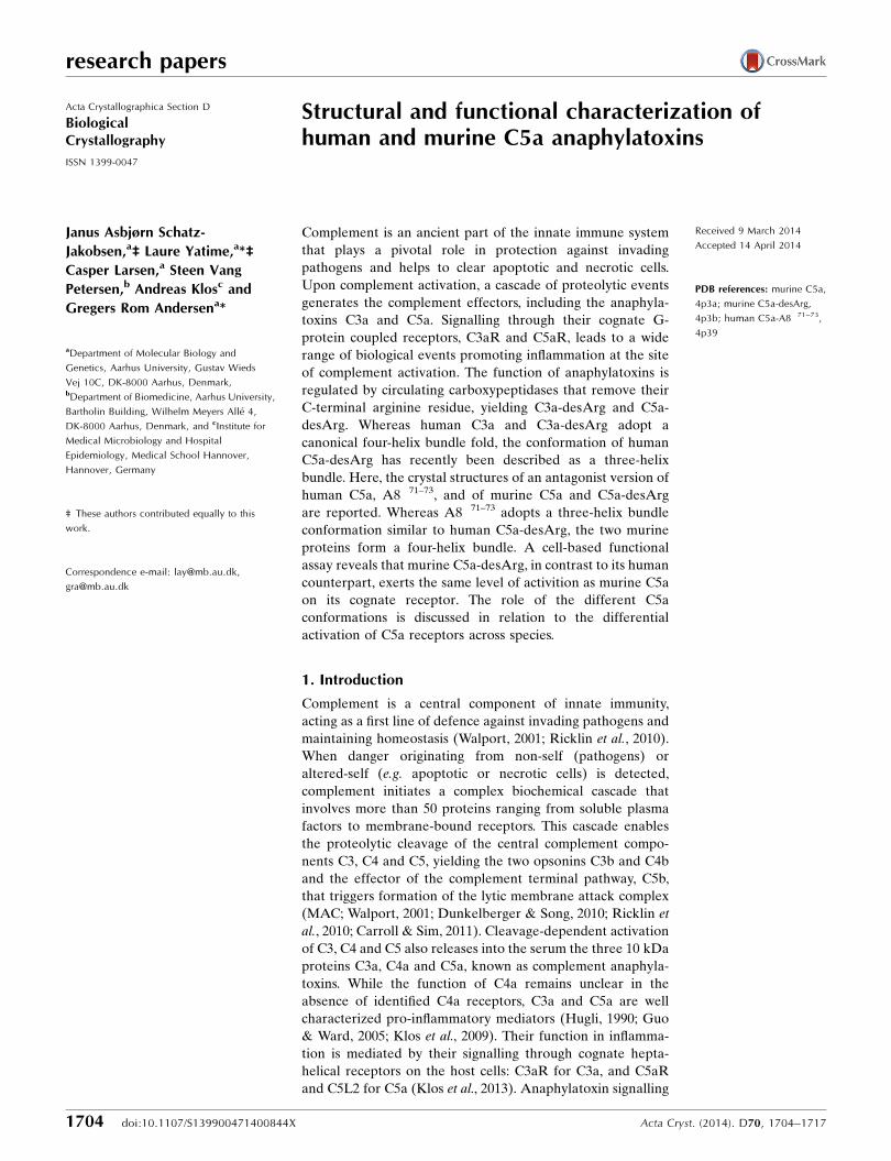
Figure 5General overview of mC5a and mC5a-desArg structures. (a) Elution profile of recombinant mC5a (purple) and mC5a-desArg (orange) on the SOURCE15S column. (b) Overall structure of mouse C5a at 1.4 A resolution. The four �-helices are labelled according to the hC5a nomenclature and the threeinternal disulfide bridges stabilizing the four-helix bundle are highlighted in yellow. (c) Final model and electron-density maps for the mC5a structure.The 2mFo�DFc map is shown as a blue mesh and contoured at 1�. The mFo�DFc map is show in blue/green and contoured at 3�. (d) As in (c) but forthe mC5a-desArg structure.
superimpose well with hC5a (Fig. 6b), with r.m.s.d.s on C�
atoms of 1.24 and 1.23 A, respectively, compared with the C5a
moiety from human C5 (Fredslund et al., 2008). Small differ-
ences are mostly encountered for the loop regions connecting
the different helices. Thus, the canonical four-helix bundle fold
is well conserved between species.
The C-terminus of mC5a and mC5a-desArg (region
Pro747–Arg755/Gly754) was not visible in the electron
density, again suggesting its high flexibility. Surprisingly, a blob
of density that assumed the shape of an arginine was visible in
the mC5a structure (Supplementary Fig. S5). This density is
observed in the groove between the helix H1 N-terminus and
the helix H2 C-terminus of monomer A in the asymmetric unit
and is located within hydrogen-bonding distance of Arg743 of
the same monomer (Supplementary Fig. S5a). The density is
less prominent around this position in the other monomers of
the mC5a asymmetric unit, but this density is not observed
around any of the molecules present in the mC5a-desArg
crystals, although mC5a-desArg crystallizes under the same
conditions and according to the same packing as mC5a
(Supplementary Fig. S5b). Furthermore, none of the compo-
nents present either in the protein buffers or in the crystal-
lization conditions could account for such a density. Although
it cannot be ruled out that this corresponds to a ligand trapped
by the protein during bacterial expression, modelling of an
arginine residue at this position matches the density perfectly
(Supplementary Fig. S5c). Inspection of the crystal packing
reveals that this density could represent the C-terminal
Arg755 of mC5a from a neighbouring symmetry-related
molecule, in which the C� atom of the last modelled residue,
Glu745, is separated by 21 A from the putative position of
this Arg755 C� atom (Supplementary Fig. S5d), a distance that
could accommodate a stretch of nine residues assuming a
random-coil nonlinear conformation. Thus, the position of
Arg755 could have been stabilized by packing against Arg743
from a symmetry-related molecule, while the connecting
residues Ser746–Gly754 adopt multiple conformations within
the solvent separating the two layers of molecules. As we
research papers
Acta Cryst. (2014). D70, 1704–1717 Schatz-Jakobsen et al. � Human and murine C5a anaphylatoxins 1713
Figure 6Comparison of the mC5a/mC5a-desArg structures with hC5a. (a) Superimposition of the mC5a (blue) and mC5a-desArg (beige) structures. (b)Superimposition of mC5a (blue) with the structure of hC5a from human C5 (green; Fredslund et al., 2008) or from the NMR model (orange; Zhang et al.,1997). (c, d) Stabilizing interactions between helices H1 and H4 in the four-helix bundle models of mC5a (blue) and hC5a (green). The residues involvedin hydrogen bonds, salt bridges and hydrophobic interactions at the H1–H4 interface are represented as sticks in both structures.
cannot attribute this density to Arg755 without ambiguity, the
density was left unmodelled in the final mC5a structure.
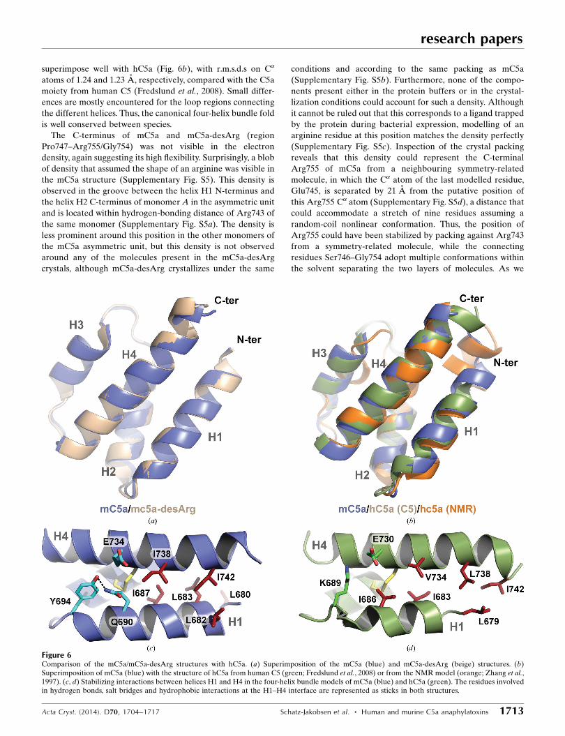
3.4. Differential activity of C5a/C5a-desArg anaphylatoxinsacross species
In the light of the structural differences observed between
human and mouse C5a-desArg, we decided to evaluate
whether these differences could reflect their respective acti-
vation properties towards both human and murine C5a
receptors. For this purpose, mC5a and mC5a-desArg were
first tested in the GARA against RBL cells expressing human
C5aR. As shown in Figs. 7(a) and 7(c), recombinant mC5a can
activate hC5aR in a dose-dependent manner to the same level
as commercially available recombinant hC5a and with an even
lower EC50 (0.64 � 0.11 nM on average compared with 1.05 �
0.27 nM for commercial hC5a). mC5a-desArg could also
activate hC5aR but to a maximal NAG release activity that
reached only half of the activity observed for mC5a and hC5a.
Thus, as hC5a-desArg, mC5a-desArg also acts as a partial
agonist of hC5aR. However, while hC5a-desArg retains an
EC50 equivalent to that measured for hC5a, mC5a-desArg has
a tenfold higher EC50 than mC5a.
All four anaphylatoxins were then tested against the murine
C5a receptor in the GARA (Figs. 7b and 7d). As expected,
mC5a is a full agonist of mC5aR and can activate the receptor
with an EC50 of 0.57 � 0.13 nM. Surprisingly, mC5a-desArg
could activate mC5aR to the same level as mC5a, although
with a much increased EC50 of 5.35 � 3.94 nM. Thus, in
contrast to the activity ofC5a-desArg towards hC5aR, mC5a-
desArg seems to act as a full agonist of mC5aR but with a
lower potency. For comparison, the biological activity of
commercially available recombinant hC5a and hC5a-desArg
towards mC5aR was then assessed. Intriguingly, while mC5a
and mC5a-desArg can substitute for their human counterparts
with respect to hC5aR activation, the reverse does not hold
true. Indeed, hC5a could activate mC5aR to the same extent
as mC5a but with a fivefold higher EC50, thereby acting more
similarly to mC5a-desArg. In contrast, the maximal NAG
release observed upon incubation with up to 10 mM hC5a-
desArg reached only 60% of the maximal activity obtained
with any of the three other anaphylatoxins, with an EC50 that
was on average 100 times higher than for hC5a or mC5a-
desArg. Thus, hC5a-desArg only acts as a partial agonist
towards mC5aR, thereby failing to match the behaviour of
mC5a-desArg with respect to mC5aR.
Taken together, these data show that hC5a and mC5a both
act as C5aR agonists independently of the receptor species
and that in both cases mC5a is a more potent agonist than
research papers
1714 Schatz-Jakobsen et al. � Human and murine C5a anaphylatoxins Acta Cryst. (2014). D70, 1704–1717
Figure 7GARA for recombinant mC5a and mC5a-desArg against both hC5aR and mC5aR and comparison with commercial hC5a/hC5a-desArg. All results areindicated as the mean � standard deviation of at least eight individual experiments (except for hC5a-desArg, where n = 4). The NAG release activity isexpressed as a percentage of the maximal NAG release activity obtained with commercial hC5a. The statistical significance of the differential behaviourobserved between hC5a and mC5a towards both receptors is as follows: p-value (hC5aR) = 0.0016; p-value (mC5aR) = 0.006. (a) Activation of hC5aR-expressing RBL cells by recombinant mC5a and mC5a-desArg compared with commercially available recombinant hC5a and hC5a-desArg. (b) As in (a)but with mC5aR-expressing RBL cells. (c) EC50 and maximal NAG release activity observed for all proteins upon incubation with hC5aR-expressingRBL cells. (d) As in (c) but with mC5aR-expressing RBL cells.

hC5a. In contrast, the desArg versions of these anaphylatoxins
have differential effects depending on the receptor species.
Indeed, hC5a-desArg is a partial agonist of both hC5aR and
mC5aR but has a much reduced potency towards mC5aR,
whereas mC5a-desArg only acts as a partial agonist towards
hC5aR and is a full agonist towards mC5aR but with a reduced
potency compared with mC5a.
4. Discussion
Here, we have reported the crystal structures of the human
C5aR antagonist hC5a-A8 and of the two murine anaphyla-
toxins mC5a and mC5a-desArg. The proteins were produced
recombinantly in bacteria using a fast and simple protocol
previously employed to purify recombinant bioactive hC3a
and hC3a-desArg (Bajic et al., 2013). As judged for the human
proteins, this procedure yielded recombinant proteins
possessing the same biological activity towards hC5aR as
commercially available C5a proteins, with potencies and
agonist/antagonist effects that are in agreement with the
current literature (Otto et al., 2004; Higginbottom et al., 2005).
Interestingly, the hC5a-A8 antagonist crystallized as a three-
helix bundle similar to that recently described for hC5a-
desArg (Cook et al., 2010), whereas both mC5a and mC5a-
desArg adopted the canonical four-helix bundle conformation
also observed for hC5a both as a part of C5 (Fredslund et al.,
2008) and as an isolated protein in solution (Zhang et al.,
1997). Furthermore, evaluation of the anaphylatoxic proper-
ties of both human and murine proteins in an N-acetyl-�-d-
glucosaminidase release assay revealed strong differences
between species, i.e. of human versus murine proteins towards
both hC5aR and mC5aR, and for the same species but towards
cognate versus noncognate receptors. Such differences
between species have also been reported for the human and
porcine systems (Bubeck et al., 1994). Since C5a sequences
are well conserved across species (Supplementary Fig. S1),
modulation of their activity towards C5a receptors could arise
from subtle differences in both anaphylatoxins and receptors,
in addition to different downstream effectors of C5aR acti-
vation. Furthermore, since C5a is rapidly desarginated in vivo
after release from its inert C5 precursor, our findings that
hC5a-desArg and mC5a-desArg have differential activation
properties towards their cognate receptors indicate that
desargination acts as a partial control mechanism mainly in
humans but not in mice. Therefore, the biological effects of
C5a/C5a-desArg in this rodent (e.g. in animal models versus
diseases) might be more prominent and/or less locally
restricted to the site of complement activation owing to the
higher remaining biological activity of the C5a-desArg form,
which represents 99% of the circulating C5a form in the blood.
This might also compensate for the relatively low levels of
complement factors, including C5, reported in mice (Ong &
Mattes, 1989). In consequence, a clearly visible differential
effect of C5a/C5a-desArg in C5aR�/� versus wild-type mice
can be expected, thereby validating the use of mouse models
to study C5a/C5a-desArg-dependent effects despite the low
C5/C5a levels in mice.
Extensive mutational studies have mapped the C5aR
binding site on C5a (Mollison et al., 1989; Toth et al., 1994;
Bubeck et al., 1994; Hagemann et al., 2006, 2008). It is now
commonly accepted that C5a binding to its cognate receptor
occurs through a two-site binding mode (Siciliano et al., 1994).
The C5a core first docks against the receptor N-terminus,
allowing proper positioning of the C5a molecule with respect
to the GPCR transmembrane region. The C5a C-terminus
can then insert into a binding pocket within the C5aR trans-
membrane domain, thereby triggering receptor activation.
The C5a core residues interacting with the receptor
N-terminus are located either in the H1–H2 loop and at the
beginning of helix H2 (four-helix bundle nomenclature) or in
the region encompassing the end of helix H3, the H3–H4 loop
and the beginning of helix H4 (Supplementary Fig. S1), i.e.
on the opposite face of C5a compared with the C-terminal
extension. This patch is mostly composed of positively charged
residues, such as His692, Lys696, Lys697, Arg714, Arg717,
Arg723 and Lys726, that interact with acidic residues and
sulfotyrosines contained in the C5aR N-terminus (DeMartino
et al., 1994; Mery & Boulay, 1994; Chen et al., 1998; Farzan et
al., 2001). Interestingly, none of these C5a residues are located
within helix H1. This could indicate that C5a binding can occur
independently of whether the anaphylatoxin adopts a three-
helix or four-helix bundle conformation. In agreement with
this idea, hC5a-desArg and mC5a-desArg display similar
activation properties towards hC5aR, although their respec-
tive crystal structures (Cook et al., 2010 and this study) reveal
quite distinct structural arrangements of the two proteins.
Evidently, one cannot rule out that the conformations of the
molecules in the crystal structures differ from the conforma-
tions that they adopt upon receptor binding. Thus, although
mC5a and mC5a-desArg adopt a four-helix bundle fold in the
crystal structure, their conformation could change to a three-
helix bundle upon receptor binding. Another possibility, as
suggested previously (Cook et al., 2010; Bajic et al., 2013), is
that the three-helix bundle conformation may be relevant for
other C5a functions such as the processing of pro-C5 into
separate � and � chains or the cleavage of C5a by carboxy-
peptidases. Comparison of the H1–H4 packing in both hC5a
and mC5a (Figs. 6c and 6d) reveals that the stabilization of the
H1–H4 interface in mC5a is strengthened by the presence of
an additional helical turn in the H1 N-terminus owing to the
N-terminal extension by three residues (Asn-Leu-His) of
mC5a compared with hC5a. However, this property may only
be specific to mouse and rat C5a, since C5a proteins from
other species have a length similar to hC5a in their N-termini
(Supplementary Fig. S1). A similar situation was encountered
for the hC3a and hC3a-desArg proteins (Bajic et al., 2013),
which both display a four-helix bundle conformation while
acting as expected towards hC3aR (Wilken et al., 1999). In the
case of hC3a and hC3a-desArg, the H1–H4 packing again
seemed to be more strongly stabilized than for hC5a, in which
H1–H4 interactions are exclusively hydrophobic. In particular,
hydrogen bonds between Lys682 (H1), Tyr686 (H1) and
Asp726 (H2) further held helices H1 and H2 in place in the
hC3a and hC3a-desArg structures. A similar triad is encoun-
research papers
Acta Cryst. (2014). D70, 1704–1717 Schatz-Jakobsen et al. � Human and murine C5a anaphylatoxins 1715

tered in mC5a and mC5a-desArg with Gln690 (H1), Tyr694
(H1) and Glu734, although the Glu734 side chain points away
from the residues in H1 and interacts with a symmetry-related
molecule. Thus, the propensity of helix H1 from hC5a to
separate from the four-helix bundle might be an intrinsic
property of the molecule allowing this to occur more spon-
taneously, while a specific context (binding partner or biolo-
gical function) may be required for mC5a-derived and/or
hC3a-derived molecules.
In any case, the in vivo relevance of the three-helix bundle
conformation with respect to C5aR activation and C5L2
binding remains to be established. Our study also revealed
that anaphylatoxin properties are not equivalent among
species. Thus, conformational modulation of the anaphyla-
toxin three-dimensional architecture may have differential
effects depending on the receptor considered, as examplified
by mC5a-desArg and hC5a-desArg, which behave similarly
towards hC5aR but have quite distinct activities towards
mC5aR. Such differential effects across species are quite
intriguing, notably since both the C5a and C5aR sequences
are quite well conserved (Supplementary Figs. S1 and S6). In
particular, both the sulfotyrosines and the aspartate residues
present in the C5aR N-terminus and important for C5a
binding are well conserved across species (Supplementary Fig.
S6). Furthermore, the second binding site in hC5aR, which is
composed of charged residues on the extracellular membrane
side such as Arg206 and of the hydrophobic pocket formed
between helices H3 and H7 in the transmembrane region, is
also well conserved between species (Supplementary Fig. S6)
(Raffetseder et al., 1996; Gerber et al., 2001; Higginbottom et
al., 2005; Hagemann et al., 2008). Recent data have suggested
that the role of hC5a-desArg may have to be revisited (Reis et
al., 2012). Indeed, in a novel label-free cellular assay evalu-
ating the global downstream cellular response to C5aR acti-
vation, physiological concentrations of hC5a-desArg could
induce an overall cellular activation as high as, if not higher
than, the intact hC5a anaphylatoxin (Reis et al., 2012). Thus,
the differential activation properties of these anaphylatoxins
may also depend on the cell context and on the type of
biological activity investigated. The sequence and geometry of
the C5a C-terminus are also essential parameters in triggering
the activation switch that conditions C5aR-mediated cellular
response. Indeed, downstream G-protein coupling and acti-
vation seem to involve subtle changes in the relative orien-
tation of C5aR helices H3 and H7 (Gerber et al., 2001). For
example, a single Ile116Ala substitution in the hydrophobic
pocket of hC5aR constituting the second binding site for C5a
transforms an antagonistic peptide mimicking the C5a
C-terminus into an agonist. Inversely, changes in the C5a
C-terminal topography will also have dramatic effects on
C5aR activation. In this context, the structuring of hC5a-A8
into a �-strand motif extending outside the helix-bundle core
could provide a mechanistic model for the two-step binding
mode of hC5a to its receptor. Indeed, while the hC5a
C-terminus may adopt the helical conformation of the NMR
model (Zhang et al., 1997) in solution and possibly keep this
conformation throughout binding to the receptor, interaction
of the hC5a core with its first binding site on the hC5aR
N-terminus could also induce release of the hC5a C-terminus
from the bundle core, and an extended conformation as
observed for this region in hC5a-A8 may be required to allow
the C-terminus to anchor into the transmembrane binding site
on hC5aR. Nevertheless, it is not known whether such release
is necessary and hence it remains difficult to speculate on
precise mechanisms at this stage. Thus, deeper structural
investigations, and notably a detailed atomic structure
revealing the C5a–C5aR interaction, are required to gain
detailed insights into C5aR activation by complement
anaphylatoxins.
5. Related literature
The following references are cited in the Supporting Infor-
mation for this article: Bond & Schuttelkopf (2009), Bubeck et
al. (1994), Chen et al. (1998), Cook et al. (2010), Corpet (1988),
DeMartino et al. (1994), Farzan et al. (2001), Hagemann et al.
(2006, 2008), McCoy et al. (2005), Mery & Boulay (1994),
Mollison et al. (1989), Raffetseder et al. (1996), Siciliano et al.
(1994) and Toth et al. (1994).
We would like to thank the beamline staff at MAX-lab and
SLS for support during data collection. We are grateful to
Yangzi He and Mickael Blaise for data collection on the hC5a-
A8 and mC5a crystals and to Claudia Rheinheimer for tech-
nical assistance with the functional characterization of the
anaphylatoxins. We thank the Lundbeck Foundation for
supporting this work through a Lundbeck Foundation Nano-
medicine Center for Individualized Management of Tissue
Damage and Regeneration grant. This project was also
supported by DANSCATT, by the Danish Cancer Society and
by the Novo-Nordisk Foundation through a Hallas-Møller
Fellowship to GRA.
References
Adams, P. D. et al. (2010). Acta Cryst. D66, 213–221.Amara, U., Flierl, M. A., Rittirsch, D., Klos, A., Chen, H., Acker, B.,
Bruckner, U. B., Nilsson, B., Gebhard, F., Lambris, J. D. & Huber-Lang, M. (2010). J. Immunol. 185, 5628–5636.
Bajic, G., Yatime, L., Klos, A. & Andersen, G. R. (2013). Protein Sci.22, 204–212.
Bokisch, V. A. & Muller-Eberhard, H. J. (1970). J. Clin. Investig. 49,2427–2436.
Bond, C. S. & Schuttelkopf, A. W. (2009). Acta Cryst. D65, 510–512.Bubeck, P., Grotzinger, J., Winkler, M., Kohl, J., Wollmer, A., Klos, A.
& Bautsch, W. (1994). Eur. J. Biochem. 219, 897–904.Burgi, B., Brunner, T. & Dahinden, C. A. (1994). Eur. J. Immunol. 24,
1583–1589.Carroll, M. V. & Sim, R. B. (2011). Adv. Drug Deliv. Rev. 63, 965–975.Chen, V. B., Arendall, W. B., Headd, J. J., Keedy, D. A., Immormino,
R. M., Kapral, G. J., Murray, L. W., Richardson, J. S. & Richardson,D. C. (2010). Acta Cryst. D66, 12–21.
Chen, Z., Zhang, X., Gonnella, N. C., Pellas, T. C., Boyar, W. C. & Ni,F. (1998). J. Biol. Chem. 273, 10411–10419.
Cook, W. J., Galakatos, N., Boyar, W. C., Walter, R. L. & Ealick, S. E.(2010). Acta Cryst. D66, 190–197.
Corpet, F. (1988). Nucleic Acids Res. 16, 10881–10890.
research papers
1716 Schatz-Jakobsen et al. � Human and murine C5a anaphylatoxins Acta Cryst. (2014). D70, 1704–1717

DeMartino, J. A., Van Riper, G., Siciliano, S. J., Molineaux, C. J.,Konteatis, Z. D., Rosen, H. & Springer, M. S. (1994). J. Biol. Chem.269, 14446–14450.
Dunkelberger, J. R. & Song, W. C. (2010). Cell Res. 20, 34–50.Eglite, S., Pluss, K. & Dahinden, C. A. (2000). J. Immunol. 165, 2183–
2189.Emsley, P., Lohkamp, B., Scott, W. G. & Cowtan, K. (2010). Acta
Cryst. D66, 486–501.Farzan, M., Schnitzler, C. E., Vasilieva, N., Leung, D., Kuhn, J.,
Gerard, C., Gerard, N. P. & Choe, H. (2001). J. Exp. Med. 193,1059–1066.
Finch, A. M., Wong, A. K., Paczkowski, N. J., Wadi, S. K., Craik, D. J.,Fairlie, D. P. & Taylor, S. M. (1999). J. Med. Chem. 42, 1965–1974.
Fredslund, F., Laursen, N. S., Roversi, P., Jenner, L., Oliveira, C. L.,Pedersen, J. S., Nunn, M. A., Lea, S. M., Discipio, R., Sottrup-Jensen, L. & Andersen, G. R. (2008). Nature Immunol. 9, 753–760.
Gerard, N. P. & Gerard, C. (1990). Biochemistry, 29, 9274–9281.Gerber, B. O., Meng, E. C., Dotsch, V., Baranski, T. J. & Bourne, H. R.
(2001). J. Biol. Chem. 276, 3394–3400.Guo, R.-F. & Ward, P. A. (2005). Annu. Rev. Immunol. 23, 821–852.Hagemann, I. S., Miller, D. L., Klco, J. M., Nikiforovich, G. V. &
Baranski, T. J. (2008). J. Biol. Chem. 283, 7763–7775.Hagemann, I. S., Narzinski, K. D., Floyd, D. H. & Baranski, T. J.
(2006). J. Biol. Chem. 281, 36783–36792.Higginbottom, A., Cain, S. A., Woodruff, T. M., Proctor, L. M.,
Madala, P. K., Tyndall, J. D., Taylor, S. M., Fairlie, D. P. & Monk,P. N. (2005). J. Biol. Chem. 280, 17831–17840.
Hoehlig, K., Maasch, C., Shushakova, N., Buchner, K., Huber-Lang,M., Purschke, W. G., Vater, A. & Klussmann, S. (2013). Mol. Ther.21, 2236–2246.
Huber-Lang, M., Sarma, J. V., Zetoune, F. S., Rittirsch, D., Neff, T. A.,McGuire, S. R., Lambris, J. D., Warner, R. L., Flierl, M. A., Hoesel,L. M., Gebhard, F., Younger, J. G., Drouin, S. M., Wetsel, R. A. &Ward, P. A. (2006). Nature Med. 12, 682–687.
Hugli, T. E. (1990). Curr. Top. Microbiol. Immunol. 153, 181–208.Kabsch, W. (1993). J. Appl. Cryst. 26, 795–800.Kemper, C. & Kohl, J. (2013). Mol. Immunol. 56, 181–190.Kessel, C., Nandakumar, K. S., Peters, F. B., Gauba, V., Schultz, P. G.
& Holmdahl, R. (2014). Arthritis. Rheumatol. 66, 610–621.Klos, A., Tenner, A. J., Johswich, K. O., Ager, R. R., Reis, E. S. &
Kohl, J. (2009). Mol. Immunol. 46, 2753–2766.Klos, A., Wende, E., Wareham, K. J. & Monk, P. N. (2013). Pharmacol.
Rev. 65, 500–543.Krissinel, E. & Henrick, K. (2007). J. Mol. Biol. 372, 774–797.Laursen, N. S., Magnani, F., Gottfredsen, R. H., Petersen, S. V. &
Andersen, G. R. (2012). Curr. Mol. Med. 12, 1083–1097.Lee, H., Zahra, D., Vogelzang, A., Newton, R., Thatcher, J., Quan, A.,
So, T., Zwirner, J., Koentgen, F., Padkjaer, S. B., Mackay, F.,Whitfeld, P. L. & Mackay, C. R. (2006). Nature Biotechnol. 24,1279–1284.
Li, R., Coulthard, L. G., Wu, M. C. L., Taylor, S. M. & Woodruff, T. M.(2013). FASEB J. 27, 855–864.
Liszewski, M. K. et al. (2013). Immunity, 39, 1143–1157.MacLaren, R., Cui, W. & Cianflone, K. (2008). Adv. Exp. Med. Biol.
632, 1–21.Markiewski, M. M., DeAngelis, R. A., Benencia, F., Ricklin-
Lichtsteiner, S. K., Koutoulaki, A., Gerard, C., Coukos, G. &Lambris, J. D. (2008). Nature Immunol. 9, 1225–1235.
McCoy, A. J., Grosse-Kunstleve, R. W., Storoni, L. C. & Read, R. J.(2005). Acta Cryst. D61, 458–464.
Mery, L. & Boulay, F. (1994). J. Biol. Chem. 269, 3457–3463.Mollison, K. W., Mandecki, W., Zuiderweg, E. R. P., Fayer, L., Fey,
T. A., Krause, R. A., Conway, R. G., Miller, L., Edalji, R. P.,Shallcross, M. A., Lane, B., Fox, J. L., Greer, J. & Carter, G. W.(1989). Proc. Natl Acad. Sci. USA, 86, 292–296.
Ong, G. L. & Mattes, M. J. (1989). J. Immunol. Methods, 125, 147–158.Otto, M., Hawlisch, H., Monk, P. N., Muller, M., Klos, A., Karp, C. L.
& Kohl, J. (2004). J. Biol. Chem. 279, 142–151.Raffetseder, U., Roper, D., Mery, L., Gietz, C., Klos, A., Grotzinger,
J., Wollmer, A., Boulay, F., Kohl, J. & Bautsch, W. (1996). Eur. J.Biochem. 235, 82–90.
Reis, E. S., Chen, H., Sfyroera, G., Monk, P. N., Kohl, J., Ricklin, D. &Lambris, J. D. (2012). J. Immunol. 189, 4797–4805.
Ricklin, D., Hajishengallis, G., Yang, K. & Lambris, J. D. (2010).Nature Immunol. 11, 785–797.
Siciliano, S. J., Rollins, T. E., DeMartino, J., Konteatis, Z., Malkowitz,L., Van Riper, G., Bondy, S., Rosen, H. & Springer, M. S. (1994).Proc. Natl Acad. Sci. USA, 91, 1214–1218.
Sprong, T., Brandtzaeg, P., Fung, M., Pharo, A. M., Høiby, E. A.,Michaelsen, T. E., Aase, A., van der Meer, J. W., van Deuren, M. &Mollnes, T. E. (2003). Blood, 102, 3702–3710.
Strainic, M. G., Shevach, E. M., An, F., Lin, F. & Medof, M. E. (2013).Nature Immunol. 14, 162–171.
Tokodai, K., Goto, M., Inagaki, A., Nakanishi, W., Okada, N., Okada,H. & Satomi, S. (2010). Transplant. Proc. 42, 2102–2103.
Toth, M. J., Huwyler, L., Boyar, W. C., Braunwalder, A. F., Yarwood,D., Hadala, J., Haston, W. O., Sills, M. A., Seligmann, B. &Galakatos, N. (1994). Protein Sci. 3, 1159–1168.
Touw, W. van der, Cravedi, P., Kwan, W. H., Paz-Artal, E., Merad, M.& Heeger, P. S. (2013). J. Immunol. 190, 5921–5925.
Walport, M. J. (2001). N. Engl. J. Med. 344, 1058–1066.Wenzel, T., Sparbier, K., Mieruch, T. & Kostrzewa, M. (2006). Rapid
Commun. Mass Spectrom. 20, 785–789.Wilken, H. C., Gotze, O., Werfel, T. & Zwirner, J. (1999). Immunol.
Lett. 67, 141–145.Williamson, M. P. & Madison, V. S. (1990). Biochemistry, 29, 2895–
2905.Woodruff, T. M., Nandakumar, K. S. & Tedesco, F. (2011). Mol.
Immunol. 48, 1631–1642.Zhang, X., Boyar, W., Toth, M. J., Wennogle, L. & Gonnella, N. C.
(1997). Proteins, 28, 261–267.Zhou, W. (2012). Immunobiology, 217, 225–234.Zuiderweg, E. R., Nettesheim, D. G., Mollison, K. W. & Carter, G. W.
(1989). Biochemistry, 28, 172–185.
research papers
Acta Cryst. (2014). D70, 1704–1717 Schatz-Jakobsen et al. � Human and murine C5a anaphylatoxins 1717