structural basis for backtracking by the sars-cov-2
TRANSCRIPT

Structural basis for backtracking by the SARS-CoV-2replication–transcription complexBrandon Malonea,1, James Chena,1
, Qi Wangb, Eliza Llewellyna, Young Joo Choia, Paul Dominic B. Olinaresc,Xinyun Caod, Carolina Hernandeze, Edward T. Enge
, Brian T. Chaitc, David E. Shawb,f, Robert Landickd,g,
Seth A. Darsta,2, and Elizabeth A. Campbella,2
aLaboratory of Molecular Biophysics, The Rockefeller University, New York, NY 10065; bD. E. Shaw Research, New York, NY 10036; cLaboratory of MassSpectrometry and Gaseous Ion Chemistry, The Rockefeller University, New York, NY, 10065; dDepartment of Biochemistry, University of Wisconsin–Madison,Madison, WI 53706; eThe National Resource for Automated Molecular Microscopy, Simons Electron Microscopy Center, New York Structural Biology Center,New York, NY 10027; fDepartment of Biochemistry and Molecular Biophysics, Columbia University, New York, NY 10032; and gDepartment of Bacteriology,University of Wisconsin–Madison, Madison, WI 53706
Edited by Eva Nogales, University of California, Berkeley, CA, and approved April 7, 2021 (received for review February 7, 2021)
Backtracking, the reverse motion of the transcriptase enzyme onthe nucleic acid template, is a universal regulatory feature oftranscription in cellular organisms but its role in viruses is notestablished. Here we present evidence that backtracking extendsinto the viral realm, where backtracking by the severe acuterespiratory syndrome coronavirus 2 (SARS-CoV-2) RNA-dependentRNA polymerase (RdRp) may aid viral transcription and replication.Structures of SARS-CoV-2 RdRp bound to the essential nsp13 helicaseand RNA suggested the helicase facilitates backtracking. We usecryo-electron microscopy, RNA–protein cross-linking, and unbiasedmolecular dynamics simulations to characterize SARS-CoV-2 RdRpbacktracking. The results establish that the single-stranded 3′ seg-ment of the product RNA generated by backtracking extrudesthrough the RdRp nucleoside triphosphate (NTP) entry tunnel, thata mismatched nucleotide at the product RNA 3′ end frays and entersthe NTP entry tunnel to initiate backtracking, and that nsp13 stimu-lates RdRp backtracking. Backtracking may aid proofreading, a crucialprocess for SARS-CoV-2 resistance against antivirals.
coronavirus | backtracking | cryo-electron microscopy | moleculardynamics | RNA-dependent RNA polymerase
Severe acute respiratory syndrome coronavirus 2 (SARS-CoV-2) is the causative agent of the current COVID-19 pandemic
(1, 2). The SARS-CoV-2 genome is replicated and transcribed byits RNA-dependent RNA polymerase holoenzyme [holo-RdRp,subunit composition nsp7/nsp82/nsp12 (3, 4)] in a replication–transcription complex (RTC), which is the target for antiviralssuch as remdesivir (Rdv) (5). The holo-RdRp is thought to co-ordinate with many cofactors to carry out its function (6, 7).Some of these cofactors, such as the nsp13 helicase (8) and thensp10/nsp14 proofreading assembly (9, 10), are also essential forviral replication and are antiviral targets (11–13).We recently reported views of the SARS-CoV-2 RTC in
complex with the nsp13 helicase [cryo-electron microscopy(cryo-EM) structures at a nominal resolution of 3.5 Å (14)]. Theoverall architecture of the nsp13-RTC places the nucleic acidbinding site of nsp13 directly in the path of the downstreamtemplate-strand RNA (t-RNA), and cryo-EM difference mapsrevealed the 5′-single-stranded t-RNA overhang engaged withnsp13 before entering the RdRp active site (14). The nsp13helicase translocates on single-stranded nucleic acid in the 5′→3′direction (15–22). Thus, this structural arrangement presents aconundrum: The RdRp translocates in the 3′→5′ direction onthe t-RNA strand, while nsp13 translocates on the same strand inthe opposite direction. Translocation of each enzyme opposeseach other, and if the helicase prevails it is expected to push theRdRp backward on the t-RNA (14). This reversible backwardsliding, termed backtracking, is a well-studied feature of thecellular DNA-dependent RNA polymerases (DdRps) (23–30).
Backtracking by the cellular DdRps plays important roles intranscription regulation, including the control of DdRp pausingduring transcription elongation, termination, DNA repair, andtranscription fidelity (25). In backtracking, the DdRp and associ-ated transcription bubble move backward on the DNA, while theRNA transcript reverse-threads through the complex to maintainthe register of the RNA–DNA hybrid (23–30). This movementgenerates a single-stranded 3′ segment of the RNA transcript whichis extruded out the secondary or nucleoside triphosphate (NTP)entry tunnel that branches off from the primary DdRp active-sitecleft around the conserved bridge helix (27–31).Although evolutionarily unrelated to the DdRps, a secondary
channel, formed by the RdRp motif F β-hairpin loop and pro-posed to serve as an NTP entry tunnel, branches off from themain SARS-CoV-2 RdRp active-site channel (32). This NTPentry tunnel is well positioned to receive the single-stranded 3′segment of backtracked RNA, a structural architecture analo-gous to the DdRps (14). We envisaged that translocation by thehelicase could mediate backtracking of the RdRp, an otherwiseenergetically unfavorable process, enabling the key viral func-tions such as proofreading (9, 10, 12, 33) and template switchingduring subgenomic transcription (7, 34). Here we outline thestructural basis for SARS-CoV-2 RTC backtracking and describethe role of nsp13 in stimulating backtracking.
Significance
The COVID-19 pandemic is caused by the severe acute respi-ratory syndrome coronavirus 2 (SARS-CoV-2). The SARS-CoV-2genome is replicated and transcribed by its RNA-dependent RNApolymerase (RdRp), which is the target for antivirals such asremdesivir. We use a combination of approaches to show thatbacktracking (backward motion of the RdRp on the templateRNA) is a feature of SARS-CoV-2 replication/transcription. Back-tracking may play a critical role in proofreading, a crucial processfor SARS-CoV-2 resistance against many antivirals.
Author contributions: B.M., J.C., Q.W., D.E.S., R.L., S.A.D., and E.A.C. designed research;B.M., J.C., Q.W., E.L., Y.J.C., P.D.B.O., X.C., C.H., E.T.E., S.A.D., and E.A.C. performed re-search; X.C. contributed new reagents/analytic tools; B.M., J.C., Q.W., P.D.B.O., B.T.C.,D.E.S., R.L., S.A.D., and E.A.C. analyzed data; and B.M., J.C., Q.W., P.D.B.O., B.C., D.E.S.,R.L., S.A.D., and E.A.C. wrote the paper.
The authors declare no competing interest.
This article is a PNAS Direct Submission.
This open access article is distributed under Creative Commons Attribution License 4.0(CC BY).1B.M. and J.C. contributed equally to this work.2To whom correspondence may be addressed. Email: [email protected] or [email protected].
This article contains supporting information online at https://www.pnas.org/lookup/suppl/doi:10.1073/pnas.2102516118/-/DCSupplemental.
Published April 21, 2021.
PNAS 2021 Vol. 118 No. 19 e2102516118 https://doi.org/10.1073/pnas.2102516118 | 1 of 8
BIOPH
YSICSAND
COMPU
TATIONALBIOLO
GY
Dow
nloa
ded
by g
uest
on
Oct
ober
27,
202
1

ResultsSARS-CoV-2 RdRp Backtracked Complexes for Cryo-EM. Previously,DdRp backtracked complexes (BTCs) were generated forstructural studies by direct incubation of the DdRp with DNA–
RNA scaffolds containing mismatched nucleotides at the RNA3′ end (27, 28, 30); these BTC scaffolds bind with the down-stream Watson–Crick base pairs of the RNA–DNA hybrid po-sitioned in the DdRp active site and the single-stranded 3′segment of mismatched RNA extruding out the DdRp NTPentry tunnel. To study RdRp BTCs, we therefore designed andtested RNA scaffolds based on our original SARS-CoV-2 RTCscaffold but with three or five mismatched cytosine nucleotidesadded to the product RNA (p-RNA) 3′ end (BTC3 and BTC5scaffolds; Fig. 1A). Consecutive mismatches at the p-RNA 3′ endwere designed to generate stable, homogeneous BTCs for bio-chemical and structural analysis—we do not propose that con-secutive mismatches are biologically relevant.Native electrophoretic mobility shift assays revealed that al-
though the holo-RdRp (nsp7/nsp82/nsp12) bound the RTCscaffold as observed previously (Fig. 1B, lane 1, SI Appendix, Fig.S1A, and ref. 14), nsp13 was required for efficient binding to theBTC scaffolds (Fig. 1B). Stable nsp13–holo-RdRp complexeswith BTC scaffolds were also observed by native mass spec-trometry (SI Appendix, Fig. S1 B and C).Modeling suggested that about five nucleotides of backtracked
single-stranded RNA at the p-RNA 3′ end would be sufficient totraverse the RdRp NTP entry tunnel. Therefore, to determinethe structural organization of the SARS-CoV-2 BTC, we assemblednsp13(ADP-AlF3) and holo-RdRp with the BTC5 scaffold (Fig. 1A;hereafter called BTC5) and analyzed the samples by single-particle
cryo-EM. The sample comprised two major classes: nsp131-BTC5(3.4-Å nominal resolution) and nsp132-BTC5 (3.6 Å; Fig. 1C and SIAppendix, Figs. S2 and S3). Analysis of the two refined structuresrevealed that the RdRp portion of each structure was essentiallyidentical (rmsd of 927 nsp12 α-carbon positions <0.3 Å; SI Appendix,Table S2), while the disposition of the common nsp13 protomer(nsp13.1) was divergent (rmsd of 590 nsp13 α-carbon positions >8 Å;SI Appendix, Table S2). To eliminate structural heterogeneity in thensp13 subunits and obtain a higher-resolution view of the BTC, theparticles from both classes were combined and locally refined insidea mask applied around the holo-RdRp and RNA (excluding thensp13 subunits), leading to the BTC5(local) combined map (3.2 Å;Fig. 1C and SI Appendix, Figs. S2 and S3 and Table S1).The cryo-EM maps (Figs. 1C and 2) revealed two significant
differences with the nsp13-RTC structures (14): 1) The single-stranded downstream template RNA (t-RNA) engaged withnsp13.1 was resolved (Fig. 2A), and 2) a single-stranded p-RNA 3′segment was extruded into the RdRp NTP entry tunnel (Fig. 2B).
Nsp13 Binds the Downstream Single-Stranded t-RNA. In the nsp131-BTC5 and nsp132-BTC5 cryo-EM maps, the single-stranded 5′segment of the t-RNA was engaged with nsp13.1. This region of thecryo-EM density was well-resolved (Fig. 2A), allowing identificationof the t-RNA segment engaged within the helicase as +14 to +8(numbering defined in Fig. 1A), 5′CCCAUGU3′. The five-nucleotidesegment connecting the t-RNA between the helicase and theRdRp (+7 to +3) was disordered and not modeled.
The SARS-CoV-2 RdRp NTP Entry Tunnel Accommodates the BacktrackedRNA. The cryo-EM maps also resolved a single-stranded p-RNA 3′segment of the BTC5 scaffold extruding into the RdRp NTP entry
Fig. 1. SARS-CoV-2 backtrack complex. (A) RNA scaffolds: (Top) RTC scaffold (14); (Bottom) backtrack complex scaffolds (BTC3 and BTC5). (B) A native gelelectrophoretic mobility shift assay reveals that holo-RdRp requires nsp13(ADP-AlF3) to bind the BTC scaffolds efficiently. (C) Cryo-EM structures ofSARS-CoV-2 BTCs. Shown is the transparent cryo-EM density [local-resolution-filtered (47)] with the refined models superimposed (SI Appendix, Table S1). Themodels and density are colored according to the key. Two major BTCs were observed (SI Appendix, Fig. S2), one containing one nsp13 protomer (nsp131-BTC5),and one containing two nsp13 promoters (nsp132-BTC5). We designate the nsp13 promoter common to both structures nsp13.1 and the other nsp13.2 (14).The cyan spheres denote the path of the single-stranded t-RNA 5′ segment, some of which is engaged with nsp13.1 in both structures.
2 of 8 | PNAS Malone et al.https://doi.org/10.1073/pnas.2102516118 Structural basis for backtracking by the SARS-CoV-2 replication–transcription complex
Dow
nloa
ded
by g
uest
on
Oct
ober
27,
202
1

tunnel (Fig. 2B), confirming the formation of a BTC (Fig. 3A). Theoverall architecture of the SARS-CoV-2 BTC is analogous toDdRp BTCs (Fig. 3 and ref. 14). The DdRp bridge helix (BH) (35)separates the DdRp active site cleft into a channel for the down-stream template DNA (over the top of the BH; Fig. 3B) and theNTP entry tunnel (underneath the BH; Fig. 3B). Similarly, the viralRdRp motif F (SI Appendix, Fig. S4A and ref. 32) serves as thestrand-separating structural element for the backtracked RNA(Fig. 3A). The downstream t-RNA passes over the top of motif F,while the backtracked RNA extrudes out the NTP entry tunnelunderneath motif F (Fig. 3A).The RdRp NTP entry tunnel provides a steric and electrostatic
environment conducive to channeling the backtracked RNA outof the active site without specific polar protein–RNA interac-tions that could hinder the RNA movement (Figs. 3C and 4).Comparing the electrostatic surface potential of the NTP entrytunnels of the SARS-CoV-2 RdRp with eukaryotic and bacterialDdRps reveals a similar overall electrostatic surface environ-ment that may facilitate backtracked RNA entry (Fig. 3C and SIAppendix, Fig. S4B), including a “track” of conserved positively
charged Arg and Lys residues of motif F (SARS-CoV-2 nsp12K545, K551, R553, and R555; Fig. 4 and SI Appendix, Fig. S4A).Conserved residues of RdRp motifs C and E complete the ac-tive-site/NTP entry tunnel environment surrounding the back-tracked RNA (Fig. 4 and SI Appendix, Fig. S4A).In the nsp13-RTCs, the RTC scaffold (Fig. 1A) is bound in a
posttranslocated state (14); the 3′ p-RNA A is base-paired to thet-RNA U at the −1 site near the catalytic nsp12-D760 (Fig. 5A).The next t-RNA base (A at +1) is positioned to receive the in-coming NTP substrate, but the site for the incoming NTP sub-strate is empty (Fig. 5A). By contrast, the BTC structures weretranslocated by one base pair compared to the RTCs; the basepair corresponding to the A–U Watson–Crick base pair at the 3′end of the p-RNA (located in the −1 site of the RTCs) was inthe −2 position of the BTCs (Figs. 1A, 4, and 5B). The −1 po-sition of the BTC was occupied by the first C–A mismatch; thep-RNA −1C made a non-Watson–Crick hydrogen bond with theopposing t-RNA A (Figs. 4 and 5B). The next three mismatchedp-RNA nucleotides (+1C, +2C, and +3C) trailed into the NTPentry tunnel (Figs. 4 and 5B). The 3′ nucleotide of the BTC5scaffold p-RNA (+4C; Fig. 1A) was solvent-exposed at theoutward-facing end of the NTP entry tunnel and lacked densityand was therefore not modeled (Fig. 2B). The trajectory of thebacktracked nucleotides at positions +1/+2 was sharply bent dueto spatial constraints of motif F residues (Fig. 4A).
Nsp13 Stimulates Backtracking. The SARS-CoV-2 wild-type holo-RdRp required the nsp13 helicase to bind the BTC scaffoldsefficiently (Fig. 1B). However, we observed that the holo-RdRpcontaining nsp12 with a single amino acid substitution (D760A)did not require nsp13 to bind the BTC scaffolds (SI Appendix,Fig. S1A, lane 4). Nsp12-D760 is a conserved residue of theRdRp motif C that chelates a crucial Mg2+ ion in catalyticcomplexes (SI Appendix, Fig. S4A and ref. 32), but in RdRpstructures lacking substrate (including the BTC structures) theMg2+ ions are absent (14, 36, 37). The catalytic Asp residues ofthe DdRps typically chelate the Mg2+ ion even in the absence ofsubstrate (31, 38), and this Mg2+ is retained in DdRp back-tracked structures (27–30). Our RdRp BTC structures suggestthat in the absence of a Mg2+ ion D760 presents an electrostaticbarrier to the phosphate backbone of the backtracked RNA(Fig. 5B), explaining the requirement for the helicase to sur-mount this barrier and why removal of D760 stabilizes binding tothe BTC scaffolds.To generate the SARS-CoV-2 BTCs for structural studies, we
used the BTC5 scaffold with five mismatched Cs at the p-RNA 3′end (Fig. 1A). To study the formation of SARS-CoV-2 BTCsfrom an RTC scaffold (fully Watson–Crick base-paired p-RNA3′ end), we analyzed ultraviolet (UV)-induced cross-linking from4-thio-U incorporated penultimate to the p-RNA 3′ end[RTC(4-thio-U)-scaffold; SI Appendix, Fig. S5A and ref. 39].Cross-linking was absolutely dependent on the presence of4-thio-U in the RNA, establishing specificity (SI Appendix, Fig. S5B).RTCs assembled with wild-type nsp12 and the RTC(4-thio-U)scaffold gave weak nsp12-RNA cross-linking upon UV expo-sure (SI Appendix, Fig. S5A, lane 1). These conditions favor aposttranslocated RTC (14, 36, 37) with the 4-thio-U seques-tered in the RNA–RNA hybrid and thus not available forprotein–RNA cross-linking. Cross-linking of the p-RNA tonsp12 was substantially increased by the addition of nsp13 with2 mM adenosine 5′-triphosphate (ATP) (SI Appendix, Fig. S5A,lane 2). Under these conditions, we propose that the translo-cation activity of nsp13 backtracked a fraction of the complexes,freeing the 4-thio-U from the RNA–RNA hybrid for cross-linking to nsp12. Cross-linking in the presence of nsp13 but inthe absence of ATP reduced nsp12 cross-linking (SI Appendix,Fig. S5A, lane 7 versus lane 2), supporting the proposal thatnsp13 translocation activity facilitates backtracking. Replacing
Fig. 2. Cryo-EM density maps. (A, Left) Overall view of nsp132-BTC5. Nsp13.2is removed (outline) for clarity. The boxed region is magnified on the right.(A, Right) Magnified view of the t-RNA segment (+14-5′-CCCAUGU-3′-+8)enclosed in the nsp13.1 helicase subunit. The cryo-EM density map (from thensp132-BTC structure) for the RNA is shown (blue mesh). (B, Left) Overallview of the BTC5(local) structure. The boxed region is magnified on the right.(B, Right) Magnified view of the region around the RdRp active site, showingthe t-RNA (cyan) and p-RNA (red) with the backtracked RNA segment. Thecryo-EM density map for the RNA [from BTC5(local)] is shown (blue mesh). (C)BTC5(local) cryo-EM density maps around nsp12 conserved motifs F, C, and E.Selected residues are labeled.
Malone et al. PNAS | 3 of 8Structural basis for backtracking by the SARS-CoV-2 replication–transcription complex https://doi.org/10.1073/pnas.2102516118
BIOPH
YSICSAND
COMPU
TATIONALBIOLO
GY
Dow
nloa
ded
by g
uest
on
Oct
ober
27,
202
1

wild-type nsp12 with nsp12-D760A (nsp12*; SI Appendix, Fig.S5A, lanes 4 to 6, 9, and 10), which is more prone to back-tracking (SI Appendix, Fig. S1A), showed the same trends butwith increased UV-dependent nsp12-RNA cross-linking, withthe maximal cross-linking occurring under the conditions expectedto favor backtracking the most (SI Appendix, Fig. S5A, lane 5).These results affirm the view that nsp13 facilitates backtracking ofthe SARS-CoV-2 RdRp.
A Mismatched Nucleotide at the p-RNA 3′ End Spontaneously Fraysand Enters into the RdRp NTP Entry Tunnel. The SARS-CoV-2 RTCis a highly processive and rapid replicase/transcriptase, capableof replicating a ∼1-kb RNA template at an average rate of ∼170nt/s (40). However, studies of other viral RdRps suggest that mis-incorporation slows the overall elongation rate and may induce
backtracking (41–43). We used molecular dynamics simulations toexplore the fate of a mismatched nucleotide incorporated at thep-RNA 3′ end. Starting with the nsp132-BTC5 structure, the −1Cwas mutated to U, and the +2 to +4 Cs were removed. Theresulting pretranslocated p-RNA had a matched −1U and a mis-matched +1C (−1U + 1C; Fig. 5C). In three 5-μs simulations weobserved the 3′-mismatched +1C alternating between two positions,either remaining in the vicinity of the active site (rmsd <3.5 Å) orfraying away from the p-RNA:t-RNA hybrid toward or into theNTP entry tunnel (rmsd >3.5 Å; Fig. 5C). Based on analysis ofthe aggregated −1U + 1C simulations, the mismatched +1C spentabout 40% of the time near the active site and about 60% of thetime frayed toward or in the NTP entry tunnel. In control simula-tions with a fully matched p-RNA 3′ end (−1U + 1U), the matched
Fig. 3. SARS-CoV-2 RdRp and DdRp BTCs. (A and B) SARS-CoV-2 RdRp (A) and DdRp (B) BTCs. (Top) Proteins are shown as transparent molecular surfaces andnucleic acids as atomic spheres. The boxed regions are magnified on the bottom. (Bottom) Magnified, cross-sectional view. Proteins are shown as molecularsurfaces and nucleic acids in stick format with transparent molecular surface. (A) The SARS-CoV-2 BTC5(local). Nsp8a and nsp12 are shown (nsp7 and nsp8b areremoved for clarity). Nsp12 motif F is shown as a magenta backbone ribbon (Top). Backtracked RNA (+1C to +3C of the BTC5-scaffold; Fig. 1A) extrudes out theNTP entry tunnel. (B) A DdRp (Saccharomyces cerevisiae Pol II) BTC [Protein Data Bank (PDB) ID code 3PO2 (29)]. The BH is shown as a magenta backboneribbon. The backtracked RNA extrudes out the NTP entry tunnel/secondary channel/funnel. (C) Views from the outside into the NTP entry tunnels of theSARS-CoV-2 (Left) and an S. cerevisiae DdRp [PDB ID code 3GTP (27)] BTC. Protein surfaces are colored by the electrostatic surface potential [calculated usingAPBS (48)]. Backtracked RNA is shown as atomic spheres with yellow carbon atoms.
4 of 8 | PNAS Malone et al.https://doi.org/10.1073/pnas.2102516118 Structural basis for backtracking by the SARS-CoV-2 replication–transcription complex
Dow
nloa
ded
by g
uest
on
Oct
ober
27,
202
1

+1U at the p-RNA 3′ end did not fray and spent 100% of thetime in the active-site pocket (SI Appendix, Fig. S6).Nucleotides −36 to +14 of the BTC5 scaffold t-RNA (as de-
fined in Fig. 1A) were included in the simulations. The nsp13.1-bound (+8 to +14) and the nsp12-bound (−36 to +2) regionswere stable over the course of the simulation time. The t-RNAnucleotides +3 to +7 (the portion connecting the nsp12-boundand nsp13.1-bound t-RNA) were highly dynamic, consistent withthe absence of well-defined cryo-EM density for this region ofthe t-RNA. We note that the simulations inform on the path offrayed RNAs but not on the role of nsp13 in backtracking.
DiscussionOur results establish that the SARS-CoV-2 RTC backtracks, thatbacktracking is facilitated by the nsp13 helicase, and that theresulting single-stranded 3′ segment of the p-RNA extrudes outthe RdRp NTP entry tunnel in a manner analogous to the evo-lutionarily unrelated cellular DdRps (Fig. 3). Thus, a secondarytunnel to accommodate backtracked RNA, facilitating fidelityand possibly other functions (Fig. 6), appears to be a crucialfeature of transcriptase enzymes that evolved independently.Backtracking of Φ6 and poliovirus RdRps has been reported
based on analysis of single-molecule observations (41–43). Thensp13 helicase facilitates efficient backtracking of the SARS-CoV-2RTC (SI Appendix, Fig. S5). We note that in bacteria the UvrDhelicase has been shown to induce DdRp backtracking, sug-gesting that a role for helicases in backtracking may be wide-spread (44). Here we envision the helicase translocating on thedownstream t-RNA, facilitating unwinding of the duplex t-RNA/p-RNA and entry of the p-RNA 3′-single-stranded fragment intothe NTP entry tunnel. This process could be triggered by amismatched nucleotide at the p-RNA 3′ end.Our results are consistent with the view that a matched nu-
cleotide at the pretranslocated p-RNA 3′ end remains basepaired to the t-RNA (Fig. 5 and SI Appendix, Fig. S6), facilitatingtranslocation and subsequent NTP addition and thus rapidelongation (at a maximum elongation rate of ∼170 nt/s a trans-location event would occur approximately every 6 ms, on aver-age, explaining why translocation was not observed in our 5-μs
simulations; Fig. 5 and SI Appendix, Fig. S6). However, uponmisincorporation, the pretranslocated, mismatched nucleotide atthe p-RNA 3′ end spends more than half the time frayed fromthe t-RNA and toward or in the NTP entry tunnel (Fig. 5C), aconfiguration that is likely recalcitrant to translocation andsubsequent elongation. The favorable environment of the NTPentry tunnel (Figs. 3 and 4) may further encourage backtracking.The resulting inhibition of translocation may enable the tightengagement of the nsp13.1 helicase with the downstream single-stranded t-RNA (Fig. 2A), allowing the 5′→3′ translocation ac-tivity of the helicase to more robustly backtrack the complex(SI Appendix, Fig. S5).Our findings have implications for the processes of sub-
genomic transcription and proofreading in SARS-CoV-2 (Fig. 6and ref. 14). Generation of messenger RNAs for the viralstructural proteins begins with transcription initiation at the 3′-poly(A) tail of the (+)-strand RNA genome. The process ofsubgenomic transcription ultimately generates a nested set oftranscripts that are both 5′- and 3′-coterminal with the viral ge-nome and involves a remarkable template switch from the 3′portion of the genome to the 5′ leader (7, 34). The template-switching event is thought to involve stalling of the RdRp thenbase-pairing between the 3′ end of the nascent transcript and acomplementary sequence (the transcription regulatory sequence,or TRS) near the (+)-strand 5′ leader (45). The 3′ end of thenascent transcript is base-paired to the t-RNA and is sequesteredin the stalled RdRp active site; for template switching to occurthe 3′ end of the nascent transcript must be separated from thet-RNA and from the RdRp active site so that it is available forbase pairing to the TRS near the 5′ leader. Backtracking wouldseparate the p-RNA 3′ end from the t-RNA and would alsoextrude the 3′ end of the nascent transcript out the NTP entrytunnel, making it available for base pairing to the 5′ TRS (Fig. 6).Our results establishing that the SARS-CoV-2 RTC can back-track validates a key prediction of this model for the mechanismof template switching during subgenomic transcription (14).Nucleotide analogs that function by being incorporated into
product RNA by viral RdRps are important antiviral therapeu-tics (46). Notably, their incorporation may induce backtracking
Fig. 4. Protein–RNA interactions in the BTC. (A, Top) Overall view of BTC5(local). Proteins are shown as transparent molecular surfaces and nucleic acids asatomic spheres. Nsp8a and nsp12 are shown (nsp7 and nsp8b are removed for clarity). Nsp12 motifs C, E, and F are shown as backbone ribbons (coloredaccording to the key on the bottom). The boxed region is magnified below. (A, Bottom) RNA is shown from −2 to +3. Proteins are shown as transparentmolecular surfaces. RdRp motifs C, E, and F are shown as transparent backbone ribbons (colored according to the key) with side chains of residues thatapproach the backtracked RNA (≤4.5 Å) shown. (B) Schematic illustrating the same protein–RNA interactions as A. Drawn using Nucplot (49).
Malone et al. PNAS | 5 of 8Structural basis for backtracking by the SARS-CoV-2 replication–transcription complex https://doi.org/10.1073/pnas.2102516118
BIOPH
YSICSAND
COMPU
TATIONALBIOLO
GY
Dow
nloa
ded
by g
uest
on
Oct
ober
27,
202
1

Fig. 5. Comparison of active-site proximal RNA in the RTC and BTC structures and from simulations of a mismatched nucleotide at the p-RNA 3′ end. (A andB) Comparison of the active-site proximal RNA in the RTC [A; PDB ID code 6XEZ (14)], BTC5(local) (B), and from selected snapshots of molecular dynamicssimulations of a −1U + 1C complex (C). The schematics denote the nucleotides shown in the context of the RTC (A) and BTC5 scaffolds (B; full scaffold se-quences shown in Fig. 1A) or generated from the BTC5 scaffold for the simulations (C). Carbon atoms of the t-RNA are colored cyan and p-RNA are coloredsalmon except in the case of mismatched Cs at the 3′ end, which are colored dark red. Watson–Crick base-pairing hydrogen bonds are denoted as dark graydashed lines; other hydrogen-bonds as red dashed lines. Nsp12 motif C is shown as a yellow-orange backbone ribbon, and the side chain of D760 is shown asatomic spheres. (A) The RTC is in a posttranslocated state, with the A–U base pair at the p-RNA 3′ end in the −1 position (14). (B) The BTC5(local) RNA istranslocated compared to the RTC; the base pair corresponding to A–U at the 3′ end of the RTC RNA in the −1 position is in the −2 position of the BTC RNA. AC–A mismatch occupies the BTC −1 site. The +1, +2, and +3 mismatched Cs trail into the RdRp NTP entry tunnel (denoted by black squiggly lines). The +4C(present in the BTC5 scaffold; Fig. 1A) is exposed to solvent, disordered, and not modeled. (C) Molecular dynamics simulations of the nsp132–BTC−1U+1C
complex. The complex was simulated with three replicates (green, blue, and orange traces). Rmsd values plotted as a function of time represent the heavy-atom rmsd of the +1C of the p-RNA compared with the starting configuration (Materials and Methods). The rmsd histograms (plotted on the right) are anaggregate of all three replicates. Two structures taken from one of the simulations are shown, one showing the +1C of the p-RNA in the active site (t = 0 μs)and the other showing the +1C frayed into the NTP entry tunnel (t = 4.5 μs).
6 of 8 | PNAS Malone et al.https://doi.org/10.1073/pnas.2102516118 Structural basis for backtracking by the SARS-CoV-2 replication–transcription complex
Dow
nloa
ded
by g
uest
on
Oct
ober
27,
202
1
by the RdRp (43). Rdv, a nucleotide analog, is the only Food andDrug Administration–approved drug for COVID-19 treatment(5). Our results support a model in which RdRp misincorporation
or incorporation of nucleotide analogs can pause the RdRp,allowing nsp13 to engage with the downstream single-strandedt-RNA to induce backtracking (14). The resulting exposure ofthe p-RNA 3′ end out the NTP entry tunnel (Figs. 3A and 6) couldprovide access for the SARS-CoV-2 proofreading machinery(nsp10/14) (9, 12) to degrade the p-RNA 3′ end, thus removingthe misincorporation or analog. This proofreading activity, whichis unique to the nidovirus order to which CoVs belong (10), is amajor determinant for the resistance of CoVs against many nu-cleotide analog inhibitors (13). Thus, understanding RdRp back-tracking and its potential role in CoV proofreading can facilitatethe development of therapeutics.
Materials and MethodsDetailed descriptions of SARS-CoV-2 nsp12, 7, 8, and 13 protein purification,assembly of the RTC complexes, native electrophoretic mobility shift assays,native mass spectrometry, cross-linking, specimen preparation for cryo-EM,cryo-EM data acquisition and processing, model building and refinement,and molecular dynamics simulations are provided in SI Appendix.
Data Availability. The cryo-EM density maps have been deposited in theEMDataBank: EMD-23007 (nsp131-BTC5), EMD-23008 (nsp132-BTC5), andEMD-23009 [BTC5(local)]. Atomic coordinates have been deposited in theProtein Data Bank: 7KRN (nsp131-BTC5), 7KRO (nsp132-BTC5), and 7KRP[BTC5(local)]. The molecular dynamics trajectories described in this work areavailable at https://www.deshawresearch.com/downloads/download_trajectory_sarscov2.cgi/.
ACKNOWLEDGMENTS. We thank M. Ebrahim, J. Sotiris, and H. Ng at TheRockefeller University Evelyn Gruss Lipper Cryo-electron Microscopy Re-source Center for help with cryo-EM. Some of the work reported here wasconducted at the Simons Electron Microscopy Center and the NationalResource for Automated Molecular Microscopy and National Center forCryoEM Access and Training located at the New York Structural BiologyCenter, supported by grants from the NIH National Institute of GeneralMedical Sciences (P41 GM103310), NYSTAR, the Simons Foundation(SF349247), the NIH Common Fund Transformative High Resolution Cryo-Electron Microscopy program (U24 GM129539), and New York State Assem-bly Majority. This work was supported by the Pels Family Center forBiochemistry and Structural Biology (The Rockefeller University) and NIHgrants P41 GM109824 and P41 GM103314 to B.T.C., R35 GM118130 to S.A.D.,and R01 GM114450 to E.A.C.
1. F. Wu et al., A new coronavirus associated with human respiratory disease in China.Nature 579, 265–269 (2020).
2. P. Zhou et al., A pneumonia outbreak associated with a new coronavirus of probablebat origin. Nature 579, 270–273 (2020).
3. L. Subissi et al., One severe acute respiratory syndrome coronavirus protein complexintegrates processive RNA polymerase and exonuclease activities. Proc. Natl. Acad. Sci.U.S.A. 111, E3900–E3909 (2014).
4. R. N. Kirchdoerfer, A. B. Ward, Structure of the SARS-CoV nsp12 polymerase bound tonsp7 and nsp8 co-factors. Nat. Commun. 10, 2342–2349 (2019).
5. US FDA, Remdesivir Emergency Use Authorization letter (2020). https://www.fda.gov/media/137564/download. Accessed 18 April 2021.
6. E. J. Snijder, E. Decroly, J. Ziebuhr, The nonstructural proteins directing coronavirusRNA synthesis and processing. Adv. Virus Res. 96, 59–126 (2016).
7. I. Sola, F. Almazán, S. Zúñiga, L. Enjuanes, Continuous and discontinuous RNA syn-thesis in coronaviruses. Annu. Rev. Virol. 2, 265–288 (2015).
8. K. C. Lehmann, E. J. Snijder, C. C. Posthuma, A. E. Gorbalenya, What we know but donot understand about nidovirus helicases. Virus Res. 202, 12–32 (2015).
9. E. Minskaia et al., Discovery of an RNA virus 3′->5′ exoribonuclease that is criticallyinvolved in coronavirus RNA synthesis. Proc. Natl. Acad. Sci. U.S.A. 103, 5108–5113(2006).
10. A. E. Gorbalenya, L. Enjuanes, J. Ziebuhr, E. J. Snijder, Nidovirales: Evolving the largestRNA virus genome. Virus Res. 117, 17–37 (2006).
11. R. Zhang et al., The nsp1, nsp13, and M proteins contribute to the hepatotropism ofmurine coronavirus JHM.WU. J. Virol. 89, 3598–3609 (2015).
12. M. R. Denison, R. L. Graham, E. F. Donaldson, L. D. Eckerle, R. S. Baric, Coronaviruses:An RNA proofreading machine regulates replication fidelity and diversity. RNA Biol.8, 270–279 (2011).
13. E. C. Smith, H. Blanc, M. C. Surdel, M. Vignuzzi, M. R. Denison, Coronaviruses lackingexoribonuclease activity are susceptible to lethal mutagenesis: Evidence for proof-reading and potential therapeutics. PLoS Pathog. 9, e1003565 (2013).
14. J. Chen et al., Structural basis for helicase-polymerase coupling in the SARS-CoV-2 replication-transcription complex. Cell 182, 1560–1573.e13 (2020).
15. A. O. Adedeji et al., Mechanism of nucleic acid unwinding by SARS-CoV helicase. PLoSOne 7, e36521 (2012).
16. E. M. Bautista, K. S. Faaberg, D. Mickelson, E. D. McGruder, Functional properties ofthe predicted helicase of porcine reproductive and respiratory syndrome virus. Vi-rology 298, 258–270 (2002).
17. K. A. Ivanov, J. Ziebuhr, Human coronavirus 229E nonstructural protein 13: Charac-terization of duplex-unwinding, nucleoside triphosphatase, and RNA 5′-triphosphataseactivities. J. Virol. 78, 7833–7838 (2004).
18. N.-R. Lee et al., Cooperative translocation enhances the unwinding of duplex DNA bySARS coronavirus helicase nsP13. Nucleic Acids Res. 38, 7626–7636 (2010).
19. K. J. Mickolajczyk et al., Force-dependent stimulation of RNA unwinding by SARS-CoV-2nsp13 helicase. Biophys. J. 120, 1020–1030 (2020).
20. A. Seybert, L. C. van Dinten, E. J. Snijder, J. Ziebuhr, Biochemical characterization ofthe equine arteritis virus helicase suggests a close functional relationship betweenarterivirus and coronavirus helicases. J. Virol. 74, 9586–9593 (2000).
21. A. Seybert, A. Hegyi, S. G. Siddell, J. Ziebuhr, The human coronavirus 229E superfamily1 helicase has RNA and DNA duplex-unwinding activities with 5′-to-3′ polarity. RNA 6,1056–1068 (2000).
22. J. A. Tanner et al., The severe acute respiratory syndrome (SARS) coronavirus NTPase/helicase belongs to a distinct class of 5′ to 3′ viral helicases. J. Biol. Chem. 278,39578–39582 (2003).
23. N. Komissarova, M. Kashlev, RNA polymerase switches between inactivated and ac-tivated states by translocating back and forth along the DNA and the RNA. J. Biol.Chem. 272, 15329–15338 (1997).
24. N. Komissarova, M. Kashlev, Transcriptional arrest: Escherichia coli RNA polymerasetranslocates backward, leaving the 3′ end of the RNA intact and extruded. Proc. Natl.Acad. Sci. U.S.A. 94, 1755–1760 (1997).
25. E. Nudler, RNA polymerase backtracking in gene regulation and genome instability.Cell 149, 1438–1445 (2012).
26. E. Nudler, A. Mustaev, E. Lukhtanov, A. Goldfarb, The RNA-DNA hybrid maintains theregister of transcription by preventing backtracking of RNA polymerase. Cell 89,33–41 (1997).
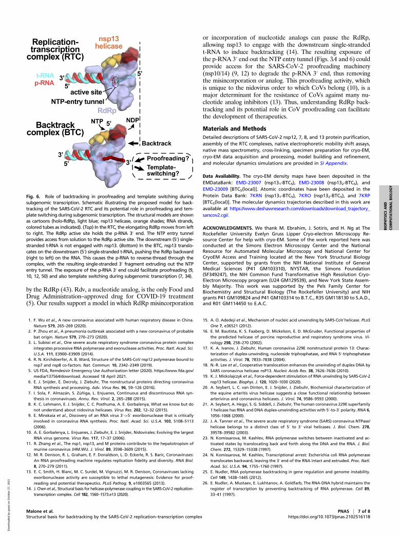
Fig. 6. Role of backtracking in proofreading and template switching duringsubgenomic transcription. Schematic illustrating the proposed model for back-tracking of the SARS-CoV-2 RTC and its potential role in proofreading and tem-plate switching during subgenomic transcription. The structural models are shownas cartoons (holo-RdRp, light blue; nsp13 helicase, orange shades; RNA strands,colored tubes as indicated). (Top) In the RTC, the elongating RdRpmoves from leftto right. The RdRp active site holds the p-RNA 3′ end. The NTP entry tunnelprovides access from solution to the RdRp active site. The downstream (5′) single-stranded t-RNA is not engaged with nsp13. (Bottom) In the BTC, nsp13 translo-cates on the downstream (5′) single-stranded t-RNA, pushing the RdRp backward(right to left) on the RNA. This causes the p-RNA to reverse-thread through thecomplex, with the resulting single-stranded 3′ fragment extruding out the NTPentry tunnel. The exposure of the p-RNA 3′ end could facilitate proofreading (9,10, 12, 50) and also template switching during subgenomic transcription (7, 34).
Malone et al. PNAS | 7 of 8Structural basis for backtracking by the SARS-CoV-2 replication–transcription complex https://doi.org/10.1073/pnas.2102516118
BIOPH
YSICSAND
COMPU
TATIONALBIOLO
GY
Dow
nloa
ded
by g
uest
on
Oct
ober
27,
202
1

27. D. Wang et al., Structural basis of transcription: Backtracked RNA polymerase II at 3.4angstrom resolution. Science 324, 1203–1206 (2009).
28. S. Sekine, Y. Murayama, V. Svetlov, E. Nudler, S. Yokoyama, The ratcheted andratchetable structural states of RNA polymerase underlie multiple transcriptionalfunctions. Mol. Cell 57, 408–421 (2015).
29. A. C. M. Cheung, P. Cramer, Structural basis of RNA polymerase II backtracking, arrestand reactivation. Nature 471, 249–253 (2011).
30. M. Abdelkareem et al., Structural basis of transcription: RNA polymerase backtrackingand its reactivation. Mol. Cell 75, 298–309.e4 (2019).
31. G. Zhang et al., Crystal structure of Thermus aquaticus core RNA polymerase at 3.3 Aresolution. Cell 98, 811–824 (1999).
32. A. J. W. te Velthuis, Common and unique features of viral RNA-dependent poly-merases. Cell. Mol. Life Sci. 71, 4403–4420 (2014).
33. M. L. Agostini et al., Coronavirus susceptibility to the antiviral remdesivir (GS-5734) ismediated by the viral polymerase and the proofreading exoribonuclease. mBio 9,e00221–e18 (2018).
34. S. G. Sawicki, D. L. Sawicki, Advances in experimental medicine and Biology. Adv. Exp.Med. Biol. 440, 215–219 (1998).
35. W. J. Lane, S. A. Darst, Molecular evolution of multisubunit RNA polymerases:Structural analysis. J. Mol. Biol. 395, 686–704 (2010).
36. Q. Wang et al., Structural basis for RNA replication by the SARS-CoV-2 polymerase.Cell 182, 417–428.e13 (2020).
37. H. S. Hillen et al., Structure of replicating SARS-CoV-2 polymerase. Nature 584,154–156 (2020).
38. P. Cramer et al., Architecture of RNA polymerase II and implications for the tran-scription mechanism. Science 288, 640–649 (2000).
39. E. J. Sontheimer, Site-specific RNA crosslinking with 4-thiouridine. Mol. Biol. Rep. 20,35–44 (1994).
40. M. Seifert et al., Signatures and mechanisms of efficacious therapeutic ribonucleotidesagainst SARS-CoV-2 revealed by analysis of its replicase using magnetic tweezers.bioRxiv [Preprint] (2020). https://www.biorxiv.org/content/10.1101/2020.08.06.240325v2(Accessed 18 April 2021).
41. D. Dulin et al., Backtracking behavior in viral RNA-dependent RNA polymerase pro-vides the basis for a second initiation site. Nucleic Acids Res. 43, 10421–10429 (2015).
42. D. Dulin et al., Elongation-competent pauses govern the fidelity of a viral RNA-dependent RNA polymerase. Cell Rep. 10, 983–992 (2015).
43. D. Dulin et al., Signatures of nucleotide analog incorporation by an RNA-dependentRNA polymerase revealed using high-throughput magnetic tweezers. Cell Rep. 21,1063–1076 (2017).
44. V. Epshtein et al., UvrD facilitates DNA repair by pulling RNA polymerase backwards.Nature 505, 372–377 (2014).
45. A. O. Pasternak, E. van den Born, W. J. M. Spaan, E. J. Snijder, Sequence requirementsfor RNA strand transfer during nidovirus discontinuous subgenomic RNA synthesis.EMBO J. 20, 7220–7228 (2001).
46. E. De Clercq, G. Li, Approved antiviral drugs over the past 50 years. Clin. Microbiol.Rev. 29, 695–747 (2016).
47. G. Cardone, J. B. Heymann, A. C. Steven, One number does not fit all: Mapping localvariations in resolution in cryo-EM reconstructions. J. Struct. Biol. 184, 226–236 (2013).
48. E. Jurrus et al., Improvements to the APBS biomolecular solvation software suite.Protein Sci. 27, 112–128 (2018).
49. N. M. Luscombe, R. A. Laskowski, J. M. Thornton, NUCPLOT: A program to generateschematic diagrams of protein-nucleic acid interactions. Nucleic Acids Res. 25,4940–4945 (1997).
50. E. C. Smith, N. R. Sexton, M. R. Denison, Thinking outside the triangle: Replicationfidelity of the largest RNA viruses. Annu. Rev. Virol. 1, 111–132 (2014).
8 of 8 | PNAS Malone et al.https://doi.org/10.1073/pnas.2102516118 Structural basis for backtracking by the SARS-CoV-2 replication–transcription complex
Dow
nloa
ded
by g
uest
on
Oct
ober
27,
202
1