structural basis for high selectivity of a rice silicon
TRANSCRIPT

ARTICLE
Structural basis for high selectivity of a rice siliconchannel Lsi1Yasunori Saitoh 1,7, Namiki Mitani-Ueno2,7, Keisuke Saito 3,4,7, Kengo Matsuki 5, Sheng Huang2,
Lingli Yang 1, Naoki Yamaji2, Hiroshi Ishikita 3,4, Jian-Ren Shen 1,5, Jian Feng Ma 2✉ &
Michihiro Suga 1,5,6✉
Silicon (Si), the most abundant mineral element in the earth’s crust, is taken up by plant roots
in the form of silicic acid through Low silicon rice 1 (Lsi1). Lsi1 belongs to the Nodulin 26-like
intrinsic protein subfamily in aquaporin and shows high selectivity for silicic acid. To uncover
the structural basis for this high selectivity, here we show the crystal structure of the rice Lsi1
at a resolution of 1.8 Å. The structure reveals transmembrane helical orientations different
from other aquaporins, characterized by a unique, widely opened, and hydrophilic selectivity
filter (SF) composed of five residues. Our structural, functional, and theoretical investigations
provide a solid structural basis for the Si uptake mechanism in plants, which will contribute to
secure and sustainable rice production by manipulating Lsi1 selectivity for different
metalloids.
https://doi.org/10.1038/s41467-021-26535-x OPEN
1 Research Institute for Interdisciplinary Science, Okayama University, Tsushima Naka 3-1-1, Kita, Okayama 700-8530, Japan. 2 Institute of Plant Science andResources, Okayama University, Chuo 2-20-1, Kurashiki 710-0046, Japan. 3 Research Center for Advanced Science and Technology, The University of Tokyo,4-6-1 Komaba, Meguro-Ku, Tokyo 153-8904, Japan. 4Department of Applied Chemistry, Graduate School of Engineering, The University of Tokyo, 7-3-1Hongo, Bunkyo-Ku, Tokyo 113-8654, Japan. 5 Graduate School of Natural Science and Technology, Okayama University, 3-1-1 Tsushima Naka, Okayama 700-8530, Japan. 6 Japan Science and Technology Agency, PRESTO, 4-1-8 Honcho, Kawaguchi, Saitama 332-0012, Japan. 7These authors contributed equally:Yasunori Saitoh, Namiki Mitani-Ueno, Keisuke Saito. ✉email: [email protected]; [email protected]
NATURE COMMUNICATIONS | (2021) 12:6236 | https://doi.org/10.1038/s41467-021-26535-x | www.nature.com/naturecommunications 1
1234
5678
90():,;

A ll plants rooting in soil contain a significant amount ofsilicon (Si) in their bodies1–3. Although Si has not beenrecognized as an essential element for plant growth, its
beneficial effects have been observed in many plant species. Si isespecially essential for the high and stable production of rice(Oryza sativa), which is able to accumulate Si in the shoots to upto 10% Si of dry weight3. This high Si accumulation helps plantsto overcome various biotic (e.g., pest, disease) and abiotic (e.g.,lodging, nutrient imbalance, metal toxicity) stresses1–4. Due to itsimportance in rice production, Si has been recognized as an“agronomically essential element”, in Japan and Si fertilizers areroutinely applied to the paddy field3.
Plant roots take up Si from soil solution as silicic acid Si(OH)4,an uncharged monomeric molecule at a pH below 9. Highaccumulation of Si in rice shoots is achieved by the cooperation oftwo different transporters for silicic acid; Low Si rice 1 and 2 (Lsi1and Lsi2)5–7. Lsi1 and Lsi2 are polarly localized at the distal andproximal sides of both exodermis and endodermis of the roots,respectively, and are responsible for Si uptake5–7. Knockout ofeither Lsi1 or Lsi2 results in a significant decrease in Si uptake andrice grain yield5–7. Lsi1 belongs to the Nodulin 26-like intrinsicproteins (NIPs) subfamily in the aquaporin (AQP) family6, whileLsi2 belongs to the ion transporter superfamily7. NIPs are uniquemembers of AQP because they are only present in plants but notin animals. Furthermore, among NIPs, only a small group (NIPIII), including Lsi1, shows transport activity for silicic acid8–10,while other members transport boric acid, arsenite, andglycerol11,12 (Fig. 1a and Supplementary Table 1). These findingsindicate that Lsi1 has a distinct selectivity for silicic acid, but thestructural basis for this high selectivity is unknown.
In the present study, we show the crystal structure of rice Lsi1at 1.8 Å resolution and compare it with that of water-specificAQP113 and glycerol permeable aquaglyceroporin GlpF14, andother known AQP structures. The structure of Lsi1 reveals uniquetransmembrane (TM) helical orientations, the selectivity filter(SF), and water molecules in the channel that are distinct fromthe other structurally characterized AQPs. Mutational studiesbased on the high-resolution structure and theoretical calcula-tions uncover the principles of silicic acid permeability.
ResultsThe overall structure of Lsi1. To obtain the crystal of Lsi1, weused fluorescence-detection size-exclusion chromatography(FSEC)15. However, we failed to obtain crystals of full-length Lsi1,so we screened a large variety of mutant Lsi1 and found that amutant starting at Leu47 and ending at Arg264 (ΔN44/ΔC24/K50R/C66A/T93V/C139A/K232R/T253V/K264R) gave rise tocrystals (Supplementary Figs. 1 and 2). This Lsi1 variant, Lsi1cryst,is functional in transporting Ge (Si analog) based on assay in Sf9insect cells (Supplementary Fig. 3). Compared to the C-terminallyEGFP-tagged full-length Lsi1 (CE-Lsi1), both CE-Lsi1cryst and theN- and C- terminally truncated constructs (CE-Lsi1_ΔNC)showed a slightly reduced Ge transport activity (about 60% ofwild type) (Supplementary Fig. 3). Crystals of Lsi1cryst diffractedto 1.6 Å resolution but suffered from lattice-translocationdefects16. We corrected X-ray intensities with the approach ofWang et al.16 and solved the structure of Lsi1cryst at 1.8 Å reso-lution (Table 1). The structure of Lsi1cryst reveals a tetrameric foldsimilar to other AQPs from bacteria14, plants17,18, andanimals13,19. Each monomer contains six transmembrane helices(TM1-TM6), five connecting loops (loop A- loop E), and two halfhelices (HB and HE) with N and C-terminus located on thecytoplasmic side of the membrane (Fig. 1b, c). The high-resolution electron density map allowed us to build all side-chainresidues (Ala46 through Arg264) unambiguously. About 120
water molecules per monomer were also identified in the high-resolution structure (Figs. 1d and 2e). The channel pore exists ineach monomer with a constriction on the extracellular side,similar to the other AQP structures13,14,17.
However, there are notable differences between water-specificAQPs and aquaglyceroporins in the loop regions and the tilting ofTM helices. Compared to other AQP structures, includingAQP113 (PDB 1J4N) and GlpF14 (PDB 1FX8), Lsi1cryst has ashift in TM1, TM4, TM5, and HE at the extracellular side towardsthe center of the channel, whereas its TM2 shifts towards apseudo-c4 axis of the tetramer (Fig. 2a, b and SupplementaryFig. 4). A few residues unique to the Si permeable AQPs(Gly88TM2, Val173, Thr206, Ser207TM5, and Gly216LE1) canexplain such shifts well. AQP1 has bulky residues Phe58TM2 andHis182TM5 in SF, whereas they are replaced by smaller ones inLsi1cryst, Gly88TM2, and Ser207TM5 (Fig. 3a, b). Two bulkyresidues in SF of GlpF, Trp48TM2, and Phe200LE1, are alsoreplaced by Gly residues in Lsi1cryst, Gly88TM2, and Gly216LE1(Fig. 3a, c). The smaller residues in Lsi1cryst (Gly88TM2,Ser207TM5, and Gly216LE1) alleviate steric restriction and allowthe shifts of the TM1, TM2, TM5, and HE. In addition,extracellular loop A of Lsi1cryst (Gly73 to Ser80) is shorter thanother AQPs, which may be related to the close approach of TM1and TM2. AQP1 has Gly147 in TM4 and Gly181 in TM5, makinga close contact between TM4 and TM5 possible, whereas theequivalent residues in Lsi1cryst are Val173 and Thr206, whichdisable the close approach in the center of the bilayer region.Instead, loop C interacts with the tips of TM4 and TM5, assisting
Table 1 Crystallographic data collection and refinementstatistics.
Data collection
Wavelength (Å) 1.112181Space group P1211Cell dimensions a = 89.5 Å, b= 91.4 Å, c= 166.1 Å,
β= 102.1°Resolution (Å) 40−1.80 (1.91−1.80)No. of unique reflections 237,514 (37,334)Completeness (%) 96.9 (94.7)R-factor (%) 12.0 (104.5)Multiplicity 3.2 (3.0)CC1/2 99.6 (78.4)Mean I/ơ (I) 5.73 (1.02)RefinementResolution (Å) 20−1.80 (1.86−1.80)Rwork/Rfree (%) 0.2475 (0.4022)/0.2758 (0.4179)Willson B-factor (Å2) 23.62No. of non-H atoms 14,182Macromolecules 12,920Ligands 278Water 984Protein residues 1,731Average B-factor (Å2) 28.6Macromolecules 27.0Ligands 63.3Water 39.8RMSDsBond length (Å) 0.017Bond angles (°) 1.57Ramachandran plot (%)Favored 95.8Allowed 4.2Disallowed 0.0PDB code 7CJS
Values in parenthesis are those of the highest resolution shell.
ARTICLE NATURE COMMUNICATIONS | https://doi.org/10.1038/s41467-021-26535-x
2 NATURE COMMUNICATIONS | (2021) 12:6236 | https://doi.org/10.1038/s41467-021-26535-x | www.nature.com/naturecommunications
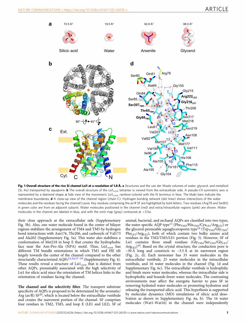
their close approach at the extracellular side (SupplementaryFig. 5b). Also, one water molecule found in the center of bilayerregions stabilizes the arrangement of TM4 and TM5 by hydrogenbond interactions with Asn176, Thr206, and carbonyls of Val173and Ala202 (Supplementary Fig. 5a). This water also stabilizes aconformation of Met218 in loop E that creates the hydrophobicface near the Asn-Pro-Ala (NPA) motif. Thus, Lsi1cryst hasdifferent TM bundle orientations in which TM1 and HE tiltlargely towards the center of the channel compared to the otherstructurally characterized AQPs13,14,17–19 (Supplementary Fig. 4).These results reveal a structure of Lsi1cryst that is distinct fromother AQPs, presumably associated with the high selectivity ofLsi1 for silicic acid since the orientation of TM helices links to theorientation of residues facing the channel.
The channel and the selectivity filter. The transport substratespecificity of AQPs is proposed to be determined by the aromatic/Arg (ar/R) SF20, which is located below the extracellular vestibuleand creates the narrowest portion of the channel. SF comprisesfour residues in TM2, TM5, and loop E (LE1 and LE2). SF of
animal, bacterial, and archaeal AQPs are classified into two types,the water-specific AQP type13 (PheTM2/HisTM5/CysLE1/ArgLE2) orthe glycerol-permeable aquaglyceroporin type14 (TrpTM2/GlyTM5/PheLE1/ArgLE2), both of which contain two bulky amino acidresidues in the TM2/TM5/LE1 portion (Fig. 3). However, SF ofLsi1 contains three small residues (GlyTM2/SerTM5/GlyLE1/ArgLE2)10. Based on the crystal structure, the conduction pore is~30 Å long and constricts to ~3.5 Å at its narrowest region(Fig. 2c, d). Each monomer has 33 water molecules in theextracellular vestibule, 25 water molecules in the intracellularvestibule, and 16 water molecules in the channel (Fig. 1d andSupplementary Fig. 6c). The extracellular vestibule is hydrophilicand binds more water molecules, whereas the intracellular side ishydrophobic and bounds fewer water molecules. The contrastingenvironments may affect the energetic barrier to pass SF inremoving hydrated water molecules or promoting hydration andreleasing the transported silicic acid. This hypothesis is supportedby molecular dynamics (MD) simulations of silicic acid deso-lvation as shown in Supplementary Fig. 6a, b). The 16 watermolecules (Wat1-Wat16) in the channel were independently
a
Silicic acid Water Arsenite Glycerol
b
c
NC
Ext.
Cyt.
d
Gly215
Gly216LE1
Ser217Met218
Asn219
Asn108
Met177Val111
His106
Phe115Thr181Ala195
Ile96
Ala105
Val199
Met107Val92
Leu61
Gly88TM2
Thr65TM1
Gln84
Gly155
Arg222LE2
Ser207TM5Thr206
3 2 1
4
56
97
108
1112
13
1415
16
Val203
Ser85
Ser80 Gln81 Arg78
Gly104
Ala69
d
72.5 Å3 19.5 Å3 62.8 Å3 88.0 Å3
4590
Fig. 1 Overall structure of the rice Si channel Lsi1 at a resolution of 1.8 Å. a Structures and the van der Waals volumes of water, glycerol, and metalloid(Si, As) transported by aquaporin. b The overall structure of the Lsi1cryst tetramer is viewed from the extracellular side. A pseudo-C4 symmetric axis isrepresented by a diamond shape. c Side view of the monomeric Lsi1cryst, rainbow-colored with the N terminus in blue. The khaki bars indicate themembrane boundaries. d A close-up view of the channel region (chain C). Hydrogen bonding network (dot lines) shows interactions of the watermolecules and the residues facing the channel’s pore. Key residues comprising the ar/R SF are highlighted by bold letters. Two residues (Arg78 and Ser80)in green color are from an adjacent subunit. Water molecules positioned in the channel (red) and extra/intracellular regions (pink) are shown. Watermolecules in the channel are labeled in blue, and with the omit map (gray) contoured at +3.5ơ.
NATURE COMMUNICATIONS | https://doi.org/10.1038/s41467-021-26535-x ARTICLE
NATURE COMMUNICATIONS | (2021) 12:6236 | https://doi.org/10.1038/s41467-021-26535-x | www.nature.com/naturecommunications 3

identified in two non-crystallographic symmetry-related Lsi1crysttetramers, indicating that they are intrinsically associated with themonomer. Wat7 and Wat8 had relatively higher temperaturefactors, and their positions were too closely spaced (2.3 Å) to besimultaneously occupied, suggesting that a single water moleculeoccupies in rapid equilibrium between adjacent sites. Similarly,several pairs of two adjacent water molecules, Wat6-Wat10,Wat11-Wat12, Wat12-Wat13, Wat13-Wat14, and Wat14-Wat15,may be occupied by a single water molecule. By contrast, Wat3and Wat9 have typical hydrogen bonds interactions shorter than2.8 Å with the low-temperature factors, suggesting their stablebinding to the channel.
A striking feature of the channel of Lsi1cryst is that a largenumber of water molecules at the extracellular half of the pore arenot in a single file, which is brought by the hydrophilic SF in thebroad channel (Figs. 1d and 2e). In chain A, three watermolecules in SF (Wat4, Wat5, and Wat6) and two watermolecules beneath it (Wat7 and Wat8) are separated by morethan 3.4 Å along the channel, which no longer contributes to thehydrogen bond interactions. In addition, since Wat9 donateshydrogen bonds to the carbonyls, it cannot transfer protons towater molecules Wat5 and Wat8. It has been proposed that
strongly correlated movements of the well-oriented single-filewater in orthodox AQP family proteins prevent proton transfervia the grotthuss mechanism21. Lsi1 must have a differentmechanism that prevents the fast proton transport since the watermolecules in SF of Lsi1 are not single-file. The breakage of theconnectivity between the SF waters and nearby water moleculesmay prevent proton translocation. The water molecules at theNPA motifs and the intracellular half, by contrast, are in a singlefile like the other AQP structures (Figs. 1d and 2e). The NPAmotifs stabilize each other creating a positive electrostaticpotential that functions as a barrier against proton transportacross the membrane21–23. Therefore, the NPA motifs and watermolecules nearby function to prevent protons in the rice Sichannel like the other AQPs21–23.
Unlike AQP1 and GlpF, the channel of Lsi1cryst has a broaderpore diameter at the constricted region with a slight distortion(An arrowhead in Fig. 2c). The shift of HE provides this channel’sdistortion, which could prevent the transport of substrates largerthan silicic acid, such as silicic acid oligomer, as they are unlikelyto rotate freely in this distortion. The narrowest region of theLsi1 structure is located at SF, as observed in other AQPs. Twocarbonyls of Gly215 and Gly216LE1 create the constrictions that
a b
45 45
Lsi1 AQP1 Lsi1 GlpF
SF
NPA
AQP1 GlpFLsi1silicic acid water glycerol
Ext.
Int.
e
Aqy1 (water)Lsi1 (silicic acid) GlpF (glycerol)
c
-25
-15
-5
5
15
25
0 1 2 3 4 5 6 7 8 9 10
Por
e po
sitio
n ( Å
)
Pore diameter (Å)
Lsi1BtAQP1GlpF
d
NPA
SF
55º
Å
Å Å
Å
Å
Å
Å
Å
Å
Fig. 2 Comparison of the structures of Si channel Lsi1 and other AQPs. Superposition of the structure of Lsi1cryst (cyan) with AQP1 (pink, PDB 1J4N) (a),and GlpF (light green, PDB 1FX8) (b). Top view from the extracellular side (left) and a rotated view by 45° against the membrane normal (right).Arrowheads indicate loop A and red arrows represent the shift of TM helices (in Å). A diamond shape represents a pseudo-C4 symmetric axis. Channelprofile (c) and diameters (d) along the pore for Lsi1cryst, AQP1, and GlpF, calculated using the program HOLE2, are shown. The regions for SF and NPAmotifs are colored in plum and khaki, respectively. An arrowhead indicates the distortion in the channel. e Cross-section of the channel of Lsi1cryst, Aqy1(wheat, PDB 3ZOJ), and GlpF with a 55° rotation relative to (c). Water molecules in the channel (red), extra/intracellular regions (pink), and glycerolmolecules are shown. In e, N-terminal and C-terminals of Aqy1 were omitted for clarity. The color coding shown here is used for all Figures unlessotherwise noted.
ARTICLE NATURE COMMUNICATIONS | https://doi.org/10.1038/s41467-021-26535-x
4 NATURE COMMUNICATIONS | (2021) 12:6236 | https://doi.org/10.1038/s41467-021-26535-x | www.nature.com/naturecommunications

point towards the channel. SF of AQP1 is narrower andhydrophilic (Phe58TM2/His182TM5/Cys191LE1/Arg197LE2), andthat of GlpF is wider and amphipathic (Trp48TM2/Gly191TM5/Phe200LE1/Arg206LE2), whereas that of Lsi1cryst is the widest andhydrophilic (Thr65TM1/Gly88TM2/Ser207TM5/Gly216LE1/Arg222LE2) (Fig. 3). The most striking feature of the Lsi1cryst’sSF arises from an additional “fifth residue” Thr65 in TM1(Thr65TM1) and a water molecule hydrogen-bonded to Thr65TM1
(Wat9) (Fig. 3a). The significance of Thr65TM1 in SF has not beenrecognized since, in canonical AQPs, a bulky hydrophobic residuein TM2 (Phe58TM2 in AQP1 and Trp48TM2 in GlpF, respectively)shields the residue equivalent to Thr65TM1. In Lsi1cryst, however,Gly88TM2 exposes Thr65TM1 to the channel, thereby making SFwide and hydrophilic. Thr65TM1 donates a strong hydrogen bond
to the carbonyl of Leu61 and acts as a suitable hydrogen acceptorfrom a nearby water molecule Wat9 (Fig. 4a). Wat9 donateshydrogen bonds to both Thr65TM1 and carbonyl of Gly88TM2,thereby pointing its oxygen atom towards the channel (Fig. 4).Similarly, Wat3 can donate hydrogen bonds to both carbonyls ofGln84 and Thr65TM1, thereby directing its oxygen atom towardsthe channel as well (Fig. 4). Wat3 and Wat9 are located on oneside of the channel and are separated by 6 Å, facing oppositely tocarbonyls of Gly215 and Ser217 (Figs. 1d and 4). Therefore,unlike other AQPs, the channel of Lsi1cryst has two polar faces.The additional polar face’s water molecules likely act as hydrogenbond acceptors during the substrate transport in a similar fashionto the consecutive carbonyls protruding the channel. This notionis supported by the lower temperature factors of Wat3 and Wat9.
Arg222LE2
Wat9
a
Thr65TM1
Gly88TM2
Ser207TM5
Gly216LE1 Arg197LE2
Ser28TM1
Phe58TM2
His182TM5
Cys191LE1 Arg206LE2
Gly25TM1Trp48TM2
Gly191TM5
Phe200LE1
FplG1PQAtB1isLb c
Arg196LE2
Gly27TM1Trp50TM2
Gly181TM5
Phe190LE1 Arg202LE2
Gly22TM1
Phe62TM2
Ile187TM5
Ser196LE1 Arg217LE2
Thr38TM1
Gly62TM2
Gly202TM5
Ile211LE1
Wat
Arg225LE2
Thr55TM1
Phe81TM2
His210TM5
Thr219LE1
Arg200LE2
Gly35TM1
His63TM2
Ile185TM5
Gly194LE1 His131LC
d01PQAhMPQAPQAfP
e f
g1;2PITtA1;2PIPoS
h
Fig. 3 Comparison of SFs of Lsi1 and other AQPs. SF of Lsi1cryst (a, cyan, PDB 7CJS), BtAQP1 (b, pink, PDB 1J4N), GlpF (c, light green, PDB 1FX8), PfAQP(d, white, PDB 3C02), AQPM (e, khaki, PDB 2F2B), hAQP10 (f, yellow, PDB 6F7H), SoPIP2;1 (g, green, PDB 1Z98), and AtTIP2;1 (h, light purple PDB 5I32).The view directions are the same as Fig. 2a.
NATURE COMMUNICATIONS | https://doi.org/10.1038/s41467-021-26535-x ARTICLE
NATURE COMMUNICATIONS | (2021) 12:6236 | https://doi.org/10.1038/s41467-021-26535-x | www.nature.com/naturecommunications 5

Interestingly, the channel of Lsi1cryst has Thr181 in a pseudo-c2-symmetrically related position (N- and C- terminal three TMrepeats) corresponding to the Thr65TM1. Thr181 makes thechannel hydrophilic likewise by having hydrogen bonds with thecarbonyl of Met177 and Wat15 (Fig. 1d). These two Thr residues(Thr65TM1 and Thr181) highlight the unique characteristic of theSi-channel Lsi1.
To examine the role of the Thr65TM1 in transport substratespecificity, we generated ten variants of Lsi1. We investigatedtheir transport activity for germanic acid (Ge) as a Si analog andarsenite (As) in Xenopus oocytes. The transport activities for bothGe and As were unaffected by the substitutions of Thr65TM1 toAla, Gly, and Ser (Fig. 5a, b), although expression of the Alamutant was lower than wild type or the other two mutants inXenopus oocytes (Supplementary Fig. 7a, b). One possibleinterpretation is that water molecules occupying free spacecreated by the substitutions can compensate for a polarenvironment made by hydroxyl of Thr65TM1 and Wat9. In
contrast, the activity was substantially decreased or lost by theother substitutions (Fig. 5a, b). By T65VTM1 substitution, whichincreases the hydrophobicity of SF but keeps its size unchanged,transport activities for Ge and As were decreased. On the otherhand, the T65ITM1 substitution that mimics the size of Thrresidue plus Wat9 severely decreased the activity for Ge, whereasthe activity for As was similar to the T65VTM1 mutant. Theseresults suggested that Thr65TM1, together with Wat9, constituteSF and play a key role in the specificity of transport substrate.
We also examined the role of the Thr181 in the substratespecificity by generating five site-directed mutants (T181S,T181V, T181N, T181Q, and T181D) and determined transportactivity of Ge and As likewise in Sf9 cells. While the substitutionsof Thr181 to Ser, Val, Asn, and Asp unaffected or slightlydecreased the Ge transport activity, they slightly increased activityfor As transport (Fig. 5c, d and Supplementary Fig. 7c, d). T181Qsubstitution, which narrows the size of the channel, completelyabolished the Ge transport activity, whereas the As transport
Fig. 4 Unique water molecules and two Thr residues in the Lsi1 structure. Hydrogen bonding interactions between the two water molecules comprisingSF (Wat3 and Wat9) and TMs (TM1 and TM2) are shown with view directions facing the channel (a) or from the extracellular side (b). c A panel isshowing the positions of Thr65 and Thr181. Hydrogen bond interactions that make the oxygen atoms of the waters pointing towards the channel are shownin yellow dashes.
ARTICLE NATURE COMMUNICATIONS | https://doi.org/10.1038/s41467-021-26535-x
6 NATURE COMMUNICATIONS | (2021) 12:6236 | https://doi.org/10.1038/s41467-021-26535-x | www.nature.com/naturecommunications

Sf9 ce
lls
CE-Lsi1
T181S
T181V
T181N
T181Q
T181D
0102030405060708090
100
Sf9 ce
lls
CE-Lsi1
T181S
T181V
T181N
T181Q
T181D
0
50
100
150
200
250
waterLs
i1T65
AT65
GT65
ST65
VT65
IT65
CT65
DT65
HT65
NT65
Y0
2
4
6
8
10
12
14
waterLs
i1T65
AT65
GT65
ST65
VT65
IT65
CT65
DT65
HT65
NT65
Y0
10
20
30
40
50
60
70
e
ad
b
ad
c
ad
a
b
c
d
e
aAs
upta
ke (n
gm
g dr
ied
Sf9
cel
ls-1
)
Ge
upta
ke (n
gm
g dr
ied
Sf9
cel
ls-1
)
fsAeG
baG
e(O
H) 4
upta
ke a
ctiv
ity (n
goo
cyte
-1 3
0 m
in-1
)
As(
OH
) 3up
take
act
ivity
(ng
oocy
te-1
30
min
-1)
dc
As
upta
ke (n
gm
g dr
ied
Sf9
cel
ls-1
)
Ge
upta
ke (n
gm
g dr
ied
Sf9
cel
ls-1
) sAeG
sAeG
a
b
c
b
c
a
d
a
b
c c c
d
bc
a
bb b
b
cd
ef
d
a ae
cfg
a
a
bb b
b
cc
c
d
aa
a
Fig. 5 Effect of Lsi1 mutation on the transport activity for Ge and As. Transport activity of germanic acid (Ge) (a, Si analog) and arsenite (As) (b) inXenopus oocytes. The oocytes were injected with water (as a control) or cRNA of Lsi1 WT or Thr65TM1 mutants. Oocytes were exposed to a solutioncontaining Ge or As for 30min. Transport activity of Ge (c, e) and As (d, f) in Sf9 cells. C-terminally EGFP-tagged Lsi1 or Thr181 mutants (c, d) or otheraquaglyceroporins (e, f) were expressed in the cells. The cells were exposed to a solution containing Ge or As for 5 min. In a−f, different letters above thecolumns indicate statistically significant differences at P < 0.01 by Tukey−Kramer’s test, and the test was two-sided. Values are means ± s.d., n= 3 forT65C in (a) and n= 4 for the others, n is independent experiments.
NATURE COMMUNICATIONS | https://doi.org/10.1038/s41467-021-26535-x ARTICLE
NATURE COMMUNICATIONS | (2021) 12:6236 | https://doi.org/10.1038/s41467-021-26535-x | www.nature.com/naturecommunications 7

activity remained about 35% of native Lsi1 (Fig. 5d). It should benoted that the effect of Thr181 mutations is moderate comparedto Thr65TM1 and its function is still not clear. Nevertheless, theresults suggest that the specificity of transport substrate can bemodified by manipulating the two Thr residues (Thr65TM1 andThr181) identified in the present study.
Transport specificity of Lsi1 for silicic acid. What is the struc-tural basis of Lsi1 yielding the high selectivity for silicic acidrather than smaller glycerol? Our structural analysis showed that
the oppositely located two polar faces found in the channel mightbe responsible for this difference in the transport substrate spe-cificity (Fig. 3a). Since silicic acid is a hydrophilic tetrahedron, thetwo polar faces can provide an energetically preferable environ-ment by surrounding it. In contrast, linear carbohydrates areamphipathic, and the two polar faces in Lsi1cryst may impairconductivity for glycerol. Indeed, SF of GlpF has an amphipathicproperty that fits well for glycerol with hydrophobic interactionsby Trp48TM2 and Phe200LE1 and hydrogen bonds formed byArg206LE2 and carbonyl of Phe200LE1 with the glycerol’s hydroxyl
Gly215 Gly216LE1
Arg222LE2
Thr65TM1
Gln84
Ser207TM5
Gly88TM2
H H
H
1
2
4
3
9
Si2(modeled)
dc
1Ext 4Ext13245
610
Arg222LE2798Asn219
Asn10811
1213
141516
Si1Si2
Si3Si4Si5Si6
Si7Si8
a
e
b
Fig. 6 Structural determinant for the silicic acid permeability and modeled eight silicic acid positions in the Lsi1 channel. SF of Lsi1cryst (a), and apossible conformation of silicic acid during the passage through SF (b). Water molecules in the channel (Wat1, Wat2, Wat3, Wat4, and Wat9) are shownas spheres and numbered in blue. Among them, three (Wat1, Wat2, and Wat4) are superposition from the eight Lsi1cryst protomers of the two tetramers.Putative hydrogens of hydroxyl groups of the silicic acid in the position Si2 are encircled by dashed lines. The view direction is the same as Fig. 2a. Watermolecules used for the assignment of possible silicic acid positions (c) and the assigned eight silicic acids (Si1-Si8) in the channel (d) are shown in a cross-section view. Water molecules in the channel region (red spheres) and the extracellular region (pink spheres) are labeled in numbers. Arg222LE2 in the SFregion and two Asn residues in the NPA motifs are shown as the stick. e Water molecules employed for the assignment of four hydroxyl groups in silicicacid are listed. Ext in c and e indicates water molecules at the extracellular side.
ARTICLE NATURE COMMUNICATIONS | https://doi.org/10.1038/s41467-021-26535-x
8 NATURE COMMUNICATIONS | (2021) 12:6236 | https://doi.org/10.1038/s41467-021-26535-x | www.nature.com/naturecommunications

groups (Fig. 3c). To test this possibility, we focused on gain-of-function mutants of the structurally characterized aqua-glyceroporin GlpF14 in an attempt to mimic the Si-permeabilityof Lsi1. Wild-type GlpF, PfAQP24, and AQPM25 did not showtransport activity for Ge in Sf9 cells or oocytes (Fig. 5e andSupplementary Figs. 7c, d, 8e). However, with the substitutions oftheir SF residues into Lsi1 type, GlpF variant GlpF_SFLsi1(G25TTM1, W48GTM2, G191STM5, and F200GLE1) showed ahigher Ge transport activity (Fig. 5e and Supplementary Fig. 8e).Interestingly, GlpF_SFLsi1 also gained the As transport activity butshowed the decreased glycerol transport activity (Fig. 5f andSupplementary Fig. 8c, f).
Deshmukh et al. reported that number of residues connectingtwo NPA motifs is essential for the Si permeability26. Since thespacing is 108 residues in Lsi1 and 132 residues in the Si-permeable GlpF variant GlpF_SFLsi1, the spacing itself is unlikelyto be critical for the Si specificity. This is supported by a recentstudy on Lsi1 in tomatoes that possesses 109 residues in the
spacing but shows transport activity for Si27. Instead, loop Cextensively stabilizes the consecutive carbonyls (Gly215-Ser217)and SF of Lsi1cryst via hydrogen bond interactions (Supplemen-tary Fig. 5b). Taken together, the two polar faces of the channelprovided by the unique SF have an essential role in the Sispecificity in Lsi1.
The silicic acid transport mechanism. We failed to detect any Si-or Ge- derived anomalous signals from the Lsi1cryst crystals soakedin a buffer at saturated concentrations. However, we tentativelypredicted eight positions of silicic acid (Si1 through Si8) thatpossibly occupy the channel in Si transport, based on the positionsof waters identified in the channel (Wat1 through Wat16)(Fig. 6a). We postulated that hydroxyl groups of the transportedsilicic acid should favor the hydrophilic environment that watersbound. We first investigated the modeled silicic acid locationsusing quantum mechanical/molecular mechanical (QM/MM)
Si density Si(OH)4 (modelled) a c d e
Si densityOH densityWater densityWater density
Si density OH density b Si density
Wat3
Wat9
Wat17
NPA
SF Wat9 Wat3
Wat17
-30
-20
-10
0
10
20
30
100 150 200 250 300 350 400
Por
e po
sitio
n (Å
)
Time (nsec)
f
Fig. 7 MD simulation of Lsi1 with silicic acid. Distribution of Si atoms of silicic acid (a) and its overlay with modeled Si(OH)4 molecules (Si1-Si8) (b), anddistribution of Si atoms as well as oxygen atoms of silicic acid (c), or oxygen atoms of water (d) in the 0−300 ns MD simulation. In a, structuralbottlenecks found in the channel are indicated by red arrows. In d, Wat3, Wat9, and Wat17 locations are indicated by blue arrows. Distributions of (c) and(d) are merged in (e). f The MD trajectory of the silicic acid permeation during the 100−420 ns. Positions of silicic acid (black), Wat3 (green), Wat9(blue), and Wat17 (red) in the channel are plotted. Enlarged views of (f) and the distributions of Si(OH)4 molecules at the beginning and end of thesimulation are provided in Supplementary Fig. 11.
NATURE COMMUNICATIONS | https://doi.org/10.1038/s41467-021-26535-x ARTICLE
NATURE COMMUNICATIONS | (2021) 12:6236 | https://doi.org/10.1038/s41467-021-26535-x | www.nature.com/naturecommunications 9

geometry optimization (Supplementary Fig. 9). Displacement ofthe QM/MM model ranges from 0.2 to 0.9 Å relative to themodeled silicic acid (Supplementary Fig. 9). The number ofhydrogen-bonding interactions in the channel is mainly unchan-ged after the QM/MM optimization, suggesting that silicic acidcan stably occupy the positions through Si1 to Si8.
Intriguingly, the modeled Si2 occupies SF, without any sterichindrance, with the three hydroxyl groups overlapping withthree water molecules (Wat1, Wat2, and Wat4), therebydonating hydrogen bonds to Wat3 and two carbonyls (Gly215,and Gly216 LE1) (Fig. 6a, b). Si2 thus likely represents a transientconformation during the passage through SF. Two hydroxylgroups go through a crevice formed by Arg222LE2 and the twocarbonyls (Gly215 and Gly216LE1), another one goes through acrevice by the carbonyls and Ser207TM5, and the other one goesthrough a crevice by Ser207 TM5 and waters (Wat3 and Wat9)(Fig. 6b). In this way, all hydroxyl groups of the silicic acid canform hydrogen bonds with the carbonyls or waters positioned
along the channel and with Ser207TM5 and Arg222LE2 tocompensate for the energetic cost of dehydration. The other foursilicic acids (Si1, Si3, Si4, and Si7) overlap three or four hydroxylgroups with the waters identified, and the other three (Si5, Si6,and Si8) also do so in two hydroxyl groups (Fig. 6c−e). Thisassignment identifies plausible hydrogen bonding partners of thesilicic acid and provides orientations of the former four positions.However, there may be other interpretations because we basedour hypothesis on the structure without any substrates. Thepredicted silicic acids suggested that water molecules can replaceone or two hydrogen-bonding interactions with the channel afterthe passage of SF to promote silicic acid migration.
We also performed a 450 ns MD simulation of theLsi1 structure in the presence of ~1M Si(OH)4 to investigatethe transport mechanism (Fig. 7 and Supplementary Figs. 10, 11).Two silicic acids permeated the channel during the simulation.During the MD simulation, an average of 11.6 ± 2.6 watermolecules and 0.7 ± 0.7 Si(OH)4 molecules occupied in the
0.05
OsLsi1ZmLsi1
HvLsi1TaLsi1 OsLsi6
HvLsi6
ZmLsi6
CmLsi1
CsLsi1
PtLsi1NsLsi1
SlLsi1
GmNIP2-2OsNIP1;1
OsNIP3;1
AtNIP5;1
EaNIP3;1
EaNIP3;3
EaNIP3;4a
Monocot NIP III
ThrTM1
GlyTM1
AlaTM1
Dicot NIP IIINIP I
NIP II
Equisetum Si channel
a
bTM1 TM2 TM5 LE1 LE2 Thr181
Monocot NIP III T G S G R T
Dicot NIP III without Leguminosae T G S G R T
Leguminosae NIP III G G S G R S
IRAVW)G(I PIN
VRGIAAII PIN
Equisetum Si channel A S T A R N
Fig. 8 Analysis of selectivity filters in NIP subfamilies. Phylogenetic tree of plant NIP subfamilies (a), and summary of the residues comprising theselectivity filer and Thr181 (b). The phylogenetic tree was constructed using the neighbor-joining algorithm by the software MEGAX51 after the sequencealignment using Clustal Omega52 with 1,000 bootstrap trials. The 0.05 scale indicates substitution distance.
ARTICLE NATURE COMMUNICATIONS | https://doi.org/10.1038/s41467-021-26535-x
10 NATURE COMMUNICATIONS | (2021) 12:6236 | https://doi.org/10.1038/s41467-021-26535-x | www.nature.com/naturecommunications

channel region, where we found 16 water molecules in the crystalstructure (Supplementary Fig. 10f). The simulation results alsosuggested three structural bottlenecks where silicic acid densitieswere low during the simulation and silicic acid cannot movefreely (Red arrows in Fig. 7a). Two bottlenecks were in SF; onewas consistent with the modeled Si2, and the other was betweenthe Si3 and Si4. The third one was near the Thr181 in between theSi7 and Si8. The simulation suggested that each bottleneck has acavity which the oxygen atoms of silicic acid cannot occupy.Instead, water molecules, Wat3, Wat9, and Wat17 (Wat17 wasnot identified in the crystal structure and has hydrogen bondinginteraction with Thr181 in the MD simulation, see Supplemen-tary Fig. 10b), stably exist within the cavities (Blue arrows inFig. 7d). When the Si(OH)4 moved across the Wat3 andWat17 sites, Wat3/Wat17 were dissociated from this site. Onthe other hand, Wat9 was independent of the Si(OH)4 movementand remained bound to the site (Fig. 7f and SupplementaryFigs. 10f and 11). We calculated how often these water moleculesoccupy the cavities throughout the MD simulation. Theiroccupancies were 60% for Wat3, 94% for Wat9, and 44% forWat17. Their exchange rate suggested that Wat9 bound to the sitelonger (the average exchange time, τ = 1.5 ns) than Wat3 andWat17 (τ ~ 0.3 ns) (Supplementary Table 2). In this case, Wat3formed hydrogen bonding interactions with the carbonyls ofGln84 or Thr65TM1 in most cases, whereas Wat9 mainly formedtwo hydrogen bonds with Thr65TM1 and Gly88TM2. MD snap-shots also suggested that Wat3 and Wat9 could accept hydrogenbonds from silicic acid consistent with our proposal from thecrystal structure (Supplementary Fig. 10c, e). The silicic acidpermeation trajectories and the MD snapshots support that Wat3and Wat9 remain in the vicinity of their positions during thesilicic acid passage while Wat17 will be displaced (Fig. 7f andSupplementary Figs. 10, 11). The channel diameter calculationincluding Wat9 results in a narrower channel diameter (Fig. 2cand Supplementary Fig. 12). These results indicate that duringsilicic acid permeation, Wat3 and Wat9 act as part of the channellumen, narrowing the channel and strictly selecting the orienta-tion of silicic acid, highlighting the importance of the high-resolution structure’s ability to visualize most water molecules inthe channel.
DiscussionPlant AQPs have five subfamilies; the plasma membrane intrinsicproteins (PIP) subfamily, the tonoplast intrinsic proteins (TIP)subfamily, the NIP subfamily, the small basic intrinsic proteinssubfamily (SIP), and the X intrinsic proteins (XIP) subfamily28. Inthe present study, we determined the high-resolution crystalstructure of the rice Si channel Lsi1 belonging to the unique NIPsubfamily. We compared the Lsi1cryst structure with other knownplant AQP structures, SoPIP2;1 and AtTIP2;1. TheSoPIP2;1 structure in the open and closed states have beenreported17. The striking feature of the SoPIP2;1 structure is thatloop D changes its conformation to open or occlude the pore inresponse to phosphorylation or pH change. In theSoPIP2;1 structure, loop D folds below the pore and occludes it inthe closed conformation, whereas loop D flips largely towards TM4and TM5 of the adjacent protomer, thereby opening the pore in theopen conformation. The overall structure of Lsi1 is very similar tothat of SoPIP2;1 (The RMSD values are 1.1 Å for the open and1.2 Å for the closed structures, respectively), except for loop D(Supplementary Fig. 13a). Loop D in the Lsi1 structure flips towardsTM2 of the adjacent protomer (Supplementary Fig. 13b). SF of theSoPIP2;1 is narrow and hydrophilic (Phe81TM2/His210TM5/Thr219LE1/Arg225LE2) similar to other structures of the water-specific AQPs (Fig. 3g). Thr55, which corresponds to the “fifth
residue” of the Lsi1 structure, is covered by the Phe81TM2 andtherefore does not face the pore.
On the other hand, AtTIP2;1 is water and ammonia permeable.While the Lsi1 structure is similar to the AtTIP2;1 structure (theRMSD value is 1.4 Å)18, their loops A and C are quite different. Inthe Lsi1 structure, loop A orients to the pore’s distal side, and loopC is displaced up to 5 Å towards the pore relative to theAtTIP2;1 structure (Supplementary Fig. 14). SF of AtTIP2;1 isnarrow and hydrophilic (His63TM2/His131LC/Ile185TM5/Gly194LE1/Arg200LE2) (Fig. 3h and Supplementary Fig. 15). Gly35, whichcorresponds to the “fifth residue” of the Lsi1 structure, is covered bythe bulky His63TM2. SF of AtTIP2;1 is characterized by the fact thatthe conserved Arg200LE2 adopts a unique position and that anadditional hydrophilic residue His131LC extended from loop Ccontributes to SF. Lsi1 has Thr157 at the corresponding position ofHis131LC in the AtTIP2;1 structure (Supplementary Fig. 15).Thr157 in Lsi1 has hydrogen bonding interaction with Thr223 andexposes its methyl group to the pore. Moreover, MD simulationsuggested that Si(OH)4 remains at the position of Thr157 in thecrystal structure when the Thr157 is displaced (SupplementaryFig. 15e, f). Therefore, Thr157 in Lsi1 is distinct from theAtTIP2;1 structure and may not directly contribute to substrateselectivity.
Among five residues defining SF of the Lsi1cryst structure,Thr65TM1 is unique and likely plays an essential role in thespecificity of the transport substrate (Fig. 3a). Among the NIPsubfamily (NIP I, II, and III), NIP I subgroup has a bulky TrpTM2
in SF, whereas NIP II and III subgroups have small amino acidresidues GlyTM2 or AlaTM2 or SerTM2 (Fig. 8). Therefore, the “fifthresidue” of SF corresponding to Thr65TM1 in Lsi1 is likely to facethe channel in NIP II and III subgroups but not in the NIP Isubgroup. In the NIP III subgroup, the “fifth residue” is ThrTM1
in monocot and dicot species, except GlyTM1 in legumes, whereasit is GlyTM1 or AlaTM1 in NIP I and II subgroups (Fig. 8). Also,Thr181 is conserved in NIP III subgroup except for leguminosae(Supplementary Fig. 1). Thus Thr65TM1 and Thr181 identified inthe present study are unique to the NIP III subgroup. TheEquisetum Si channel group, which also possesses the Si transportactivity, has AlaTM1, SerTM2, and ThrTM5
29 (Fig. 8). Hydroxylgroups of SerTM2 and ThrTM5 in the Equisetum Si channel maycompensate for the substrate specificity or interactions with watermolecules provided by the hydroxyl groups of Thr65TM1 andSer207TM5 in Lsi1.
Human aquaglyceroporin hAQP10 is permeable to silicicacid30, and its structure in the closed state has been reported31.The RMSD value between the hAQP10 and Lsi1cryst structures is1.7 Å, which is slightly larger than those calculated between Lsi1and the structures SoPIP2;1 and AtTIP2;1. The relatively largerRMSD value arises from different structures in loops and differentorientations of TM helices (Supplementary Fig. 16a). Never-theless, SF of hAQP10 is very similar to that of Lsi1 (Fig. 3a, f),consistent with the fact that hAQP10 is permeable to Si(OH)4. SFof hAQP10 is wide and amphiphilic (Thr35TM1/Gly62TM2/Gly202TM5/Ile211LE1/Arg217LE2). SF of hAQP10 contains manywater molecules, including two water molecules Wat3 and Wat9,in the Lsi1 structure, which create the polar face (Fig. 3f andSupplementary Fig. 16c). There are two notable differences in SFbetween hAQP10 and Lsi1. First, the pore diameter of thehAQP10 is wider in SF (Supplementary Fig. 16b). Second, Gly210and Ile211LE1 provide a row of carbonyls in the pore in thehAQP10 structure, but they are two Gly residues (Gly215 andGly216LE1) in the Lsi1 structure. The side chain of Ile211LE1renders the channel hydrophobic and displaces TM5 of hAQP10up to 5 Å towards the pore’s distal side relative to theLsi1 structure. Therefore, SF’s hydrophilicity and the tilting anglesof TM helices are different between the structures hAQP10 and
NATURE COMMUNICATIONS | https://doi.org/10.1038/s41467-021-26535-x ARTICLE
NATURE COMMUNICATIONS | (2021) 12:6236 | https://doi.org/10.1038/s41467-021-26535-x | www.nature.com/naturecommunications 11

Lsi1 (Fig. 3f and Supplementary Fig. 4e). The bulky Ile211LE1residue also affects the orientation of the carbonyls. The carbonylof Gly210 in hAQP10 rotates by 40° towards Arg217LE2 comparedto Gly215 in the Lsi1 structure. This rotation weakens thehydrogen bond to waters and narrows the region of the porethrough which silicic acid may pass. Given that SF of Lsi1 is idealfor silicic acid permeation, the selectivity for silicic acid ofhAQP10 may be different from that of Lsi1.
We have shown that the unique TM helix orientations and SFof Lsi1 are essential for silicic acid transport. Lsi1 has presumablyacquired these features during its evolution from canonical AQPsthat could not transport silicic acid. The evolution of plant AQPfamily proteins that permeate substrates other than water andglycerol, such as Lsi1 and AtTIP2;1, seems to have involveddrastic modification of SF from canonical AQPs. As described,even between the evolutionarily distant species of human(hAQP10) and rice (Lsi1), a shared structure exists in whichwater molecules create a polar face in SF to transport silicic acid.Such a common structure has likely evolved from convergentevolution.
Lsi1 is also permeable to carcinogenic arsenite11, the primaryform of As in the paddy field. Arsenite is also present in the formof a non-charged molecule and has a similar size as silicic acid11.Rice is a staple food for half of the world population but canaccumulate high As through Lsi111. Since rice is the primarydietary source of As, it is crucial to reduce As in rice grain forhuman health. However, compared with silicic acid, usually,arsenite shows broader specificity12. For example, two T65ITM1
and T181Q substitutions were identified to decrease or abolishGe-transport activity while As-transport activity was substantiallyretained (Fig. 5). Also, Lsi1 with a S207ITM5 substitution10 doesnot transport Ge but transports As. Silicic acid is a tetrahedralmolecule that forms four hydrogen bonds, whereas arsenite is atrigonal pyramid that forms three hydrogen bonds (Fig. 1a). SinceSF closely matches the transport substrates in the dehydratedform as observed in Lsi1cryst and GlpF14, or even in the KcsApotassium channel32, the larger number of possible hydrogen-bonding interactions with the channel as well as the tetrahedralstereochemistry may be the reasons why selectivity for silicic acidis stricter than that for arsenite. While other factors determiningtransport substrate specificity remain to be investigated, thestructure of Lsi1 obtained in this study could serve as a blueprintfor rational designs of transgenic crops that specifically take upsilicic acid but not arsenite through manipulating the selectivityof Lsi1. Such modification will contribute to safe food productionin the future.
MethodsProtein expression and purification of Lsi1. The Lsi1 gene from rice (Oryzasativa cv. Nipponbare) was cloned into the pFastBac1 vector for baculovirusexpression in Sf9 insect cells using standard methods. A TEV protease cleavage siteand the octa-His affinity tag were introduced between the C terminus of Lsi1 andEGFP. The functionally active construct of Lsi1 was discovered by examining N-and C-terminal deletion constructs, several point mutations, as well as additional Sipermeable AQPs from other organisms. All these constructs were created by usingthe QuikChange II site-directed mutagenesis method (Stratagene) with primers(Supplementary Table 3) and screened by FSEC15. Removing 44 residues from theN-terminus, 24 residues from the C-terminus, with seven point mutations (K50R,C66A, T93V, C139A, K232R, T253V, and K264R), yielded the construct, Lsi1cryst,used in the crystallographic studies described here. Among the seven mutations inthe construct Lsi1cryst, four mutations (C66A, T93V, C139A, and T253V) in TMhelices enhanced the thermo-stability of Lsi1 in the detergent micelle. Three lysineresidues (K50R, K232R, and K264R) in loop regions are mutated to arginine toreduce the surface entropy, hoping that mutants may improve the crystal packing.
Infected Sf9 cells were harvested by centrifugation (8000 × g, 15 min), and weredisrupted by an ultrasonic disrupter UD-211 (TOMY). After centrifugation(3000 × g, 10 min), the supernatant was ultra-centrifuged (200,000 × g, 1 h), andmembrane fraction was collected and homogenized. The crude membrane fractionswere solubilized for 1 h in a buffer containing 500 mM NaCl, 20 mM Tris-HCl pH8.0, 6% (w/v) glycerol, 1.8% (w/v) n-dodecyl-β-D-maltopyranoside (DDM),
0.06 mg/ml RNase A. Insoluble material was removed by ultracentrifugation(148,500 × g, 1.5 h) and the supernatant was incubated with TALON cobalt affinityresin (Clontech) for 3 h in the presence of 10 mM imidazole. After washing with abuffer containing 15 mM imidazole, 500 mM NaCl, 20 mM HEPES-NaOH pH 7.5,10% (w/v) glycerol, and 0.02% (w/v) DDM, Lsi1 mutants were eluted byapplication of a buffer containing 150 mM imidazole, 500 mM NaCl, 20 mMHEPES-NaOH pH 7.5, 10% (w/v) glycerol, and 0.02% (w/v) DDM. The eluateswere precipitated in the presence of 22.2% (w/v) PEG 1500, and then dissolved in abuffer composed of 500 mM NaCl, 20 mM HEPES-NaOH pH 7.5, 10% (w/v)glycerol and 1% (w/v) n-octyl-β-D-glucoside (OG). The octa-His tag was cleavedwith hexa-His-tagged TEVSH
33 (3:1 mass ratio of Lsi1 to TEVSH) overnight, andthe protein was re-chromatographed on a TALON cobalt affinity resin. The tagcleaved Lsi1 was further purified by gel filtration (Superdex 200 Increase 10/300 GLcolumn) in 500 mM NaCl, 20 mM HEPES-NaOH pH 7.5, and 1% (w/v) OG. Allsteps were performed at 4 °C unless otherwise noted.
Crystallization. The purified protein was concentrated to about 10 mg/ml using a50 kD molecular weight cut-off centrifugal filter device. The Lsi1cyst crystals wereobtained at 7 °C by vapor diffusion sitting drop method by mixing 1:1 (v/v) ratio ofprotein and a reservoir solution containing 39−50% (w/v) PEG 400, 100 mM Gly-NaOH pH 9.5, 1% (w/v) OG, and 0.1% (w/v) cholesteryl hemisuccinate (CHS).Both pyramidal and rod-shaped crystals appeared in the same crystallization drops,but the diffraction limit of the pyramidal crystals was around 7 Å resolution. Therod-shaped crystals were collected and soaked in a solution containing 41% (w/v)PEG 400, 500 mM NaCl, 20 mM HEPES-NaOH pH 7.5, 100 mM Bis-Tris HCl pH7.0, 2% (w/v) OG, 0.1% (w/v) CHS, then flash-frozen in liquid nitrogen for X-raydiffraction experiment. Some crystals were soaked in a crystallization buffer sup-plemented with 44 mM Si(OH)4 or a saturated concentration of Ge(OH)4 for 5 minprior to freezing. A fresh Si(OH)4 solution was prepared to avoid the oligomer-ization of silicic acids.
Structure determination. X-ray diffraction data were collected at SPring-8BL41XU or BL44XU and were processed with XDS34. The crystals belong to thespace group P21 (unit-cell parameters a= 89.5 Å, b= 91.4 Å, c= 166.1 Å, and ß =102.1°). While some reflections remained sharp, the others were diffused withstronger intensities. This unusual pattern was induced by so-called lattice-trans-location defects, in which two identical but translated lattices coexist as a singlemosaic block in a crystal. We thus corrected the intensities based on the methodreported previously16. In brief, if the intensity for each unit cell is Iunit, the totalintensity is
Itotal ¼ ð2κ2 � 2κþ 1Þ ½1þ 2κð1� κÞ=ð2κ2 � 2κþ 1Þ cosð2πhtdÞ�Iunit
where the translocation vector td and the fraction κ were determined to be (1/3, 0,1/3) and 0.30, respectively. The corrected intensities gave significantly smallervalues for R-factor and free R-factor. They were 0.343 for the R-factor and 0.365 forthe free R-factor before the correction and 0.245 for the R-factor, and 0.273 for thefree R-factor after the correction. The initial phase information was obtained bymolecular replacement with Phaser35 using a homology model of Lsi1 as a searchprobe, created from the crystal structure of Archaeoglobus fulgidus AQP (PDB3NE2). Two tetramers were manually built using COOT36 based on the electrondensity map calculated at a 1.8 Å resolution and refined using phenix.refine37. Thetwo Lsi1 tetramers contained eight sodium ions, four OGs, two CHSs, and elevenPEGs. During the refinement process, coordinates, temperature factors, TLS wererefined, and non-crystallographic symmetry restraints were not applied. The sta-tistics for refinement was shown in Table 1. Figures were prepared using Cuemol2(http://www.cuemol.org) or PyMOL (http://www.pymol.org), and the channel ofLsi1cryst was analyzed using HOLE238.
Functional assay in Sf9 cells. The full-length Lsi1, including the octa-His tag andEGFP at the C-terminus of the Lsi1 construct (CE-Lsi1), was cloned into thepFastBac1 vector. Similarly, codon-optimized GlpF, AQPM, and PfAQP geneswere synthesized (Integrated DNA Technologies), amplified by PCR, cloned intothe pFastBac1 vector using a seamless cloning method. All mutated Lsi1 genes (e.g.,Thr65TM1 variants, CE-Lsi1_ΔNC, and CE-Lsi1cryst) used in the functional assaywere generated using the QuikChange II site-directed mutagenesis method(Stratagene).
Sf9 cells infected with P3 viruses of interested CE-Lsi1 mutants or other AQPs(CE-GlpF, CE-AQPM, and CE-PfAQP) were grown at 27 °C for 24 h afterinfection, followed by growing at 20 °C for an additional 24 h. Cell pellets weresuspended with 900 µl of PBS buffer (200 mM NaCl, 2.68 mM KCl, 10 mMNa2HPO4, and 2 mM KH2PO4), and then mixed with about 100 µl of PBS buffer orPBS buffer containing 1 mM Ge(OH)4 or 100 µM As(OH)3. For one assay,1.5 × 107 cells were used, and the suspension was in a total volume of 1 ml. After asubsequent 5 min incubation, cells were collected and then dried in a rotaryevaporator for 2 days. After nitric acid digestion, the concentration of Ge or As ofthe dried Sf9 cells were determined with ICP-MS (inductively coupled plasma-massspectrometry 7700X; Agilent Technologies). The entire experiment was performedin triplicate.
ARTICLE NATURE COMMUNICATIONS | https://doi.org/10.1038/s41467-021-26535-x
12 NATURE COMMUNICATIONS | (2021) 12:6236 | https://doi.org/10.1038/s41467-021-26535-x | www.nature.com/naturecommunications

To determine the protein expression in the Sf9 cells, 2.0 × 10⁶ cells weresolubilized with 125 mM Tris-HCl, pH 6.8, 4% SDS, 20% glycerol, 1.5 µMaprotinin, 10 µM leupeptin, 10 µg/ml trypsin inhibitor, and 1 mM PMSF, and thensonicated. The protein contents were determined with a BCA protein assay kit(TaKaRa). A 20 µg of total protein from each sample was separated by SDS-PAGE,transferred to PVDF membrane, detected with the anti-green fluorescent proteintag polyclonal antibody-HRP-DirecT (MBL). As a loading control, the solubilizedcell was stained by CBB.
Transport activity assay in Xenopus oocytes. Oocytes for transport activity assaywere isolated from X. laevis. Procedures for defolliculation, culture conditions, andselection were the same as described previously6. The ORFs of native and mutatedLsi1, and GlpF were amplified from pFastbac1 plasmids containing the genesdescribed above by PCR. The ORFs of these genes were inserted into the BglII siteof a Xenopus oocyte expression vector, pXβG-ev1 with FLAG tag (DYKDDDDK).Capped RNA was synthesized by in vitro transcription with a mMESSAGEmMACHINE High Yield Capped RNA Transcription Kit (Ambion). A volume of50 nl (1 ng nl−1) cRNA or RNase-free water as a negative control was injected intothe oocyte. After 1−3 days of incubation in Modified Barth’s Saline (MBS) at 18 °C,oocytes were subjected to the transport activity assay.
To determine the protein expression in the oocytes, membrane protein wascollected from the oocytes by centrifugation according to the previous report10. A5 µg of a membrane protein from each sample was separated by SDS-PAGE,transferred to PVDF membrane, probed with the anti-DYKDDDDK tagmonoclonal antibody (Invitrogen), and detected with Anti-Mouse IgG HRPConjugate (Promega) or mouse monoclonal ANTI-FLAG® M2-HRP antibody(Sigma-Aldrich). As a loading control, the membrane was stained by CBB.
For Ge and As transport activity determination, oocytes were incubated in MBSwith 1 mM Ge(OH)4 or 100 µM As(OH)3 for 30 min at 18 °C. At the end of theuptake, the oocytes were washed in ice-cold MBS and digested with HNO3. Ge andAs concentrations in the digested solution were determined by ICP-MS asdescribed above. The permeability of oocytes for glycerol and water wasdetermined by a swelling assay. After cRNA injection and initial incubation incontrol MBS for 2−3 days, oocytes were transferred to a five-fold diluted MBS forwater permeability assay. Changes in the oocyte volume were monitored within180 s at 20 s intervals. For the glycerol permeability, oocytes were transferred to anisotonic solution containing five-fold diluted MBS supplemented with glycerol toadjust the osmolarity (glycerol concentration was 170 mM). Changes in the oocytevolume were recorded as described above. Permeability of glycerol and water waspresented as oocyte volume change [d(V V0
–1) dt−1]. In this study, we used twosystems (Xenopus oocytes and insect cells) for transport analysis, and thefunctional results obtained were equivalent or similar in either system.
QM/MM calculation. We placed silicic acid molecules (The Cambridge Crystal-lographic Data Center, the deposition number 1406687) by hand so that theiroxygen atoms overlapping with water molecules in the crystal structure. Thisprocess allowed us to build eight Si molecules (Si1−Si8), and water moleculesemployed for the modeling were shown in Fig. 6e. Theoretical analysis (QM/MMcalculations and the MD simulation) for Lsi1 was performed using the X-raycrystal structure. Hydrogen atoms were generated and energetically optimized withthe CHARMM program39, while the positions of all non-hydrogen atoms werefixed, and all titratable groups were maintained in their standard protonation stateat pH 7. We added additional counterions to neutralize the entire system. Atomicpartial charges of the amino acid were adopted from the all-atom CHARMM22parameter set40.
For QM/MM calculations, we used the Qsite41 code and employed the restricteddensity functional-theory method with the B3LYP functional and LACVP* basissets. The geometries were refined by constrained QM/MM optimization. To avoidthe uncertainty associated with the MM force field, we constrained most of theatoms in the surrounding MM region. Namely, the coordinates of the heavy atomsin the MM region were fixed to the original X-ray coordinates. In contrast, those ofthe H atoms in the MM region were optimized using the OPLS2005 force field. Allatomic coordinates in the QM region were fully relaxed (i.e., not fixed) in the QM/MM calculations. The QM region was defined as the modeled Si(OH)4 in thechannel (Si1−Si8; Fig. 6d), water molecules (Fig. 6c) in the channel (Fig. 6), andamino acids H-bonded with them (Thr65, Gln84, Gly104, Val203, Ser207, Gly215,Gly216, the sidechain of Asn108, Met177, Thr181, Asn219, and Arg222, and thebackbone of His106, Thr156, and Ser217).
Molecular dynamics simulation. For the MD simulation, the Lsi1 tetramer wasembedded in a lipid bilayer consisting of 314 1-palmitoyl-2-oleyl-sn-glycero-3-phosphocholine (POPC), using the CHARMM-GUI program42. Then the systemwas soaked in 385 Si(OH)4 and 24215 TIP3P water models43 (~1 M of Si(OH)4).After structural optimization with positional restraints on heavy atoms of the Lsi1tetramer, the system was heated from 0.1 to 300 K over 5.5 ps with a time step of0.01 fs, equilibrated at 300 K for 1 ns with a time step of 0.5 fs, and annealed from300 to 0 K over 5.5 ps with a time step of 0.01 fs. The same procedure was repeatedwith positional restraints on the heavy atom of the protein backbone. The sameprocedure was repeated without positional restraints on any atoms. After an
equilibrating MD run for 15 ns, a production run was conducted over 450 ns with atime step of 1.5 fs. The SHAKE algorithm was used for hydrogen constraints44. TheMD simulation was based on the CHARMM force field for protein residues40 andlipids45. For Si(OH)4, we employed the parameter set reported by Piane et al.46,except for the parameter set for the O−H bond, which was taken from the gen-eralized Amber force field (GAFF)47. The atomic partial charges of Si(OH)4 weredetermined by fitting the electrostatic potential by using the RESP procedure48.They were 1.0631 for Si atom, −0.7103 for O atom, and 0.4445 for H atom,respectively. The electronic wave functions were calculated after geometry opti-mization with the density functional theory of the B3LYP/6-31G** level by usingJAGUAR49. The MD simulation was conducted using the MD engine NAMD50.
Reporting summary. Further information on research design is available in the NatureResearch Reporting Summary linked to this article.
Data availabilityThe coordinates and structure factors for Lsi1cryst have been deposited in the ProteinData Bank (PDB) with accession number 7CJS. The source data for Figs. 2, 5, and 7, andSupplementary Figs. 3, 6−8, 10−12, and 16 have been provided as the Source Data file.Any other data associated with this manuscript are available from the authors at areasonable request. Source data are provided with this paper.
Received: 24 September 2020; Accepted: 7 October 2021;
References1. Mitani-Ueno, N. & Ma, J. F. Linking transport system of silicon with its
accumulation in different plant species. Soil Sci. Plant Nutr. 67, 10–17 (2021).2. Ma, J. F. Role of silicon in enhancing the resistance of plants to biotic and
abiotic stresses. Soil Sci. Plant Nutr. 50, 11–18 (2004).3. Ma, J. F. & Takahashi, E. Soil, Fertilizer, and Plant Silicon Research in Japan
1st edn (Elsevier, 2002).4. Savant, N. K., Snyder, G. H. & Datnoff, L. E. Silicon management and
sustainable rice production. Adv. Agron. 58, 151–199 (1997).5. Ma, J. F. & Yamaji, N. A cooperative system of silicon transport in plants.
Trends Plant Sci. 20, 435–442 (2015).6. Ma, J. F. et al. A silicon transporter in rice. Nature 440, 688–691 (2006).7. Ma, J. F. et al. An efflux transporter of silicon in rice. Nature 448, 209–212
(2007).8. Wallace, I. S., Choi, W. G. & Roberts, D. M. The structure, function, and
regulation of the nodulin 26-like intrinsic protein family of plantaquaglyceroporins. Biochim. Biophys. Acta 1758, 1165–1175 (2006).
9. Wallace, I. S. & Roberts, D. M. Distinct transport selectivity of two structuralsubclasses of the nodulin-like intrinsic protein family of plantaquaglyceroporin channels. Biochemistry 44, 16826–16834 (2005).
10. Mitani-Ueno, N., Yamaji, N., Zhao, F. J. & Ma, J. F. The aromatic/arginineselectivity filter of NIP aquaporins plays a critical role in substrate selectivityfor silicon, boron, and arsenic. J. Exp. Bot. 62, 4391–4398 (2011).
11. Ma, J. F. et al. Transporters of arsenite in rice and their role in arsenicaccumulation in rice grain. Proc. Natl Acad. Sci. USA 105, 9931–9935 (2008).
12. Pommerrenig, B., Diehn, T. A. & Bienert, G. P. Metalloido-porins: essentialityof Nodulin 26-like intrinsic proteins in metalloid transport. Plant Sci. 238,212–227 (2015).
13. Sui, H., Han, B. G., Lee, J. K., Walian, P. & Jap, B. K. Structural basis of water-specific transport through the AQP1 water channel. Nature 414, 872–878(2001).
14. Fu, D. et al. Structure of a glycerol-conducting channel and the basis for itsselectivity. Science 290, 481–486 (2000).
15. Kawate, T. & Gouaux, E. Fluorescence-detection size-exclusionchromatography for precrystallization screening of integral membraneproteins. Structure 14, 673–681 (2006).
16. Wang, J., Kamtekar, S., Berman, A. J. & Steitz, T. A. Correction of X-rayintensities from single crystals containing lattice-translocation defects. ActaCrystallogr. Sect. D., Biol. Crystallogr. 61, 67–74 (2005).
17. Tornroth-Horsefield, S. et al. Structural mechanism of plant aquaporin gating.Nature 439, 688–694 (2006).
18. Kirscht, A. et al. Crystal structure of an ammonia-permeable aquaporin. PLoSBiol. 14, e1002411 (2016).
19. Murata, K. et al. Structural determinants of water permeation throughaquaporin-1. Nature 407, 599–605 (2000).
20. Gonen, T. & Walz, T. The structure of aquaporins. Q Rev. Biophys. 39,361–396 (2006).
21. Eriksson, U. K. et al. Subangstrom resolution x-ray structure detailsaquaporin-water interactions. Science 340, 1346–1349 (2013).
NATURE COMMUNICATIONS | https://doi.org/10.1038/s41467-021-26535-x ARTICLE
NATURE COMMUNICATIONS | (2021) 12:6236 | https://doi.org/10.1038/s41467-021-26535-x | www.nature.com/naturecommunications 13
22. Tajkhorshid, E. et al. Control of the selectivity of the aquaporin water channelfamily by global orientational tuning. Science 296, 525–530 (2002).
23. de Groot, B. L. & Grubmuller, H. Water permeation across biologicalmembranes: mechanism and dynamics of aquaporin-1 and GlpF. Science 294,2353–2357 (2001).
24. Newby, Z. E. et al. Crystal structure of the aquaglyceroporin PfAQP from themalarial parasite Plasmodium falciparum. Nat. Struct. Mol. Biol. 15, 619–625(2008).
25. Lee, J. K. et al. Structural basis for conductance by the archaeal aquaporinAqpM at 1.68 A. Proc. Natl Acad. Sci. USA 102, 18932–18937 (2005).
26. Deshmukh, R. K. et al. A precise spacing between the NPA domains of aquaporinsis essential for silicon permeability in plants. Plant J. 83, 489–500 (2015).
27. Sun, H. et al. Tomato roots have a functional silicon influx transporter but nota functional silicon efflux transporter. Plant Cell Environ. 43, 732–744 (2020).
28. Abascal, F., Irisarri, I. & Zardoya, R. Diversity and evolution of membraneintrinsic proteins. Biochim. Biophys. Acta 1840, 1468–1481 (2014).
29. Gregoire, C. et al. Discovery of a multigene family of aquaporin silicontransporters in the primitive plant Equisetum arvense. Plant J. 72, 320–330 (2012).
30. Garneau, A. P. et al. Aquaporins mediate silicon transport in humans. PLoSOne 10, e0136149 (2015).
31. Gotfryd, K. et al. Human adipose glycerol flux is regulated by a pH gate inAQP10. Nat. Commun. 9, 4749 (2018).
32. Doyle, D. A. et al. The structure of the potassium channel: molecular basis ofK+ conduction and selectivity. Science 280, 69–77 (1998).
33. van den Berg, S., Lofdahl, P. A., Hard, T. & Berglund, H. Improved solubilityof TEV protease by directed evolution. J. Biotechnol. 121, 291–298 (2006).
34. Kabsch, W. XDS. Acta Crystallogr. Sect. D., Biol. Crystallogr. 66, 125–132(2010).
35. McCoy, A. J. et al. Phaser crystallographic software. J. Appl. Crystallogr. 40,658–674 (2007).
36. Emsley, P. & Cowtan, K. Coot: model-building tools for molecular graphics.Acta Crystallogr. Sect. D., Biol. Crystallogr. 60, 2126–2132 (2004).
37. Adams, P. D. et al. PHENIX: a comprehensive Python-based system formacromolecular structure solution. Acta Crystallogr. Sect. D., Biol. Crystallogr.66, 213–221 (2010).
38. Smart, O. S., Neduvelil, J. G., Wang, X., Wallace, B. A. & Sansom, M. S. P.HOLE: A program for the analysis of the pore dimensions of ion channelstructural models. J. Mol. Graph Model 14, 354–360 (1996).
39. Miller, B. T. et al. CHARMMing: a new, flexible web portal for CHARMM. J.Chem. Inf. Model 48, 1920–1929 (2008).
40. Best, R. B. et al. Optimization of the additive CHARMM all-atom proteinforce field targeting improved sampling of the backbone phi, psi and side-chain chi(1) and chi(2) dihedral angles. J. Chem. Theory Comput. 8,3257–3273 (2012).
41. QSite (Schrödinger, LLC, 2012).42. Jo, S., Kim, T., Iyer, V. G. & Im, W. CHARMM-GUI: a web-based graphical
user interface for CHARMM. J. Comput. Chem. 29, 1859–1865 (2008).43. Jorgensen, W. L., Chandrasekhar, J., Madura, J. D., Impey, R. W. & Klein, M.
L. Comparison of simple potential functions for simulating liquid water. J.Chem. Phys. 79, 926–935 (1983).
44. Ryckaert, J. P., Ciccotti, G. & Berendsen, H. J. C. Numerical-integration ofCartesian equations of motion of a system with constraints—molecular-dynamics of N-alkanes. J. Comput. Phys. 23, 327–341 (1977).
45. Klauda, J. B. et al. Update of the CHARMM all-atom additive force field forlipids: validation on six lipid types. J. Phys. Chem. B 114, 7830–7843 (2010).
46. Piane, M. D., Potthoff, S., Brinker, C. J. & Colombi, L. C. Molecular dynamicssimulations of the silica–cell membrane interaction: insights on biomineralizationand nanotoxicity. J. Phys. Chem. C 122, 21330–21343 (2018).
47. Wang, J., Wolf, R. M., Caldwell, J. W., Kollman, P. A. & Case, D. A.Development and testing of a general amber force field. J. Comput. Chem. 25,1157–1174 (2004).
48. Bayly, C. I., Cieplak, P., Cornell, W. D. & Kollman, P. A. A well-behavedelectrostatic potential based method using charge restraints for derivingatomic charges—the Resp model. J. Phys. Chem. 97, 10269–10280 (1993).
49. Jaguar, version 8.0 (Schrödinger, LLC, 2013).
50. Phillips, J. C. et al. Scalable molecular dynamics with NAMD. J. Comput.Chem. 26, 1781–1802 (2005).
51. Kumar, S., Stecher, G., Li, M., Knyaz, C. & Tamura, K. MEGA X: molecularevolutionary genetics analysis across computing platforms. Mol. Biol. Evol. 35,1547–1549 (2018).
52. Sievers, F. & Higgins, D. G. Clustal Omega, accurate alignment of very largenumbers of sequences. Methods Mol. Biol. 1079, 105–116 (2014).
AcknowledgementsThis work was supported by JSPS KAKENHI Grants JP16H06296 (M.S. and J.F.M.),JP21H05034 (M.S. and J.F.M.), JP17H06879 (Y.S.), JP19K16056 (Y.S.), 18H05155 (H.I.),18H01937 (H.I.), 20H03217 (H.I.), 20H05090 (H.I.), 16H06560 (K.S.) and 18H01186(K.S.). This work was also supported by JST CREST JPMJCR1656 (H.I.) and the Inter-disciplinary Computational Science Program in CCS, University of Tsukuba. The X-raydiffraction experiment was performed at beamlines 41XU and 44XU of SPring-8 (Hyogo,Japan) with the approval of the Japan Synchrotron Radiation Research Institute (JASRI)(proposals 2016B6621, 2017A6724, 2017B6724, 2018A2530, 2018B2530, 2019A2559 and2019B2559), and we thank the staff at SPring-8 for their help. We thank S. Yonekura forthe discussion, A. Morita, Y. Takahashi, M. Hikasa and S. Rikiishi for experimentalassistance.
Author contributionsM.S. and J.F.M. conceived the project; M.S. and K.M. screened genes by FSEC; Y.S., K.M.,L.Y., J.-R.S. and M.S. purified Lsi1. Y.S. and K.M. crystallized Lsi1; M.S. and Y.S.determined the Lsi1 structure. Y.S., N.M.-U., K.M., N.Y., S.H., M.S., and J.F.M. per-formed functional assays; K.S. and H.I. performed theoretical calculations; Y.S., M.S.,N.M.-U. and J.F.M. wrote the paper with inputs from all authors.
Competing interestsThe authors declare no competing interests.
Additional informationSupplementary information The online version contains supplementary materialavailable at https://doi.org/10.1038/s41467-021-26535-x.
Correspondence and requests for materials should be addressed to Jian Feng Ma orMichihiro Suga.
Peer review information Nature Communications thanks Susanna Törnroth Horsefield,Hai Lin, and the other, anonymous, reviewer(s) for their contribution to the peer reviewof this work. Peer reviewer reports are available.
Reprints and permission information is available at http://www.nature.com/reprints
Publisher’s note Springer Nature remains neutral with regard to jurisdictional claims inpublished maps and institutional affiliations.
Open Access This article is licensed under a Creative CommonsAttribution 4.0 International License, which permits use, sharing,
adaptation, distribution and reproduction in any medium or format, as long as you giveappropriate credit to the original author(s) and the source, provide a link to the CreativeCommons license, and indicate if changes were made. The images or other third partymaterial in this article are included in the article’s Creative Commons license, unlessindicated otherwise in a credit line to the material. If material is not included in thearticle’s Creative Commons license and your intended use is not permitted by statutoryregulation or exceeds the permitted use, you will need to obtain permission directly fromthe copyright holder. To view a copy of this license, visit http://creativecommons.org/licenses/by/4.0/.
© The Author(s) 2021
ARTICLE NATURE COMMUNICATIONS | https://doi.org/10.1038/s41467-021-26535-x
14 NATURE COMMUNICATIONS | (2021) 12:6236 | https://doi.org/10.1038/s41467-021-26535-x | www.nature.com/naturecommunications