structural biology group - kek imsspfexperimental facilities 71 4-3 beamline developments ar-nw12...
TRANSCRIPT

Experimental Facilities 69
4 Structural Biology Group
The Structural Biology Group led by Prof. Soichi Wakatsuki has continued to evolve, with activities in structural biology research and synchrotron instrumenta-tion for demanding protein crystallographic experiments during its third year (The latest information is available in the group’s web site http://pfweis.kek.jp/index_eg.html). This is the second year of the MEXT (Ministry of Educa-tion, Culture, Sports, Science and Technology) grant, the Special Coordination Funds for Promoting Science and Technology (Shinko-Chosei-hi), and we are already well into the maturation phase with a series of new systems that were initiated in 2000 or 2001 for high-throughput X-ray protein crystallography. In addition, the “Protein 3000” project offi cially started in FY2002 and solicited network research proposals in seven target areas. The KEK Structural Biology Group formed a research consor-tium with 4 research institutes, 9 university groups, and a technology transfer fi rm and submitted an application to the project. We were selected as one of eight consortia and started to pursue target-oriented structural genom-ics of protein modifi cation and transport (Fig. 1). During FY2002, our consortium produced 10 new structures and 48 research papers on the structure and function of the target proteins, a large proportion of which were pub-lished in journals with high impact factors. The goal we had set forward was to determine 5 structures in the fi rst year, which we have surpassed already by a factor of two in FY2002, and are advancing even faster in FY2003.
Endocytic pathway
Autophagic pathway
GGA
Figure 1Post-translational modifi cation and transport of proteins.
Increase in the Group MembersFY2002 has seen a dramatic increase in the num-
ber of personnel (two postdoctoral fellows, three Ph.D. students and fi ve technicians) and there are now 26 members in the group. Two Japanese Ph.D. students started their Ph.D. thesis projects on protein transport at the Graduate University of Advanced Studies (GUAS) in FY2002. Both of these projects are in collaboration with Prof. Tamotsu Yoshimori of the National Institute of Genetics, Okazaki and are in part supported by the GUAS collaborative research grant between 2002 and 2004. In Autumn 2002, a French Ph.D. student joined the group from Grenoble, France to pursue a Ph.D. course in GUAS on crystallographic studies on vesicle transport and protein modifi cation. As of the end of FY2002, the three students have already accomplished a signifi cant amount of research and are now preparing their fi rst re-search papers.
Extension of the Structural Biology BuildingUsing parts of the two grants from the MEXT, we
have made substantial improvements of the experimental facilities in the Structural Biology Building. For example, an insect cell based baculovirus protein expression sys-tem has been set up for proteins which are diffi cult to express in active forms using E. coli or yeast expression systems. To cope with the rapid increase in the number of personnel and research activities, the Structural Biolo-gy Building, which was built in March 2001, was extended from 429 m2 to 643 m2 (an increase of 214 m2) at the end of March 2003 (Fig. 2). The extension will be used for ex-periments on protein expression, purifi cation and crystalli-zation. In particular, a high-throughput automated protein crystallization system will be installed in the expanded area (see 4-4).
Figure 2 The Structural Biology Building after the extension in March 2003.
4-1 Outline

Experimental Facilities70
Figure 3Structures of the individual domains of human GGA1 and their partner proteins.
factor), thereby tethering GGA to the TGN membrane for effi cient recruitment of cargo receptors by the VHS domain [2] while it employs its C-terminal half to interact with ubiquitin and Rabaptin-5. The proline-rich hinge re-gion recruits the N-terminal β-propeller of clathrin to initi-ate clathrin cage formation. It also contains sequences capable of binding to the GAE domain and the γ-ear domain of AP-1, as well as an autoinhibitory sequence for the VHS domain. Finally, the γ-ear domain of AP-1 [3] and the GAE domain [4] adopt very similar immuno-globulin folds to interact with Rabaptin-5 and γ-synergin. Taken together these structures shed light on the mo-lecular mechanisms of this new class of adaptor proteins in vesicular transport: docking to the target membrane, recruitment of cargos, regulation through interactions with accessory proteins, and vesicle formation.
References
[1] T. Shiba, H. Takatsu, T. Nogi, N. Matsugaki, M. Kawasaki, N. Igarashi, M. Suzuki, R. Kato, T. Earnest, K. Nakayama and S. Wakatsuki, Nature 415 (2002) 937; T. Shiba et al., submitted.
[2] T. Shiba, M. Kawasaki, H. Takatsu, T. Nogi, N. Matsugaki, N. Igarashi, M. Suzuki, R. Kato, K. Nakayama and S. Wakatsuki, Nature Structural Biology 10 (2003) 386; "Highlight" 8-1 (p. 30) of this issue for details.
[3] T. Nogi, Y. Shiba, M. Kawasaki, T. Shiba, N. Matsugaki, N. Igarashi, M. Suzuki, R. Kato, H. Takatsu, K. Nakayama and S. Wakatsuki, Nature Structural Biology 9 (2002) 527; Y. Yamada et al., in preparation.
[4] M. Inoue et al., in preparation.
4-2 Structural Biology Program: Intracellular Protein Transport and Protein Glycosylation
GGAs, a New Class of Multi-potent Adaptor Proteins of the Clathrin-Coated Transport Vesicles: Collection, Packaging, and Help in Delivery of Cargos
Adaptor protein (AP) complexes play critical roles in vesicular transport through an intricate network of inter-actions with cargo receptors, clathrin and various acces-sory proteins. A novel family of adaptor proteins, called GGAs (Golgi-localizing, γ-adaptin ear homology domain, ARF-binding proteins), has been shown to be important for clathrin-coated vesicle transport between the TGN (trans-Golgi network) and endosomes/lysosomes in lieu of classical AP-1 complexes. GGA is an adapter protein with three domains and a hinge region, each of which in-teracts with other transport proteins (Fig. 3). International quests for their structures have produced a number of crystal structures of the three domains, VHS, GAT, and GAE and their complexes with signal peptides or target proteins, in just under three years. The N-terminal VHS domain binds TGN sorting receptors, such as mannose 6-phosphate receptors (MPR), sortilin and β-secretase (BACE) by recognizing their acidic cluster dileucine se-quences [1]. The GAT domain uses its hydrophobic resi-dues of the N-terminal helix-turn-helix motif to dock onto Switches I and II of GTP-bound ARF (ADP-ribosylation

Experimental Facilities 71
4-3 Beamline Developments
AR-NW12 X-Ray Undulator BeamlineIn February of 2003, we completed the construction
and commissioning of a new MAD beamline NW12 (see Sec. 2-3 on p. 64 for details of the insertion device and optics). The beamline features a highly-collimated, high fl ux, tunable monochromatic X-ray beam optimized for ef-fi cient MAD experiments. We have installed a CCD-type X-ray detector, ADSC Q210 on the table in the experi-mental hutch (Fig. 4). The active area of the detector is 210 mm × 210 mm and the pixel size is 51 µm. An image (4096 × 4096 × 2 byte) can be read out in just under one second. High-speed and high-resolution data collection can be realized with the detector. Combined with the gigabit Ethernet network, the dead time including read-out is around 2.5 s per frame. The exposure time with the undulator beam is typically around 5 s. This means that the time to collect one data set (1 degree oscillation and 180 frames) is around 20 minutes. A typical four-wave-length MAD experiment including XAFS measurements takes 1 or 2 hours to complete the data collection, which is around 10 times shorter than at BL-6A or BL-18B.
The experimental table is adjusted so that the devia-tion of the direct beam position on the detector surface is less than 51 µm when changing the detector distance
from the sample. The table has an attenuator, a four-blade slit, a shutter and a one-axis goniometer in front of the detector. In addition, the equipment around the sample, such as the beam stop and the fl uorescent de-tector are all motorized and retractable to allow for the automatic mounting and removal of crystals (Fig. 5). The zoom and focus of the telescope are also motorized for semi-automatic crystal alignment. Later this will be fur-ther improved to truly automated crystal centering using an all-in-focus method the group is currently developing. All of the components can be moved together using the beamline control software, allowing for fully automated data collection in the near future.
The shutter can handle oscillation experiments with exposure times as short as 10 ms and with a rotation ac-curacy of the sample axis of 2.3 µm, which makes mea-surements with micron size crystals in a short-time expo-sure possible. Because the synchronization of the shutter timing and the movement of the sample axis is important in such measurements, we have adjusted the timing error to less than 1 ms. However, currently, we limit the short-est exposure time to about one second. Public use of this beamline will be started from May 2003.
A Multipole Wiggler MAD Beamline BL-5We are continuing the construction of the new protein
crystallography beamline, BL-5, on the PF ring using a 2.4-m multipole wiggler as a light source for high-through-put MAD experiments (Fig. 6). Two optics hutches, an experimental hutch, and a control cabin adjacent to the experimental hutch have been constructed with a large roof deck (Bridge) covering all the hutches and the con-trol cabin (Fig. 7). In the fi rst optics hutch for the beam shutter, graphite fi lters, a water-cooled slit, and a beam monitor will be installed. In the main optics hutch, two mirror benders for X-ray collimation and focusing and a water-cooled double crystal monochromator (DCM) have been installed (Fig. 8). Furthermore, an X-Y wire monitor, water-cooled slits (Slit 2), slits for the monochromated X-rays (Slit 3), and a downstream shutter will be installed during FY2003. In the experimental hutch, the combina-tion of a diffractometer with a high precision sample spin-dle axis (the maximum sphere of confusion: 1 µm), a high
Figure 4Inside the high-throughput MAD beamline PF-AR NW12.
Figure 5User-friendly experimental set-up of PF-AR NW12.
Figure 6 Layout of the multipole wiggler MAD beamline BL-5.
Cryo-head
Rotation Axis
Motorized telescope allows
automatic crystal centering
Beam stop etc. are all motorized
and installed in “tool-box”
Maximum sphere of confusion of the axis
is about 2.2 µm at the sample position
Beam stop
Fast X-ray shutter
Slits & attenuators
CCD detector

Experimental Facilities72
4-4 High-Throughput Technologies
Large Scale Protein Crystallization RobotX-ray crystallographic techniques are very well suited
for studying complex structures at atomic resolution as long as one can obtain high quality protein crystals. Crystallization of proteins remains a labor-intensive step. One might need to set up thousands of crystallization trials with varied conditions for protein solutions and precipitants before obtaining X-ray quality crystals. The process benefi ts enormously from automation using ro-bots. We are developing an extremely large-scale protein crystallization robot which can prepare 200,000 crystal-lization trials per day (Figs. 9 and 10). The robot is based on a parallel approach; dispensing will be carried out for 96 wells simultaneously using disposable chips and crys-tallization trays which conform to conventional formats but have been designed originally for this system for maximum accuracy and reasonable running costs. We hope that it will crystallize a large number of structures at an unprecedented pace. Based on initial experiences with the crystallization robot, we will also develop much more effi cient crystallization kits to overcome the diffi cul-ties of crystallizing very large, multi-domain proteins or their complexes and membrane proteins. The system is expected to become operational in Autumn 2003.
Optics Hutch
Experimental Hutch
Bridge
Figure 7The exterior of BL-5.
Figure 9Schematic view of the protein crystallization system.
Figure 10The protein crystallization robot as installed in the extended Structural Biology Building.
speed shutter (minimum exposure: 10 ms), and a liquid nitrogen sample cooling system will ensure reliable data collection from extremely small protein crystals. A large size detector (315 mm × 315 mm, the largest CCD detec-tor installed in Japan) with high speed read-out (1 s in full resolution mode) will enable very effi cient data collection (10 to 30 minutes per data set) to the highest resolution. Appropriate sample manipulation space with incubators and computational facilities including gigabit network to a collaboratory server will be provided in the control cabin for effi cient data collection and data analyses. Commis-sioning will be started in the Autumn run of 2003 and the beamline will become available for general user experi-ments in FY2004.
“Protein 3000” Beam TimeThe PF Structural Biology Group submitted an S2
beam time proposal 2003S2-002 on target-oriented struc-tural genomics in the “Protein 3000” project on behalf of the 8 university consortia. The proposal was approved with a high score, and preparation was made for its of-fi cial start in April 2003. This beam time proposal will receive up to 30% of the beam time available from the PF structural biology beamlines, (BL-6A, BL-18B, AR-NW12 and BL-5 when completed). This amounts to 120 - 160 days of beam time per year. A web reservation system is being developed for team members to reserve beam time as late as one day before the beam time.
Figure 8DCM (center) and two mirror benders in the optics hutch.

Experimental Facilities 73
Harvesting of Protein CrystalsWe have developed a system for harvesting protein
crystals using a micromanipulator for high-throughput protein crystallographic experiments at the beamlines. It is part of a suit of fully automated robotics systems being developed at the PF, including systems for protein crystal-lization, storage, monitoring of crystal growth, harvesting and freezing crystals, mounting inside a hutch and data acquisition. One of the bottlenecks of these processes is the automatic harvesting of protein crystals from crystal-lization trays. Here we propose a novel method to pick up crystals using glass chopsticks controlled by a parallel set of piezoelectric materials and a motorized cryo-loop (Fig. 11). A prototype of the protein crystal harvesting system has been developed and the results of the initial experi-ments to harvest crystals are very encouraging (Fig. 12).
Integrated Controlling System and Unifi ed Database using STARS
STARS (Simple Transmission And Retrieval System), originally developed for use in beamline interlock systems by T. Kosuge of the PF, has been chosen for the control system of the structural biology beamlines (Fig. 13). To-wards the realization of fully-automated data collection,
Figure 11Schematic view of the protein crystal harvesting system.
Figure 12Arrangement of the protein crystal harvesting system and the micromanipulator (inset).
we fi rst developed a user-friendly experimental set-up, a “fancy-box”, using STARS for beamline AR-NW12. It con-sists of a beam stopper, the fl uorescence detector and the CCD detector shield. A fully automated crystal align-ment system is being developed and a sample exchange robot will be installed later in FY2003. These devices will be controlled by a suite of programs which communicate with each other via the STARS server.
To take best advantage of STARS, the computer environments at BL-6A and BL-18B were improved for stability and speed. For example, a Giga-bit network sys-tem was introduced and a high-speed and large-capacity RAID hard disk system was installed. The time for data backup was also made substantially shorter: download-ing 180 oscillation images takes only several minutes.
As a next step, we have developed an integrated con-trolling system and a unifi ed database for high throughput protein crystallography experiments. All of the main fea-tures of protein crystallography experiments (purifi cation, crystallization, crystal harvesting, data collection, data processing) have been integrated into the software. All
Figure 13BL-6A control software architecture using STARS.
Figure 14PCCTools GUI.
Experimental Facilities74
of the information necessary to perform protein crystal-lography experiments is stored (except for the raw X-ray data which is stored in a central data server) in a MySQL relational database. The database contains four mutually linked hierarchical trees describing the protein crystals, the data collection and experimental data processing. A database editor was designed and developed. The editor supports basic database functions to view, create, modify and delete user records in the database (Fig. 14). Two search engines were realized: a direct search of neces-sary information in the database and an object oriented search. The system is based on TCP/IP secure UNIX sockets with four predefi ned behaviors for sending and receiving commands and replies, which support commu-nications between all connected servers and clients with remote control functions (creating and modifying data for experimental conditions, data acquisition, viewing ex-perimental data, and performing data processing). Two secure login schemes were designed and developed: a direct method (using locally developed Linux clients with secure connection) and an indirect method (using secure SSL connection with secure X11 support from any op-erating system with X-terminal and SSH support). Part of the system has been implemented on the new MAD beamline, AR-NW12 at the PF-AR for general user ex-periments.
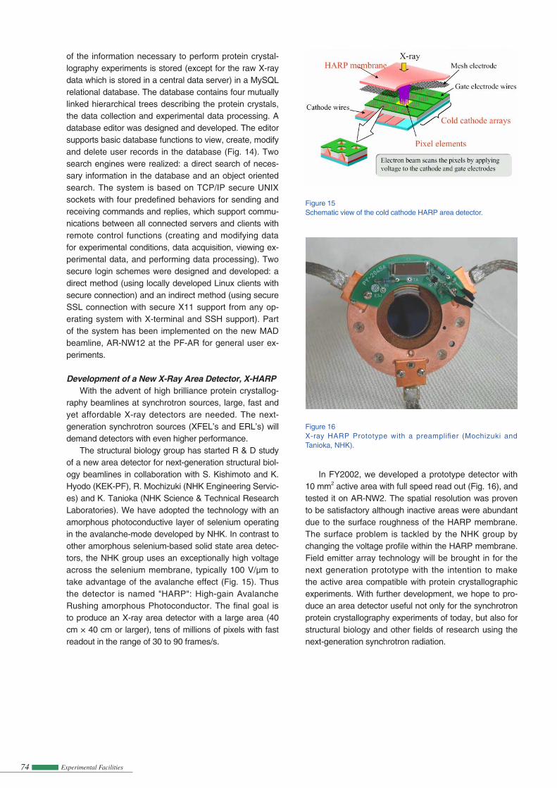
Development of a New X-Ray Area Detector, X-HARPWith the advent of high brilliance protein crystallog-
raphy beamlines at synchrotron sources, large, fast and yet affordable X-ray detectors are needed. The next-generation synchrotron sources (XFEL’s and ERL’s) will demand detectors with even higher performance.
The structural biology group has started R & D study of a new area detector for next-generation structural biol-ogy beamlines in collaboration with S. Kishimoto and K. Hyodo (KEK-PF), R. Mochizuki (NHK Engineering Servic-es) and K. Tanioka (NHK Science & Technical Research Laboratories). We have adopted the technology with an amorphous photoconductive layer of selenium operating in the avalanche-mode developed by NHK. In contrast to other amorphous selenium-based solid state area detec-tors, the NHK group uses an exceptionally high voltage across the selenium membrane, typically 100 V/µm to take advantage of the avalanche effect (Fig. 15). Thus the detector is named "HARP": High-gain Avalanche Rushing amorphous Photoconductor. The fi nal goal is to produce an X-ray area detector with a large area (40 cm × 40 cm or larger), tens of millions of pixels with fast readout in the range of 30 to 90 frames/s.
Figure 15Schematic view of the cold cathode HARP area detector.
Figure 16X-ray HARP Prototype with a preamplifi er (Mochizuki and Tanioka, NHK).
In FY2002, we developed a prototype detector with 10 mm2 active area with full speed read out (Fig. 16), and tested it on AR-NW2. The spatial resolution was proven to be satisfactory although inactive areas were abundant due to the surface roughness of the HARP membrane. The surface problem is tackled by the NHK group by changing the voltage profi le within the HARP membrane. Field emitter array technology will be brought in for the next generation prototype with the intention to make the active area compatible with protein crystallographic experiments. With further development, we hope to pro-duce an area detector useful not only for the synchrotron protein crystallography experiments of today, but also for structural biology and other fi elds of research using the next-generation synchrotron radiation.