structural determinants of oligomerization of Δ1-pyrroline...
TRANSCRIPT

Structural Determinants of Oligomerization ofΔ 1-Pyrroline-5-Carboxylate Dehydrogenase:Identification of a Hexamerization Hot Spot
Min Luo1, Ranjan K. Singh1 and John J. Tanner1,2
1 - Department of Chemistry, University of Missouri-Columbia, Columbia, MO 65211, USA2 - Department of Biochemistry, University of Missouri-Columbia, Columbia, MO 65211, USA
Correspondence to John J. Tanner: Department of Chemistry, University of Missouri-Columbia, Columbia, MO 65211,USA. [email protected]://dx.doi.org/10.1016/j.jmb.2013.05.027Edited by T. Yeates
Abstract
The aldehyde dehydrogenase (ALDH) superfamily member Δ1-pyrroline-5-carboxylate dehydrogenase(P5CDH) catalyzes the NAD+-dependent oxidation of glutamate semialdehyde to glutamate, which is the finalstep of proline catabolism. Defects in P5CDH activity lead to the metabolic disorder type II hyperprolinemia,P5CDH is essential for virulence of the fungal pathogen Cryptococcus neoformans, and bacterial P5CDHshave been targeted for vaccine development. Although the enzyme oligomeric state is known to beimportant for ALDH function, the oligomerization of P5CDH has remained relatively unstudied. Here wedetermine the oligomeric states and quaternary structures of four bacterial P5CDHs using a combination ofsmall-angle X-ray scattering, X-ray crystallography, and dynamic light scattering. The P5CDHs fromThermus thermophilus and Deinococcus radiodurans form trimer-of-dimers hexamers in solution, which isthe first observation of a hexameric ALDH in solution. In contrast, two Bacillus P5CDHs form dimers insolution but do not assemble into a higher-order oligomer. Site-directed mutagenesis was used to identify ahexamerization hot spot that is centered on an arginine residue in the NAD+-binding domain. Mutation ofthis critical Arg residue to Ala in either of the hexameric enzymes prevents hexamer formation in solution.Paradoxically, the dimeric Arg-to-Ala T. thermophilus mutant enzyme packs as a hexamer in the crystalstate, which illustrates the challenges associated with predicting the biological assembly in solution fromcrystal structures. The observation of different oligomeric states among P5CDHs suggests potentialdifferences in cooperativity and protein–protein interactions.
© 2013 Elsevier Ltd. All rights reserved.
Introduction
The enzyme Δ1-pyrroline-5-carboxylate (P5C)dehydrogenase (P5CDH; EC 1.5.1.12) catalyzesthe NAD+-dependent oxidation of glutamate semi-aldehyde to glutamate, which is the final step ofproline catabolism (Fig. 1).1 In humans, P5CDH alsocatalyzes the final step of hydroxyproline catabolism,which is the conversion of 4-hydroxyglutamatesemialdehyde to 4-erythro-hydroxy-L-glutamate.The enzyme is widely distributed in eukaryotes andbacteria. In the former organisms, P5CDH islocalized to the mitochondrial matrix. In somebacteria, mostly Gram-negative bacteria, P5CDH is
0022-2836/$ - see front matter © 2013 Elsevier Ltd. All rights reserve
combined with the first enzyme of proline catabolism,proline dehydrogenase (PRODH), into the bifunc-tional enzyme proline utilization A (PutA).2,3 How-ever, in Gram-positive bacteria, PRODH andP5CDH are separate monofunctional enzymesencoded by distinct genes. Monofunctional P5CDHis the subject of this research.Defects in P5CDH underlie the autosomal reces-
sive disorder type II hyperprolinemia.4–8Certain mis-sense and frameshift mutations in the gene encodingP5CDH abrogate enzyme function, resulting inelevated levels of P5C and proline in plasma, urine,and cerebrospinal fluid.9 Type II hyperprolinemia iscausally linked to neurologic manifestations, such as
d. J. Mol. Biol. (2013) 425, 3106–3120
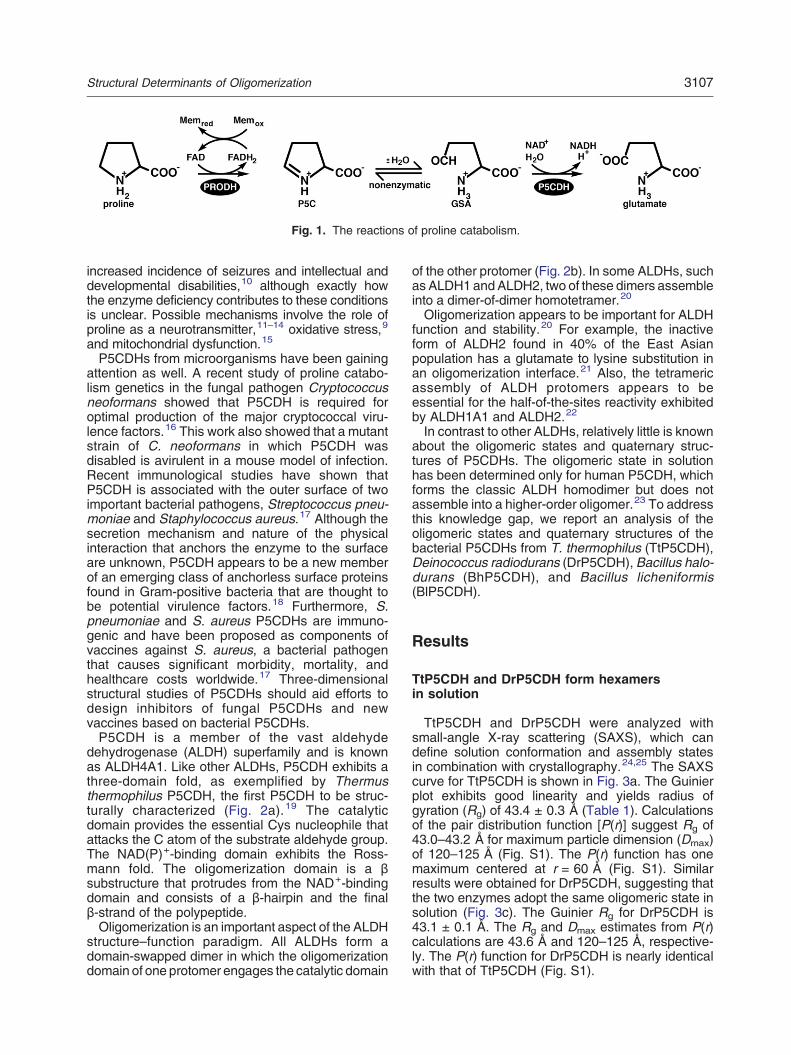
Fig. 1. The reactions of proline catabolism.
3107Structural Determinants of Oligomerization
increased incidence of seizures and intellectual anddevelopmental disabilities,10 although exactly howthe enzyme deficiency contributes to these conditionsis unclear. Possible mechanisms involve the role ofproline as a neurotransmitter,11–14 oxidative stress,9
and mitochondrial dysfunction.15
P5CDHs from microorganisms have been gainingattention as well. A recent study of proline catabo-lism genetics in the fungal pathogen Cryptococcusneoformans showed that P5CDH is required foroptimal production of the major cryptococcal viru-lence factors.16 This work also showed that a mutantstrain of C. neoformans in which P5CDH wasdisabled is avirulent in a mouse model of infection.Recent immunological studies have shown thatP5CDH is associated with the outer surface of twoimportant bacterial pathogens, Streptococcus pneu-moniae and Staphylococcus aureus.17 Although thesecretion mechanism and nature of the physicalinteraction that anchors the enzyme to the surfaceare unknown, P5CDH appears to be a new memberof an emerging class of anchorless surface proteinsfound in Gram-positive bacteria that are thought tobe potential virulence factors.18 Furthermore, S.pneumoniae and S. aureus P5CDHs are immuno-genic and have been proposed as components ofvaccines against S. aureus, a bacterial pathogenthat causes significant morbidity, mortality, andhealthcare costs worldwide.17 Three-dimensionalstructural studies of P5CDHs should aid efforts todesign inhibitors of fungal P5CDHs and newvaccines based on bacterial P5CDHs.P5CDH is a member of the vast aldehyde
dehydrogenase (ALDH) superfamily and is knownas ALDH4A1. Like other ALDHs, P5CDH exhibits athree-domain fold, as exemplified by Thermusthermophilus P5CDH, the first P5CDH to be struc-turally characterized (Fig. 2a).19 The catalyticdomain provides the essential Cys nucleophile thatattacks the C atom of the substrate aldehyde group.The NAD(P)+-binding domain exhibits the Ross-mann fold. The oligomerization domain is a βsubstructure that protrudes from the NAD+-bindingdomain and consists of a β-hairpin and the finalβ-strand of the polypeptide.Oligomerization is an important aspect of the ALDH
structure–function paradigm. All ALDHs form adomain-swapped dimer in which the oligomerizationdomain of one protomer engages the catalytic domain
of the other protomer (Fig. 2b). In some ALDHs, suchas ALDH1 andALDH2, two of these dimers assembleinto a dimer-of-dimer homotetramer.20
Oligomerization appears to be important for ALDHfunction and stability.20 For example, the inactiveform of ALDH2 found in 40% of the East Asianpopulation has a glutamate to lysine substitution inan oligomerization interface.21 Also, the tetramericassembly of ALDH protomers appears to beessential for the half-of-the-sites reactivity exhibitedby ALDH1A1 and ALDH2.22
In contrast to other ALDHs, relatively little is knownabout the oligomeric states and quaternary struc-tures of P5CDHs. The oligomeric state in solutionhas been determined only for human P5CDH, whichforms the classic ALDH homodimer but does notassemble into a higher-order oligomer.23 To addressthis knowledge gap, we report an analysis of theoligomeric states and quaternary structures of thebacterial P5CDHs from T. thermophilus (TtP5CDH),Deinococcus radiodurans (DrP5CDH), Bacillus halo-durans (BhP5CDH), and Bacillus licheniformis(BlP5CDH).
Results
TtP5CDH and DrP5CDH form hexamersin solution
TtP5CDH and DrP5CDH were analyzed withsmall-angle X-ray scattering (SAXS), which candefine solution conformation and assembly statesin combination with crystallography.24,25 The SAXScurve for TtP5CDH is shown in Fig. 3a. The Guinierplot exhibits good linearity and yields radius ofgyration (Rg) of 43.4 ± 0.3 Å (Table 1). Calculationsof the pair distribution function [P(r)] suggest Rg of43.0–43.2 Å for maximum particle dimension (Dmax)of 120–125 Å (Fig. S1). The P(r) function has onemaximum centered at r = 60 Å (Fig. S1). Similarresults were obtained for DrP5CDH, suggesting thatthe two enzymes adopt the same oligomeric state insolution (Fig. 3c). The Guinier Rg for DrP5CDH is43.1 ± 0.1 Å. The Rg and Dmax estimates from P(r)calculations are 43.6 Å and 120–125 Å, respective-ly. The P(r) function for DrP5CDH is nearly identicalwith that of TtP5CDH (Fig. S1).

Fig. 2. Structure of TtP5CDH(PDB code 2BHQ). (a) The proto-mer is shown with the catalyticdomain in green, NAD+-bindingdomain in light blue, and oligomer-ization domain in yellow. Redpatches indicate residues that formthe major dimer–dimer interface ofhexametric P5CDHs. Dark blue de-notes residues involved in the minordimer–dimer interface of hexametricP5CDHs. (b) Structure of the classicALDH dimer. (c) Two views of theTtP5CDH hexamer deduced fromSAXS and X-ray crystallography.
3108 Structural Determinants of Oligomerization

Fig. 3. SAXS analyses ofTtP5CDH and DrP5CDH. (a) Ex-perimental and calculated SAXScurves for TtP5CDH. The insetshows a Guinier plot spanning therange of qRg from 0.489 to 1.28.The linear fit of the Guinier plot hasR2 of 0.996. (b) Superposition of theTtP5CDH SAXS shape reconstruc-tion and the hexamer generatedfrom crystallographic symmetry.The SAXS envelope was calculatedusing the SASTBX server. Twoorthogonal views are shown. (c)Experimental and calculated SAXScurves for DrP5CDH. The insetshows a Guinier plot spanning therange of qRg from 0.458 to 1.30(R2 = 0.999).
3109Structural Determinants of Oligomerization

Table 1. Parameters derived from SAXS and DLS
Guinier Real space Vc SAXS DLS OligomericRG (Å) RG (Å) (Å2) M (kDa)a M (kDa)b state
TtP5CDH 43 43 1294 314 351 HexamerTtP5CDHR100A 32 33 628 100 123 DimerTtP5CDHK104A NDc NDc NDc NDc 337 HexamerTtP5CDHR111A 46 44 1346 319 314 HexamerTtP5CDHR153A NDc NDc NDc NDc 304 HexamerTtP5CDHR100A/K104A/R111A NDc NDc NDc NDc 120 DimerDrP5CDH 43 44 1295 316 361 HexamerDrP5CDHR102A 33 33 666 111 126 DimerBhP5CDH 31 32 616 98 106 DimerBlP5CDH NDc NDc NDc NDc 119 Dimer
a The molecular mass estimated from the volume of correlation as M = Vc2RG
−1/0.1231.b The molecular mass estimated from DLS.c Not determined.
3110 Structural Determinants of Oligomerization
The crystal structure of TtP5CDH was used to helpdetermine the quaternary structures of TrP5CDH andDrP5CDH. The crystal structure of TtP5CDH wasreported by Inagaki et al. in 2006.19 The space groupis H3 with one classic ALDH dimer (Fig. 2b) in theasymmetric unit. The Rg of the dimer is only 30 Å,which suggests that TtP5CDH assembles into ahigher-order oligomer in solution. Furthermore, theSAXS curve calculated from the dimer deviatessubstantially from the experimental curves (Fig. 3aand c). TheH3 crystal lattice was inspected to identifya higher-order assembly that is consistent with theSAXS data. As described previously,19 application ofthe crystallographic 3-fold rotation to the asymmetricunit generates a trimer-of-dimers hexamer (Fig. 2c).The hexamer hasRg of 42.6 Å, which agrees with theexperimentalRg of 43 Å. The theoretical SAXS curvecalculated from the hexamer exhibits good agree-ment with the experimental ones for both TtP5CDH(Fig. 3a) and DrP5CDH (Fig. 3c). Ensembles contain-ing both the hexamer and dimer models were alsoconsidered using the minimal ensemble searchmethod.26 Slightly better fits to the experimentalprofiles are obtained with ensembles consisting of94% hexamer and 6% dimer for TtP5CDH (Fig. 3a)and 87% hexamer and 13% dimer for DrP5CDH(Fig. 3c). These calculations suggest that the hex-amer is the predominant oligomer in solution.Moreover, the ab initio SAXS envelope matches thesize and shape of the hexamer (Fig. 3b). It isconcluded that TtP5CDH and DrP5CDH exist primar-ily as a trimer-of-dimers hexamer in solution. This isthe first observation of a hexameric ALDH in solution.The hexamer oligomeric state was confirmed using
dynamic light scattering (DLS) (Table 1 and TableS1) and the SAXS volume of correlation (Vc).
27 DLSdata indicate a hydrodynamic radius (RH) of 7.2 nmfor TtP5CDH, which implies a molecular mass (M) of351 kDa. The latter value is within 3% of thepredicted M of 342 kDa for a hexamer. Similarly,the estimated RH andM of DrP5CDH are 7.3 nm and361 kDa. The Vc values of TtP5CDH and DrP5CDH
are 1294 Å2 and 1295 Å2, respectively (Table 1).The corresponding M values are 314 kDa forTtP5CDH and 316 kDa for DrP5CDH (Table 1),which are within 8% of the M of the hexamer. TheDLS and Vc results confirm that TtP5CDH andDrP5CDH exist primarily as hexamers in solution.
BhP5CDH and BlP5CDH are dimeric in solution
The SAXS profile of BhP5CDH is profoundlydifferent from those of TtP5CDH and DrP5CDH(Fig. 4a). In particular, the pronounced valley at q =0.083 Å−1 and peak at q = 0.102 Å−1, which arecharacteristic of hexameric P5CDHs, are absent inthe SAXS profile for BhP5CDH. The Rg of BhP5CDHfrom Guinier analysis is 31.3 ± 0.1 Å, while the realspace Rg from calculations of P(r) is 31.8–31. 9 Å forDmax = 95–105 Å. These values are much smallerthan the Rg of 43 Å of hexameric P5CDHs,suggesting that BhP5CDH does not form a hexamer.Furthermore, the P(r) has a maximum at r = 35 Å,whereas the P(r) for hexameric P5CDH has amaximum at 60 Å (Fig. S1). The Rg calculated froma P5CDH dimer is 30 Å, implying that BhP5CDHforms the classic ALDH dimer but does notassemble into higher-order oligomers. M estimatedfrom SAXS Vc is 98 kDa (Table 1), which issuggestive of the dimer (114 kDa) and certainlyeliminates tetramers (228 kDa) and hexamers(342 kDa) from consideration. Also, DLS data areconsistent with BhP5CDH forming a dimer (RH =4.4 nm, M = 106 kDa; Table S1).The hypothesis that BhP5CDH forms a dimer in
solution was tested using crystallographic data. Thestructure of BhP5CDH was determined by the NewYork Structural Genomics Research Consortium.The enzyme crystallizes in space group C2 withthree molecules in the asymmetric unit [Protein DataBank (PDB) code 3QAN, unpublished results]. Twoof the molecules form the classic ALDH dimer, whilethe crystallographic 2-fold rotation generates theother half of the dimer for the third molecule. Analysis

Fig. 4. SAXS analysis of BhP5CDH. (a) Experimental and calculated SAXS curves. The inset shows a Guinier plotspanning the qRg range 0.352–1.30 (R2 = 0.998). (b) Superposition of the SAXS shape reconstruction and the dimer.
3111Structural Determinants of Oligomerization
of the crystal lattice with PDBePISA28 indicates thatthe dimer is the most probable assembly in solution.Inspection of crystal packing with Coot confirmedthat the hexamer is absent. The SAXS profilecalculated from the BhP5CDH dimer agrees wellwith the experimental profile (Fig. 4a), and the SAXSreconstruction exhibits good agreement with thedimer (Fig. 4b). It is concluded that BhP5CDH isdimeric in solution.The oligomeric state of BlP5CDH (75% identical
with BhP5CDH) was determined using DLS andanalysis of crystal packing. The RH is 4.6 nm, whichcorresponds to M = 119 kDa, consistent with a dimer(TableS1). The crystal structure of BlP5CDHwas alsodetermined by the New York Structural Genomics
Research Consortium. The enzyme crystallizes inspace group P21 with eight molecules in the asym-metric unit. As deposited, dimers are not evident in theasymmetric unit. However, application of crystallo-graphic symmetry allows a different choice of asym-metric unit that contains four dimers. Analysis of theBlP5CDH crystal lattice with PDBePISA indicates thatthe dimer is the most probable assembly in solution. Itis concluded that BlP5CDH is also dimeric in solution.
Site-directed mutagenesis rationale
Site-directed mutagenesis (to Ala) was used toidentify residues important for hexamerization.TtP5CDH was chosen as the model for this study

3112 Structural Determinants of Oligomerization
because high-resolution crystal structures are avail-able (e.g., PDB code 2BHQ). The major interfacebetween dimers in the hexamer is formed by a helixfrom the NAD+-binding domain (α3, residues 96–111) and the oligomerization domain (red in Fig. 2a).These structural elements form a symmetric interfacelining the inside surface of the tunnel that surroundsthe 3-fold axis (red in Fig. 5a). This interface buries1100 Å2 of surface area. For comparison, theinterfacial area of the domain-swapped dimer inter-face is 2900 Å2. A smaller dimer–dimer contactsurface (600 Å2) is located on the outside of thehexamer (blue in Fig. 5b). The α14 helix of thecatalytic domain (residues 454–463) and α3 areprominent in this interface (Fig. 2a).The α3 helix was targeted for site-directed
mutagenesis because it participates in both dimer–dimer interfaces (Fig. 5c). Specifically, three posi-tively charged residues that form dimer–dimerelectrostatic interactions were individually mutatedto Ala: Arg100, Lys104, and Arg111. Arg100 isunique in that it is the only residue in the protein thatinteracts with two protomers outside of its own dimer(3.2 Å cutoff). These dimer–dimer interactions in-clude ion pairs with Asp166, Glu168, and Glu458and a hydrogen bond with Tyr154 (Fig. 5c). Arg100also forms an intermolecular stacking interactionwith Arg461. The other two residues targeted for
mutagenesis, Lys104 and Arg111, form dimer–dimerion pairs with the carboxyl-terminus of the polypep-tide chain (Fig. 5c).Arg153 of the oligomerization domain was also
mutated to Ala. This residue is interesting because itis next to the 2-fold axis of the major dimer–dimerinterface, and thus, its guanidinium group stacks inparallel with that of the symmetry-related Arg153(Fig. 5c).
Steady-state kinetic measurements
The kinetic constants for the native and mutantenzymes were estimated using P5C as the variablesubstrate and NAD+ fixed at 1 mM in order to assesswhether mutation of the hexamer interface causes anygross change in enzyme activity (Table S2 and Fig.S2). The catalytic efficiencies (kcat/Km) of the TtP5CDHmutant enzymes are within 20% of that of the nativeenzyme, indicating that these particular mutations donot have an obvious, substantial effect on activity.
Hexamerization hot spot
SAXS analysis of TtP5CDHR100A clearly showsthat mutation of Arg100 to Ala is sufficient to disruptthe hexamer. The SAXS curve for TtP5CDHR100Alacks the valley and peak features that are
Fig. 5. Dimer–dimer interfaceswithin the P5CDH hexamer. (a andb) Orthogonal views of the hexamerwith the A–B dimer colored cyan–green, C–D dimer in gray–yellow,and E–F dimer in brown–magenta.Red and blue denote residues in themajor and minor dimer–dimer in-terfaces, respectively. (c) Dimer–dimer interactions formed by theresidues targeted for mutagenesis(Arg100, Lys104, Arg111, andArg153). As in (a) and (b), protomerA is colored cyan, and dimer C–D iscolored gray–yellow. The oval de-notes the approximate location ofone of the 2-fold axes of thehexamer.

3113Structural Determinants of Oligomerization
diagnostic of the hexamer (Fig. 6a). The Rg fromGuinier analysis is 32.1 ± 0.2 Å, which is close to thevalue of 30 Å calculated from the dimer andsubstantially smaller than that of the hexamer(43 Å). Furthermore, the SAXS curve calculatedfrom the dimer exhibits excellent agreement with theexperimental one (Fig. 6a), and the SAXS envelopematches the dimer (Fig. 6b). Also, the RH estimatedfrom DLS is 4.7 nm, which is substantially smallerthan the RH of 7.2 for the TtP5CDH hexamer (TableS1). The corresponding M from DLS is 123 kDa,which is similar to the value of 114 kDa expected forthe dimer. M estimated from SAXS Vc is 100 kDa(Table 1), which is also consistent with the dimer. It isconcluded that TtP5CDHR100A exists in solutionprimarily as a dimer.The analogous mutation was generated for
DrP5CDH (DrP5CDHR102A). The SAXS profile of
DrP5CDHR102A is likewise indicative of a dimer(Fig. 6a). The Rg is 32.6 ± 0.1 Å, and the SAXScurve calculated from the dimer agrees well with theexperimental curve. The SAXS envelope exhibitsgood agreement with the dimer (Fig. 6b). Also, theMof DrP5CDHR102A estimated from DLS is 126 kDa,which is similar to the value of 114 kDa expected fora dimer (Table S1). M estimated from SAXS Vc is111 kDa (Table 1), which is also consistent with thedimer. These data show that DrP5CDHR102Alikewise exists primarily as a dimer in solution.In contrast, mutation of Arg111, Lys104, or Arg153
individually to Ala does not disrupt the hexamer. TheSAXS curve for TtP5CDHR111A clearly exhibits thevalley and peak features that are diagnostic of thehexamer (Fig. 6a). The Rg from Guinier analysis is46.1 ± 0.2 Å, which is consistent with a hexamer.Furthermore, the profile calculated from the hexamer
Fig. 6. SAXS analysis of P5CDHmutant enzymes. (a) Experimentaland theoretical SAXS curves. Arbi-trary offsets were applied to thecurves for clarity. The theoreticalSAXS curves for TtP5CDHR100A(red) and DrP5CDHR102A (green)were calculated from a TtP5CDHdimer. The theoretical curve incyan for TtP5CDHR111A was cal-culated from a TtP5CDH hexamer,while the curve in orange wascalculated from an ensemble con-sisting of 88% hexamer and 12%dimer. The inset shows Guinierplots spanning the qRg ranges0.361–1.30 for TtP5CDHR100A(R 2 = 0.9992), 0.347–1.30 forDrP5CDHR102A (R2 = 0.9996),and 0.519–1.31 for TtP5CDHR111A(R2 = 0.9997). (b) Superpositionsof the SAXS shape reconstructionsand oligomer models for TtP5-CDHR100A, DrP5CDHR102A, andTtP5CDHR111A.

Table 2. X-ray diffraction data collection and refinementstatistics
TtP5CDHR100A
TtP5CDH R100A/K104A/R111A
Wavelength (Å) 1.000 1.542Space group H3 P1Unit cell parametersa (Å) 102.7 65.1b (Å) 102.7c (Å) 279.5 160.6α (°) 86.3β (°) 87.5γ (°) 79.4Resolution (Å) 47.3–1.54
(1.62–1.54)83.2–2.42(2.55–2.42)
Total observations 835,877 287,864Unique reflections 158,507 114,331Multiplicity 5.3 (2.8) 2.5 (1.8)Rmerge
a 0.045 (0.461) 0.038 (0.110)Rmeas
a 0.050 (0.549) 0.047 (0.156)Rpim
a 0.021 (0.291) 0.028 (0.110)⟨I/σ(I)⟩ 20.7 (2.3) 15.4 (5.2)Completeness (%) 96.8 (78.5) 73.5 (64.4)Predicted oligomeric stateb Hexamer DimerRwork 0.168 (0.282)Rfree
c 0.185 (0.291)Number of atoms 8608Protein residues 1032Water molecules 623RMSD bond lengths (Å) 0.006RMSD bond angles (°) 1.04Ramachandran plotd (%)Favored 98.64Outliers 0MolProbity score (percentile) 99Average B-factors (Å2)Protein 22.5Water 29.7Coordinate error (Å)e 0.13PDB code 4K57
Values for the outer-resolution shell of data are given in parenthesis.a Definitions ofRmerge,Rmeas, andRpim can be found inWeiss.29b Oligomeric state predicted from crystal packing using
PDBePISA.c Random test set (5%).d The Ramachandran plot was generated with MolProbity.30e Maximum-likelihood-based coordinate error from PHENIX.
3114 Structural Determinants of Oligomerization
matches the experimental profile (Fig. 6a), and theshape reconstruction resembles the hexamer (Fig. 6b).Minimum ensemble search calculations also suggestthat the hexamer is the predominant (88%) oligomer insolution (Fig. 6a). TheM valueestimated fromSAXSVcis 319 kDa (Table 1), which is just 7% lower thanM ofthe hexamer. TtP5CDHR111A, TtP5CDHK104A,and TtP5CDHR153A were analyzed with DLS.The hydrodynamic radii for these proteins spanthe range 6.8–7.2 nm, implying M = 304–337 kDa,which is consistent with the hexamer being themajor species in solution (Table S1). These resultssuggest that TtP5CDHR111A, TtP5CDHK104A,and TtP5CDHR153A exist primarily as hexamersin solution.
Crystallization of dimeric TtP5CDHs
TtP5CDHR100A was targeted for crystallization tobetter understand why mutation of Arg100 to Aladisrupts hexamerization. Surprisingly, the mutantenzyme crystallized in the same H3 lattice asTtP5CDH (Table 2), and thus, the trimer-of-dimershexamer is evident in crystalline TtP5CDHR100Adespite loss of the numerous interactions formedby Arg100.Electron density maps indicated that the
Arg100Ala mutation causes a cascade of conforma-tional changes in the vicinity of residue 100 (Fig. 7a).Glu168 rotates into the void created by removal ofthe Arg100 side chain, while Tyr154 moves into thespace vacated by Glu168 (Fig. 7b). Also, electrondensity for the side chains of Arg153 and Glu458 isweak and diffuse, implying disorder.Formation of the H3 lattice by TtP5CDHR100A
prompted the generation of double and triplemutants designed to induce a new crystal formthat is devoid of the hexamer (Table S3). This goalwas achieved with the triple mutant R100A/K104A/R111A, which crystallizes in space group P1. Amoderate-resolution diffraction data set wasobtained (Table 2), which was sufficient to deter-mine the arrangement of dimers in the crystallattice. The calculations show that the triclinic cellcontains four dimers (Fig. 8). Analysis of protein–protein interfaces using manual inspection andPDBePISA shows that the hexamer is not presentin the lattice.
Discussion
Global sequence identity apparently is not a goodpredictor of the oligomeric state of P5CDHs. Wepreviously showed using analytical ultracentrifuga-tion that human P5CDH is a dimer in solution. Thehuman enzyme has 30% sequence identity to thebacterial enzymes studied here (Table S4). TheBacillus enzymes are also dimeric despite having50% identity to the hexameric enzymes TtP5CDH
and DrP5CDH. Thus, prediction of the oligomericstate of P5CDH from sequence requires a morecareful examination of both sequence and structure.This conclusion is consistent with the hot spot theoryof protein–protein interaction, which posits that a fewcritical residues in the interface account for most ofthe binding energy.31,32
We used structure-guided alanine scanning mu-tagenesis to identify a hexamerization hot spot forbacterial P5CDHs. The hot spot is centered onArg100, which is consistent with early work showingthat protein–protein interaction hot spots tend to beenriched in arginine, surrounded by less importantresidues.31 Arg100 is unique in that it is the onlyresidue that interacts with two other protomers of thehexamer (Fig. 5c). This key residue forms dimer–

Fig. 7. Hexamer interface ofTtP5CDHR100A. (a) Electron den-sity for residues of TtP5CDHR100Athat change conformation. Thecage represents a simulatedannealing σA-weighted Fo − Fcomit map (3σ). The orientation andcoloring scheme are identical withthose of Fig. 5c. (b) Comparison ofthe hexamer interfaces of TtP5CDH(gray) and TtP5CDHR100A (pink).
3115Structural Determinants of Oligomerization
dimer interactions with five residues: Tyr154,Asp166, Glu168, Glu458, and Arg461. Arg100 isalso present in DrP5CDH but is substituted by Ala/Asn in the Bacillus enzymes and Gln in humanP5CDH (Fig. S3), consistent with it being essentialfor hexamerization. Arg461 is also found inDrP5CDH, while Asp166 is conservatively substitut-ed with Glu.On the other hand, Arg461 and Asp166 are not
conserved in the three dimeric P5CDHs. Also, aclear trend is not evident for Tyr154 or Glu168. Thisanalysis suggests that the triplet of Arg100, Asp/Glu166, and Arg461 is a hexamerization hot spot,which could be used to predict the oligomeric statesof P5CDHs. For example, S. aureus P5CDH, whichhas been proposed as a vaccine reagent, ispredicted to be dimeric since it has Glu in place ofArg100. The prediction of stable oligomeric speciesin solution from crystal structures is an importantarea of research. PDBePISA is used widely for thispurpose and has been adopted by the PDB forpredicting the biological assembly of depositedstructures (remark 350 of PDB entr ies) .TtP5CDHR100A is interesting in this regard becauseit forms a hexamer in the crystal yet is predominatelydimeric in solution under the conditions used forSAXS and DLS (1–5 mg/mL). Analysis of theTtP5CDHR100AH3 lattice with PDBePISA indicates
that the hexamer is the most probable assembly insolution, which contradicts the SAXS data. However,the PDBePISA results do suggest that the R100Ahexamer is less stable than the native one. Forexample, considering only the protein componentand omitting solvent, the mutation causes 5% and7% decreases in the surface area buried in the majorand minor hexamer interfaces, respectively. Also,the complexation significance score of the majorhexamer interface decreases from 1.0 for TtP5CDH(maximum possible value) to only 0.2 forTtP5CDHR100A. Thus, the program was able todiscern a difference in the interfaces of the twocrystal structures. Our results suggest that theprediction of the solution oligomeric state fromcrystal packing remains challenging in some cases.Consideration of dynamic self-association equilib-
rium provides plausible explanations for the discrep-ancy in the oligomeric state of TtP5CDHR100A inthe aqueous and solid states. The SAXS data showtha t the d imer–hexamer equ i l i b r i um o fTtP5CDHR100A lies far on the dimer side underconditions of 1–10 mg/mL protein in Tris–NaClbuffers at pH 7.5. It is possible that the conditionsused for crystallization shift the equilibrium to thehexamer, enabling crystal growth. However, DLSperformed on TtP5CDHR100A under solution con-ditions similar to those of the initial crystallization
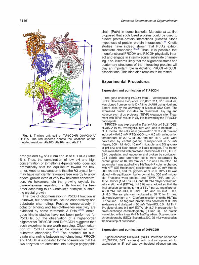
Fig. 8. Triclinic unit cell of TtP5CDHR100A/K104A/R111A. The red spheres denote the locations of themutated residues, Ala100, Ala104, and Ala111.
3116 Structural Determinants of Oligomerization
drop yielded RH of 4.3 nm and M of 101 kDa (TableS1). Thus, the combination of low pH and highconcentration of 2-methyl-2,4-pentanediol does notdramatically shift the equilibrium toward the hex-amer. Another explanation is that the H3 crystal formmay have sufficiently favorable free energy to allowcrystal growth even at very low hexamer concentra-tion. As hexamers join the growing crystal, thedimer–hexamer equilibrium shifts toward the hex-amer according to Le Chatelier's principle, sustain-ing crystal growth.The role of oligomerization in P5CDH function is
unknown, but possibilities include cooperativity andsubstrate channeling. Positive cooperatively incofactor binding and half-of-the-sites reactivity areexhibited by some tetrameric ALDHs.22,33 Analo-gous kinetic studies have not been performed forP5CDHs, but the observation of a higher-orderoligomer for TtP5CDH and DrP5CDH suggests thatsuch experiments are worth pursuing. Oligomeriza-tion of P5CDH could also be connected withsubstrate channeling.34,35 The potential for sub-strate channeling between monofunctional PRODHand P5CDH is suggested by the observation that thetwo enzymes are combined into a single polypeptide
chain (PutA) in some bacteria. Marcotte et al. firstproposed that such fused proteins could be used topredict protein–protein interactions (Rosetta Stonehypothesis of protein–protein interactions).36 Kineticstudies have indeed shown that PutAs exhibitsubstrate channeling.37,38 Thus, it is possible thatmonofunctional PRODH and P5CDH physically inter-act and engage in intermolecular substrate channel-ing. If so, it seems likely that the oligomeric states andquaternary structures of the interacting proteins willplay an important role in dictating PRODH–P5CDHassociations. This idea also remains to be tested.
Experimental Procedures
Expression and purification of TtP5CDH
The gene encoding P5CDH from T. thermophilus HB27(NCBI Reference Sequence YP_005182.1, 516 residues)was cloned from genomic DNA into pKA8H using NdeI andBamHI sites by the University of Missouri DNA Core. Theexpressed protein includes an N-terminal His8 tag andtobacco etch virus protease (TEVP) cleavage site. Treat-ment with TEVP results in Gly-His followed by the TtP5CDHpolypeptide.TtP5CDH was expressed in Escherichia coli BL21(DE3)
pLysS. A 10-mL overnight culturewas used to inoculate 1 Lof LBmedia. The cells were grown at 37 °C at 250 rpm andinducedwith 0.5 mM IPTGatOD600 = 0.6with an inductiontemperature of 22 °C at 200 rpm for 18 h. Cells wereharvested by centrifugation; resuspended in 50 mMHepes, 300 mM NaCl, 10 mM imidazole, and 5% glycerolat pH 8.0; and flash-frozen in liquid nitrogen. The frozencells were thawed with protease inhibitors (AEBSF, TPCK,E64, pepstatin, and leupeptin) and broken by sonication.Cell debris and unbroken cells were separated bycentrifugation at 16,500 rpm for 1 h in an SS34 rotor. Thesupernatant was applied to a HisTrap HP column chargedwith Ni2+ (GE Healthcare) equilibrated with 20 mM Hepes,300 mM NaCl, and 5% glycerol at pH 8.0. TtP5CDH waseluted with equilibration buffer containing 300 mM imidaz-ole. Fractions were pooled, and TEVP, THP, and 20×TEVP buffer [1 M Tris–HCl and 10 mM ethylenediamine-tetraacetic acid (EDTA), pH 8.0] were added so that thefinal solution contained 5 mg of TEVP per 30 mg of proteinin 50 mM Tris–HCl, 0.5 mM THP, and 0.5 mM EDTA,pH 8.0. The sample was incubated at 30 °C for 2 h anddialyzed overnight at 4 °C before injection onto the HisTrapHP column. The tag-free protein was collected at 30 mMimidazole and dialyzed in 50 mM Tris–HCl, 0.5 mM THP,5% glycerol, and 0.5 mM EDTA (pH 8.0) in preparation foranion-exchange chromatography (HiTrap Q). The proteinwas eluted with a linear 0–1 MNaCl gradient. Size-exclusionchromatography (SEC) (Superdex 200, 25 mL) was used asthe final step of purification.
Expression and purification of DrP5CDH
A gene encoding DrP5CDH (NCBI Reference SequenceNP_294537, 523 residues) with codons optimized forexpression in E. coli was synthesized (Genscript) and

3117Structural Determinants of Oligomerization
subcloned into pKA8H using NdeI and BamHI sites. Theexpressed protein includes an N-terminal His8 tag andTEVP site. Cleavage with TEVP produces Gly-Hisfollowed by the DrP5CDH polypeptide.DrP5CDH was expressed in BL21(DE3)pLysS (induc-
tion at OD600 = 0.8 with 0.5 mM IPTG for 5 h at 22 °C).The cells were collected by centrifugation; resuspended in50 mM Tris, 100 mM NaCl, 10 mM imidazole, and 5%glycerol at pH 7.5; and frozen at −80 °C.The frozen cells were thawed at 4 °C in the presence of
protease inhibitors (0.1 mM TPCK, 0.05 mM AEBSF,0.1 μM pepstatin, 0.01 mM leupeptin, and 5 μM E64) andbroken using sonication. The mixture was centrifuged at16,500 rpm inanSS34 rotor for 1 h at 4 °C, filtered through a0.45-μm filter (Millipore), and loadedonaHisTrapHPcolumn(5 mL) that had been charged with NiCl2 and equilibrated in50 mM Tris, 300 mM NaCl, 10 mM imidazole, and 5%glycerol at pH 7.5. Washing steps were performed using theloading buffer supplemented with 10 mM imidazole followedby 30 mM imidazole. The protein was eluted with 300 mMimidazole. The histidine tag was removed by incubating theprotein with 0.2 mg/mL TEVP for 1 h at 28 °C followed bydialysis at 4 °C against 50 mM Tris, 50 mM NaCl, and 5%glycerol at pH 7.5. The mixture was applied to the HisTrapHP column to separate the cleaved protein, which appearedin the flow-through, from the tag and TEVP. The cleavedprotein was dialyzed overnight at 4 °C into 50 mM Tris,0.5 mM EDTA, 0.5 mM DTT, and 5% glycerol at pH 7.8 inpreparation for anion-exchange chromatography (HiTrapQ).The sample was loaded onto the column using a buffer of50 mM Tris and 5% glycerol at pH 7.8, and a linear NaClgradient was applied. DrP5CDH eluted at 280–340 mMNaCl. The protein concentration was estimated using thebicinchoninic acid method (Pierce kit) with bovine serumalbumin as the standard.
Expression and purification of BhP5CDH andBlP5CDH
Expression constructs for BhP5CDH (NCBI ReferenceSequence NP_243603.1, 515 residues) and BlP5CDH(NCBI Reference Sequence YP_077616.1, 516 residues)were obtained from the New York Structural GenomicsResearch Consortium. The expressed enzymes haveC-terminal His8 tags and a TEVP cleavage site. TEVPcleavage produces the enzyme polypeptide followed byAENLYFQ. The enzymes were expressed and purifiedusing the protocols described for TtP5CDH.
Site-directed mutagenesis
Site-directed mutants of TtP5CDH and DrP5CDH weregenerated using the QuikChange II site-directed mutagen-esis kit (Agilent) using the primers listed in Table S3. Themutations were confirmed with sequencing performed bythe University of Missouri DNA core. The mutant enzymeswere purified as described above for the native enzymes.
Small-angle X-ray scattering
SAXS experiments were performed at beamline 12.3.1of the Advanced Light Source via the mail-in program.39,40
Prior to data collection, all protein samples were subjected
to SEC using a Superdex 200 column. The column bufferwas typically 50 mM Tris, 5% glycerol, 0.5 mM THP, and50 mM NaCl at pH 7.5. In some cases (e.g., DrP5CDH),the SEC fractions were pooled, concentrated to ~12 mg/mL, and dialyzed at 4 °C for 24 h against 50 mM Tris,50 mM NaCl, 0.5 mM EDTA, 0.5 mM THP, and 5%glycerol at pH 7.8. For each protein, scattering intensitieswere measured at three nominal protein concentrations(1–10 mg/mL). For each protein concentration, exposuretimes of 0.5, 1.0, 3.0, and 6.0 s were used. Scatteringcurves collected from the protein samples were correctedfor background scattering using intensity data collectedfrom the SEC effluent or dialysis buffer.The SAXS data were analyzed as follows. A composite
scattering curve for each sample was generated withPRIMUS41 by scaling and merging the high q region fromone of the longer time exposures with the low q region froma shorter time exposure. The scattering curves weremultiplied with a concentration factor and overlaid on eachother to check for concentration-dependent variation of theprofile. No substantial concentration effects were observedfor any of the samples. PRIMUS was also used to performGuinier analysis.FoXS was used to calculate theoretical scattering profiles
from atomic models.42 FoXS was also used to performminimum ensemble calculations.26 GNOM was used tocalculate pair distribution functions.43 MOLEMANwas usedto calculate Rg from atomic coordinates.44 The SASTBXserver45 was used for shape reconstruction calculations.
Estimation of molecular weight from SAXSvolume of correlation
The molecular weight was estimated from the volume ofcorrelation (Vc) as described recently by Rambo andTainer.27 Briefly, Vc is a new SAXS invariant defined as theratio of the zero angle scattering intensity, I(0), to the totalscattered intensity. The latter quantity is equal to theintegral ∫qI(q)dq performed over the entire range of thescattering data. Vc thus has units of Å
2. Rambo and Tainershowed that Vc is independent of solute concentration andthe aforementioned integral converges for both folded-compact and unfolded-flexible particles. For proteins, theyalso demonstrated that the molecular mass (M) in units ofDa can be estimated from a single SAXS curve by therelationshipM = Vc
2RG−1/0.1231. An analogous relationship
was provided for RNA.Vc was calculated as follows. First, I(0) and RG were
estimated from Guinier analysis using PRIMUS. Thesevalues were used to extrapolate the experimental SAXScurves to q = 0 using the equation I(q) = I(0)exp(−q2RG
2/3). Extrapolation to q = 0 is needed for accurate calcula-tion of the total scattered intensity. The extrapolated regionconsisted of 18–19 points with q spacing of 0.000610 Å−1,which matches the q spacing of the experimental data. Thearea under the curve of qI(q) versus qwas calculated usingthe Polygon Area utility of Origin 9 software, and Vc wascalculated as the ratio of I(0) to the area.
Crystallization of TtP5CDHR100A
Crystals of TtP5CDHR100A were grown in sitting dropsat room temperature using the protocol for TtP5CDH.46
Briefly, screening using commercially available kits

3118 Structural Determinants of Oligomerization
(Hampton Research) yielded positive results in severalconditions, which led to either the twinned form describedearlier46 or the more desired H3 form that was used forstructure determination. Space group H3 crystals ofTtP5CDHR100A were grown using sitting drops formedby mixing 1 μL of the protein stock solution [7 mg/mLprotein in 50 mMTris–HCl at pH 7.5, 100 mMNaCl, 5% (v/v)glycerol, 0.5 mM THP, and 0.5 mM EDTA] and 1 μL of thereservoir containing 45% of 2-methyl-2,4-pentanediol and0.05 M sodium citrate buffer at pH 5.2. The reservoir wasused as the cryoprotectant. The space group is H3 with unitcell dimensions of a = 173 Å and c = 279 Å and two proteinmolecules in the asymmetric unit.
Crystallization of TtP5CDHR100A/K104A/R111A
Triclinic crystals of the triple mutant TtP5CDHR100A/K104A/R111A were grown at 295 K with the sitting-dropmethod of vapor diffusion. Initial conditions were identifiedusing commercially available crystal screens (HamptonResearch). TtP5CDHR100A/K104A/R111A was crystal-lized using a reservoir of 0.2 M MgCl2, 0.1 M Tris–HCl(pH 8.5), and 30% (w/v) polyethylene glycol (PEG) 4000.The protein stock solution contained 5 mg/mL TtP5CDHtriple mutant in the buffer of 50 mM Tris, 100 mM NaCl,0.5 mM THP, 0.5 mM EDTA, and 5% glycerol at pH 7.8.Crystals were cryoprotected with 0.2 M MgCl2, 0.1 M Tris–HCl at pH 8.5, 30% (w/v) PEG 4000, and 28% PEG 200.The space group isP1with unit cell dimensions a = 65.1 Å,b = 102.7 Å, c = 160.6 Å, α = 86.3°, β = 87.5°, and γ =79.4°. The asymmetric unit includes 8 protein molecules (4dimers), which implies 48% solvent and VM of 2.35 Å3/Da.
X-ray diffraction data collection, phasing, andrefinement
X-ray diffraction data from crystals of TtP5CDHR100A inspace group H3 were collected at beamline 4.2.2 ofAdvanced Light Source. The 1.54-Å-resolution data setused for refinement consisted of 360 frames with anoscillation width of 0.5° per image, a detector distance of110 mm, and an exposure time of 2 s/image. The datawere processed with XDS47 and SCALA48 via CCP4i.49
Refinement using PHENIX50 commenced from the co-ordinates of TtP5CDH with Arg100 truncated to Ala.Coot51 was used for model building. Data collection andrefinement statistics are listed in Table 2.X-ray diffraction data from crystals of the triple mutant
TtP5CDHR100A/K104A/R111A were collected on anin-house Rigaku rotating anode generator coupled to anR-AXIS IV++ detector. Two data sets were recorded andmerged. The first one consisted of 360 frames collectedwith an oscillation width of 0.5°, a detector distance of270 mm, and an exposure time of 7 min per frame. Thesecond set consisted of 360 frames collected with anoscillation width of 0.5°, a detector distance of 200 mm,and an exposure time of 5.2 min per frame. The data wereintegrated with MOSFLM52 and merged to 2.42 Å resolu-tion with SCALA. The arrangement of the dimers in theasymmetric unit was determined using molecular replace-ment as implemented in Phaser53 with a search modelderived from a dimer of TtP5CDHR100A with Lys104 and
Arg111 truncated to Ala. A clear solution with four dimers inthe unit cell and log-likelihood gain of 31,444 was obtained.Rigid body refinement yielded Rwork = Rfree = 0.290 for alldata to 2.42 Å resolution.
DLS
These experiments were performed on a ProteinSolutions DynaPro 99 Molecular Sizing Instrument(Wyatt Technology) at 20 °C with a wavelength of836.3 nm and a scattering angle of 90°. Protein concen-trations were in the range 0.5–2 mg/mL in 50 mM Trisbuffer at pH 7.5. Prior to DLS, each sample wascentrifuged for 10 min at 13,000 g at 4 °C and passedthrough a 0.22-μm Millipore filter (Whatman). The datawere collected with acquisition time of 10 s with at least 18acquisitions. The DLS data were analyzed by the programDYNAMICS v.5.26.38 by performing regularization fitusing the regularization algorithm on the measuredautocorrelation functions.
Steady-state kinetics
The P5CDH activity of TtP5CDH and TtP5CDH mutantenzymes were measured at 20 °C by monitoring NADHproduction at 340 nm as described previously.23 The finalassay mixture (1 mL) contained 6 μg/mL P5CDH (0.1 μM),1 mM NAD+, and various concentrations of P5C in 0.1 Mpotassium phosphate buffer at pH 7.5. The pH of the P5Cstock solution was adjusted to 7.5 before adding to thereaction mixture. Kinetic constants (Table S2) wereestimated by fitting the initial rate data to the Michaelis–Menten equation using Origin 9.0 (Fig. S2).
Accession numbers
Coordinates and structure factor amplitudes forTtP5CDHR100A have been deposited in the PDB underthe accession number 4K57.Supplementary data to this article can be found online at
http://dx.doi.org/10.1016/j.jmb.2013.05.027
Acknowledgements
Research reported in this publication was sup-ported by the National Institute of General MedicalSciences of the National Institutes of Health viaGrant GM065546. We thank Dr. Tommi White forpurifying T. thermophilus genomic DNA and Dr.Mingyi Zhou for cloning the TtP5CDH gene. Wethank Prof. Steven Almo and the New YorkStructural Genomics Research Consortium for pro-viding the BhP5CDH and BlP5CDH clones and Prof.Donald Becker for providing P5C. We thank KevinDyer of the SIBYLS Mail-In SAXS Program forcollecting the SAXS data. X-ray scattering and

3119Structural Determinants of Oligomerization
diffraction technologies and their applications to thedetermination of macromolecular shapes and con-formations at the SIBYLS beamline at the AdvancedLight Source, Lawrence Berkeley National Labo-ratory, are supported in part by the Department ofEnergy program Integrated Diffraction Analysis Tech-nologies under Contract Number DE-AC02-05CH11231 with the U.S. Department of Energy. WethankDr. JayNix for help with diffraction data collectionand processing. The Advanced Light Source issupported by the Director, Office of Science, Office ofBasic Energy Sciences, of the U.S. Department ofEnergy under Contract No. DE-AC02-05CH11231.
Received 16 April 2013;Received in revised form 30 May 2013;
Accepted 31 May 2013Available online 7 June 2013
Keywords:proline catabolism;
aldehyde dehydrogenase;small-angle X-ray scattering;
X-ray crystallography
Abbreviations used:EDTA, ethylenediaminetetraacetic acid; PDB, Protein
Data Bank; PRODH, proline dehydrogenase; P5C,Δ1-pyrroline-5-carboxylate; P5CDH, Δ1-pyrroline-5-
carboxylate dehydrogenase; PutA, proline utilization A;ALDH, aldehyde dehydrogenase; SAXS, small-angle
X-ray scattering; DLS, dynamic light scattering; TEVP,tobacco etch virus protease; SEC, size-exclusion
chromatography; PEG, polyethylene glycol.
References1. Phang, J. M. (1985). The regulatory functions of
proline and pyrroline-5-carboxylic acid. Curr. Top.Cell. Regul. 25, 92–132.
2. Tanner, J. J. (2008). Structural biology of prolinecatabolism. Amino Acids, 35, 719–730.
3. Singh, R. K. & Tanner, J. J. (2012). Unique structuralfeatures and sequence motifs of proline utilization A(PutA). Front. Biosci. 17, 556–568.
4. Efron, M. L. (1965). Familial hyperprolinemia. Reportof a second case, associated with congenital renalmalformations, hereditary hematuria and mild mentalretardation, with demonstration of an enzyme defect.N. Engl. J. Med. 272, 1243–1254.
5. Baumgartner, M. R., Rabier, D., Nassogne, M. C., Dufier,J. L., Padovani, J. P., Kamoun, P. et al. (2005).Delta1-pyrroline-5-carboxylate synthase deficiency: neu-rodegeneration, cataracts and connective tissue mani-festations combined with hyperammonaemia andreduced ornithine, citrulline, arginine and proline. Eur. J.Pediatr. 164, 31–36.
6. Geraghty,M. T., Vaughn,D., Nicholson, A. J., Lin,W.W.,Jimenez-Sanchez,G.,Obie,C.et al. (1998).Mutations in
the Delta1-pyrroline 5-carboxylate dehydrogenase genecause type II hyperprolinemia. Hum. Mol. Genet. 7,1411–1415.
7. Scriver, C. R., Sly, W. S., Childs, B., Beaudet, A. L.,Valle, D., Kinzler, K. W. & Vogelstein, B. (2001). Themetabolic and molecular bases of inherited disease(Eds.), 8th edit McGraw-Hill, New York, NY.
8. Valle, D., Goodman, S. I., Applegarth, D. A., Shih, V. E.& Phang, J. M. (1976). Type II hyperprolinemia.Delta1-pyrroline-5-carboxylic acid dehydrogenase defi-ciency in cultured skin fibroblasts and circulatinglymphocytes. J. Clin. Invest. 58, 598–603.
9. Wyse, A. T. & Netto, C. A. (2011). Behavioral andneurochemical effects of proline.Metab. Brain Dis. 26,159–172.
10. Phang, J. M., Hu, C. A. & Valle, D. (2001). Disorders ofproline andhydroxyprolinemetabolism. InMetabolic andMolecular Basis of Inherited Disease (Scriver, C. R.,Beaudet, A. L., Sly, W. S. & Valle, D., eds),pp. 1821–1838, McGraw Hill, New York, NY.
11. Gogos, J. A., Santha, M., Takacs, Z., Beck, K. D.,Luine, V., Lucas, L. R. et al. (1999). The geneencoding proline dehydrogenase modulates sensori-motor gating in mice. Nat. Genet. 21, 434–439.
12. Felix, D. & Kunzle, H. (1976). The role of proline innervous transmission. Adv. Biochem. Psychopharma-col. 15, 165–173.
13. Takemoto, Y. & Semba, R. (2006). Immunohisto-chemical evidence for the localization of neuronscontaining the putative transmitter L-proline in ratbrain. Brain Res. 1073–1074, 311–315.
14. Fremeau,R.T., Jr., Caron,M.G.&Blakely,R.D. (1992).Molecular cloning and expression of a high affinityL-proline transporter expressed in putative glutamater-gic pathways of rat brain. Neuron, 8, 915–926.
15. He, F. & DiMario, P. J. (2011). Drosophila delta-1-pyrroline-5-carboxylate dehydrogenase (P5CDh) isrequired for proline breakdown and mitochondrialintegrity—establishing a fly model for human type IIhyperprolinemia. Mitochondrion, 11, 397–404.
16. Lee, I. R., Lui, E. Y., Chow, E.W., Arras, S. D., Morrow,C. A. & Fraser, J. A. (2013). Reactive oxygen specieshomeostasis and virulence of the fungal pathogenCryptococcus neoformans requires an intact prolinecatabolism pathway. Genetics, 194, 421–433.
17. Lijek, R. S., Luque, S. L., Liu, Q., Parker, D., Bae, T. &Weiser, J. N. (2012). Protection from the acquisition ofStaphylococcus aureus nasal carriage by cross-reactive antibody to a pneumococcal dehydrogenase.Proc. Natl Acad. Sci. USA, 109, 13823–13828.
18. Chhatwal, G. S. (2002). Anchorless adhesins andinvasins of Gram-positive bacteria: a new class ofvirulence factors. Trends Microbiol. 10, 205–208.
19. Inagaki, E., Ohshima, N., Takahashi, H., Kuroishi, C.,Yokoyama, S. & Tahirov, T. H. (2006). Crystalstructure of Thermus thermophilus Delta1-pyrroli-ne-5-carboxylate dehydrogenase. J. Mol. Biol. 362,490–501.
20. Rodriguez-Zavala, J. S. & Weiner, H. (2002). Struc-tural aspects of aldehyde dehydrogenase that influ-ence dimer-tetramer formation. Biochemistry, 41,8229–8237.
21. Larson, H. N., Weiner, H. & Hurley, T. D. (2005).Disruption of the coenzyme binding site and dimer

3120 Structural Determinants of Oligomerization
interface revealed in the crystal structure of mitochon-drial aldehyde dehydrogenase “Asian” variant. J. Biol.Chem. 280, 30550–30556.
22. Yoval-Sánchez, B., Pardo, J. P. & Rodríguez-Zavala,J. S. (2013). New insights into the half-of-the-sitesreactivity of human aldehyde dehydrogenase 1A1.Proteins, http://dx.doi.org/10.1002/prot.24274.
23. Srivastava, D., Singh, R. K., Moxley,M. A., Henzl,M. T.,Becker, D. F. & Tanner, J. J. (2012). The three-dimensional structural basis of type II hyperproline-mia. J. Mol. Biol. 420, 176–189.
24. Putnam, C. D., Hammel, M., Hura, G. L. & Tainer, J. A.(2007). X-ray solution scattering (SAXS) combinedwith crystallography and computation: defining ac-curate macromolecular structures, conformationsand assemblies in solution. Q. Rev. Biophys. 40,191–285.
25. Rambo, R. P. & Tainer, J. A. (2010). Bridging thesolution divide: comprehensive structural analyses ofdynamic RNA, DNA, and protein assemblies bysmall-angle X-ray scattering. Curr. Opin. Struct. Biol.20, 128–137.
26. Pelikan, M., Hura, G. L. & Hammel, M. (2009).Structure and flexibility within proteins as identifiedthrough small angle X-ray scattering. Gen. Physiol.Biophys. 28, 174–189.
27. Rambo, R. P. & Tainer, J. A. (2013). Accurateassessment of mass, models and resolution bysmall-angle scattering. Nature, 496, 477–481.
28. Krissinel, E. & Henrick, K. (2007). Inference ofmacromolecular assemblies from crystalline state. J.Mol. Biol. 372, 774–797.
29. Weiss, M. (2001). Global indicators of X-ray dataquality. J. Appl. Crystallogr. 34, 130–135.
30. Chen, V. B., Arendall, W. B., 3rd, Headd, J. J., Keedy,D. A., Immormino, R. M., Kapral, G. J. et al. (2010).MolProbity: all-atom structure validation for macromo-lecular crystallography. Acta Crystallogr., Sect. D:Biol. Crystallogr. 66, 12–21.
31. Bogan, A. A. & Thorn, K. S. (1998). Anatomy of hotspots in protein interfaces. J. Mol. Biol. 280, 1–9.
32. Moreira, I. S., Fernandes, P. A. & Ramos, M. J. (2007).Hot spots—a review of the protein–protein interfacedeterminant amino-acid residues. Proteins, 68,803–812.
33. Wei, B. & Weiner, H. (2001). Making an Orientalequivalent of the yeast cytosolic aldehyde dehydro-genase as well as making one with positive coopera-tivity in coenzyme binding by mutations of glutamate492 and arginine 480. Chem.-Biol. Interact. 130–132,173–179.
34. Arentson, B. W., Sanyal, N. & Becker, D. F. (2012).Substrate channeling in proline metabolism. Front.Biosci. 17, 375–388.
35. Anderson, K. S. (1999). Fundamental mechanisms ofsubstrate channeling. Methods Enzymol. 308,111–145.
36. Marcotte, E. M., Pellegrini, M., Ng, H. L., Rice, D. W.,Yeates, T. O. & Eisenberg, D. (1999). Detectingprotein function and protein–protein interactions fromgenome sequences. Science, 285, 751–753.
37. Srivastava, D., Schuermann, J. P., White, T. A.,Krishnan, N., Sanyal, N., Hura, G. L. et al. (2010).Crystal structure of the bifunctional proline utilization A
flavoenzyme from Bradyrhizobium japonicum. Proc.Natl Acad. Sci. USA, 107, 2878–2883.
38. Surber, M. W. & Maloy, S. (1998). The PutA protein ofSalmonella typhimurium catalyzes the two steps ofproline degradation via a leaky channel. Arch.Biochem. Biophys. 354, 281–287.
39. Hura, G. L., Menon, A. L., Hammel, M., Rambo, R. P.,Poole, F. L., 2nd, Tsutakawa, S. E. et al. (2009).Robust, high-throughput solution structural analysesby small angle X-ray scattering (SAXS).Nat. Methods,6, 606–612.
40. Classen, S., Hura, G. L., Holton, J. M., Rambo, R. P.,Rodic, I., McGuire, P. J. et al. (2013). Implementationand performance of SIBYLS: a dual endstationsmall-angle X-ray scattering and macromolecularcrystallography beamline at the Advanced LightSource. J. Appl. Crystallogr. 46, 1–13.
41. Konarev, P. V., Volkov, V. V., Sokolova, A. V., Koch,M. H. J. & Svergun, D. I. (2003). PRIMUS: a WindowsPC-based system for small-angle scattering dataanalysis. J. Appl. Crystallogr. 36, 1277–1282.
42. Schneidman-Duhovny, D., Hammel, M. & Sali, A.(2010). FoXS: a web server for rapid computationand fitting of SAXS profiles. Nucleic Acids Res. 38,W540–W544.
43. Svergun, D. (1992). Determination of the regulariza-tion parameter in indirect-transform methods usingperceptual criteria. J. Appl. Crystallogr. 25, 495–503.
44. Kleywegt,G. J. (1997). Validation of proteinmodels fromCalpha coordinates alone. J. Mol. Biol. 273, 371–376.
45. Liu, H., Hexemer, A. & Zwart, P. H. (2012). The SmallAngle Scattering ToolBox (SASTBX): an open-sourcesoftware for biomolecular small-angle scattering. J.Appl. Crystallogr. 45, 587–593.
46. Inagaki, E., Takahashi, H., Kuroishi, C. & Tahirov, T.H. (2005). Crystallization and avoiding the problem ofhemihedral twinning in crystals of Delta1-pyrroline-5-carboxylate dehydrogenase from Thermus thermo-philus. Acta Crystallogr., Sect. F: Struct. Biol. Cryst.Commun. 61, 609–611.
47. Kabsch, W. (2010). XDS. Acta Crystallogr., Sect. D:Biol. Crystallogr. 66, 125–132.
48. Evans, P. (2006). Scaling and assessment of dataquality. Acta Crystallogr., Sect. D: Biol. Crystallogr. 62,72–82.
49. Potterton, E., Briggs, P., Turkenburg, M. & Dodson, E.(2003). A graphical user interface to the CCP4program suite. Acta Crystallogr., Sect. D: Biol.Crystallogr. 59, 1131–1137.
50. Adams, P. D., Gopal, K., Grosse-Kunstleve, R. W.,Hung, L. W., Ioerger, T. R., McCoy, A. J. et al. (2004).Recent developments in the PHENIX software forautomated crystallographic structure determination. J.Synchrotron Radiat. 11, 53–55.
51. Emsley, P. & Cowtan, K. (2004). Coot: model-buildingtools for molecular graphics. Acta Crystallogr., Sect.D: Biol. Crystallogr. 60, 2126–2132.
52. Leslie, A. G. (2006). The integration of macromolec-ular diffraction data. Acta Crystallogr., Sect. D: Biol.Crystallogr. 62, 48–57.
53. McCoy, A. J., Grosse-Kunstleve, R. W., Adams, P. D.,Winn, M. D., Storoni, L. C. & Read, R. J. (2007).Phaser crystallographic software. J. Appl. Crystallogr.40, 658–674.