structural history of human srgap2...
TRANSCRIPT

Structural History of Human SRGAP2 Proteins
Michael Spornydagger1 Julia Guez-Haddaddagger1 Annett Kreusch2 Sivan Shakartzi1 Avi Neznansky1 Alice Cross3
Michail N Isupov3 Britta Qualmann2 Michael M Kessels2 and Yarden Opatowsky1
1The Mina amp Everard Goodman Faculty of Life Sciences Bar-Ilan University Ramat Gan Israel2Institute for Biochemistry I Jena University Hospital Friedrich Schiller University Jena Jena Germany3Department of Biosciences College of Life and Environmental Sciences University of Exeter Exeter United KingdomdaggerThese authors contributed equally to this work
Corresponding author E-mail yardenopatowskybiuacil
Associate editor Banu Ozkan
Abstract
In the development of the human brain human-specific genes are considered to play key roles conferring its uniqueadvantages and vulnerabilities At the time of Homo lineage divergence from Australopithecus SRGAP2C graduallyemerged through a process of serial duplications and mutagenesis from ancestral SRGAP2A (34ndash24 Ma) Remarkablyectopic expression of SRGAP2C endows cultured mouse brain cells with human-like characteristics specifically in-creased dendritic spine length and density To understand the molecular mechanisms underlying this change inneuronal morphology we determined the structure of SRGAP2A and studied the interplay between SRGAP2A andSRGAP2C We found that 1) SRGAP2A homo-dimerizes through a large interface that includes an F-BAR domain anewly identified F-BAR extension (Fx) and RhoGAP-SH3 domains 2) SRGAP2A has an unusual inverse geometryenabling associations with lamellipodia and dendritic spine heads in vivo and scaffolding of membrane protrusions incell culture 3) As a result of the initial partial duplication event (34 Ma) SRGAP2C carries a defective Fx-domainthat severely compromises its solubility and membrane-scaffolding ability Consistently SRGAP2ASRAGP2C hetero-dimers form but are insoluble inhibiting SRGAP2A activity 4) Inactivation of SRGAP2A is sensitive to the level ofhetero-dimerization with SRGAP2C 5) The primal form of SRGAP2C (P-SRGAP2C existing between 34 and 24 Ma)is less effective in hetero-dimerizing with SRGAP2A than the modern SRGAP2C which carries several substitutions(from24 Ma) Thus the genetic mutagenesis phase contributed to modulation of SRGAP2Arsquos inhibition of neuronalexpansion by introducing and improving the formation of inactive SRGAP2ASRGAP2C hetero-dimers indicating astepwise involvement of SRGAP2C in human evolutionary history
Key words SRGAP2 X-ray crystallography F-BAR domain human evolution structural biology
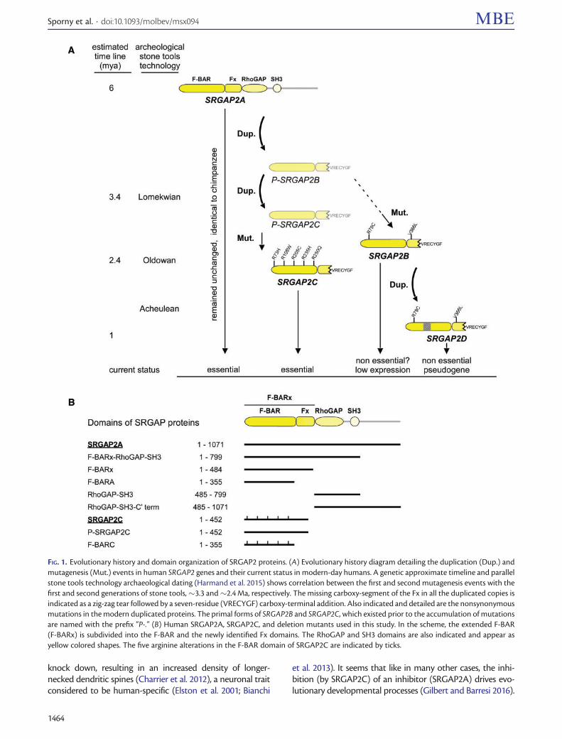
IntroductionEmergence of new genes is a powerful driving force of evolu-tion and speciation Gene duplication allows daughter copiesto acquire function-altering mutations without compromis-ing the original activity of the mother gene In recent yearscomparing the modern human genome sequences to those ofarchaic hominins and great apes has allowed the discovery ofduplicated genes unique to the Homo lineage These genes areconsidered to have played key roles in human speciationparticularly in the development of the highly advanced humanbrain (Zhang and Long 2014 Dennis and Eichler 2016) One ofthese human-specific genes is SRGAP2C which encodes for afunctional protein and originated from the ancestralSRGAP2A (fig 1) (Sudmant et al 2010 Dennis et al 2012)
Slit-Robo GTPase Activating Protein (SRGAPs) couple theplasma membrane topology to remodeling of the actin cyto-skeleton thereby regulating neuronal migration axon guid-ance and branching and dendritic spine maturation Toachieve this task SRGAPs utilize a unique domain composi-tion an amino-terminal extended F-BAR (F-BARx) followedby RhoGAP and SH3 domains (fig 1B) which mediate
membrane association cytoskeleton remodeling and pro-teinndashprotein interactions respectively A model for theSRGAP2A mechanism of action can be deduced from recent(Fritz et al 2015 Guez-Haddad et al 2015) and previous stud-ies (Wong et al 2001 Pertz et al 2008 Guerrier et al 2009Endris et al 2011 Yamazaki et al 2013) as well as from ourcurrent work According to this model SRGAP2A acts as aninhibitor of cell migration and protrusion extension The ac-tivation of SRGAP2A begins with its recruitment to the vicin-ity of the plasma membrane by one of several target proteins(eg Robo1 Wong et al 2001) Through its F-BARx domainSRGAP2A directly binds to the plasma membrane specificallyat sites of protruding curvatures (Guerrier et al 2009Coutinho-Budd et al 2012 Yamazaki et al 2013) There inan elegant example of negative feedback the RhoGAP do-main inactivates local pools of Rac1 and CDC42 which inturn leads to the subsequent breakdown of nearby actincytoskeleton and the retraction of membrane protrusions(Wong et al 2001 Fritz et al 2015) SRGAP2C interactswith SRGAP2A and inhibits its activities (Charrier et al2012 Fossati et al 2016) Expression of human SRGAP2C incultured murine cortical neurons phenocopies SRGAP2A
Article
The Author 2017 Published by Oxford University Press on behalf of the Society for Molecular Biology and EvolutionThis is an Open Access article distributed under the terms of the Creative Commons Attribution Non-Commercial License(httpcreativecommonsorglicensesby-nc40) which permits non-commercial re-use distribution and reproduction in anymedium provided the original work is properly cited For commercial re-use please contact journalspermissionsoupcom Open AccessMol Biol Evol 34(6)1463ndash1478 doi101093molbevmsx094 Advance Access publication February 21 2017 1463
knock down resulting in an increased density of longer-necked dendritic spines (Charrier et al 2012) a neuronal traitconsidered to be human-specific (Elston et al 2001 Bianchi
et al 2013) It seems that like in many other cases the inhi-bition (by SRGAP2C) of an inhibitor (SRGAP2A) drives evo-lutionary developmental processes (Gilbert and Barresi 2016)
FIG 1 Evolutionary history and domain organization of SRGAP2 proteins (A) Evolutionary history diagram detailing the duplication (Dup) andmutagenesis (Mut) events in human SRGAP2 genes and their current status in modern-day humans A genetic approximate timeline and parallelstone tools technology archaeological dating (Harmand et al 2015) shows correlation between the first and second mutagenesis events with thefirst and second generations of stone tools33 and24 Ma respectively The missing carboxy-segment of the Fx in all the duplicated copies isindicated as a zig-zag tear followed by a seven-residue (VRECYGF) carboxy-terminal addition Also indicated and detailed are the nonsynonymousmutations in the modern duplicated proteins The primal forms of SRGAP2B and SRGAP2C which existed prior to the accumulation of mutationsare named with the prefix ldquoP-rdquo (B) Human SRGAP2A SRGAP2C and deletion mutants used in this study In the scheme the extended F-BAR(F-BARx) is subdivided into the F-BAR and the newly identified Fx domains The RhoGAP and SH3 domains are also indicated and appear asyellow colored shapes The five arginine alterations in the F-BAR domain of SRGAP2C are indicated by ticks
Sporny et al doi101093molbevmsx094 MBE
1464
SRGAP2C reached its current form after several evolution-ary steps determined by (Dennis et al 2012) and are elabo-rated here in figure 1A First SRGAP2A underwentincomplete segmental duplication from chromosome1q321 into 1q211 and then from 1q211 into 1p12 Laternonsynonymous mutations accumulated on the duplicatedcopies R79C and V366L on 1q211 giving rise to the contem-porary SRGAP2B and R73H R108W R205C R235H R250Qon the 1p12 copy giving rise to the contemporary SRGAP2CHere we refer to the duplicated copies prior to the accumu-lation of the nonsynonymous mutations as ldquoprimalrdquo or P-SRGAP2B and P-SRGAP2C SRGAP2B was later duplicatedonce more into 1q211 to give rise to SRGAP2D The emer-gence of SRGAP2 B C and D are estimated to have occurred34 24 and 1 Ma respectively
The function of F-BAR (FES-CIP4 homology) domains aswell as those of the other members of the BAR superfamily[ie BAR (BinamphiphysinRVS) I-BAR (inverse BAR) andN-BAR (N-terminal BAR) domains] is thought to be directlylinked to their three-dimensional shape and oligomeric orga-nization BAR-superfamily domains form anti-parallel dimersthat interact with membranes through their ldquoN-surfacerdquowhich is either concave convex or flat and includes theamino-terminal helix (ldquohelix 1rdquo) from the two anti-parallelBAR protomers In this way BAR N-BAR and certain F-BAR domains that have a concave N-surface associate withcellular membrane invaginations while I-BARs that have con-vex N-surfaces associate with membrane protrusionsConsistent with their functions in endocytic processes thecrystal structures of membrane invagination-associating F-BAR family members for example FBP17 Syndapin andFCHo2 (PDB codes 2EFL 3HAH and 2V0O respectively) dis-play a concave N-surface (Kessels and Qualmann 2015) It istherefore surprising to see that the endogenous SRGAP2A islocalized to and active in cellular protrusions such as den-dritic spine heads and lamellipodia and scaffold membraneprotrusions when overexpressed in COS-7 cells [(Guerrieret al 2009 Charrier et al 2012 Fritz et al 2015) and this work]
Here we show that dimeric SRGAP2A has an invertedconvex curvature that can promote membrane protrusionsWe further show that SRGAP2A dimers exhibit a domain-swapping Fx coiled-coil extension domain and that theRhoGAP-SH3 domains also participate in dimeric interac-tions We found that in spite of its smaller size and reduceddimerization interface SRGAP2C forms stable hetero-dimerswith SRGAP2A which are insoluble cannot scaffold mem-branes and have a reduced affinity for Robo1 We show thatSRGAP2C acquired its full dominant negative function overtime while P-SRGAP2C was already insoluble due to a criticalcarboxy-truncation in the Fx it was by the later mutagenesisstage that SRGAP2C gained its ability to form more stableSRGAP2ASRGAP2C hetero-dimers
ResultsTo reveal the molecular mechanism by which SRGAP2A as-sociates with membrane protrusions such as dendritic spineheads and to understand how SRGAP2C inhibits SRGAP2Arsquos
activity during human brain development we first deter-mined the X-ray structure of the F-BARx and used the re-sulting atomic coordinates to study the interplay betweenSRGAP2A and SRGAP2C We docked the separate crystalstructures of the F-BARx RhoGAP (homology model) andSH3 domains into our previously determined SAXS structureof intact SRGAP4 (Guez-Haddad et al 2015) and measuredSRGAP2A inter-domain interactions to map the structuralarrangement and dimeric interface within intact SRGAP2Ahomo-dimers We employed liposome sedimentation assaysto quantitate membrane binding affinity single-turnoverGTPase assay to measure GAP catalysis and surface plasmonresonance (SPR) to measure SRGAP-Robo1 interactions Weused expression in Sf9 cells to evaluate protein solubility andexpression in COS-7 cells to assess the impact of SRGAP2Amutations on membrane-binding and remodeling activities amethod previously applied by other groups (Guerrier et al2009 Coutinho-Budd et al 2012 Yamazaki et al 2013)
F-BARx CrystallographyWe solved the F-BARx crystal structure by applying a varia-tion of an exhaustive molecular symmetry search approachthat took advantage of the 2-fold anti-parallel F-BAR con-served symmetry (Sporny et al 2016) This allowed solutionof a structure (refined to 27 A resolution) using a remotesearch model [FBP17 PDB 2EFL (Shimada et al 2007)] withonly 19 sequence identity which represented 13 (as apoly-ala model) of the asymmetric unit contents This mo-lecular symmetry-constrained systematic search approachwas vital for structure solution as it enabled the resolutionof characteristic problems common for coiled coil proteins(Dauter 2015) The exhaustive search was followed by elec-tron density modification procedures and cycles of modelrefinement and re-building (table 1) The quality of the re-sulting electron density has allowed us to ensure correct as-signment of all amino acid side chains
Crystal Structure of F-BARxThe crystal structure reveals that the F-BARx (residues 1ndash484supplementary fig S1 Supplementary Material online) iscomposed of a noncanonical F-BAR (residues 1ndash355) domainfollowed by a coiled-coil domain extension (residues 356ndash484) designated here as the F-BAR extension (Fx) The F-BARx is the largest BAR superfamily member characterizedthus far The tip-to-tip distance of the F-BARx dimer is ap-proximately 275 A [electron density in the tip regions is weakdue to high B-factor values (supplementary fig S2BSupplementary Material online) and cannot be defined ac-curately] reaching a width of 55 A The buried surface inter-face (BSI) between the two protomers is 8370 A2 Incomparison the F-BAR domain of FBP17 has 288 residuesa dimeric tip-to-tip distance of 222 A a width of 36 A and aBSI of 4647 A2
Regardless of the size difference the same structural com-ponents that form the F-BAR domains of FBP17 CIP4 andFCHo2 are also found in SRGAP2A These include three pri-mary helices (a1 a2 and a3) and one short helix (a4) (fig 2Aand B and supplementary figs S1 and S2 Supplementary
StructurendashFunction of SRGAP2 Proteins doi101093molbevmsx094 MBE
1465
Material online) In SRGAP2A the carboxy-half of a1 fromeach protomer together with approximately one third ofeach a2 and a3 form a six-helix bundle at the center of thedimer in which a1 and a3 are engaged in homotypic inter-actions with their dimeric counterpart
Two arms protrude from the central helix bundle eachincludes the long amino-half of a1 and a coiled-coil com-posed by the remaining two-thirds of a2 and a3
The membrane-binding N-surface is composed by a1 andmost of a2 together with small portions of a3 and a6 On theopposing side to the N-surface the short a4 extends directlyfrom a3 and further continues as a long random-coil towardsthe tip of the second protomer
A Homer-EVH1 binding motif 339PPMKF is located on thea4ndasha5 connecting loop (supplementary figs S1 and S3Supplementary Material online) It was recently demon-strated that through this motif SRGAP2A directly interactswith the Homer protein in excitatory synapses (Fossati et al2016) However our structural data reveals that because thePhe343 side-chain is buried in a cleft formed between thethree main a1 2 and 3 helices of the reciprocal protomer itcan actually not be engaged with EVH1 domain binding un-less a considerable conformational change takes place in thisregion On the Homer side this assessment is based on thecrystal structure and analysis of the Homer EVH1 domaincomplexed with a peptide from mGluR (TPPSPF) (PDB1DDV) (Beneken et al 2000)
The Fx a 72 A long coiled-coil composed by a5 and a6extends from one SRGAP2A protomer and packs against the
a2ndasha3 arm of the reciprocal protomer in the SRGAP2A dimer(fig 2 and supplementary fig S2 Supplementary Materialonline) The Fx is arranged in such a way that a5 is predom-inantly surface exposed and has only minor direct interac-tions with the F-BAR while a6 is mostly buried between a5and the a2ndasha3 coiled-coil arm Similar coiled-coil extensionwith reminiscent packing is also observed in the crystal struc-ture of the Fes-kinase F-BAR domain (PDB 4DYL)
SRGAP2A Has an Extended Dimerization InterfaceLike other BAR-superfamily members the isolated F-BAR do-main (F-BARA residues 1ndash355) of SRGAP2A is dimeric as canbe demonstrated by size-exclusion chromatography (SEC)(supplementary fig S4 Supplementary Material online) TheF-BARx crystal structure reveals an elaborated dimerizationinterface that involves the Fx in addition to the F-BAR
Furthermore we found that the RhoGAP-SH3 domains arealso likely to be involved in dimerization (fig 2C and D) Withthe F-BARx crystal structure at hand we could fit it into thelow resolution Small Angle X-ray Scattering (SAXS) structureof the full-length SRGAP4 that we determined recently(Guez-Haddad et al 2015) (fig 2D) Adjustment of theF-BARx arms angle by 10 and docking of RhoGAP (the ho-mologous RhoGAP of Beta2-chimaerin PDB 1XA6Canagarajah et al 2004) and SH3 (from our recent workPDB 4RUG Guez-Haddad et al 2015) crystal structures intothe densities next to the tips of the F-BARx arms results in avery good fit using the fit-in-map application in Chimera(Yang et al 2012) with real-time mean correla-tionfrac14 90438e05 SDfrac14 000066913 RMSfrac14 000067521Domain fitting was also guided by distance constraints be-tween the carboxy end of the Fx which is visible in the crystalstructure and the amino terminus of the RhoGAP modelThe resulting domain arrangement implies that the RhoGAPand SH3 domains interact directly with the F-BAR arms of thereciprocal protomers
To confirm this observation we measured the bindingaffinity of an isolated RhoGAP-SH3 to a fluorescently labeledF-BAR and F-BARx using microscale thermophoresis (MST)(fig 2C) Our measurements show that the RhoGAP-SH3 do-mains specifically bind the F-BAR domain with KDfrac14 3 mMand the F-BARx with KDfrac14 35 mM This shows that theRhoGAP-SH3 directly interacts with the F-BAR and that theFx does not contribute to the binding affinity These resultsare also consistent with our recent binding measurements ofRobo1 to SRGAP2A (Guez-Haddad et al 2015) Therein wediscovered that the F-BARx RhoGAP and SH3 domains co-operatively participate in Robo1 binding further supportingdirect interactions between the three domains
In conclusion the dimerization interface in SRGAP2A ex-tends beyond the F-BARs and includes the Fx and probablythe RhoGAP-SH3 domains of one SRGAP2 protomer thatwrap around the F-BAR domain of the reciprocal SRGAP2A(fig 2D)
SRGAP2A Inverse CurvatureAs mentioned earlier it is typical for F-BAR proteins to asso-ciate with membrane invaginations however in concordance
Table 1 Summary of Crystallographic Statistics
Data Collection Statistics
Crystal SRGAP2A F-BARxBeamline ID-29 ESRFWavelength (A) 0976Space group C2Unit Cell Parameters (A) afrac14 2032 bfrac14 299 cfrac14 948 bfrac14 919
Total reflectionsa 64564 (2660)Unique reflectionsa 16118 (726)Completeness ()a 988 (922)Rmeas ()ab 126 (1211)Mean Ira 81 (11)Resolution range (A) 4736ndash270CC12a 099 (062)
Refinement StatisticsRwork ()a 021 (038)Rfree()a 026 (040)Number of nonhydrogen atoms 3386Macromolecules 3367Water 19Protein residues 409RMS bond lengths (A) 0013RMS bond angles () 168Ramachandran favored () 93Ramachandran outliers () 02Clashscore 2 8Average B-factor 8005RCBS PDB code 5I6J
aValues for the highest resolution shell are given in parenthesesbRmeas frac14 Rhfrac12m=ethm 1THORN1=2RjiIhi lt Ih gt j=RhRiIhi
Sporny et al doi101093molbevmsx094 MBE
1466
FIG 2 SRGAP2A structure and dimerization (A) Domains of SRGAP2A and focus on F-BARx The F-BARx helices and encoding exons are drawn inscale and designated by their numbers The carboxy-half of helix 6 encoded by exon 10 which is missing in SRGAP2C is depicted in black(B) Ribbon diagrams and cartoons of the dimeric SRGAP2A F-BARx crystal structure in which the two identical protomers are colored in yellowand in cyan Labels indicating structural elements of the cyan protomer are underscored The large ribbon diagram represents a view facing the N-surfaceand delineates the 6-helix core arms and tip regions (C) The RhoGAP-SH3 domains bind directly to the F-BAR domain Microscale thermophoresis(MST) titration curves and apparent KD values for RhoGAP-SH3 binding to the F-BAR or the F-BARx that were fluorescently labeled Measured
StructurendashFunction of SRGAP2 Proteins doi101093molbevmsx094 MBE
1467
with prior studies we find that the F-BAR-containingSRGAP2A associates with membrane protrusions (fig 3Band C) (Frost et al 2008 Guerrier et al 2009 Yamazakiet al 2013 Fritz et al 2015) In order to explain how thismight be it is necessary to look at the structures of theSRGAP2A F-BAR domain in comparison to that of other F-BAR proteins In general the six-helix core region of F-BARdomains is essentially flat and the overall curved shape of F-BAR dimers is generated by the direction and angle of the a2ndasha3 arms relative to the central region In FBP17 CIP4Syndapins and FCHo2 the a2-a3 arms are bent towardsthe direction of the N-surface thereby generating a concavemembrane binding surface Unlike these our structural datafor SRGAP2A reveal that the a2ndasha3 arms are bent in theopposite direction away from the N-surface resulting in aconvex membrane binding surface (fig 3A) Structure-basedsequence comparison between the F-BAR domain ofSRGAP2A and its closest available F-BAR homolog FBP17(PDB 2ELF 19 sequence identity) revealed a 25-residuelong helical insertion (residues 127RFV LQD151) into a2 atthe point that links the central region and the a2ndasha3 armSince the bases of the a2ndasha3 arms are held with a1 in the six-helix core region the 25 aa insertion would push the a2ndasha3arm to point away from the insertion Indeed when we re-moved the 25-residue helix from the F-BARx of SRGAP2Aand expressed the truncated construct in COS-7 cells nomembrane protrusions were observed (supplementary figS5 Supplementary Material online) Taken together suchinverse geometry of the membrane-binding surface readilyexplains how contrary to other F-BAR proteins SRGAPs as-sociate with membrane protrusions rather than with invag-inations (fig 3)
Membrane Binding Is Driven and Maintained byElectrostatic InteractionsIn most BAR superfamily domains membrane association isdriven by electrostatic interactions between negativelycharged membrane surfaces and positively charged residueson the membrane-binding surface of the protein (Mim andUnger 2012 Kessels and Qualmann 2015) For the SRGAP2A F-BARx the convex N-surface is predominantly electropositivewhile the opposite concave surface is electronegative (fig 4A)We identified 46 lysine and arginine side chains that contributeto the electropositive potential of the dimeric N-surface (23 oneach protomer) These positively charged residues are spreadthroughout the N-surface with higher concentrations aroundthe six-helix center and the two tip regions (fig 4A and sup-plementary fig S1 Supplementary Material online)
The Fx does not encroach upon the membrane-binding N-surface In contrast it constitutes a lateral addition that
generates a transverse curvature in addition to the moreconspicuous longitudinal F-BARx curvature To evaluatewhether and to what extent the Fx contributes to mem-brane binding we compared liposome cosedimentation withF-BARA (residues 1ndash355 fig 1B) to that of the F-BARx (res-idues 1ndash484) The results show that the presence of the Fxincreases liposome binding by25 (fig 4B and supplemen-tary fig S6 Supplementary Material online) This result dem-onstrates that although the F-BAR is sufficient for membranebinding the Fx strengthens membrane-association forSRGAP2A
In order to experimentally test whether membrane bind-ing is indeed mediated by electrostatic interactions we firstmeasured SRGAP2A F-BARx binding to liposomes under dif-ferent conditions of phosphate concentration In line withelectrostatic interactions binding was weaker as phosphateconcentrations in the reaction buffer increased About140 mM phosphate almost completely dissociated F-BARxfrom liposomes after their prior association (fig 4C) Theseexperiments show that the F-BARx membrane association isprimarily driven and maintained by electrostatic interactionsTo confirm that the convex N-surface is the membrane-binding surface we substituted five arginine and lysine resi-dues with glutamates The resulting RKKKR 5455234235238EEEEE (designated F-BARx-R5E) contains charge alterations ofthree residues on each of the tips and four in the six-helixcenter of the dimer (fig 4A and supplementary fig S1Supplementary Material online) Liposome cosedimentationassays demonstrated that membrane binding of F-BARx-R5Ewas abolished (fig 4B and supplementary fig S6Supplementary Material online) as was its ability to formmembrane protrusions when expressed in COS-7 cells (fig 4D)
Taken together these experiments verify that the principleof membrane binding through the N-surface is retained inSRGAP2A and demonstrate that membrane binding is a pre-requisite for membrane scaffolding
SRGAP2C Is Insoluble and Inflicts Insolubility onCoexpressed SRGAP2AWith the SRGAP2A F-BARx crystal structure at hand wecould readily investigate the molecular mechanism by whichSRGAP2C antagonizes the activity of SRGAP2A
SRGAP2C roughly spans the F-BARx of SRGAP2A withthree differences (fig 1) First SRGAP2C is missing exon 10onwards The F-BARx crystal structure now reveals that thisleaves a carboxy truncation that disturbs the Fx fold Secondit has a seven-residue (VRECYGF) carboxy-terminal additiontranslated from intron 9 Third it has five arginine alterations(R73H R108W R205C R235H and R250Q) all located on theF-BAR domain and none on the Fx The first two
FIG 2 Continuedthermophoresis values (symbols) fitted to a one-to-one binding model (solid lines) (D) 3D domain organization of SRGAP2A homo-dimers (leftand center) and SRGAP2ASRGAP2C hetero-dimers (right) SAXS 3D volume of full-length SRGAP4 (first shown in Guez-Haddad et al [2015]) isrepresented as a transparent envelope into which the crystal structures of the F-BARx and the RhoGAP-SH3 domains were docked after being reducedto a 25 A resolution 3D volumes by CHIMERA (Yang et al 2012) SRGAP2C is colored in red and is missing the RhoGAP-SH3 domains as well as part ofthe Fx Therefore it forms an asymmetric hetero-dimer with SRGAP2A that has lesser dimeric interface than the SRGAP2A homo-dimer
Sporny et al doi101093molbevmsx094 MBE
1468
modifications were inflicted simultaneously by the partial du-plication of SRGAP2A (34 Ma) (Dennis et al 2012 Dennisand Eichler 2016) and were therefore present in the primalforms P-SRGAP2B and P-SRGAP2C (fig 1A) Because each ofthe contemporary forms (SRGAP2B and SRGAP2C) have dif-ferent nonsynonymous substitutions we conclude that alltheir substitutions (two on SRGAP2B and five onSRGAP2C) were introduced after SRGAP2C duplicationfrom SRGAP2B (24 Ma) As the contemporary F-BARx re-gion in the human SRGAP2A sequence is 100 identical tothose of chimpanzees (supplementary fig S1 SupplementaryMaterial online) bonobos and orangutans we also concludethat SRGAP2A was unchanged at least in the past 6 My ofhuman evolution (fig 1A)
We first expressed SRGAP2C alone in COS-7 and Sf9 cellsSRGAP2C failed to induce membrane protrusions in COS-7cells displaying a granulated distribution in the cellsrsquo cyto-plasm and unlike SRGAP2A was insoluble in the Sf9 culture(fig 5AndashC) We re-created P-SRGAP2C which is an SRGAP2Cthat lacks the five arginine substitutions and is identical toP-SRGAP2B (fig 1B and supplementary fig S1 SupplementaryMaterial online) P-SRGAP2C was also insoluble in Sf9 culture(fig 5D) Consistently with our findings Polleux and col-leagues showed that an F-BARx that is missing exon 10 butlacks the five arginine substitutions does not generate mem-brane protrusions in COS-7 cells (Guerrier et al 2009)
Analysis of the F-BARx crystal structure explains the reasonfor SRGAP2C and P-SRGAP2C insolubility Since the part ofhelix 6 that is encoded by exon 10 supports the Fx structureits absence in SRGAP2C (and P-SRGAP2C) would inflictFx misfolding and insolubility due to exposure of significanthydrophobic patches (fig 5E)
The insolubility of SRGAP2C and P-SRGAP2C is in sharpcontrast to the full solubility of SRGAP2A (fig 5) Howeverwhen coexpressed with either SRGAP2C or P-SRGAP2C 60and 40 of the SRGAP2A are found in the insoluble fractionrespectively (fig 6AndashC) It seems that SRGAP2C and P-SRGAP2C interact with SRGAP2A to pull it out of solubility
We next investigated the properties of these interactionsGuided by the propencity of the SRGAP2A F-BAR domain to
FIG 3 An inverse F-BAR curvature in SRGAP2A (A) The crystal struc-tures of the SRGAP2A F-BARx homodimer (yellow and cyan) and theFBP17 F-BAR (PDB code 2EFL both protomers colored in gray) aresuperimposed While the six-helix core segments are well aligned thearms of the SRGAP2A F-BARx point in opposite directions to those ofthe FBP17 thereby generating a convex N-surface This view of thesuperimposed a2-a3 arms shows a30 inversion in the SRGAP2A F-BARx arm curvature Colored in red is the 25 aa insertion segment inhelix a2 that imposes the curvature inversion Residue Val100 of FBP-17 is colored in green (B) Maximum intensity projections of COS-7
FIG 3 Continuedcells transfected with GFP and GFP-fusion of the SRGAP2A F-BARxUpper panel is a magnification of an area boxed in the image Notethat F-BARx-expressing cells are marked by high frequencies of pro-trusions from the plasma membrane Bars 10 mm Quantitative de-terminations of protrusion frequencies at the perimeter of cellstransfected as indicated Data are mean thorn SEM Statistical signifi-cances were tested using one-way ANOVA with KruskalndashWallisand Dunnrsquos post-tests Plt 005 Plt 0001 (C) Maximum intensityprojections of COS-7 cells transfected with GFP-fusion of the FBP17 F-BAR domain shows formation of membrane invaginations a typicalphenotype of canonical F-BAR domains (D) While canonical F-BARdomains (eg FBP-17) that have a concave membrane binding sur-faces associate with membrane invaginations and participate inendocytic processes SRGAP proteins have an inverse convex mem-brane binding surface that allows them to associate with membraneprotrusions for example lamellipodia and dendritic spine heads
StructurendashFunction of SRGAP2 Proteins doi101093molbevmsx094 MBE
1469
form dimers the underlying assumption is that nativeSRGAP2C protomers associate to form hetero-dimers withSRGAP2A protomers (Charrier et al 2012 Dennis et al 2012)Indeed Polleux and coworkers coexpressed SRGAP2A andSRGAP2C in HEK293T cells and demonstrated that the twoproteins interact observed through coprecipitation (Charrieret al 2012) suggesting direct interactions between the twoproteins but does not necessarily prove hetero-dimerizationNotably the experiment by Charrier et al does not contradictour finding that both SRGAP2C and the SRGAP2ASRGAP2Ccomplex are insoluble since they used RIPA buffer containingdenaturing ionic detergents (SDS and sodium-deoxycholate)capable of solubilizing the SRGAP2ASRGAP2C complex
The SRGAP2ASRGAP2C Hetero-DimerizationConundrumConsidering SRGAP2Crsquos smaller dimeric contact interface andsize (fig 2D) the mechanism of hetero-dimerization that out-competes the SRGAP2A homo-dimerization is not straight-forward We sought to investigate whether SRGAP2Cinteraction with SRGAP2A involves hetero-dimerizationand to further characterize the biochemical properties ofSRGAP2C and the SRGAP2ASRGAP2C complex BecauseSRGAP2C and the complex that it forms with SRGAP2Aare insoluble and therefore cannot be thoroughly investi-gated we truncated the residual Fx of SRGAP2C leaving asoluble F-BAR portion (residues 1ndash355 designated F-BARCfig 1B) This construct differs from its F-BARA counterpart bythe five SRGAP2C arginine substitutions We then coex-pressed F-BARA and F-BARC (separately) with SRGAP2A (aconstruct spanning residues 1ndash799 the F-BARx-RhoGAP-SH3domains fig 1B) in Ecoli and purified the SRGAP2AF-BARAand SRGAP2AF-BARC complexes using metal-chelate andsize exclusion chromatography (supplementary fig S7Supplementary Material online) Strikingly SEC-MALS (SECin-line with multi-angle light scattering) analysis (fig 6DndashF)showed that while the SRGAP2AF-BARA complexes tend todissociate during elution (fig 6E) F-BARC forms stable hetero-dimers with SRGAP2A (fig 6F) that even withstand stringention exchange MonoQ chromatography conditions (supple-mentary fig S7E Supplementary Material online) Only coex-pressed proteins form hetero-dimers while proteins thatwere expressed and purified separately could not exchange
FIG 4 SRGAP2A-membrane electrostatic interactions (A)Electrostatic surface potential of F-BARx homodimer projectedonto its molecular surface using PyMOL (Schrodinger LLC) Notethe contrast between the predominantly electropositive N-surfaceand the electronegative opposite surface Key residues are indicatedand residues from one protomer are underlined while those of thesecond protomer are not Fx regions are circled (B) SRGAP2A F-BARxassociation with membranes is mediated by electrostatic
FIG 4 Continuedinteractions as SRGAP2A F-BARx cosedimentation with liposomes isreduced by increasing concentrations of phosphate Moreover a pre-bound F-BARx is released by addition of phosphate (right panel)Supernatant (S) and pellet (P) fractions are indicated (C) Gel densi-tometry quantification of liposome-bound fractions from cosedi-mentation experiments of SRGAP2A and SRGAP2C proteinsincubated with increasing concentrations of 400 nm sized liposomescomposed from porcine whole brain extract lipids The error barsindicate SEM see supplementary figure S6 Supplementary Materialonline for SDS-PAGE Coomassie stained gels (D) COS-7 cells express-ing the F-BARx-R5E mutant show smooth surfaces when comparedwith the F-BARx-WT in figure 3B Quantitative protrusion analysis asin figure 3B
Sporny et al doi101093molbevmsx094 MBE
1470
FIG 5 SRGAP2C is insoluble due to Fx truncation (A) GFP fusion of SRGAP2C expressed in COS-7 cells does not form membrane protrusions whencompared with F-BARx and shows a granulated cytosolic distribution (B) When expressed alone in Sf9 cells SRGAP2A is found exclusively in thesoluble fraction Coomassie-stained SDS-PAGE (left) and anti-SRGAP2A (abcam ab124958 directed against the carboxy-tail of the protein that isnot present in the SRGAP2C) western-blot analysis SRGAP2A migrates next to the 135kDa marker His-tagged SRGAP2A was expressed in Sf9 cellsusing the Bac-to-Bac system P3 infected Sf9 cells (from one 2-l roller bottle Greiner Bio-One) were harvested 72 h postinfection and lyzed inphosphate lysis buffer The cell extract was centrifuged and the soluble fraction loaded onto a Ni-chelate column Imidazole gradient was appliedand the His-tagged SRGAP2A eluted at 100 mM imidazole (C and D) His-tagged SRGAP2C (C) and His-tagged P-SRGAP2C (D) are foundexclusively in the insoluble fraction Both proteins were expressed as in (B) and analyzed using Coomassie-stained SDS-PAGE and anti-His
StructurendashFunction of SRGAP2 Proteins doi101093molbevmsx094 MBE
1471
protomers when mixed as evident from SEC analysis (sup-plementary fig S7F Supplementary Material online) indicat-ing that pre-formed SRGAP2A homo-dimers dissociate veryweakly if at all
These results are consistent with our observation that P-SRGAP2C (which does not have the five arginine substitu-tions and is represented by F-BARA) is less effective thanSRGAP2C (represented by F-BARC) in insolubilizing coex-pressed SRGAP2A (fig 6AndashC)
In conclusion the five arginine substitutions that wereintroduced to SRGAP2C in the later phase of its evolutionfacilitate tighter hetero-dimerization between F-BARC andSRGAP2A In the case of the full-length SRGAP2C thesesubstitutions allow more effective insolubilization of coex-pressed SRGAP2A
SRGAP2C Substitutions Are Destabilizing andFacilitate Tight Hetero-DimerizationWe considered three possible mechanisms by which the argininesubstitutions of SRGAP2C may promote hetero-dimerizationwith SRGAP2A (1) creation of new direct interactions withSRGAP2A (2) elimination of repulsive forces that may exist inan SRGAP2A homo-dimer and (3) indirect effects
AnalysisoftheF-BARxcrystalstructuredoesnotsupportthepossibility that the substitutions are engaged in new hetero-dimeric contacts with SRGAP2A However because we do nothave an SRGAP2ASRGAP2C crystal structure (the complex isinsoluble)wecannotruleoutthispossibilityThesecondmech-anism that is eliminating repulsive forces appears more plau-sible As homo-dimerization may involve overcoming same-charge repulsive forces elimination of positive charges (inter-estingly all five substitutions are of arginine residues) fromSRGAP2C would promote hetero-dimerization The third pos-sibility indirecteffectsconcernsalteredstructuralpropertiesofSRGAP2C itself To investigate the structural changes that thearginine substitutions may inflict we compared the observedsolubilities and stabilities of F-BARC and F-BARA after express-ing them individually in E coli (fig 7A and B) While F-BARA ismostly found in the soluble fraction F-BARC is mostly (but notentirely) insolubleNeverthelessonce isolatedtohomogeneityF-BARA and F-BARC show similar SEC elution and CD spectraprofiles (supplementary fig S4 Supplementary Material onlineand fig 7C respectively) demonstrating that both have thesame general fold and dimeric arrangement
We next measured the thermal stability of purified F-BARCand F-BARA and found that they have profoundly differentdenaturation profiles (fig 7D) The F-BARC denaturationslope is wider and is considerably less thermo-stable than F-BARA with denaturation temperature values of 32 and 38 Crespectively (fig 7E) The wider denaturation slope of F-BARC
demonstrate a loss of cooperativity in the F-BARC structure(Kuwajima 1989) in which the F-BARC retains an F-BARA-like fold but with a looser packing of the three long helices inthe tertiary structure
We next introduced each one of the five arginine substitu-tions separately into F-BARA and found that the greatest de-stabilization effect is inflicted by the R108W mutation (fig 7F)We next showed that R108K R108L and R108S substitutions didnot have the same effect which implies that the destabilizingeffect of R108W is due not only to loss of arginine interactionsbut is rather inflicted by a tryptophan-specific contribution (sup-plementary fig S8 Supplementary Material online)
Taken together these results reveal a fundamental bio-chemical property of SRGAP2C structural instability of theF-BAR fold This property was acquired at the final stage ofSRGAP2C evolution with the mutagenesis of R108 to tryp-tophan and is not present in P-SRGAP2B and P-SRGAP2C
SRGAP2AF-BARC Hetero-Dimers Are Also Defectivein Membrane Binding and Robo1 Association but Notin RhoGAP ActivityUp to this point we showed that SRGAP2C hetero-dimerizeswith SRGAP2A and that once formed these hetero-dimersdo not dissociate and are insoluble which explains SRGAP2Ainactivation by SRGAP2C Next we investigated whether ad-ditional properties of SRGAP2A (critical for its cell biologicalfunctions) are modulated by hetero-dimer formation withSRGAP2C Since SRGAP2C and SRGAP2ASRGAP2C hetero-dimers are insoluble and cannot be isolated for biochemicalinvestigations SRGAP2AF-BARC heterodimers were used formeasurements of membrane binding and scaffolding RhoGAPcatalysis activity and Robo1-CC3 binding as these are soluble
Liposome cosedimentation experiments showed thatF-BARC and SRGAP2AF-BARC hetero-dimers have a reducedaffinity for membranes in comparison to F-BARA F-BARx andSRGAP2A (fig 4B and supplementary fig S6 SupplementaryMaterial online) In addition while F-BARA shows some mem-brane scaffolding activity in COS-7 cells F-BARC does not(supplementary fig S9A Supplementary Material online)
We considered that the SRGAP2C arginine substitutionsin particular those engaged in direct electrostatic contactswith the negatively charged membrane inflict weakerSRGAP2C membrane binding Mapping the five substitutedarginine residues of SRGAP2C onto the F-BARx crystal struc-ture (supplementary fig S10 Supplementary Material online)reveals that two of them R73 and R235 point at the mem-brane binding surface The third arginine residue (R250) ispositioned on the opposite surface the fourth (R108) ispacked against the moleculersquos side and the fifth (R205) is lo-cated at the arm tip region where electron density is not
FIG 5 Continuedwestern-blot analysis Both SRGAP2C and P-SRGAP2C migrates below the 63 kDa marker (E) Cartoon ribbon and electron density map repre-sentation of the missing-in-SRGAP2C exon 10 structure Exon 10 encodes for the carboxy-half of helix a6 that mediates the Fx interactions with theF-BAR a2ndasha3 arm and is thereby essential for the Fx fold integrity In its absence the amino-half of helix a6 and the entire a5 helix loses their holdon the F-BAR arm and the structural integrity of the Fx Fx misfolding due to the missing exon 10 readily explains SRGAP2C and P-SRGAP2Cinsolubility A simulated annealing composite omit electron density map contoured to 13r highlighting the close contacts of helix 6 with helix 5 ofthe Fx and with the F-BAR portion of the reciprocal F-BAR protomer
Sporny et al doi101093molbevmsx094 MBE
1472
FIG 6 SRGAP2ASRGAP2C hetero-dimerization (AndashC) Coomassie-stained SDS-PAGE (A) and western-blot analysis (B) show that in sharpcontrast to SRGAP2A solubility (fig 5B) large amounts of (nontagged) SRGAP2A are in the insoluble fraction when coexpressed with either His-tagged SRGAP2C or His-tagged P-SRGAP2C in Sf9 cells Important to note that the nonHis-tagged SRGAP2A binds to the Ni-chelate column andelutes at 50 mM imidazole SRGAP2C and P-SRGAP2C remain insoluble under coexpression conditions (C) Gel densitometry quantification ofSRGAP2A soluble fraction (calculated from [A] and fig 5B) as the ratio between the total amount of protein in the Ni-column elution and theinsoluble fraction The error bars indicate SEM (DndashF) Coomassie stained SDS-PAGE of superose6 10300 elution profiles and SEC-MALS analysis ofSRGAP2A (residues 1ndash799 F-BARx-RhoGAP-SH3) alone (D) coexpressed with F-BARA (SRGAP2A residues 1ndash355) (E) or coexpressed with F-BARC (SRGAP2C residues 1ndash355) (F) Note that while the SRGAP2AF-BARA complex dissociates into a homo- and hetero-dimers mixture under
StructurendashFunction of SRGAP2 Proteins doi101093molbevmsx094 MBE
1473
clearly visible The SRGAP2C substitutions R73H and R235Hdeprive the membrane binding surface of two of its positivecharges and are therefore likely to reduce the proteinrsquos affinityfor negatively charged membranes We introduced SRGAP2CrsquosR73H and R235H substitutions to the F-BARx construct(termed F-BARx-R2H) and compared the mutantrsquos membranebinding to that of F-BARx-WT F-BARx-R2H showed mildly re-duced binding to liposomes (fig 4B and supplementary fig S6Supplementary Material online) and reduced protrusion forma-tion in COS-7 cells (supplementary fig S9B SupplementaryMaterial online)
We next measured and compared the binding affinity ofRobo1 for SRGAP2A and the SRGAP2F-BARC hetero-dimerPreviously we have found that the SRGAP2A SH3 domain isnecessary but not sufficient for an effective Robo1ndashSRGAP2Ainteraction and that the addition of the RhoGAP and F-BARxdomains change the binding kinetics and greatly strengthenRobo1ndashSRGAP2A association (Guez-Haddad et al 2015)Specifically measured KD values for Robo1ndashCC3 associationwith SH3 RhoGAPndashSH3 and F-BARxndashRhoGAPndashSH3 were 1345 and 06 mM respectively We concluded that the F-BARxRhoGAP and SH3 domains form a composite surface thatbinds the Robo1-CC3 better than the isolated SH3 domainSurface plasmon resonance (SPR) studies of the SRGAP2AF-BARC hetero-dimer interactions with Robo1-CC3 revealed akinetic profile significantly different to that of SRGAP2A TheRobo1 interaction was significantly weaker and the obtainedkinetic profile merely reached that of a RhoGAP-SH3 fragmentwith a calculated KD value of 56 mM (supplementary fig S11ASupplementary Material online) These results indicate thatthe composite F-BARxndashRhoGAPndashSH3 Robo1 binding surfacein the SRGAP2ASRGAP2C hetero-dimer is disturbed thushampering potential to be recruited to Robo1 signaling sites
In contrast the enzymatic GAP activity (Rac1 GTP hydro-lysis rate evaluated using the single-turnover GTPase assay) ofan isolated SRGAP2A RhoGAP domain was similar to that ofSRGAP2AF-BARC hetero-dimers (supplementary fig S11BSupplementary Material online)
Taken together these experiments show that arginine sub-stitutions in SRGAP2C reduce its affinity for negatively chargedmembranes and that a soluble truncation of SRGAP2C(F-BARC) is incapable of membrane scaffolding in COS7 cellsA soluble version of the SRGAP2ASRGAP2C hetero-dimer(SRGAP2AF-BARC) also shows weaker affinity for membranesFurthermore this SRGAP2AF-BARC hetero-dimer has a weakeraffinity for Robo1 which reflects a defective F-BARxndashRhoGAPndashSH3 domain arrangement disturbing the ligand-binding mech-anism and thereby effectively antagonizing SRGAP2A functions
Discussion and ConclusionsIn 2012 Polleux and colleagues (Charrier et al 2012) showedthat SRGAP2C ectopic expression in cultured mouse neurons
has a similar effect to SRGAP2A knock down suggesting thatthis antagonizing function played a key role in the develop-ment of unique features of the human brain In this work wepresent and analyze the structure of SRGAP2A the interplaybetween SRGAP2A with the human specific paralogSRGAP2C and unveil a molecular mechanism by which thehuman specific SRGAP2C progressively acquired the ability toantagonize SRGAP2A
A New BAR Domain FoldOur work reveals a new BAR domain fold the ldquoinverseF-BARrdquo which explains how SRGAP2A interacts with mem-branes in protruding sub-cellular regions as opposed to in-vaginated endocytic sites the typical interaction for canonicalF-BAR proteins (fig 3)
The crystal structure of the extended F-BAR domain (F-BARx) of SRGAP2A also reveals domain-swapped coiled-coilF-BAR extensions (Fx) that pack on the lateral sides of thereciprocal F-BAR protomers Direct MST binding measure-ments and fitting of crystal structures into the 3D SAXS en-velope of full-length SRGAP4 shows that the RhoGAP-SH3domains are also likely to participate in dimeric interactionsrevealing a large SRGAP2A dimerization interface that in-cludes the F-BAR Fx and RhoGAP-SH3 domains (fig 2)
SRGAP2C Hetero-Dimerization with and Inactivationof SRGAP2AWe show that the antagonism of SRGAP2A by SRGAP2C hastwo separate yet equally imperative components One is thehetero-dimerization of the two proteins and the second isthe inactivation of the hetero-dimerized SRGAP2A
We demonstrate that unlike the soluble SRGAP2ASRGAP2C is insoluble and that consequentlySRGAP2ASRGAP2C hetero-dimers are also insolubleSRGAP2Crsquos insolubility is inflicted by a carboxy-truncationin the Fx that most likely results in Fx misfolding As aninsoluble complex SRGAP2ASRGAP2C hetero-dimers aggre-gate unable to reach designated recruitment sites an imped-iment that by itself renders the SRGAP2ASRGAP2C complexinactive Nonetheless by engineering a soluble version ofSRGAP2C (F-BARC) we could to some extent probe thebiochemical properties of the SRGAP2AF-BARC hetero-dimers and show that these hetero-dimers are additionallyimpaired in other aspects besides solubility they have weakermembrane- and Robo1-binding affinities
SRGAP2AndashSRGAP2C hetero-dimerization poses a thermo-dynamic puzzle Evidence for the contribution of the Fx andRhoGAPndashSH3 domains to dimerization raises the question ofhow does SRGAP2C that lacks the RhoGAPndashSH3 domainsand has an impaired Fx form hetero-dimers while competingwith the larger and seemingly favorable SRGAP2A homo-dimers We show that unlike the stable SRGAP2AF-BARChetero-dimers dimers between SRGAP2A and F-BARA are
FIG 6 Continuedgel-filtration conditions the SRGAP2AF-BARC hetero-dimers are remarkably stable An elaborate description of the SRGAP2AF-BARC andSRGAP2AF-BARA hetero-dimers production is presented in supplementary figure S7 Supplementary Material online (G) Coomassie stained SDS-PAGE of superose6 10300 elution profiles and SEC-MALS analysis of F-BARC
Sporny et al doi101093molbevmsx094 MBE
1474
FIG 7 The SRGAP2C R108W substitution inflicts structural instability F-BARA (A) and F-BARC (B) were expressed as -TEV-His-Trx fusions in E coli Theproteins were eluted by an imidazole gradient from Ni-NTA column digested by TEV protease and further purified by a superdex200 2060 gel filtrationcolumn (C) The purified F-BARA and F-BARC as well as each one of the SRGAP2C point mutants introduced to the F-BARA template (which wereexpressed and purified like F-BARA and F-BARC) were analyzed by circular dichroism (CD) spectroscopy in 10 C All seven F-BAR constructs showsimilar CD spectra with a predominantly alpha helical secondary structure content (D) Temperature gradient CD analysis (average of three runs) showsa reduced thermal stability and structural cooperativity of F-BARC and the R108W mutant in comparison to F-BARA (E) Calculated denaturationtemperatures (Td) and denaturation slopes are presented for each F-BAR construct (F) Zoom in on the R108 region and interactions
StructurendashFunction of SRGAP2 Proteins doi101093molbevmsx094 MBE
1475
not stable and dissociate into separate SRGAP2A and F-BARApopulations during SEC elution (fig 6 and supplementary figS7 Supplementary Material online) Evidently F-BARC and F-BARA behave very differently in respect to association withSRGAP2A We found that this difference stems from the fivearginine substitutions present in SRGAP2C R108W whichhas a dramatic effect on the thermal stability of the F-BARin SRGAP2C causes loosening of structural cooperativity anda decrease in the denaturation temperature of the F-BAR (fig7) We suggest that the stabilization of SRGAP2C in theSRGAP2ASRGAP2C hetero-dimers brings a thermodynamicgain that allows SRGAP2C despite its smaller dimerizationsurface to compete with thermodynamically favorableSRGAP2A homo-dimerization
Were the Primal P-SRGAP2BP-SRGAP2CBiochemically Different to Modern SRGAP2C and IfSo HowThe five arginine substitutions in modern SRGAP2C (includ-ing R108W) were acquired in the final stage of SRGAP2Cevolution (fig 1) Thus we propose that the primal formsof SRGAP2C if they were indeed active during the periodof their existence were less effective in SRGAP2A hetero-dimerization and therefore in antagonizing SRGAP2Afunctions
Throughout an estimated 1 My (34ndash24 Ma) SRGAP2Band SRGAP2C existed in their primal forms The amino acidsequences of these partially duplicated SRGAP2A copies wereidentical to each other free from nonsynonymous substitu-tions (fig 1A) Later each acquired a different set of non-synonymous mutations giving rise to the modern formsSRGAP2B and SRGAP2C (Dennis et al 2012) An interestingquestion is whether the primal copies differed from the mod-ern ones In this work we show that the biochemical prop-erties of P-SRGAP2BP-SRGAP2C were different from those ofmodern SRGAP2C Although both SRGAP2C and P-SRGAP2BP-SRGAP2C appear insoluble on isolation the pri-mal forms are less effective in bringing coexpressed SRGAP2Aout of solubility (fig 6AndashC) As a result more SRGAP2Ahomo-dimers were able to form and signal effectively whilepresence of coexpressed modern SRGAP2C brought aboutstronger attenuation of SRGAP2A functions
The final question is could it be possible that SRGAP2Cmutants designed to make stronger heterodimeric contactswith SRGAP2A would further attenuate activity and whatmight be the physiological effects of this
Experimental
Design and Cloning of SRGAP ConstructsThe human SRGAP2A full-length cDNA clone (KIAA0456)was purchased from ImaGenes GmbH and its internalBamHI digestion site was mutated by polymerase cycling as-sembly SRGAP2C was cloned from a human cDNA libraryand P-SRGAP2C was prepared using the SRGAP2A cDNAtemplate for PCR amplification (SRGAP2A 1ndash452) with re-verse primer that adds the carboxy-extension 453VRECYGF459
(primal sequences derived from Dennis et al 2012)
Mutants were generated by PCR with appropriate primersintroducing the mutations Multiple mutations were intro-duced by multiple rounds of mutagenesis using templatesalready carrying some of the desired mutations All constructsgenerated by PCR were checked by sequencing
For E coli expression SRGAP2 constructs were digestedwith BamHI and XhoI and ligated into a modified pHis-parallel2 vector (Novagen) with an amino-terminal His-TagTRX fusion protein and TEV cleavage sequence TheRhoGAP-SH3-Crsquo term (SRGAP2A 485ndash1071) construct wasdigested with BamHI and XhoI and ligated into a His-tagdeleted pET28 vector (Novagen) The F-BARxndashRhoGAPndashSH3 (SRGAP2A 1ndash799) construct was digested with BamHIand NotI and ligated into a modified pRSFDuet-1 vector(Novagen) with the N-terminal His-tag replaced by aSUMO fusion protein
For Sf9 expression full length SRGAP2A SRGAP2C and P-SRGAP2C cDNA was digested with BamHI and XhoI andligated into the pFastBac-HTB vector (Invitrogen) with acarboxy-terminal HisndashTag The pFastBac Dual vector(Invitrogen) was used to coexpress SRGAP2A (no tag ligatedat the EcoRI and SalI sites) under the polyhedrin promoterwith either SRGAP2C or P-SRGAP2C (both His-Tagged li-gated at the XhoI and NheI sites) under the p10 promoter
For COS-7 expression SRGAP2constructs were GFP-fusions encoded by pEGFP
Expression and PurificationFor bacterial expression all constructs were expressed in E coliBL21 Tuner strain (Novagen) also expressing the RIL CodonPlus plasmid (Barak and Opatowsky 2013) Transformed cellswere grown for 3ndash4 h at 37 C in 2xYT media containing100 lgmL ampicillin and 34ndash50 lgmL chloramphenicolProtein expression was induced with 200 lM IPTG over a16 h period at 16 C Cells were harvested and frozen priorto lysis and centrifugation For in vitro assays F-BARx-RhoGAP-SH3 F-BARx F-BARA F-BARC and coexpressedprotein constructs were purified by the following steps su-pernatant was loaded onto a pre-equilibrated Ni-chelate col-umn (HisTrap GE Healthcare) with buffer A [50 mMPhosphate buffer pH 8 400 mM NaCl 5 glycerol 5 mMb-MercaptoEthanol (bME)] washed and eluted with a bufferB gradient (50 mM Phosphate buffer pH 8 400 mM NaCl 5glycerol 500 mM imidazole 5 mM bME) Protein-containingfractions were pooled incubated with TEV protease (150 vv)and dialyzed ON at 4 C against buffer A The protein wasthen passed through a Ni-chelate column and further isolatedby SEC (HiLoad 2660 Superdex 200 column GE Healthcare)pre-equilibrated with buffer C (120 mM NaCl 50 mM phos-phate buffer pH 8 1 mM DTT) Proteins were concentratedusing Vivaspin 20 (Sartorius) When required SUMO and TRXfusions were removed by TEV andor SENP proteolysis Forquality control purified proteins were analyzed by SEC andcircular dichroism
Baculoviruses ware generated using the Bac-to-BacVR system(Invitrogen) Proteins were expressed in baculovirus-infectedSf9 cells grown in 50ml ESF 921 culture medium (Expressionsystems) using a 2 L roller bottle (Greiner Bio-One) P3 infected
Sporny et al doi101093molbevmsx094 MBE
1476
Sf9 cells were grown at 27 C and harvested after 72 h bycentrifugation (200 g 10 min) then frozen in liquid N2Cell pellets were thawed and re-suspended in 10 ml bufferD (50 mM Phosphate buffer pH 75 200 mM NaCl 5 mMbME 5 glycerol (wv) 05 mM EDTA) About 2 mM PMSFwas added after lysis using a dounce homogenizer Celllysates were clarified by centrifugation at 20000 g for20 min and the supernatant treated as above for Ni-chelatecolumn purification using buffer D for equilibration andbuffer E for a gradient elution [50 mM Phosphate bufferpH 75 200 mM NaCl 5 mM bME 5 glycerol (wv) 05Mimidazole] Eluted fractions were analyzed by SDS-PAGEand immunoblotting with anti-His (Santa Cruz) and anti-SRGAP2A (abcam ab124958) antibodies
Cell Culture Transfection and StainingHEK293 cells and COS-7 cells were maintained in 10 mlDMEM containing 2 mM L-glutamine 10 (vv) fetal bovineserum and gentamycin at 37 C and 5 CO2
HEK293 cells were seeded in six-well plates and transfectedwith TurboFect according to manufacturerrsquos instructions(Thermo Scientific) 12 h later Twenty-four hours after trans-fection cells were harvested resuspended in homogenizationbuffer [5 mM HEPES pH74 032 M sucrose 1 mM EDTAsupplemented with CompleteVR protease inhibitor (Roche)]and homogenized by passing through a syringe and pottering(12 strokes) The lysates were centrifuged for 10 min at 1000 g at 4 C to remove cell debris and nuclei The supernatantwas then spun at 11700 g for 20 min at 4 C to obtain acrude membrane fraction The membrane fraction was thenresuspended in homogenization buffer and again spun toyield a washed membrane fraction as a pellet Aliquotswere analyzed by anti-GFP immunoblotting
COS-7 cells were seeded in 24-well plates and transfected8 h later using TurboFect (Thermo Scientific) After 18 hthe cells were fixed with 4 PFA for 7 min and processed forimmunofluorescence microscopy (Schneider et al 2014) withPhalloidin AlexaFluorVR 568 (Molecular Probes)
Light Microscopy and Quantitative Assessment ofCOS7 Cell Membrane TopologyConfocal images were recorded using a Zeiss AxioObserverZ1microscope equipped with an ApoTome and AxioCam MRmCCD camera (Zeiss) Digital images were acquired as a z-series(02ndash03 mm intervals) by AxioVision Software (Vs40 4820)Transfected cells with perimeters available for quantitativeanalysis of membrane topology were imaged in systematicsweeps across the coverslips using a Plan-Apochromat 63x14oil objective Quantitative image processing was done byImageJ 146r software using maximum intensity projections ofconfocal image stacks at high magnification The perimeter ofeach cell was outlined (segmented line tool) and the lengthdetermined (ROI manager) The number of protrusions fromthe cell perimeter (irrespective of their varying individuallength) was determined and expressed per mm cell perimeterIn most cases 15ndash27 cells were analyzed per condition in 3ndash4assays reaching sum n-numbers of cells evaluated rangingfrom 54 to 87 The analyses of the srGAP2 F-BARdel25
mutant in comparison to wild-type and GFP control includeonly two assays and 32ndash34 cells per condition Data is pre-sented as meanthorn SEM Statistical significance was calculatedusing Prism 503 software (GraphPad) Nonquantitative im-age processing was done by Adobe Photoshop
Docking of Crystal Structures and Molecular GraphicsDocking of crystal structures into SAXS densities was per-formed manually using COOT (Emsley et al 2010) andUCSF Chimera (Yang et al 2012) Images were produced us-ing Pymol (httpwwwpymolorg) and UCSF Chimera
Supplementary MaterialSupplementary data are available at Molecular Biology andEvolution online
AcknowledgmentsWe thank Adam Frost and Eckart Gundelfinger for valuableadvice on the manuscript Michaela Vogel Lada Gevorkyan-Airapetov Rinat Vasserman and Tomer Orevi for technicalassistance and Hadar Amartely and Mario Lebendiker forhelp with SEC-MALS experiments and analysis Thanks tothe staff of beamlines ID14 ID23 and ID29 of ESRF and thestaff of BESSY II BL141 This work was supported by fundsfrom the ISF (Grants no 18210 and 142515 to YO) and BSF(Grant no 2013310 to YO and Adam Frost) as well as by theDFG grants QU1166-2 to BQ and KE6854-2 to MMK
ReferencesBarak R Opatowsky Y 2013 Expression derivatization crystallization
and experimental phasing of an extracellular segment of the humanRobo1 receptorActa Crystallogr Sect F Struct Biol Cryst Commun69771ndash775
Beneken J Tu JC Xiao B Nuriya M Yuan JP Worley PF Leahy DJ 2000Structure of the Homer EVH1 domain-peptide complex reveals anew twist in polyproline recognition Neuron 26143ndash154
Bianchi S Stimpson CD Bauernfeind AL Schapiro SJ Baze WBMcArthur MJ Bronson E Hopkins WD Semendeferi K Jacobs Bet al 2013 Dendritic morphology of pyramidal neurons in the chim-panzee neocortex regional specializations and comparison to hu-mans Cereb Cortex 232429ndash2436
Canagarajah B Leskow FC Ho JY Mischak H Saidi LF Kazanietz MGHurley JH 2004 Structural mechanism for lipid activation of the Rac-specific GAP beta2-chimaerin Cell 119407ndash418
Charrier C Joshi K Coutinho-Budd J Kim JE Lambert N de Marchena JJin WL Vanderhaeghen P Ghosh A Sassa T Polleux F 2012Inhibition of SRGAP2 function by its human-specific paralogs indu-ces neoteny during spine maturation Cell 149923ndash935
Coutinho-Budd J Ghukasyan V Zylka MJ Polleux F 2012 The F-BARdomains from srGAP1 srGAP2 and srGAP3 regulate membranedeformation differently J Cell Sci 1253390ndash3401
Dauter Z 2015 Solving coiled-coil protein structures IUCr J 2164ndash165Dennis MY Eichler EE 2016 Human adaptation and evolution by seg-
mental duplication Curr Opin Genet Dev 4144ndash52Dennis MY Nuttle X Sudmant PH Antonacci F Graves TA Nefedov M
Rosenfeld JA Sajjadian S Malig M Kotkiewicz H et al 2012Evolution of human-specific neural SRGAP2 genes by incompletesegmental duplication Cell 149912ndash922
Elston GN Benavides-Piccione R DeFelipe J 2001 The pyramidal cell incognition a comparative study in human and monkey J Neurosci21RC163
StructurendashFunction of SRGAP2 Proteins doi101093molbevmsx094 MBE
1477
Emsley P Lohkamp B Scott WG Cowtan K 2010 Features and devel-opment of Coot Acta Crystallogr D Biol Crystallogr 66486ndash501
Endris V Haussmann L Buss E Bacon C Bartsch D Rappold G 2011SrGAP3 interacts with lamellipodin at the cell membrane and reg-ulates Rac-dependent cellular protrusions J Cell Sci 1243941ndash3955
Fossati M Pizzarelli R Schmidt ER Kupferman JV Stroebel D Polleux FCharrier C 2016 SRGAP2 and its human-specific paralog co-regulatethe development of excitatory and inhibitory synapses Neuron91356ndash369
Fritz RD Menshykau D Martin K Reimann A Pontelli V Pertz O 2015SrGAP2-dependent integration of membrane geometry and slit-Robo-repulsive cues regulates fibroblast contact inhibition of loco-motion Dev Cell 3578ndash92
Frost A Perera R Roux A Spasov K Destaing O Egelman EH De CamilliP Unger VM 2008 Structural basis of membrane invagination byF-BAR domains Cell 132807ndash817
Gilbert SF Barresi MJF 2016 Developmental biology Chapter 26 11thed Sunderland (MA) Sinauer Associates Inc
Guerrier S Coutinho-Budd J Sassa T Gresset A Jordan NV Chen K JinWL Frost A Polleux F 2009 The F-BAR domain of srGAP2 inducesmembrane protrusions required for neuronal migration and mor-phogenesis Cell 138990ndash1004
Guez-Haddad J Sporny M Sasson Y Gevorkyan-Airapetov L Lahav-Mankovski N Margulies D Radzimanowski J Opatowsky Y 2015The neuronal migration factor srGAP2 achieves specificity in ligandbinding through a two-component molecular mechanism Structure231989ndash2000
Harmand S Lewis JE Feibel CS Lepre CJ Prat S Lenoble A Boes X QuinnRL Brenet M Arroyo A et al 2015 33-million-year-old stone toolsfrom Lomekwi 3 West Turkana Kenya Nature 521310ndash315
Kessels MM Qualmann B 2015 Different functional modes of BARdomain proteins in formation and plasticity of mammalian post-synapses J Cell Sci 1283177ndash3185
Kuwajima K 1989 The molten globule state as a clue for understandingthe folding and cooperativity of globular-protein structure Proteins687ndash103
Mim C Unger VM 2012 Membrane curvature and its generation byBAR proteins Trends Biochem Sci 37526ndash533
Pertz OC Wang Y Yang F Wang W Gay LJ Gristenko MA ClaussTR Anderson DJ Liu T Auberry KJ et al 2008 Spatial mappingof the neurite and soma proteomes reveals a functional Cdc42Rac regulatory network Proc Natl Acad Sci USA1051931ndash1936
Schneider K Seemann E Liebmann L Ahuja R Koch D Westermann MHubner CA Kessels MM Qualmann B 2014 ProSAP1 and mem-brane nanodomain-associated syndapin I promote postsynapse for-mation and function J Cell Biol 205197ndash215
Shimada A Niwa H Tsujita K Suetsugu S Nitta K Hanawa-Suetsugu KAkasaka R Nishino Y Toyama M Chen L et al 2007 Curved EFCF-BAR-domain dimers are joined end to end into a filament for mem-brane invagination in endocytosis Cell 129761ndash772
Sporny M Guez-Haddad J Waterman DG Isupov MN Opatowsky Y2016 Molecular symmetry-constrained systematic search approachto structure solution of the coiled-coil SRGAP2 F-BARx domainActa Crystallogr D Struct Biol 721241ndash1253
Sudmant PH Kitzman JO Antonacci F Alkan C Malig M Tsalenko ASampas N Bruhn L Shendure J Genomes P Eichler EE 2010Diversity of human copy number variation and multicopy genesScience 330641ndash646
Wong K Ren XR Huang YZ Xie Y Liu G Saito H Tang H Wen L Brady-Kalnay SM Mei L et al 2001 Signal transduction in neuronal mi-gration roles of GTPase activating proteins and the small GTPaseCdc42 in the Slit-Robo pathway Cell 107209ndash221
Yamazaki D Itoh T Miki H Takenawa T 2013 srGAP1 regulates lamel-lipodial dynamics and cell migratory behavior by modulating Rac1activity Mol Biol Cell 243393ndash3405
Yang Z Lasker K Schneidman-Duhovny D Webb B Huang CCPettersen EF Goddard TD Meng EC Sali A Ferrin TE 2012 UCSFChimera MODELLER and IMP an integrated modeling system JStruct Biol 179269ndash278
Zhang YE Long M 2014 New genes contribute to genetic and pheno-typic novelties in human evolution Curr Opin Genet Dev 2990ndash96
Sporny et al doi101093molbevmsx094 MBE
1478
- msx094-TF1
- msx094-TF2
-
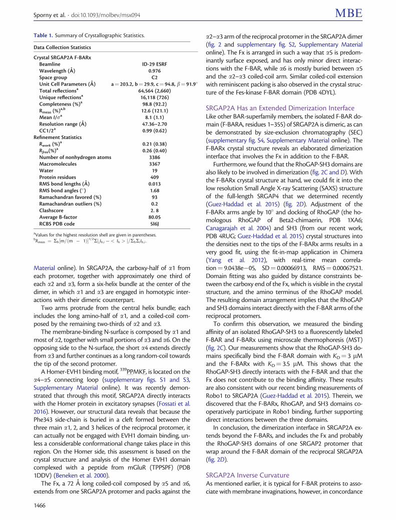
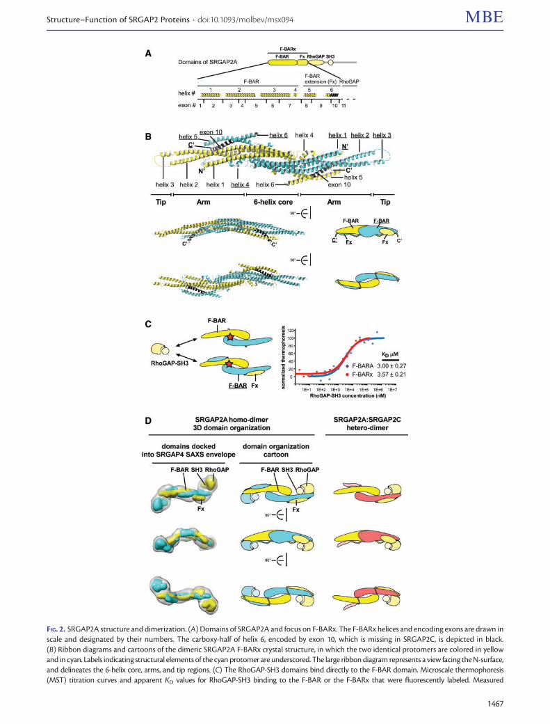
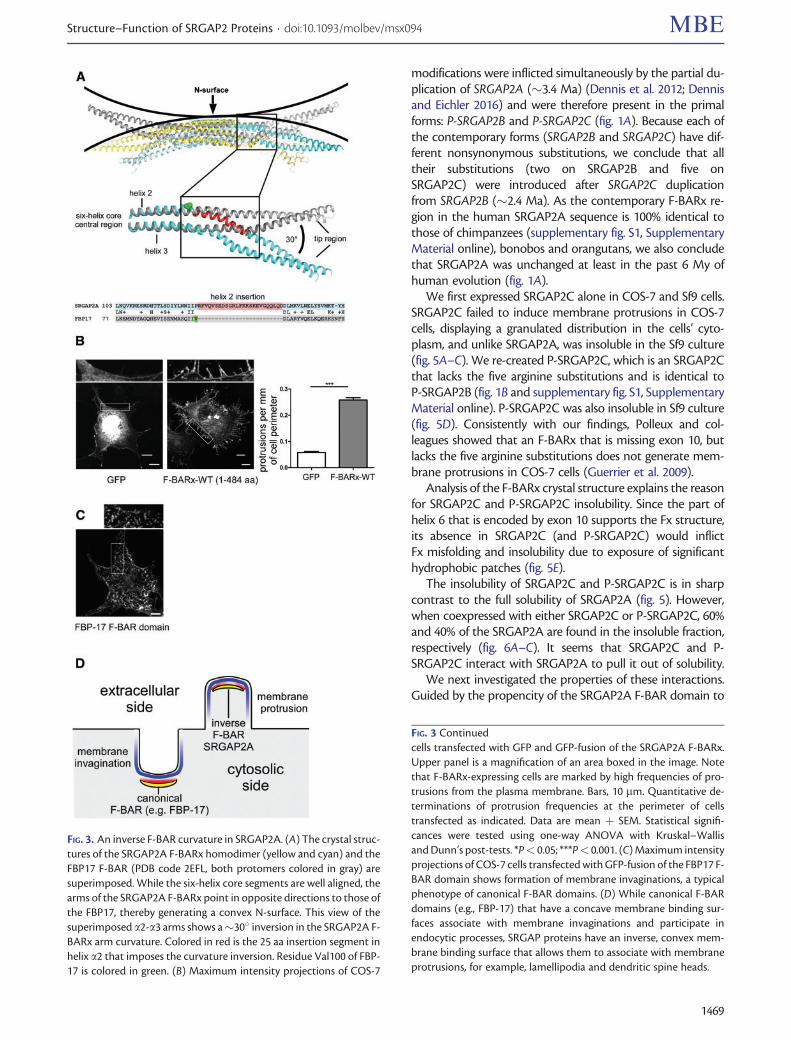
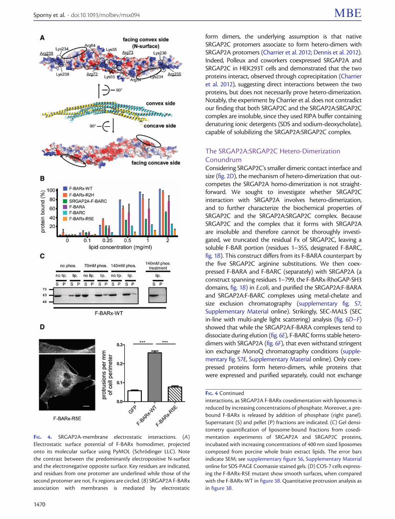
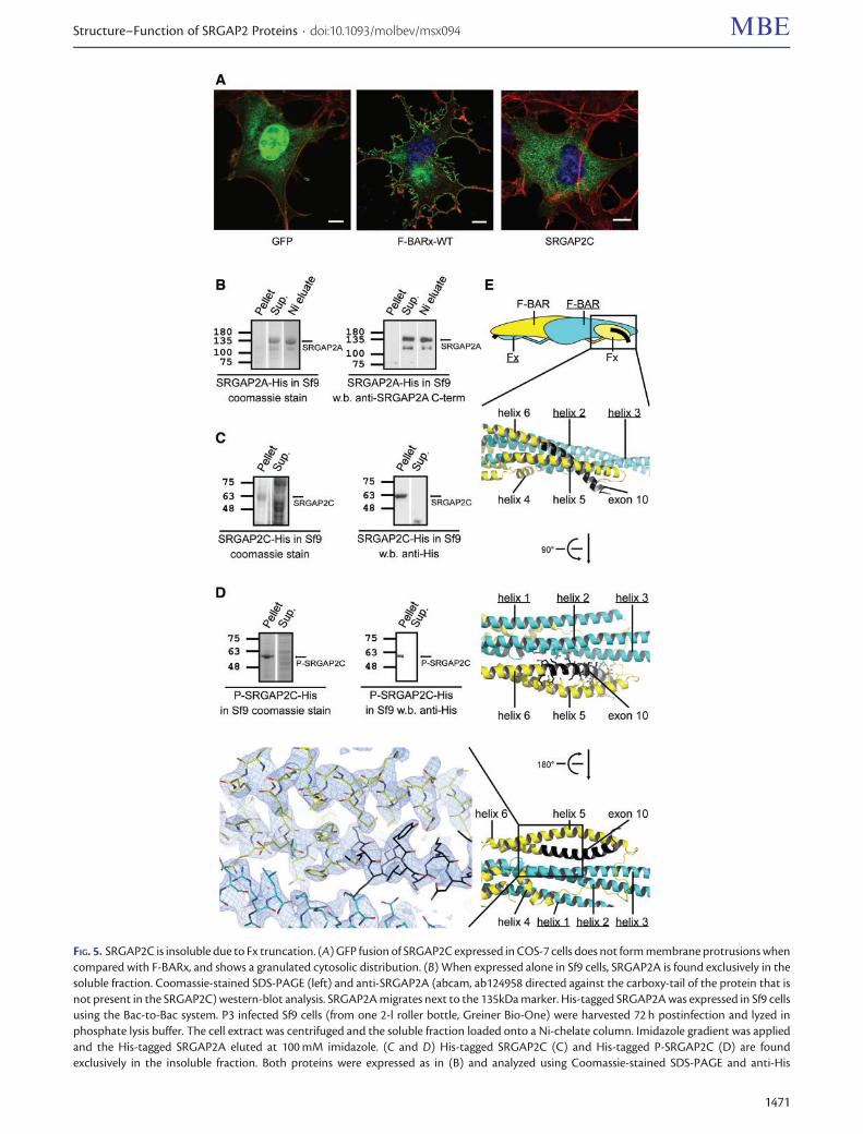
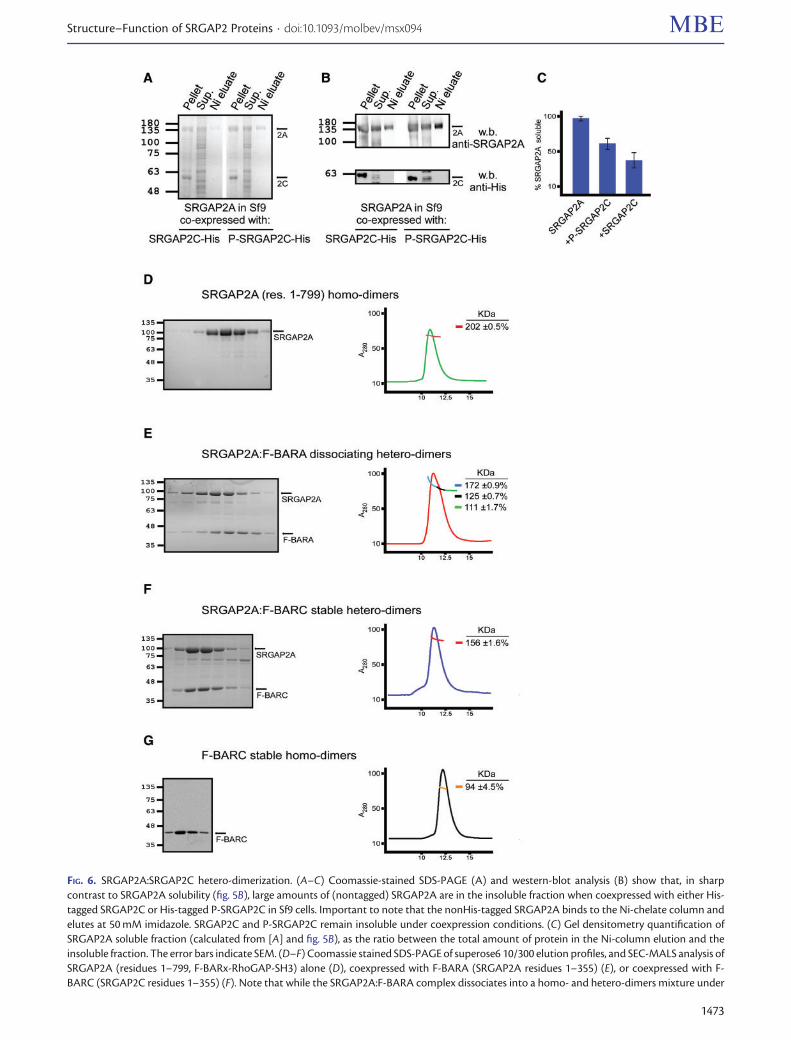