structural investigation of the thermostability … · structural investigation of the...
TRANSCRIPT

1
STRUCTURAL INVESTIGATION OF THE THERMOSTABILITY AND PRODUCT SPECIFICITY OF
AMYLOSUCRASE FROM THE BACTERIUM DEINOCOCCUS GEOTHERMALIS Frédéric Guérin1-,5, Sophie Barbe1-,3, Sandra Pizzut-Serin1-3, Gabrielle Potocki-Véronèse1-3,
David Guieysse1-3, Valérie Guillet4,5, Pierre Monsan1-3,6, Lionel Mourey4,5, Magali Remaud-Siméon1-3, Isabelle André1-3* and Samuel Tranier4,5*
From 1 Université de Toulouse; INSA,UPS,INP; LISBP, 135 Avenue de Rangueil, F-31077 Toulouse, France 2 CNRS, UMR5504, F-31400 Toulouse, France
3 INRA, UMR792 Ingénierie des Systèmes Biologiques et des Procédés, F-31400 Toulouse, France 4 CNRS, IPBS (Institut de Pharmacologie et de Biologie Structurale), Département de Biologie Structurale et
Biophysique, 205 Route de Narbonne, BP 64182, F-31077 Toulouse 5 Université de Toulouse, UPS, IPBS, F-31077 Toulouse; France
6 Institut Universitaire de France, 103 Boulevard Saint-Michel, F-75005 Paris, France
Running title: Structural investigation of Deinococcus geothermalis amylosucrase * To whom correspondence should be addressed: Isabelle André, Laboratoire d'Ingénierie des Systèmes Biologiques et des Procédés – INSA; CNRS UMR5504; UMR INRA 792; 135, Avenue de Rangueil; F-31077 Toulouse cedex 4, France. Tel: +33 561 559 963; Fax: +33 561 559 400; E-mail: [email protected] and Samuel Tranier, Institut de Pharmacologie et de Biologie Structurale, Département Biologie Structurale et Biophysique, 205 Route de Narbonne, BP 64182, F-31077 Toulouse, France; Tel: +33 561 175 438; Fax: E-mail: [email protected] Keywords: amylosucrase, thermostability, crystal structure, molecular dynamics, sucrose
isomerisation, glucosylation
Background: Amylosucrases (AS) hold great potential for glycodiversification. Results: The first 3D-structure of AS from Deinococcus geothermalis solved here revealed an unusual dimer organization. Structures of complex of AS with turanose were also determined. Conclusion: Dimerization may contribute to thermostability. Turanose versus trehalulose formation is controlled by residues from subsite +1. Significance: This study improves the comprehension of AS properties and provides new insight for AS design. Amylosucrases are sucrose-utilizing α-transglucosidases that naturally catalyze the synthesis of α-glucans, linked exclusively through α-1,4 linkages. Side-products and in particular sucrose isomers such as turanose and trehalulose are also produced by these enzymes. Here, we report the first structural and biophysical characterization of the most thermostable amylosucrase identified so far, the amylosucrase from Deinoccocus geothermalis (DgAS). The 3D-structure revealed a homodimeric quaternary organization, never reported before for other amylosucrases. A sequence signature of dimerization was identified from the analysis
of the dimer interface and sequence alignments. By rigidifying DgAS structure, the quaternary organization is likely to participate in the enhanced thermal stability of the protein. Amylosucrase specificity with respect to sucrose isomer formation (turanose or trehalulose) was also investigated. We report the first structures of the amylosucrases from Deinococcus geothermalis and Neisseria polysaccharea in complex with turanose. In the amylosucrase from N. polysaccharea (NpAS), key residues were found to force the fructosyl moiety to bind in an open state with the O3' ideally positioned to explain the preferential formation of turanose by NpAS. Such residues are either not present or not similarly placed in DgAS. As a consequence, DgAS binds the furanoid tautomers of fructose through a weak network of interactions to enable turanose formation. Such topology at subsite +1 is likely favoring other possible fructose binding modes in agreement with the higher amount of trehalulose formed by DgAS. Our findings help to understand the inter-relationships between amylosucrase structure, flexibility, function and stability and provide new insight for amylosucrase design.
http://www.jbc.org/cgi/doi/10.1074/jbc.M111.322917The latest version is at JBC Papers in Press. Published on December 29, 2011 as Manuscript M111.322917
Copyright 2011 by The American Society for Biochemistry and Molecular Biology, Inc.
by guest on September 9, 2018
http://ww
w.jbc.org/
Dow
nloaded from

2
Amylosucrases (AS, E.C. number 2.4.1.4) are glucansucrases belonging to Glycoside-Hydrolase Family 13 of the Carbohydrate-Active EnZymes classification (1-3). The main reaction catalyzed by these enzymes is the formation of an amylose-like glucan with a concomitant release of fructose from sucrose substrate (Fig. 1) (4). Side-reactions including sucrose hydrolysis and sucrose isomer synthesis (i.e. turanose: α-D-Glcp(1→3)-β-D-Fru and trehalulose: α-D-Glcp(1→1)-β-D-Fru) also occur (4,5) although other GH13 enzymes are known to be more specific for these types of reaction (6-8). These molecules are known to be less cariogenic than sucrose and are thus of interest for nutritional applications (9). AS are also able to transfer the glucosyl moieties from sucrose to exogenous acceptors such as maltose, glycogen, maltodextrins, arbutin and others (10,11). AS ability to convert sucrose (an abundant and inexpensive substrate) into valuable derivatives is a great advantage in comparison to Leloir glucosyltransferases which use nucleotide-activated sugars as glucosyl donor substrates (12). Moreover, protein engineering techniques also enabled the creation of novel AS with tailored specificity toward unnatural acceptor molecules (13).
AS that were characterized so far are produced by various species from the genus Neisseria (4), Deinococcus (14,15) and Alteromonas (16). However, Neisseria polysaccharea amylosucrase (NpAS) is the only AS for which several structures, alone or in complex with sucrose substrate or products are available to date (17-20). The three-dimensional structure of NpAS is organized in five domains, namely A, B, C, N and B’. B’ and N domains are only found in AS whereas the three other ones are conserved amongst GH13 enzymes. Although the potential of NpAS for glycodiversification is large, this enzyme suffers from a low catalytic efficiency and a weak thermostability, limiting its industrial development (15). Directed evolution has been attempted to improve NpAS catalytic efficiency and thermostability (21). Searching for more thermostable and efficient enzymes in the natural diversity is another alternative that has motivated the biochemical characterization of the amylosucrases from Deinococcus geothermalis (DgAS) (15), Deinococcus radiodurans (DrAS) (14) and Alteromonas macleodii (AmAS) (15).
With a specific activity of 44 U.mg-1 at the optimal temperature of 50 °C, the recombinant DgAS is the most thermostable AS characterized to date (15). The size distribution of the α-glucan chains produced by DgAS and NpAS differs and the two enzymes do not synthesize equivalent amounts of turanose and trehalulose. Indeed, DgAS produces significantly higher amounts of trehalulose than NpAS (15). Glucosylation of unnatural acceptors such as salicin also revealed that DgAS only produces a monoglucosylated form whereas NpAS is able to produce a di-glucosylated compound (22).
Here, we report the first 3D-structure of DgAS in its apo form that reveals an unusual homodimeric arrangement, thereby allowing identification of determinants possibly controlling thermostability. Furthermore, the structures of both DgAS and NpAS in complex with turanose (a fructose acceptor reaction product) were also determined and their analysis highlighted major differences upon fructose binding which are discussed with regard to the enzyme product specificities. EXPERIMENTAL PROCEDURES
Expression and purification. Expression and purification of recombinant glutathione S-transferase-amylosucrase fusion proteins (GST-DgAS, GST-DrAS and GST-NpAS) were performed as previously described by Emond et al. (15), Pizzut et al. (14) and Potocki de Montalk et al. (23), respectively. The GST-AS proteins were purified by affinity chromatography using Glutathion-Sepharose-4B support (GE Healthcare, Little Chalfont, UK). The GST tag was then removed using Prescission protease (GE Healthcare, Little Chalfont, UK) which left 5 residues (GPLGS) of the cutting site at the N-ter extremity.
Thermostability of amylosucrases. The melting point (Tm) of amylosucrases was assayed by differential scanning fluorimetry (DSF). A mix of enzyme (2 µM), Sypro-orange (5 X) (Invitrogen, Paisley, UK) and 50 mM Tris, pH 7.0, 150 mM NaCl, 1 mM DTT, 1 mM EDTA were incubated using a temperature gradient from 20 to 80 °C with a 0.3 °C increment. The thermal transition was monitored using a RTQ-PCR CFX96 Real-Time System (Biorad, Marnes-la-Coquette, France). Tm was given by the inflexion point of the curve RFU=f(T), with RFU standing for Relative Fluorescence Unit. Circular dichroïsm spectra were recorded on a
by guest on September 9, 2018
http://ww
w.jbc.org/
Dow
nloaded from

3
JASCO J815 spectropolarimeter equipped with a Peltier cell temperature controller. Tm values were obtained by heating the sample (enzyme 2 µM in 50 mM Tris, pH 7.0, 150 mM NaCl, 70 µM DTT, 70 µM EDTA) at 1 °C/min and recording the ellipticity value at 220 nm from 25 °C to 80 °C with delays of 30 seconds. The SigmaPlot 10.0 software was used for all graphic analyses, Tm determination and statistics. The half-life (t1/2) at 50 °C of DgAS and NpAS preparations were also determined by incubating AS pure enzymes (300 mg.L-1) in 50 mM Tris, pH 7.0, 150 mM NaCl, 1 mM DTT, 1 mM EDTA at 50 °C. At various intervals, aliquots were taken and the enzyme activity was determined as previously described (22).
Size exclusion chromatography multiangle laser light scattering (SEC-MALLS) experiments. NpAS, DgAS and DrAS protein samples buffered in 50 mM Tris pH 7.5, 150 mM NaCl were analyzed on a Shodex KW-803 column (Showa Denko Europe GmbH, Munich, Germany) with multiangle laser light scattering (MALLS). The column was equilibrated in a 0.1 µm filtered 50 mM Tris pH 7.5, 150 mM NaCl, 0.02 % (w/v) NaN3 buffer on a Agilent 1260 Infinity LC chromatographic system (Agilent Technology, Massy, France). Data were collected using a DAWN HELEOS-II 18-angle and Optilab T-rEX refractive index detector (Wyatt technology Corp., Toulouse France). Sample concentrations were 1.55 g.L-1, 2.1 g.L-1 and 1.85 g.L-1 for NpAS, DgAS and DrAS solutions respectively. Protein samples were prepared in the Tris buffer used as the mobile phase to equilibrate the column. 20 µl of each protein sample was loaded on the column and the separation was performed at a flow rate of 0.5 mL.min-1 at 22 °C. Results were analyzed using the ASTRA V software (Wyatt technology Corp., Toulouse France).
Native-PAGE. Native-PAGE was performed with a two-phase gel composed of a 4 % stacking gel (4 % acrylamide, 125 mM Tris, pH 6.8, 0.1 % APS, 0.15 % TEMES) and a 10 % separating gel (10 % acrylamide, 375 mM Tris, pH 8.8, 0.1 % APS, 0.15 % TEMED). 15 µL of the protein samples (2 g.L-1) mixed with 2 X loading buffer (40 mM Tris, pH 6.8, 50 % glycerol (v/v), 0.04 % bromophenol blue (w/v)) were loaded onto the gel. Electrophoresis was performed at room temperature using a mini-PROTEAN system (BioRad, Marnes-la-Coquette, France) at a voltage of 100 V with an electrophoresis buffer composed of 15 g.L-1 Tris,
pH 8.3 and 72 g.L-1 glycine. The gels were stained with Coomassie Brilliant Blue. Molecular weight estimation was performed using NativeMark native protein markers (Invitrogen, Paisley, UK) as standards.
Crystallization. NpAS was crystallized using conditions previously described by Skov et al. (17,18). DgAS crystallization experiments were carried out at 12 °C using the hanging drop vapour diffusion method. Best crystals were obtained with a 1:1 (v/v) ratio of protein (6 mg.mL-1 in 20 mM Tris, pH 8.0) to precipitant solution (1.5 M sodium Acetate, 0.1 M sodium cacodylate, pH 7.0). Lens shape crystals appeared after two weeks and grew to a maximal size of 140×60×40 µm3.
Soaking experiments. Crystals of NpAS were soaked for 20 min in the reservoir solution supplemented by 250 mM turanose (Sigma, Saint-Martin-d'Hères, France). DgAS crystals quickly fractured in the presence of such a high concentration of turanose, so they were soaked for few seconds in the reservoir solution supplemented with only 14 mM of turanose.
Data collection and structure determination. X-ray experiments were carried out at 100 K. Prior to flash cooling, native crystals of DgAS were soaked for few seconds in the reservoir solution supplemented with 20 % (v/v) glycerol to avoid ice formation. Conversely, due to the cryoprotection effect of turanose, crystals of AS-turanose complexes were intrinsically cryoprotected. Native DgAS and NpAS-turanose diffraction datasets were collected to a maximum resolution of 1.97 Å and 1.85 Å, respectively on beamline ID14-1 at the European Synchrotron Radiation Facility (ESRF, Grenoble, France). DgAS-turanose complex dataset was collected to 2.10 Å on the ESRF beamline ID29. Diffracted intensities were integrated using iMOSFLM (24) and scaled with SCALA (25) from the CCP4 software suite (26,27) and 5 % of the scaled amplitudes were randomly selected and excluded from the refinement procedure. Crystals of DgAS in apo form and in complex with turanose belong to the C2221 spacegroup with 1 molecule per asymmetric unit giving a Matthews coefficient of 2.3 Å3/Da. Crystal of NpAS-turanose belongs to the P21212 spacegroup with 1 molecule per asymmetric unit and a Matthews coefficient of 2.4 Å3/Da. Data collection statistics are given in Table 1. The native structure of DgAS was solved by the molecular replacement method using PHASER (28) and the structure of NpAS
by guest on September 9, 2018
http://ww
w.jbc.org/
Dow
nloaded from

4
(PDB code: 1G5A) (18) as a search model. The translation function Z-score was 16.5 and R and Rfree of the refined molecular replacement solution were 0.39 and 0.47, respectively. Structures of the complexes NpAS-turanose or DgAS-turanose were straight refined from their native structures using refmac5 (29).
Building and refinement. Structure refinement was performed with refmac5 from the CCP4 GUI (29) and models were manually reconstructed in SigmaA weighted electron density maps using COOT (30). Water molecules were automatically assigned and ligand molecules were manually fitted in residual maps. Final DgAS structures in apo form and in complex with turanose contain 651 residues out of the 655 theoretical residues with four missing residues at the C-terminal extremity. The final model of NpAS in complex with turanose contains 628 residues out of the 632 theoretical residues with four missing residues at the N-ter extremity. Refinement statistics are given in Table 1.
Coordinates. Coordinates have been deposited at the protein data bank (PDB codes: 3UCQ, 3UER and 3UEQ for DgAS, DgAS-turanose complex and NpAS-turanose complex, respectively).
In vitro synthesis of sucrose isomers. Synthesis of sucrose isomers (turanose and trehalulose) was performed with 100 mM sucrose in the presence of 0 or 100 mM fructose at 30 °C or at enzyme optimum temperature (50 °C for DgAS and 37 °C for NpAs). At the end of the reaction (24 h), samples were centrifuged at 12,000 rpm for 10 min. Concentration of sucrose isomers was determined by HPAEC-PAD using an analytical CarboPACTM PA100 (4x250 mm) column with a CarboPACTM PA-100 Guard (4×50 mm). Detection was performed using a Dionex ED40 module with a gold working electrode and an Ag/AgCl pH reference electrode (Dionex, Sonnyvale, CA, USA).
Molecular Dynamics (MD) simulations. All MD simulations were carried out using the AMBER 9 suite of programs and the all-atom ff03 force field (31,32). The starting models were derived from the high resolution crystal structures of DgAS and NpAS (PDB code: 1G5A) (18). Fifteen and twenty Na+ cations were added to neutralize the DgAS and the NpAS monomers, respectively. Each protein together with its counter-ions was embedded in a rectangular parallelepipedal solvent box that left
a space of 0.12 nm around the solute. TIP3P water molecules (approximately 28,000) were added using the LEaP module integrated in the AMBER9 package (33). Simulation preparation consisted in different phases of minimization, heating, equilibration under different type of restraints. The simulation was carried out at constant temperature (303 K) and pressure (1 bar) conditions over 60 ns. The temperature and pressure were s controlled using Langevin thermostat (34) and Berendsen barostat (35) with a collision frequency (2 ps-1) and pressure relaxation time (2 ps). Long-range electrostatic forces were handled by using the particle-mesh Ewald method (36). The time step of the simulations was 2.0 fs and the SHAKE algorithm was used to constrain the lengths of all chemical bonds involving hydrogen atoms to their equilibrium values (37). To avoid artefacts, MD simulations were run three times with different starting velocity distribution. The resulting trajectories were analyzed using the Ptraj module of the AMBER9 package. The RMSD was calculated for the protein backbone atoms using least squares fitting. Distances between protein loops were calculated with respect to their center of mass. Atomic positional fluctuations (∆ri
2) of protein backbone were calculated using the coordinates of the 60 ns trajectories. A mass-weighted average value was then calculated for each residue. These parameters are related to the B-factors through the following relationship: Bi = 8π2 <∆ri
2> 3
Sequence and structure alignments. Multiple sequence alignment was performed using Clustal W2 (38) and represented using ESPript (39). Structural alignments were generated using SwissPDB-viewer (40) and subsequently served as a basis to align sequences of homologous proteins of unknown 3D-structures. Multiple sequence alignment was then manually corrected. The root mean-square deviations (RMSDs) among three-dimensional structures were calculated using Superpose (41) from the CCP4 suite of programs (26,27). RESULTS AND DISCUSSION Overall structure and dynamics
Tertiary structure of DgAS. The three-dimensional structure of DgAS is organized into
by guest on September 9, 2018
http://ww
w.jbc.org/
Dow
nloaded from

5
the five distinct domains already described for NpAS (18) (Fig. 2): the all helical N-terminal domain (residues 1 to 83), the central catalytic domain A (residues 84-177, 259-398 and 468-565), two regions protruding from domain A, constituting respectively domains B (178-258) and B’ (399-467), and finally the β-stranded C-terminal domain (566-646) adopting a Greek key motif. DgAS and NpAS share 38 % sequence identity. Amongst the five regions, domains A, B and B’ are the most conserved with a sequence identity of 43 %, 58 % and 52 %, respectively. Domains N and C only share 21 % and 19 % sequence identity, respectively. The overall RMSD value obtained after secondary structure superimposition of DgAS with NpAS was 1.3 Å. Lower RMSD values, i.e. 1.0 Å, 1.1 Å and 0.9 Å, were observed for domains A, B and B’, respectively. More significant structural differences occurred between the less conserved domains N and C leading to larger RMSD values of 1.9 Å and 1.5 Å, respectively.
The catalytic domain A of DgAS adopts the typical (β/α)8-barrel of GH family 13 (2). The active site is highly conserved with respect to that of NpAS (Fig. 3A). Residues E326 and D284 of DgAS were identified as the general acid/base and the nucleophile, respectively, involved in the formation of the β-glucosyl intermediate occurring in the α-retaining mechanism (corresponding to E328 and D286 in NpAS (42)). Three additional important residues conserved in GH13 family and known to assist in catalysis are D396, H180 and H395 (corresponding to D393, H187 and H392 in NpAS, respectively). A salt bridge formed by residues R520 and D137 (equivalent to R509 and D144 in NpAS) blocks the bottom of the catalytic pocket. All the residues defining the catalytic site are conserved between NpAS and DgAS with the exception of R226, which is substituted by P219 in DgAS. As a result, the active site pocket of DgAS shows an enlarged aperture around subsite +2 compared to NpAS that may facilitate ligand accessibility and binding into the active site (Fig. 3B). Two ligands were found in the active site (Fig. 3A), a glycerol molecule (used as cryoprotectant for X-ray experiments) at subsite -1 and a Tris molecule at subsite +1, which are interacting together through a polar contact involving Glycerol-O2 and Tris-O3. Both B and B’ domains are found longer in DgAS than in
NpAS. Domain B is composed of 81 amino acid residues in DgAS (vs 76 residues in NpAS), organized in two antiparallel β-strands and two α-helices. As for domain B’, it is composed of four α-helices comprising 69 amino acid residues (vs 65 in NpAS). The domain C of DgAS is composed of 82 amino acid residues folded as an eight-stranded β-sandwich as previously observed in the NpAS structure. The domains C of both enzymes differ in the short α-helix between strands β4 and β5 observed only in DgAS. Additionally, the loop interconnecting strands β2 and β3 is three residues longer in DgAS than in NpAS. The main difference between the DgAS and NpAS helical domains N resides in the N-terminal extremity which is composed by a long α-helix in DgAS and two small α-helices in NpAS (Fig. 3C). The remaining α-helices of the N-terminal domain (H3, H4 and H5) are structurally conserved in DgAS and NpAS.
Insight into enzyme plasticity through Molecular Dynamics (MD) simulations. To complete our structural comparison of amylosucrases, we undertook the investigation of their dynamic properties. Large scale MD simulations (60 ns) were thus carried out in explicit water for DgAS and NpAS. As a check for the stability of protein structures during the course of MD simulations, the time evolution of the backbone atoms RMSD was calculated after least square fitting (Supplementary Fig. S1A). Variations of backbone RMSD indicated a distinct conformational behaviour of the two enzymes. The RMSD of DgAS rapidly increased by 3 Å within the first 12 ns of the simulation and then stabilized for the rest of the simulation. As for NpAS, the RMSD slowly increased by 2.2 Å over the first 30 ns before reaching a plateau (Supplementary Fig. S1A). Thus, faster (12 ns vs 30 ns) and larger-amplitude (3 Å vs 2.2 Å) changes were observed for DgAS compared to NpAS. MD simulations further revealed that RMSD deviation of DgAS mainly resulted from a high mobility of the N-terminal part of the protein (residues 1-83). As shown in Supplementary Fig. S1B, the RMSD of the domain N increased by as much as 5.2 Å over the first 12 ns of the simulation for DgAS whereas it remained unchanged for NpAS. The high flexibility of the N-extremity was also shown by the large calculated B-factor values (Supplementary Fig. S1C). Such large
by guest on September 9, 2018
http://ww
w.jbc.org/
Dow
nloaded from

6
conformational fluctuations of DgAS are due to the rearrangement of the first long helix (residues 1-24) into two shorter helices (composed of residues [1-14] and [15-24], respectively). Such conformational changes are not observed for NpAS as its first α-helix is already composed of two short helices in the X-ray structure. These helices are strongly stabilized through a large network of ionic interactions involving charged residues (R, K, D, E) from the three first α-helices belonging to the N-terminal region of NpAS (Fig. 3C). Noteworthy, there are no equivalent charged residues in DgAS. Even if other structural regions also appear flexible, as indicated by the calculated B-factors, differences between both enzymes are, however, less significant. Most flexible regions correspond to loops of the (β/α)8-barrel that confer a pocket topology to the active site. In particular, loops 7 and 3 (encompassed in domains B’ and B, respectively) moved away from each other during the MD simulations (Supplementary Fig. S1D) resulting in the opening of the catalytic pocket. With the exception of the N domain in DgAS and surface loops (2, 3, 4, 7 and 8) in both enzymes, remaining parts of DgAs and NpAS did not exhibit any significant fluctuation (Supplementary Fig. S1E and S1F). Oligomerization and thermostability
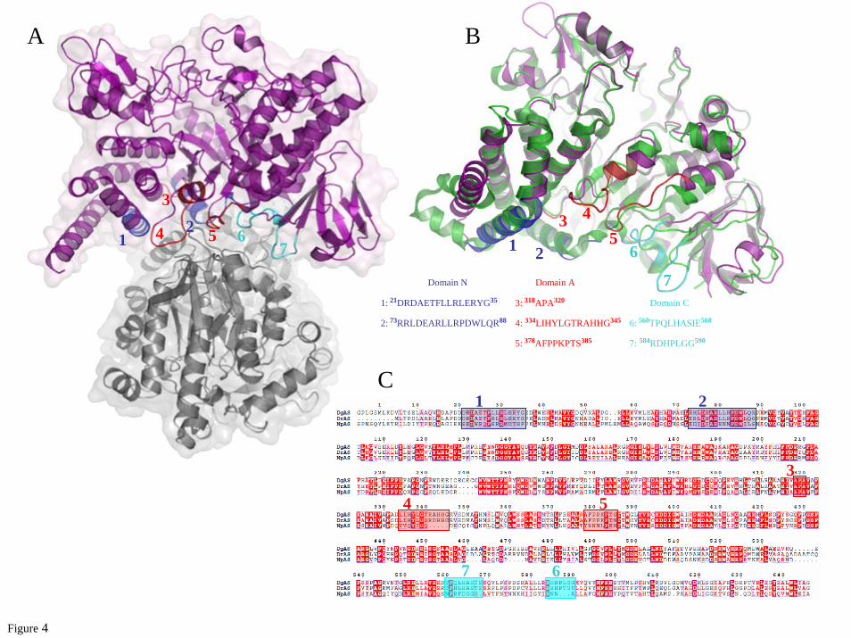
The first dimeric structure of an amylosucrase. While the asymmetric unit of DgAS is composed of a single peptide chain, exploration of symmetry related molecular interfaces using the PISA server (43) indicated a stable dimeric organization with a buried interface of 2,950 A² out of a total accessible surface area of 45,330 A² (Fig. 4A). The buried surface represents 6.1 % of the solvent accessible area. The interface is defined by a set of residues located in the catalytic domain (A318-A320; L334-H344; A378-S385) and also in domains N (D21-G35; R73-R88) and C (T560-E568; R584-G590) (Fig. 4B). Stabilization between the protomer units of DgAS benefits from strong salt bridge interactions formed between R74 and E25 and between R341 and D84, and from a network of direct and water-mediated hydrogen-bonding interactions (Supplementary Fig. S2). The residues involved in the interface stabilization of DgAS are not conserved in NpAS. In particular,
R341 which appears to play a key role in stabilizing the DgAS interface, is one of the five additional residues of the α5-β6 interconnecting loop from the (β/α)8-barrel, which is found longer compared to NpAS (Fig. 2C and 4B). Of note, such insertion region in DgAS is also found to interact through polar interactions with loop R584-M591 from the domain C of the second protomer, which is four residues shorter in NpAS (Fig. 4C).
The dimerization interface displays a strong shape complementarity between the protomers. First, a bundle of hydrophobic residues from the N domain involving L80 and L81 from α-helix H5 of the first protomer fits nicely into the groove formed by the two α-helices, H3 and H5, from the second protomer (Supplementary Fig. S2). Residues L80, L81 and L29, belonging to these α-helices and only present in DgAS, are involved in these hydrophobic contacts (Supplementary Fig. S2A). In addition, the extremity of the loop R584-G590 (insertion region situated between strands β3 and β4 of the C-terminal domain in DgAS as described earlier) is found interlocked between two loops, 339-344 (another insertion region in DgAS) and 379-385 (highly conserved in the Deinococcus genus). Interestingly, in such contacting amino acid residues, we observe a bunch of prolines, which are generally known to rigidify the polypeptide backbone and that may constrain the polypeptide chain conformation to facilitate the dimer formation. P587 from the first protomer faces up P380, P381 and P383 from the second one. Although oligomeric states have been previously observed for other members of the GH13 family, such as cyclomaltodextrinases (CDase; EC 3.2.1.54, PDB code: 1H3G (44)), maltogenic amylase (MAase; EC 3.2.1.133, PDB code: 1SMA (45)), and neopullulanase (NPase, EC, 3.2.1.135, PDB code: 1J0H (46)), the structure of DgAS is, to our knowledge, the first homodimeric amylosucrase reported to date. Comparison of electrophoresis under denaturating (SDS-PAGE) and native conditions argue for a dimeric assembly of DgAS in solution (Fig. 5A and 5B). The dimeric conformation of DgAS in solution was confirmed by SEC-MALLS/RI experiments. As shown in Fig 5C, the elution volume of DgAS differed significantly from that of NpAS, corresponding to a molecule of molecular weight of 151.7 kDa compatible with a dimeric assembly of DgAS (theoretical molecular weight
by guest on September 9, 2018
http://ww
w.jbc.org/
Dow
nloaded from

7
of 146.3 kDa). As expected, the NpAS mainly eluted as a monomer with a molecular weight of 74.5 kDa (theoretical molecular weight of 71.5 kDa). The monomeric NpAS and the dimeric DgAS remained stable over few weeks at 4 °C. To search for a sequence signature of dimeric forms in other amylosucrases, we performed a sequence alignment of DgAS, NpAS and DrAS (Fig. 4C). Among the 7 regions found to be involved in DgAS dimerization from structural analysis (listed in Fig. 4B), regions 4, 6 and 7 are well conserved in DrAS. Notably, the corresponding loops in NpAS are shorter and could prevent dimer formation. To get some evidence of DrAS dimerization, SEC-MALLS analysis of DrAS was performed and it confirmed the homodimeric organization of this enzyme with a molecular weight of 136.0 kDa (theoretical molecular weight of 143.3 kDa) (Fig. 5C). The pattern composed of regions 1 to 7 could thus be proposed as a signature of dimerization. We screened the identified pattern against sequences of GH13 enzymes (sequence alignment is provided in Supplementary Fig. S3). The proposed dimerization patern was found in Deinococcus desertii DdAS, Deinococcus maripensis DmAS, α-amylases from Meiothermus rubber, Meiothermus silvanus and Truepera radiovictrix sequences, suggesting a similar dimeric organization to DgAS. On the other hand, sequences of amylosucrases from Neisseria and Alteromonas groups did not contain this molecular pattern. Like NpAS, one could infer that they might adopt a monomeric form (23).
DgAS thermostability. The optimum temperature of DgAS is 50 °C whereas an optimal temperature of 37 °C was found for NpAS (15). The t1/2 of freshly purified DgAS and NpAS was 69 h and 15 min at 50 °C, respectively. The higher stability of DgAS was confirmed by Tm measurements which gave 58.9 +/- 0.2 °C for DgAS versus 49.6 +/- 0.4 °C for NpAS when measured by DSF and 62.0 +/- 0.2 °C versus 51.3 +/- 0.2 °C when determined by circular dichroïsm. Close inspection of the primary sequences of DgAS and NpAS gave rise to the following observation: DgAS contains i) a higher proportion of charged residues (K, R, H, D, E), which represent 28.8 % of the total number of residues compared to only 22.9 % in NpAS, ii) a lower number of polar uncharged residues (G, S, T, N, Q, Y, C) which represent 24.3 % of the total number of residues for DgAS
and 33.4 % for NpAS, iii) more hydrophobic residues (L, M, I, V, W, P, A, F) representing 46.9 % of the total number of residues compared to 43.7 % for NpAS and iv) six additional proline residues. All aforementioned features are usually observed in proteins from thermophilic organisms and could contribute to the higher thermostability of DgAS (47,48). In addition, we have shown using MD simulations on DgAS and NpAS monomers that the N-terminal extremity of DgAS exhibits a higher flexibility. DgAS dimerization might then constrain the movement of helix H1-H2 at the N-terminus of the protein and one could thus assume that dimerization is likely to contribute significantly to enzyme stabilization of this enzyme. Specificity toward sucrose isomer formation
Previous characterization of DgAS and NpAS revealed that both enzymes catalyze turanose and trehalulose synthesis as side-products when incubated with sucrose but the ratio of turanose versus trehalulose production was radically different for the two enzymes (15). Other enzymes catalyzing exclusively sucrose isomerization reactions were previously characterized (7). Their primary structures contain a highly conserved motif named “charged isomerization motif” composed of residues RLDRD which are involved in the product specificity (49). Additionally, these enzymes contain two conserved phenylalanine forming the so-called “aromatic clamp” motif, which is involved in the tautomerization of fructose occuring during the isomerization reaction (8). Both sites are neither conserved in primary nor in tertiary structures of amylosucrases what might help to explain differences observed between product profiles of amylosucrases and sucrose isomerases. To further investigate the specificity for sucrose isomerization of DgAS and NpAS, experiments were carried out in the presence of 100 mM sucrose and of exogenous fructose acceptor at either 30 °C or at the optimal temperature of each enzyme, i.e. 50 °C and 37 °C for DgAS and NpAS, respectively. The results reported in Table 2 show that, for all operational conditions, NpAS preferentially synthesized turanose. In contrast, equivalent amounts of turanose and trehalulose were produced by DgAS. Increasing initial fructose concentration enabled sucrose isomer production yield to be enhanced. Acceptor reaction onto fructose was forced at the
by guest on September 9, 2018
http://ww
w.jbc.org/
Dow
nloaded from

8
detriment of α-glucan chain formation. At 30 °C, 69 % and 64 % of the glucosyl units coming from sucrose were transferred onto D-Fructose by DgAS and NpAS, respectively. However, the ratio of turanose versus trehalulose remained unchanged compared to that obtained in the absence of exogenous fructose acceptor. Similarly, increasing the temperature had no significant effect on sucrose isomer ratio. To go further in the understanding of this feature, the crystal structures of DgAS and NpAS in complex with turanose were determined. These are the first complexes of amylosucrases with sucrose isomers and electron density maps showed in both cases density which corresponds to turanose.
The NpAS-turanose complex was first compared to the structure of inactive E328Q NpAS mutant in complex with sucrose (PDB code: 1JGI) and no difference exceeding the structure coordinate errors was observed between the two structures (19). The structure of the NpAS-turanose complex revealed two turanose-binding sites, which were named TB1 and TB2 for the site located in the catalytic pocket and at the surface of the C domain, respectively. The electron density corresponding to the turanose bound at subsite -1 and +1 of the catalytic pocket is shown in Figure 6A. The glucosyl moiety occupies the catalytic subsite -1 in a 4C1 chair conformation. Its pyranosyl ring can be nicely superimposed onto the sucrose glucosyl ring from X-ray structure (PDB code: 1JGI) and is maintained by the same dense network of hydrogen bonding interactions involving residues D144, H187, E328, R284, D286, H392, D393, R509 (Supplementary Table S1). Glucosyl binding is also reinforced by a stacking interaction with Y147 and hydrogen bonds with R513 and D144 mediated by a water molecule (Supplementary Fig. S4). The most striking observation arose from the fructosyl moiety binding conformation. Indeed, at subsite +1, the electron density of the ligand unequivocally revealed that the fructose unit of turanose is bound in the open state. This is quite surprising, considering that in water solution, turanose adopts an equilibria with a predominant proportion of β pyranoid (47 %) and β furanoid (37 %) tautomers after 20 min at 20 °C as determined by 1H NMR spectroscopy (50). The hydroxyl groups O4', O5' and O6' of the open state fructose form direct hydrogen bonds with residues A287, E328 and I330, respectively. Noteworthy, a dense network of additional
hydrogen bonds mediated by water molecules is also observed around fructose (Table 3). Within this network, the water molecule Wat1134 mediates two hydrogen bonds between R226 and both O1' and O6'. In addition O6' is also within hydrogen bond distance of the main chain NH of I330 and water molecule Wat1366. These interactions involving O1' and O6' of the fructose unit probably favor fructose binding in the open state by preventing hemi-acetal formation. Such a binding mode allows a perfect positioning of the O3' for the nucleophilic attack of the β-glucosyl intermediate and the formation of turanose. It is likely responsible for the predominant turanose formation by NpAS. In the turanose molecule found in TB2, the fructose ring adopts a β-D-furanosyl conformation (Supplementary Fig. S5). In DgAS, only one turanose-binding site, equivalent to TB1 of NpAS, was observed (Fig. 6B). The absence of TB2 is likely due to the fact that residues corresponding to F559, N560 and N562 from domain C of NpAS are substituted by three hydrophobic residues: L572, P573 and P575, respectively. Such substitutions prevent stacking and hydrogen bonding interactions with turanose. In the TB1 site of DgAS, the glucosyl moiety of turanose occupies subsite -1 and adopts a 4C1 configuration with a very clear electron density. Interactions between the glucopyranosyl ring and the protein are similar to those described for the NpAS-turanose complex (Supplementary Table S1). The electron density of the fructosyl ring is less clear at subsite +1. However, it defines an envelope that fits best with the α-anomer of fructofuranose (Fig. 6B). The β-anomer also probably bound subsite +1 but to a lesser extent. In addition, we cannot exclude the presence of fructose either in open or pyranoid forms. The O3' glucosylated-α−D–fructofuranose is reported to be one of the less prevalent tautomer of turanose in water solution (1 % at 20 °C (50)). Accommodation of this conformation in subsite +1 involves H-bonds with the catalytic residues E326 and D396 and two water-mediated interactions with O1', O4' and O6'. DgAS binds the furanoid tautomers of fructose to form turanose but via a considerably weaker network of interactions than that observed in NpAS. Further comparisons of DgAS and NpAS subsite +1 revealed structural traits that prevent open state conformation of fructose to bind DgAS subsite +1 (Fig. 6C). Indeed, residue R226 of
by guest on September 9, 2018
http://ww
w.jbc.org/
Dow
nloaded from

9
NpAS, which interacts via a water molecule with both O1' and O6' of the open form of fructose, is replaced by P219 in DgAS. Furthermore, residue I328 corresponding to I330 in NpAS, which was directly involved in H-bond interaction with O6' of fructose in open state, cannot play the same role in DgAS due to a slight motion of the loop bearing this residue. The discrimination between the various tautomers of turanose is less drastic in DgAS than in NpAS. DgAS subsite +1 accommodates various fructose tautomers in order to optimally arrange O1' or O3' of fructose unit for nucleophilic attack of the glucosyl enzyme intermediate therefore yielding equivalent amounts of trehalulose and turanose. DgAS- and NpAS-turanose complex analysis thus enabled us to explain differences observed in DgAS and NpAS product specificity. This work opens new perspectives for the rational and/or semi-rational redesign of this thermostable amylosucrase in order to modulate sucrose isomer synthesis with the view of reducing side-product formation or controlling
sucrose isomer profile for the development of novel syrups enriched in sucrose substitutes. Acknowledgments Frédéric Guérin is supported by a PhD grant from the Pôles de Recherche et d'Enseignement Supérieur de l'Université de Toulouse and the Région Midi-Pyrénées, France. We thank the Computing Center of Region Midi-Pyrénées (CALMIP, Toulouse, France) and the Center for Computing Resources (CRI) of INSA-Toulouse for providing computing resources and support. We are grateful to the staff of synchrotron beamlines ID14-1 and ID29 at European Synchrotron Radiation Facility (Grenoble, France) for providing assistance in using the beamlines. Authors are grateful to Javier Perez (Synchrotron SOLEIL) for the fruitful discussions. We also thank Stéphanie Terme (Wyatt technology France) for her helpful assistance in the SEC-MALLS experiments.
by guest on September 9, 2018
http://ww
w.jbc.org/
Dow
nloaded from

10
REFERENCES 1. Henrissat, B. (1991) Biochem J 280, 309-316 2. Henrissat, B., and Davies, G. (1997) Curr Opin Struct Biol 7(5), 637-644 3. Cantarel, B. L., Coutinho, P. M., Rancurel, C., Bernard, T., Lombard, V., and
Henrissat, B. (2009) Nucleic Acids Res 37, D233-238 4. Potocki de Montalk, G., Remaud-Simeon, M., Willemot, R. M., Sarcabal, P.,
Planchot, V., and Monsan, P. (2000) FEBS Lett 471(2-3), 219-223 5. Okada, G., and Hehre, E. J. (1974) J Biol Chem 249(1), 126-135 6. Goulter, K. C., Hashimi, S. M., and Birch, R. G. Enzyme Microb Technol 50(1), 57-64 7. Ravaud, S., Robert, X., Watzlawick, H., Haser, R., Mattes, R., and Aghajari, N. (2007)
J Biol Chem 282(38), 28126-28136 8. Ravaud, S., Robert, X., Watzlawick, H., Haser, R., Mattes, R., and Aghajari, N. (2009)
FEBS Lett 583(12), 1964-1968 9. Thompson, J., and Pikis, A. (2011) Mol Oral Microbiol 26, 1-11 10. Park, C. S., Seo, D. H., Jung, J. H., Ha, S. J., Song, M. C., Cha, J., Yoo, S. H., Kim, T.
J., and Baek, N. I. (2009) J Mol Catal B 60(3-4), 113-118 11. André, I., Potocki-Veronèse, G., Morel, S., Monsan, P., and Remaud-Siméon, M.
(2010) Top Curr Chem 294, 25-48 12. Chang, A., Singh, S., Phillips, A. N., and Thorson, J. S. (2011) Curr Opin Biotechnol
22, 800-808 13. Champion, E., André, I., Moulis, C., Boutet, J., Descroix, K., Morel, S., Monsan, P.,
Mulard, L. A., and Remaud-Siméon, M. (2009) J Am Chem Soc 131(21), 7379-7389 14. Pizzut-Serin, S., Potocki-Veronese, G., van der Veen, B. A., Albenne, C., Monsan, P.,
and Remaud-Simeon, M. (2005) FEBS Lett 579(6), 1405-1410 15. Emond, S., Mondeil, S., Jaziri, K., André, I., Monsan, P., Remaud-Siméon, M., and
Potocki-Veronèse, G. (2008) FEMS Microbiol Lett 285(1), 25-32 16. Ha, S. J., Seo, D. H., Jung, J. H., Cha, J., Kim, T. J., Kim, Y. W., and Park, C. S.
(2009) Biosci Biotechnol Biochem 73(7), 1505-1512 17. Skov, L. K., Mirza, O., Henriksen, A., Potocki de Montalk, G., Remaud-Siméon, M.,
Sarçabal, P., Willemot, R. M., Monsan, P., and Gajhede, M. (2000) Acta Crystallogr D 56, 203-205
18. Skov, L. K., Mirza, O., Henriksen, A., De Montalk, G. P., Remaud-Siméon, M., Sarçabal, P., Willemot, R. M., Monsan, P., and Gajhede, M. (2001) J Biol Chem 276(27), 25273-25278
19. Skov, L. K., Mirza, O., Sprogoe, D., Dar, I., Remaud-Siméon, M., Albenne, C., Monsan, P., and Gajhede, M. (2002) J Biol Chem 277(49), 47741-47747
20. Jensen, M. H., Mirza, O., Albenne, C., Remaud-Simeon, M., Monsan, P., Gajhede, M., and Skov, L. K. (2004) Biochemistry 43(11), 3104-3110
21. Emond, S., André, I., Jaziri, K., Potocki-Veronèse, G., Mondon, P., Bouayadi, K., Kharrat, H., Monsan, P., and Remaud-Siméon, M. (2008) Protein Sci 17(6), 967-976
22. Jung, J. H., Seo, D. H., Ha, S. J., Song, M. C., Cha, J., Yoo, S. H., Kim, T. J., Baek, N. I., Baik, M. Y., and Park, C. S. (2009) Carbohydr Res 344(13), 1612-1619
23. De Montalk, G. P., Remaud-Siméon, M., Willemot, R. M., Planchot, V., and Monsan, P. (1999) J Bacteriol 181(2), 375-381
24. Battye, T. G., Kontogiannis, L., Johnson, O., Powell, H. R., and Leslie, A. G. Acta Crystallogr D 67, 271-281
25. Evans, P. (2006) Acta Crystallogr D 62, 72-82 26. Collaborative Computational Project, N. (1994) Acta Crystallogr D 50, 760-763
by guest on September 9, 2018
http://ww
w.jbc.org/
Dow
nloaded from

11
27. Potterton, E., Briggs, P., Turkenburg, M., and Dodson, E. (2003) Acta Crystallogr D 59, 1131-1137
28. McCoy, A. J. (2007) Acta Crystallogr D 63, 32-41 29. Murshudov, G. N., Vagin, A. A., and Dodson, E. J. (1997) Acta Crystallogr D 53,
240-255 30. Emsley, P., and Cowtan, K. (2004) Acta Crystallogr D 60, 2126-2132 31. Duan, Y., Wu, C., Chowdhury, S., Lee, M. C., Xiong, G., Zhang, W., Yang, R.,
Cieplak, P., Luo, R., Lee, T., Caldwell, J., Wang, J., and Kollman, P. (2003) J Comput Chem 24(16), 1999-2012
32. Lee, M. C., and Duan, Y. (2004) Proteins 55(3), 620-634 33. Case, D. A., Darden, T. E., Cheatham, I. T. E., Simmerling, C. L., Wang, J., Duke, R.
E., Luo, R., Merz, K. M., Pearlman, D. A., Crowley, M., Walker, R. C., Zhang, W., Wang, B., Hayik, S., Roitberg, A., Seabra, G., Wong, K. F., Paesani, F., Wu, X., Brozell, S., Tsui, V., Gohlke, H., Yang, L., Tan, C., Mongan, J., Hornak, V., Cui, G., Beroza, P., Mathews, D. H., Schafmeister, C., Ross, W. S., and Kollman, P. A. (2006) AMBER 9, University of California, San Francisco.
34. Pastor, R. W., Brooks, B. R., and Szabo, A. (1988) Mol Phys 65(6), 1409-1419 35. Berendsen, H. J., Postma, J. P., Van Gunsteren, W. F., Di Nola, A., and Haak, J. R.
(1984) J Chem Phys 81(8), 3684 36. Essmann, U., Perera, L., Berkowitz, M. L., Darden, T., Lee, H., and Pedersen, L. G.
(1995) J Chem Phys 103(19), 8577-8593 37. Ryckaert, J.-P., Ciccotti, G., and Berendsen, H. J. C. (1977) J Comput Phys 23(3),
327-341 38. Larkin, M. A., Blackshields, G., Brown, N. P., Chenna, R., McGettigan, P. A.,
McWilliam, H., Valentin, F., Wallace, I. M., Wilm, A., Lopez, R., Thompson, J. D., Gibson, T. J., and Higgins, D. G. (2007) Bioinformatics 23(21), 2947-2948
39. Gouet, P., Robert, X., and Courcelle, E. (2003) Nucleic Acids Res 31(13), 3320-3323 40. Guex, N., and Peitsch, M. C. (1997) Electrophoresis 18(15), 2714-2723 41. Krissinel, E., and Henrick, K. (2004) Acta Crystallogr D 60, 2256-2268 42. Sarçabal, P., Remaud-Siméon, M., Willemot, R., Potocki de Montalk, G., Svensson,
B., and Monsan, P. (2000) FEBS Lett 474(1), 33-37 43. Krissinel, E., and Henrick, K. (2007) J Mol Biol 372(3), 774-797 44. Fritzsche, H. B., Schwede, T., and Schulz, G. E. (2003) Eur J Biochem 270(10), 2332-
2341 45. Kim, J. S., Cha, S. S., Kim, H. J., Kim, T. J., Ha, N. C., Oh, S. T., Cho, H. S., Cho, M.
J., Kim, M. J., Lee, H. S., Kim, J. W., Choi, K. Y., Park, K. H., and Oh, B. H. (1999) J Biol Chem 274(37), 26279-26286
46. Hondoh, H., Kuriki, T., and Matsuura, Y. (2003) J Mol Biol 326(1), 177-188 47. Jaenicke, R., and Bohm, G. (1998) Curr Opin Struct Biol 8(6), 738-748 48. Prakash, O., and Jaiswal, N. (2010) Appl Biochem Biotechnol 160(8), 2401-2414 49. Zhang, D., Li, N., Swaminathan, K., and Zhang, L. H. (2003) FEBS Lett 534(1-3),
151-155 50. Lichtenthaler, F. W., and Ronninger, S. (1990) J. Chem. Soc., Perkin Trans. 2, (8),
1489-1497
by guest on September 9, 2018
http://ww
w.jbc.org/
Dow
nloaded from

12
FIGURE CAPTIONS Figure 1: Reactions catalyzed by amylosucrases from sole sucrose. Glc: Glucose; Fru: Fructose; Sucrose: α-D-glucopyranosyl-1,2-β-D-fructofuranoside; Turanose: α-D-glucopyranosyl-1,3-β-D-fructose; Trehalulose: α-D-glucopyranosyl-1,1-β-D-fructose. Figure 2: Comparison of the three dimensional architecture of DgAS (PDB code: 3UCQ) and NpAS (PDB code: 1G5A (18)). Panel A: DgAS. Panel B: NpAS. Panel C: schematic view of the DgAS and NpAS primary and secondary structures. The limits of the different domains are given in purple for DgAS and in green for NpAS. The same color code is used throughout the different views: catalytic domain A (red), domain N (blue), domain C (cyan), domain B (orange) and domain B’ (yellow). Figure 3: Comparison of DgAS (PDB code: 3UCQ) and NpAS (PDB code 1G5A (18)) crystallographic structures. Panel A: superimposition of the active sites of DgAS (purple) and NpAS (green). Catalytic residues are shown in stick. Ligands (Tris and Glycerol) were omitted for clarity purpose. Panel B: molecular surface representation of the DgAS (purple) and NpAS (green) catalytic sites. Panel C: representation of the polar interactions involved in the stabilization of the first helix of domain N in DgAS (purple) and NpAS (green). Figure 4: Quaternary structure organization of DgAS (PDB code: 3UCQ). Panel A: visualization of the DgAS dimer. The protomers are symmetry-related following a two-fold crystallographic symmetry. Panel B: structural superimposition of DgAS (purple) and NpAS (PDB code 1G5A (18)) (green) monomeric units. Structural elements involved in the dimeric interface of DgAS involve 7 regions of domains N, A and C. The amino acid sequences composing the 7 regions are listed. Panel C: sequence alignment of DgAS, DrAS and NpAS. The 7 regions are shown on the alignment using the same color code as in panel B. Figure 5: Biochemical analysis of DgAS and NpAS. Panel A: SDS-PAGE of protein markers (lane 1), DgAS (lane 2) and NpAS (lane 3). Panel B: Native PAGE of protein markers (lanes 1 and 8), NpAS (lanes 2, 3, 4) and DgAS (lanes 5, 6, 7); for both enzymes, no effect of dilution is visible (concentration in proteins are 0.5, 1 and 2 g.L-1, respectively). Panel C: SEC-MALLS/RI experiments for NpAS (green line), DgAS (purple line) and DrAS (blue line). Continuous lines represent the light scattering curve, dash lines define the variation of refractive index. The experimentally measured molecular weight distribution (as horizontal lines) and the average molecular weight are indicated for each elution peaks. Figure 6: Stereoview of turanose (orange) conformation bound to NpAS (PDB code: 3UEQ) (green) and DgAS (PDB code: 3UER) (purple) with a sigmaA weighted 2Fo-Fc electron density map contoured at 1.0 σ around the ligand. Hydrogen bonding interactions are shown as red dashed line and water molecules as red spheres; Panel A: TB1 turanose-binding site of NpAS. Panel B: TB1 turanose-binding site of DgAS, the two alternative conformations of the ligand corresponding to the two turanose anomers α and β are represented. Panel C:.Stereoview of the structural superimposition of TB1 turanose binding sites of NpAS and DgAS using the same colour code except for the turanose-NpAS complex where the turanose and water molecules are brown coloured for clarity purpose. Residue numbering has been done according to DgAS sequence numbering with the exception of I330 and A287 (in green) that are only present in the interaction between NpAS and turanose.
by guest on September 9, 2018
http://ww
w.jbc.org/
Dow
nloaded from

13
Table 1: Data collection and refinement statistics NpAS-turanose DgAS DgAS-turanose
Data collection
Space group P21212 C2221 C2221
a, b, c (Å) 96.0, 116.3, 60.5 105.3, 110.2, 115.5 104.7, 110.4, 115.3
α, β, γ (°) 90.0, 90.0, 90.0 90.0, 90.0, 90.0 90.0, 90.0, 90.0
Resolution (Å) 29.27-1.85 (1.95-
1.85)*
33.38-1.97 (2.08-
1.97)
63.45-2.10 (2.21-
2.10)
Rsym 0.096 (0.332) 0.086 (0.376) 0.099 (0.324)
I/σI 6.5 (2.2) 8.3 (2.0) 4.9 (2.1)
Completeness (%) 99.6 (100) 99.6 (99.8) 100 (98.9)
Redundancy 3.6 (3.6) 4.0 (3.9) 6.3 (4.2)
Nb of molecule / AU 1 1 1
Matthews coefficient
(Å3/Da) 2.4 2.3 2.3
Refinement
Resolution (Å) 29.10-1.85 33.22-1.97 13-2.10
No. of unique reflections 58,309 (8,452) 47,355 (6,837) 39,319 (5,648) Rwork / Rfree (%) 14.7/18.6 14.4/18.7 14.6/20.7
Total number of atoms 5,867 5,956 5,712
Number of residues in the
protein 632 651 651
Number of ligand molecules 2 Turanoses
1 PEG
1 Tris
13 Glycerols 1 Turanose
Number of water molecules 767 602 394
B-factors (Å2)
Protein 11.6 17.9 20.7
Ligand 19.9 36.9 26.4
Water 21.2 27.5 26.1
Rmsd
Bond lengths (Å) 0.022 0.024 0.023
Bond angles (°) 1.8 1.8 1.9
*Values in parentheses are for the outer resolution shell
by guest on September 9, 2018
http://ww
w.jbc.org/
Dow
nloaded from

14
Table 2: Comparison of turanose and trehalulose formation by DgAS and NpAS expressed as the percentage of the glucosyl units coming from
sucrose transferred onto fructose
Enzyme NpAS DgAS
Temperature 30 °C 37 °C
(Optimum temperature)
30 °C 50 °C
(Optimum temperature)
Reaction
conditions
Suc 100 mM Suc 100 mM
Fru 100 mM
Suc 100 mM Suc 100 mM
Fru 100 mM
Suc 100 mM Suc 100 mM
Fru 100 mM
Suc 100 mM Suc 100 mM
Fru 100 mM
% of glucosyl residues in turanose
20.6 51.9 19.4 44.0 12.9 34.1 12.4 33.4
% of glucosyl residues in trehalulose
4.5 11.9 4.1 9.0 12.8 35.2 10.6 26.5
by guest on September 9, 2018 http://www.jbc.org/ Downloaded from

15
Table 3: Interactions of fructose unit from sucrose and turanose with NpAS and DgAS Fructosyl moiety in NpAS sucrose
complex (PDB: 1JGI) (19)
Fructose unit of NpAS
turanose complex
α-Fructose unit of DgAS
turanose complex
β-Fructose unit of DgAS
turanose complex A287(N)•••Fru(O4') 3.2 Å
Q328(OE1)•••Fru(O1') 2.6 Å E328(OE1)•••Fru(O4') 2.6 Å E326(OE1)•••Fru(O1') 3.4 Å E326(OE1)•••Fru(O2') 3.4 Å
Q328(NE2)•••Fru(O1) 3.2 Å E328(OE2)•••Fru(O5') 3.1 Å E326(OE2)•••Fru(O2') 2.6 Å E326(OE2)•••Fru(O2')3.2 Å
Q328(NE2)•••Fru(O1') 2.8 Å
Q328(NE2)•••Fru(O3') 3.4 Å
I330(N)•••Fru(O6') 3.0 Å
D393(OD2)•••Fru(O3') 2.8 Å D396(OD2)•••Fru(O4') 3.5 Å D396(OD2)•••Fru(O4') 3.5 Å
D394(OD1)•••Fru(O6') 2.8 Å
D394(OD2)•••Fru(O6') 3.4 Å
R446(NH2)•••Fru(O6') 2.8 Å
Water molecules
Wat808•••Fru(O6') 2.8 Å Wat808•••Fru(O1') 3.2 Å
Wat838•••Fru(O2') 3.1 Å Wat838•••Fru(O4') 2.6 Å Wat838•••Fru(O4') 2.6 Å
Wat1102•••Fru(O3') 3.5 Å
Wat1111•••Fru(O3') 3.0 Å
Wat1134•••Fru(O4') 2.8 Å Wat1134•••Fru(O1') 3.0 Å
Wat1134•••Fru(O6') 2.6 Å
W1151•••Fru(O6') 3.0 Å W1151•••Fru(O6') 3.0 Å
Wat1273•••Fru(O1') 2.7 Å
Wat1379•••Fru(O4') 2.7 Å Wat1379•••Fru(O1') 2.8 Å
Wat1447•••Fru(O1') 2.7 Å
Wat1447•••Fru(O5') 2.8 Å
Wat1448•••Fru(O5') 3.1 Å
by guest on September 9, 2018 http://www.jbc.org/ Downloaded from

SucroseGlc-amylosucrase
Fru
Glc
Hydrolysis
H2O
Glucose
Trehalulose
Fru
Glc-FruTuranose
Isomerisation
Polymerisation
(Glc)n
(Glc)n+1
+ Insoluble AmyloseSoluble Malto-oligosaccharides
β-D-glucosyl enzyme covalent intermediate
Glc-FruSucrose
Fru
Glc-Fru
Fru
2’ 1’
3’
Glc-Fru + amylosucrase
Figure 1
by guest on September 9, 2018 http://www.jbc.org/ Downloaded from

β1 β2 β3 β4 β5 β6 β7 β8α1 α2 α3 α4 α5 α6 α7 α8
N-ter C-ter Domain CDomain N 11
84 89
399396
467460
566 554
646628
171 178
176 183
280 282
283 285
320 324
325 327
387 384338 340
369 366
489 478
391 388 541 529259 261
276 278
300 302
318 320
329 332
350 347
468 461
378 376
496 485
490 481 560 548
352 349
92 99
96 103
122 129
104 111 125 132
119 126
154 161
168 175
Domain A (β/α)8-barrel
258260
Domain B Domain B’178185
A B
C
N-ter N-ter
C-ter C-ter
Figure 2
by guest on September 9, 2018 http://www.jbc.org/ Downloaded from

D137D144
R520R509
D284D286 E326
E328
D396D393
TrisH187
R226D144
R509
D393
H392
E328
D286
Glycerol
H180
P219 D137R520
D396
H395
E326
D284
G55
L10
H66
A17 V14
Q13
A B
C
-1
-1
Figure 3
+1
Tris
Helix H5
Helix H4Helix H3
Helix H1-H2
E62
R10
S4
D37
S33
K8
T9
D13
Y15 L12R20 Helix H5
Helix H4Helix H3
Helix H2
Helix H1
H180H187
H395H392
+2
by guest on September 9, 2018 http://www.jbc.org/ Downloaded from
1: 21DRDAETFLLRLERYG35 3: 318APA320
2: 73RRLDEARLLRPDWLQR88
5: 378AFPPKPTS385
6: 560TPQLHASIE5684: 334LIHYLGTRAHHG345
7: 584RDHPLGG590
1 2
3 45
6
7Domain N Domain A
Domain C
A B
12
3
4 5 67
Figure 4
C1 2
3
4 5
67
by guest on September 9, 2018 http://www.jbc.org/ Downloaded from

DgASDrAS NpAS
C
BA
75 kDa
50 kDa
1 2 3
Figure 5
1 2 3 4 5 6 7 8
74.5 kDa
136.0 kDa
151.7 kDa
by guest on September 9, 2018 http://www.jbc.org/ Downloaded from

Figure 6
R284
A287
E328
I330
D393
H392
Y147
H187
D144
R509A289
R526
R546
D394
R284
A287
E328
I330
D393
H392
Y147
H187
D144
R509A289
R526
R546
D394
D286 D286
A
P219
I328
R449
D397
R524
A287
R520D396
H395
D137
Y140
H180R282
D284
E326
P219
I328
R449
D397
R524
A287
R520D396
H395
D137
Y140
H180R282
D284
E326
B
CY140
D137
H180D284
R282
E326
R520
H395
D396
Y140
D137
H180D284
R282
E326
R520
H395
D396
I330 I330
A287 A287
by guest on September 9, 2018
http://ww
w.jbc.org/
Dow
nloaded from

Isabelle André and Samuel TranierGuieysse, Valérie Guillet, Pierre Monsan, Lionel Mourey, Magali Remaud-Siméon,
Frédéric Guérin, Sophie Barbe, Sandra Pizzut-Serin, Gabrielle Potocki-Véronèse, DavidDeinococcus geothermalis amylosucrase from the bacterium
Structural investigation of the thermostability and product specificity of
published online December 29, 2011J. Biol. Chem.
10.1074/jbc.M111.322917Access the most updated version of this article at doi:
Alerts:
When a correction for this article is posted•
When this article is cited•
to choose from all of JBC's e-mail alertsClick here
Supplemental material:
http://www.jbc.org/content/suppl/2011/12/29/M111.322917.DC1
by guest on September 9, 2018
http://ww
w.jbc.org/
Dow
nloaded from