structural mechanism for bacterial oxidation of...
TRANSCRIPT
warwick.ac.uk/lib-publications
Original citation: Li, Chun-Yang, Chen, Xiu-Lan, Zhang, Dian, Wang, Peng, Sheng, Qi, Peng, Ming, Xie, Bin-Bin, Qin, Qi-Long, Li, Ping-Yi, Zhang, Xi-Ying, Su, Hai-Nan, Song, Xiao-Yan, Shi, Mei, Zhou, Bai-Cheng, Xun, Lu-Ying, Chen, Yin and Zhang, Yu-Zhong. (2017) Structural mechanism for bacterial oxidation of oceanic trimethylamine into trimethylamine N -oxide. Molecular Microbiology. http://dx.doi.org/10.1111/mmi.13605 Permanent WRAP URL: http://wrap.warwick.ac.uk/85443 Copyright and reuse: The Warwick Research Archive Portal (WRAP) makes this work of researchers of the University of Warwick available open access under the following conditions. This article is made available under the Creative Commons Attribution 4.0 International license (CC BY 4.0) and may be reused according to the conditions of the license. For more details see: http://creativecommons.org/licenses/by/4.0/ A note on versions: The version presented in WRAP is the published version, or, version of record, and may be cited as it appears here. For more information, please contact the WRAP Team at: [email protected]
Structural mechanism for bacterial oxidation of oceanictrimethylamine into trimethylamine N-oxide
Chun-Yang Li,1 Xiu-Lan Chen,1 Dian Zhang,1
Peng Wang,1 Qi Sheng,1 Ming Peng,1 Bin-Bin Xie,1
Qi-Long Qin,1 Ping-Yi Li,1 Xi-Ying Zhang,1
Hai-Nan Su,1 Xiao-Yan Song,1 Mei Shi,1
Bai-Cheng Zhou,1 Lu-Ying Xun,1 Yin Chen2 and
Yu-Zhong Zhang1,3*1State Key Laboratory of Microbial Technology,
Marine Biotechnology Research Center, Institute of
Marine Science and Technology, Shandong University,
Jinan 250100, China.2School of Life Sciences, University of Warwick,
Coventry CV4 7AL, UK.3Laboratory for Marine Biology and Biotechnology,
Qingdao National Laboratory for Marine Science and
Technology, Qingdao, China.
Summary
Trimethylamine (TMA) and trimethylamine N-oxide
(TMAO) are widespread in the ocean and are
important nitrogen source for bacteria. TMA monoox-
ygenase (Tmm), a bacterial flavin-containing mono-
oxygenase (FMO), is found widespread in marine
bacteria and is responsible for converting TMA to
TMAO. However, the molecular mechanism of TMA
oxygenation by Tmm has not been explained. Here,
we determined the crystal structures of two reaction
intermediates of a marine bacterial Tmm (RnTmm)
and elucidated the catalytic mechanism of TMA oxi-
dation by RnTmm. The catalytic process of Tmm con-
sists of a reductive half-reaction and an oxidative
half-reaction. In the reductive half-reaction, FAD is
reduced and a C4a-hydroperoxyflavin intermediate
forms. In the oxidative half-reaction, this intermediate
attracts TMA through electronic interactions. After
TMA binding, NADP1 bends and interacts with D317,
shutting off the entrance to create a protected
micro-environment for catalysis and exposing C4a-
hydroperoxyflavin to TMA for oxidation. Sequence
analysis suggests that the proposed catalytic
mechanism is common for bacterial Tmms. These
findings reveal the catalytic process of TMA oxida-
tion by marine bacterial Tmm and first show that
NADP1 undergoes a conformational change in the
oxidative half-reaction of FMOs.
Introduction
Methylated amines (MAs), such as trimethylamine
(TMA) and trimethylamine N-oxide (TMAO), are ubiqui-
tous in marine environments, and represent a significant
pool of carbon and nitrogen in the ocean (Gibb and
Hatton, 2004; Chen et al., 2011; Carpenter et al., 2012).
Volatile MAs are precursors of nitrous oxide (a green-
house gas) in marine atmospheres and marine aerosols
(Quinn et al., 1988; Carpenter et al., 2012). TMAO plays
important roles in many physiological processes (Seibel
and Walsh, 2002). For example, TMAO acts as a potent
protein stabilizer in deep-sea organisms (Ma et al.,
2014; Yancey et al., 2014). TMAO can also act as an
electron acceptor for anaerobic respiration (Arata et al.,
1992; Gon et al., 2001).
Flavin-containing monooxygenases (FMOs) and cyto-
chrome P450 are two effective families involved in the
metabolism of xenobiotics in eukaryotes. FMOs belong
to the class B of flavoprotein monooxygenases and can
oxygenate a wide range of substrates, such as nitrogen-
containing and sulfur-containing compounds (Van Berkel
et al., 2006). FMOs have been well studied in eukar-
yotes, including plants, fungi and mammals (Cashman,
1995; Suh et al., 1999; Suh and Robertus, 2000;
Krueger and Williams, 2005; Koch et al., 2006; Schlaich,
2007). Notably, there are five functional FMO isoforms
in human (Cashman and Zhang, 2006), and some muta-
tions in isoform 3 of human FMO (hFMO3) can cause
an inheritable disease, trimethylaminuria (TMAU), also
known as “fish-odor syndrome” (Mitchell and Smith,
2001; Zhou and Shephard, 2006).
Bacterial FMOs homologous to hFMO3 oxidize TMA
to TMAO (Choi et al., 2003; Chen et al., 2011), and
they are TMA monooxygenases (Tmms) (Chen et al.,
2011). Physiological analysis suggests that Tmms are
essential for bacteria to utilize TMA as a nitrogen source
Accepted 6 December, 2016. *For correspondence. [email protected]; Tel. 186 531 88564326; Fax 186 53188564326.
VC 2016 The Authors. Molecular Microbiology Published by John Wiley & Sons LtdThis is an open access article under the terms of the Creative Commons Attribution License, which permits use, distribution andreproduction in any medium, provided the original work is properly cited.
Molecular Microbiology (2016) 00(00), 00–00 � doi:10.1111/mmi.13605First published online 2016
(Chen et al., 2011). The tmm gene is found highly abun-
dant in the metagenomes of the Global Ocean Sampling
data set, particularly in marine Roseobacter clade
(MRC) and the SAR11 clade, which are both abundant
in marine environments and are important participants
in marine C, S and N cycles (Morris et al., 2002;
Buchan et al., 2005; Rusch et al., 2007; Chen et al.,
2011; Chen, 2012). It is estimated that approximately
20% of bacteria in the surface ocean contain tmm
(Chen et al., 2011), suggesting that Tmm plays an
important role in marine N and C cycles. However, the
molecular mechanism of Tmm to oxidize TMA to TMAO
still remains unclear.
Tmm is able to catalyze oxygenations on a wide range
of substrates, such as TMA, indole and methimazole
(Alfieri et al., 2008; Orru et al., 2010; Cho et al., 2011).
The catalytic process of Tmm to oxidize indole or methi-
mazole has been studied and can be divided into a reduc-
tive half-reaction and a followed oxidative half-reaction
(Beaty and Ballou, 1981a,b; Cho et al., 2011). In the
reductive half-reaction, flavin adenine dinucleotide (FAD)
is reduced by NADPH, and the reduced FAD accepts an
oxygen molecule to generate C4a-hydroperoxyflavin.
The C4a-hydroperoxyflavin intermediate of Tmm is a
stable form in vivo (Alfieri et al., 2008). In the oxidative
half-reaction, an oxygen atom is transferred from the
C4a-hydroperoxyflavin to the substrate. It has been estab-
lished that in the reductive half-reaction, NADP1 remains
bound to Tmm, shielding the catalytic site of Tmm after
FAD reduction, and promotes the stabilization of the
C4a-hydroperoxyflavin intermediate (Alfieri et al., 2008;
Orru et al., 2010). During the oxidative half-reaction, some
studies indicated that NADP1 remains bound to Tmm
(Alfieri et al., 2008; Orru et al., 2010). However, later
structural analysis suggested that NADP1 dissociates
and the substrate binds before the oxidation (Cho et al.,
2011). Based on previous studies on indole and methima-
zole monooxygenation (Cho et al., 2011; Beaty and
Ballou, 1981a,b), it is reasonable to suppose that the
catalysis of TMA by Tmm can be divided into a reductive
half and a followed oxidative half (Fig. 1). However,
because TMA is smaller and lacks a ring structure com-
pared with indole and methimazole, the underpinning
mechanism of Tmm oxidizing TMA may be different. Till
now, the structural basis for TMA oxidation is still lacking.
In this study, we cloned a tmm gene from Roseovar-
ius nubinhibens ISM, an MRC strain isolated from sur-
face waters of the Caribbean Sea (Gonzalez et al.,
2003). The tmm gene was over-expressed in Esche-
richia coli, and the recombinant Tmm (RnTmm) was
characterized. The crystal structures of RnTmm/FAD/
NADPH complex, a mutant Y207S with marginal activity
in complex with FAD and NADPH (the Y207S/FAD/
NADPH complex), the Y207S/FAD/NADPH complex
soaked with TMA and the Y207S/FAD/NADPH/methima-
zole complex were obtained. Structural and biochemical
analyses suggest that NADP1 binds to RnTmm through-
out the catalytic process. In addition to reducing FAD
and stabilizing the C4a-hydroperoxyflavin intermediate in
the reductive half-reaction, NADP1 is also involved in
the oxidative half-reaction of RnTmm. Our results reveal
the molecular mechanism of TMA oxidation by marine
bacterial Tmms and provide novel insight into the cata-
lytic mechanism of FMOs.
Results
Expression and characterization of RnTmm
Full-length tmm was amplified from R. nubinhibens ISM
and was expressed in E. coli BL21(DE3) cells. The
gene tmm contains 1344 nucleotides and encodes a
protein of 447 residues. The optimal temperature for
RnTmm enzymatic activity was � 308C (Fig. 2A), and
the optimal pH was � 8.0 (Fig. 2B). Since FMOs exhibit
striking substrate promiscuity, we analyzed the substrate
specificity of RnTmm (Table 1). RnTmm can oxidize
TMA, methimazole, indole and dimethylamine (DMA),
and TMA is the best substrate.
Overall structure of RnTmm
To study the catalytic mechanism of RnTmm to oxygen-
ate TMA, we tried to obtain the crystal structure of
RnTmm. However, X-ray analysis showed that WT
RnTmm crystals suffered from severe twinning. We
noticed that bacterial FMO from Methylophaga sp. strain
SK1 (mFMO) also encountered the twinning problem
(Alfieri et al., 2008), and that mutation of two solvent-
exposed charged residues (E158A/E159A) gave mFMO
Fig. 1. The reaction scheme for TMA oxidation by Tmm. The catalytic process of Tmm can be divided into a reductive half-reaction and afollowed oxidative half-reaction. In the oxidative half-reaction, NADP1 is colored in gray because its function is controversial.
2 C.-Y. Li et al. �
VC 2016 The Authors. Molecular Microbiology Published by John Wiley & Sons Ltd, Molecular Microbiology, 00, 00–00

crystals free from twinning (Alfieri et al., 2008).
Sequence alignment indicated that RnTmm possesses
E153/D154 at the equivalent position to E158/E159 of
mFMO. We, therefore, generated an E153A/D154A dou-
ble mutant of RnTmm. This mutation enabled RnTmm
crystals free from twinning, and we obtained the 1.5 A
crystal structure of this mutant in complex with FAD and
NADPH (Table 2). The biochemical properties of this
mutant exhibited no substantial difference from those of
WT RnTmm (Table 1), indicating that the E153A/D154A
mutation had little effect on the catalytic properties of
RnTmm. Thus, the E153A/D154A mutant was used to
create additional mutants. For clarity, the crystal struc-
ture of E153A/D154A in complex with FAD and NADPH
was simply termed as the crystal structure of WT
RnTmm hereafter.
The overall structure of WT RnTmm is similar to those
of other reported bacterial FMOs (Eswaramoorthy et al.,
2006; Alfieri et al., 2008; Orru et al., 2010; Cho et al.,
2011). WT RnTmm contains a smaller NADP1 binding
domain and a larger FAD binding domain, which are con-
nected by two hinge regions (H164 to N169 and C271 to
H276) (Fig. 3A). FAD molecule and NADP1 molecule can
be clearly observed in the structure (Fig. 3A). To obtain
the crystal structure of RnTmm in complex with TMA, we
constructed a mutant Y207S, which had significantly
lower activity compared with that of WT RnTmm (Fig. 3B).
Although our attempt to obtain the Y207S/FAD/NADPH in
complex with TMA by co-crystallization was not success-
ful, the crystal structures of Y207S/FAD/NADPH complex
without ligand and of Y207S/FAD/NADPH complex
soaked with TMA were determined to 1.75 A and 1.5 A
respectively (Table 2). For briefness, the crystal structure
of Y207S/FAD/NADPH complex without ligand was
termed the crystal structure of Y207S and that of Y207S/
FAD/NADPH complex soaked with TMA termed the crys-
tal structure of Y207S soaking hereafter. The overall
structures of Y207S and Y207S soaking are quite similar
to that of WT RnTmm, with the root mean square devia-
tion (RMSD) between WT RnTmm and Y207S of 0.42 A,
and the RMSD between WT RnTmm and Y207S soaking
of 0.44 A.
Entrance for NADPH and substrates
The electron density map of RnTmm shows the location
of the FAD molecule and the NADP1 molecule. The
FAD molecule is bound deeply in the protein (Fig. 4A).
Residues N73 and T321 form hydrogen bonds with the
isoalloxazine ring, the backbone carbonyl of V126 forms
interaction with the adenine moiety, residues E38 and
Q40 form hydrogen bonds with the ribose moiety, and
residues S13, L46, W47, H63 and Q318 interact with
the other parts of FAD (Fig. 4B). For the NADP1 mole-
cule, only the nicotinamide ring is located inside
RnTmm, forming pi–pi stacking interaction with the iso-
alloxazine ring of FAD, and the other parts nest the sur-
face of RnTmm (Fig. 4A). Residues W71, D211, and
R413 form hydrogen bonds with the nicotinamide ring,
residues Y173 and R229 form interactions with the ade-
nine moiety, and residues N73, A205 and S208 interact
with the other parts of NADP1 (Fig. 4C).
Analysis of WT RnTmm structure indicated that
the entrance is still partially solvent accessible after the
NADP1 molecule binds to RnTmm, and that the
entrance is obviously divided into two zones, the basic
Fig. 2. Characterization ofrecombinant RnTmm.A. Effect of temperature onthe enzymatic activity ofRnTmm. B. Effect of pH onthe enzymatic activity ofRnTmm.
Table 1. Kinetic parameters for recombinant RnTmm with
substrates.
Substrate Km (lM) kcat (s21)
TMA 110.5 6 14.5 0.53 6 0.04TMA (E153A/D154A) 85.1 6 11.3 0.45 6 0.03methimazole 123.3 6 44.6 0.22 6 0.03indole 244.0 6 36.0 0.15 6 0.01DMA 164.9 6 36.5 0.17 6 0.02
NADP1 bending is important for TMA oxidation 3
VC 2016 The Authors. Molecular Microbiology Published by John Wiley & Sons Ltd, Molecular Microbiology, 00, 00–00

zone and the acidic zone (Fig. 4A). The basic zone is
formed mainly by the basic side-chain of R229 and the
nitrogen atoms from the main-chain of residues G204,
A205 and G273; the acidic zone contains several acidic
residues, such as D317, E360, E361 and D364. The
NADP1 molecule nests in the basic zone of the
entrance by forming hydrogen bonds with RnTmm resi-
dues. Among Tmm’s substrates, most contain a basic
amine group, such as TMA, methimazole, N,N dimethy-
laniline and indole. Therefore, the acidic zone can
attract these basic substrates, bringing an appropriate
substrate into the catalytic site.
Table 2. Crystallographic data collection and refinement.
Parameters WT RnTmm Y207S TMA soaking Y207S-Methimazole
Diffraction dataSpace group P21 P21 P21 P21
Unit cella, b, c (A) 73.5, 85.4, 79.5 72.8, 60.9, 104.7 73.0, 61.4, 104.7 60.8, 207.6, 72.5a, b, g (˚) 90.0, 113.0, 90.0 90.0, 93.7, 90. 0 90.0, 94.0, 90.0 90.0, 90.3, 90.0Resolution range(A)
50.0–1.5(1.55–1.50)a
50.0–1.75(1.81–1.75)
50.0–1.5(1.53–1.50)
50.0–2.2(2.28–2.20)
Redundancy 6.2 (5.9) 6.8 (6.3) 3.6 (3.7) 3.1 (2.9)Completeness (%) 98.8 (98.3) 98.7 (98.2) 88.2 (98.4) 93.2 (92.1)Rmerge
b 0.1 (0.3) 0.1 (0.5) 0.1 (0.4) 0.2 (0.4)I/rI 31.1 (7.8) 14.2 (2.8) 36.8 (5.3) 10.5 (2.7)Refinement statisticsR-factor 0.15 0.18 0.16 0.20Free R-factor 0.17 0.22 0.21 0.27RMSD from ideal geometryBond lengths (A) 0.006 0.015 0.005 0.008Bond angles (˚) 1.17 1.10 1.08 1.163Ramachandran plot (%)Favored 94.9 95.4 94.4 92.7Allowed 5.1 4.6 5.3 7.1Outliers 0.3 0.2Overall B-factors (A2) 14.1 23.6 24.3 33.07
a. Numbers in parentheses refer to data in the highest resolution shell.b. Rmerge 5
Phkl
Pi|I(hkl)i 2<I(hkl)>|/
Phkl
Pi< I(hkl)i>.
Fig. 3. Overall structure of WT RnTmm.A. The overall structure of WT RnTmm. The RnTmm molecule contains a smaller NADP1 binding domain (colored in cyan) and a larger FADbinding domain (colored in bluewhite). The NADP1 molecule (colored in purple) and the FAD molecule (colored in yellow) can be clearlyidentified in the structure. Residue Y207 is colored in green.B. The effect of mutation Y207S on the enzymatic activity of RnTmm. The activity of WT RnTmm was defined as 100%.
4 C.-Y. Li et al. �
VC 2016 The Authors. Molecular Microbiology Published by John Wiley & Sons Ltd, Molecular Microbiology, 00, 00–00
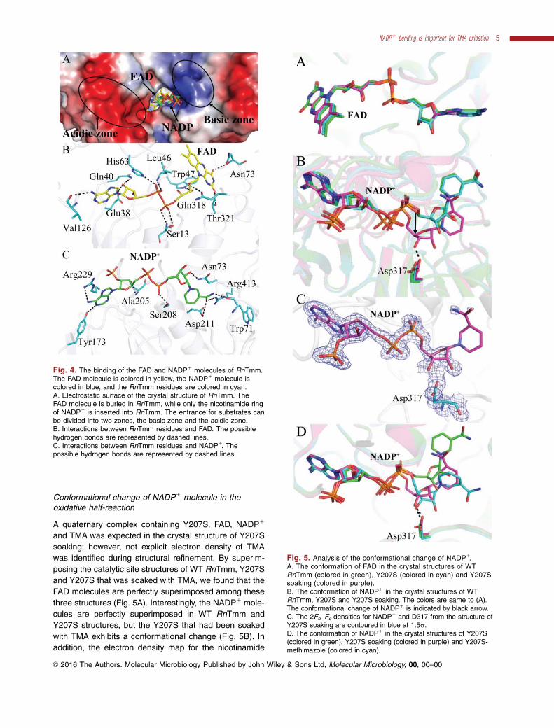
Conformational change of NADP1 molecule in theoxidative half-reaction
A quaternary complex containing Y207S, FAD, NADP1
and TMA was expected in the crystal structure of Y207S
soaking; however, not explicit electron density of TMA
was identified during structural refinement. By superim-
posing the catalytic site structures of WT RnTmm, Y207S
and Y207S that was soaked with TMA, we found that the
FAD molecules are perfectly superimposed among these
three structures (Fig. 5A). Interestingly, the NADP1 mole-
cules are perfectly superimposed in WT RnTmm and
Y207S structures, but the Y207S that had been soaked
with TMA exhibits a conformational change (Fig. 5B). In
addition, the electron density map for the nicotinamide
Fig. 4. The binding of the FAD and NADP1 molecules of RnTmm.The FAD molecule is colored in yellow, the NADP1 molecule iscolored in blue, and the RnTmm residues are colored in cyan.A. Electrostatic surface of the crystal structure of RnTmm. TheFAD molecule is buried in RnTmm, while only the nicotinamide ringof NADP1 is inserted into RnTmm. The entrance for substrates canbe divided into two zones, the basic zone and the acidic zone.B. Interactions between RnTmm residues and FAD. The possiblehydrogen bonds are represented by dashed lines.C. Interactions between RnTmm residues and NADP1. Thepossible hydrogen bonds are represented by dashed lines.
Fig. 5. Analysis of the conformational change of NADP1.A. The conformation of FAD in the crystal structures of WTRnTmm (colored in green), Y207S (colored in cyan) and Y207Ssoaking (colored in purple).B. The conformation of NADP1 in the crystal structures of WTRnTmm, Y207S and Y207S soaking. The colors are same to (A).The conformational change of NADP1 is indicated by black arrow.C. The 2Fo–Fc densities for NADP1 and D317 from the structure ofY207S soaking are contoured in blue at 1.5r.D. The conformation of NADP1 in the crystal structures of Y207S(colored in green), Y207S soaking (colored in purple) and Y207S-methimazole (colored in cyan).
NADP1 bending is important for TMA oxidation 5
VC 2016 The Authors. Molecular Microbiology Published by John Wiley & Sons Ltd, Molecular Microbiology, 00, 00–00
ring of NADP1 in the structure of Y207S that had been
soaked with TMA is rather poor under the 1.5 A resolution,
whereas the electron density map for the other parts of
NADP1 is clear (Fig. 5C), indicating that the nicotinamide
ring of NADP1 becomes mobile during soaking. Notably,
the electron density map strongly indicates an interaction
between NADP1 and D317 in Y207S soaking (Fig. 5C).
Compared with those in WT RnTmm and Y207S, the
ribose ring connecting to the nicotinamide ring of NADP1
in Y207S soaking moves approximate 2.7 A toward D317,
and forms a hydrogen bond with the side-chain of D317
(Fig. 5B). Because D317 is one of the residues constitut-
ing the acidic zone of the substrate entrance, the forma-
tion of the hydrogen bond between NADP1 and D317 can
“shut off” the substrate entrance of RnTmm and make the
catalytic site solvent inaccessible, thereby forming a rela-
tively enclosed micro-environment for the catalytic reac-
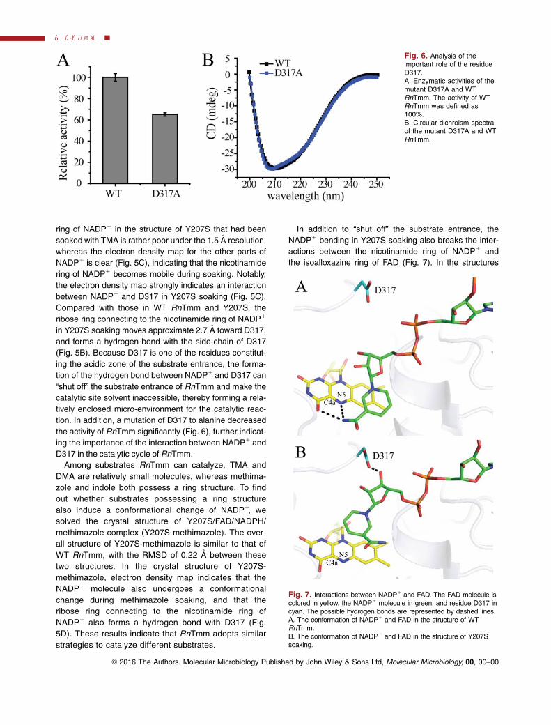
tion. In addition, a mutation of D317 to alanine decreased
the activity of RnTmm significantly (Fig. 6), further indicat-
ing the importance of the interaction between NADP1 and
D317 in the catalytic cycle of RnTmm.
Among substrates RnTmm can catalyze, TMA and
DMA are relatively small molecules, whereas methima-
zole and indole both possess a ring structure. To find
out whether substrates possessing a ring structure
also induce a conformational change of NADP1, we
solved the crystal structure of Y207S/FAD/NADPH/
methimazole complex (Y207S-methimazole). The over-
all structure of Y207S-methimazole is similar to that of
WT RnTmm, with the RMSD of 0.22 A between these
two structures. In the crystal structure of Y207S-
methimazole, electron density map indicates that the
NADP1 molecule also undergoes a conformational
change during methimazole soaking, and that the
ribose ring connecting to the nicotinamide ring of
NADP1 also forms a hydrogen bond with D317 (Fig.
5D). These results indicate that RnTmm adopts similar
strategies to catalyze different substrates.
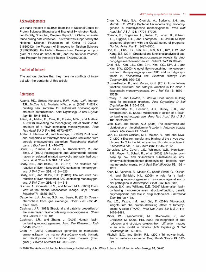
In addition to “shut off” the substrate entrance, the
NADP1 bending in Y207S soaking also breaks the inter-
actions between the nicotinamide ring of NADP1 and
the isoalloxazine ring of FAD (Fig. 7). In the structures
Fig. 6. Analysis of theimportant role of the residueD317.A. Enzymatic activities of themutant D317A and WTRnTmm. The activity of WTRnTmm was defined as100%.B. Circular-dichroism spectraof the mutant D317A and WTRnTmm.
Fig. 7. Interactions between NADP1 and FAD. The FAD molecule iscolored in yellow, the NADP1 molecule in green, and residue D317 incyan. The possible hydrogen bonds are represented by dashed lines.A. The conformation of NADP1 and FAD in the structure of WTRnTmm.B. The conformation of NADP1 and FAD in the structure of Y207Ssoaking.
6 C.-Y. Li et al. �
VC 2016 The Authors. Molecular Microbiology Published by John Wiley & Sons Ltd, Molecular Microbiology, 00, 00–00

of WT RnTmm and Y207S, the nicotinamide ring of
NADP1 forms stacking interactions with the isoalloxa-
zine ring of FAD, and the nitrogen atom of the nicotina-
mide ring of NADP1 forms hydrogen bonds with the
nitrogen atom or the oxygen atom of the isoalloxazine
ring of FAD (Fig. 7A). The interactions between the two
rings are essential for stabilization of the C4a-
hydroperoxyflavin intermediate by NADP1 (Alfieri et al.,
2008; Orru et al., 2010). In the structure of Y207S soak-
ing, the electron density of the nicotinamide ring is poor
(Fig. 5C), suggesting that there may be no direct inter-
actions between NADP1 and FAD (Fig. 7B). However,
the NADP1 does seem to close off the entrance to pro-
tect the C4a-hydroperoxyflavin.
The catalytic cycle of RnTmm with TMA as a substrate
Based on previous studies on the reductive half-reaction
of Tmm catalysis (Alfieri et al., 2008; Orru et al., 2010)
and our results on the oxidative half-reaction, we pro-
pose a relatively complete catalytic cycle of RnTmm
with TMA as a substrate (Fig. 8). In the reductive half-
reaction, NADPH reduces FAD and the C4a-
hydroperoxyflavin intermediate forms. It is believed that
Tmm spends most of the time in this intermediate state
in vivo (Alfieri et al., 2008). In the intermediate state, the
NADP1 molecule nests the basic zone of the entrance
for substrates, and the catalytic pocket of RnTmm is still
partially solvent accessible. The nicotinamide ring of
NADP1 inserted in RnTmm protects the C4a-
hydroperoxyflavin intermediate from solvent attack
(Beaty and Ballou, 1981b). The acidic zone of RnTmm
attracts a TMA molecule and directs it into the catalytic
pocket. Once TMA enters the catalytic pocket, it occu-
pies the catalytic site and makes NADP1 bend to start
the oxidative half-reaction. NADP1 bending would result
in two consequences: Firstly, the ribose ring connecting
to the nicotinamide ring of NADP1 forms a hydrogen
bond with D317, shutting off the substrate entrance and
promoting a protected micro-environment for the cata-
lytic reaction; Secondly, the nicotinamide ring of NADP1
that protects the C4a-hydroperoxyflavin shifts, exposing
the C4a-hydroperoxyflavin to the substrate for catalysis
(Fig. 8). After the oxygenation, the C4a-position of the
FAD bears an AOH group, which loses water, NADP1
and TMAO to regenerate the oxidized FAD. In the next
catalytic cycle, an NADPH molecule re-binds to RnTmm,
reduces the FAD, and shields the C4a-hydroperoxyflavin
intermediate, enabling RnTmm to get ready for the oxy-
genation of the next substrate.
Universality of the catalytic cycle of RnTmm to oxidize
TMA into TMAO in bacteria
The tmm gene is widespread in many marine bacteria,
especially in MRC and the SAR11 clade. Most bacterial
strains containing tmm homologs can grow on TMA as
a sole nitrogen source (Chen et al., 2011), implying the
importance of these bacterial strains in metabolizing
TMA in the ocean. In addition, many soil bacteria also
contain tmm homologs, for example, Rhizobium and
Mesorhizobium. To analyze the universality of the cata-
lytic mechanism of RnTmm to oxidize TMA into TMAO
in bacteria, we performed sequence alignment of
Fig. 8. The catalytic cycle of RnTmm to oxidize TMA into TMAO. In the reductive half-reaction, the C4a-hydroperoxyflavin intermediate isformed. The NADP1 molecule remains in the active site, shielding the C4a-hydroperoxyflavin intermediate from solvent attack. Once TMAcomes into the catalytic pocket, the NADP1 undergoes a conformational change to form a hydrogen bond with D317, shutting off the entranceand exposing the C4a-hydroperoxyflavin to oxidize TMA. After the reaction, NADP1 and the produced TMAO are released, and the FAD isregenerated. FAD intermediate indicates the C4a-hydroperoxyflavin.
NADP1 bending is important for TMA oxidation 7
VC 2016 The Authors. Molecular Microbiology Published by John Wiley & Sons Ltd, Molecular Microbiology, 00, 00–00

Fig. 9. Sequence alignment of bacterial Tmm proteins. Black dots indicate residues involved in NADPH binding, black triangles indicateresidues involved in FAD binding, and residue D317 is marked by a black star. Sequences 1–8 are Tmm’s from marine bacteria, including 1–4from MRC and 5–6 from the SAR11 clade. Sequences 9–10 are Tmm’s from soil bacteria. 1, Roseovarius nubinhibens ISM, EAP78254.1; 2,Ruegeria pomeroyi DSS3, WP_011047288.1; 3, Roseobacter denitrificans, WP_044032905.1; 4, Roseovarius sp. 217, WP_009818593.1; 5,Pelagibacter ubique HTCC1002, WP_006997992.1; 6, Pelagibacter ubique HTCC7211, WP_008544347.1; 7, Marinobacterium stanieri,WP_029511274.1; 8, Marinobacterium litorale, WP_027855190.1; 9, Rhizobium leguminosarum, WP_025397603.1; and 10, Mesorhizobiumsp. 1M-11, WP_054310147.1. The alignment was done with ClustalW (Chenna et al., 2003) and ESPript (Robert and Gouet, 2014).
8 C.-Y. Li et al. �

bacterial Tmms. The result showed that residue D317
and most residues involved in binding FAD and NADP1
in RnTmm are highly conserved in Tmms from both
marine bacteria and soil bacteria (Fig. 9). This indicates
that the proposed catalytic cycle of RnTmm to oxidize
TMA into TMAO is likely adopted by most, if not all, bac-
terial Tmms.
Discussion
Many studies on the catalytic mechanisms of both
eukaryotic and bacterial FMOs have been reported and
it is regarded as a general characteristic of FMOs that
the coenzyme NADP1 remains bound to the enzyme
throughout catalysis (Van Berkel et al., 2006; Alfieri
et al., 2008; Orru et al., 2010; Crozier-Reabe and
Moran, 2012). However, a structural study by Cho et al.
suggested that for the catalysis of indole by Tmm from
Methylophaga aminosulfidovorans MPT, indole competes
with NADP1 for binding to the catalytic site and NADP1
is released before the oxidative half-reaction (Cho et al.,
2011). After the departure of NADP1, the C4a-
hydroperoxyflavin intermediate might be transitorily
stabilized by residues of the enzyme (Cho et al., 2011).
Here, our data showed that TMA only competes the nic-
otinamide ring of NADP1, and the NADP1 molecule
remains bound to Tmm until the catalytic reaction
finishes, which is in accordance with the general
characteristics of FMOs (Van Berkel et al., 2006;
Crozier-Reabe and Moran, 2012). This is the first struc-
tural evidence that NADP1 binds to Tmm throughout the
catalysis of TMA.
In addition to reducing FAD, it has been reported
that NADP(H) also exhibited a moonlighting activity to
protect the C4a-hydroperoxyflavin intermediate during
the reductive half-reaction of the catalysis (Alfieri
et al., 2008; Orru et al., 2010). Although studies indi-
cated that NADP1 binds to FMOs throughout the catal-
ysis (Van Berkel et al., 2006; Alfieri et al., 2008; Orru
et al., 2010; Crozier-Reabe and Moran, 2012), the role
of NADP1 in the oxidative half-reaction is not explicit
yet. Our results indicate that the NADP1 molecule
undergoes a conformational change in the oxidative
half-reaction, which exposes the catalytic C4a-
hydroperoxyflavin to TMA, and promotes a protected
micro-environment for the catalytic reaction of Tmm by
forming a hydrogen bond with a conserved aspartic
acid residue. Therefore, in addition to functioning in
the reductive half-reaction, NADPH/NADP1 also plays
an important role in the oxidative half-reaction of Tmm
for TMA oxidation. NADP1 bending was also identified
in Baeyer-Villiger monooxygenases (BVMOs) (Yachnin
et al., 2012), another subclass of the class B
flavoprotein monooxygenases (Riebel et al., 2014). For
BVMOs, intramolecular hydrogen bonds are important
in stabilizing the rotated conformation of NADP1
(Yachnin et al., 2012). Here for FMOs, we highlight the
importance of interactions between NADP1 and Tmm.
In both cases, NADP1 bending exposes the catalytic
site and promotes the oxidative reaction (Yachnin
et al., 2012). Therefore, our study on FMOs should
enrich our understanding of the catalytic cycle of flavo-
protein monooxygenases.
Structural analysis demonstrated that there is no
direct interaction between the substrate and the resi-
dues of Tmm (Eswaramoorthy et al., 2006; Cho et al.,
2011). Substrates containing a ring structure, such as
indole and methimazole, can form stacking interactions
with the isoalloxazine ring of FAD (Eswaramoorthy
et al., 2006; Cho et al., 2011). Because TMA does not
possess a ring structure, there may be no effective inter-
action to stabilize its conformation when TMA enters the
catalytic pocket, and the positive charge on TMA prob-
ably repels the positive charge on the pyridinium ring of
NADP1 to drive the movement of the nicotinamide dur-
ing the reaction. In the catalytic cycle of Tmm, when
TMA enters the catalytic site, it should be directed to
the C4a-hydroperoxyflavin, triggering the oxidative half-
reaction. Because TMA molecule cannot form stacking
interactions with FAD, we suggest that the catalytic reac-
tion of Tmm proceeds quickly, and a prolonged steady-
conformation of TMA is not necessary. This may be the
reason why we have observed the conformational
change of NADP1, but cannot identify the location of
TMA in the structure of Y207S soaked with TMA. The
aromatic ring of Y207 residue forms a pi-pi stacking with
the isoalloxazine ring of FAD in the Tmm without
NADPH (Orru et al., 2010; Cho et al., 2011). When
NADPH binds to the catalytic site, it competes for the
isoalloxazine ring and frees Y207 (Orru et al., 2010;
Cho et al., 2011). Thus, the isoalloxazine ring
moves between the aromatic ring of Y207 and the nico-
tinamide ring of NADP1, and Y207 plays a role in
releasing NADP1 after each catalytic cycle during TMA
oxidation.
In conclusion, this study illustrated the catalytic cycle
of TMA oxidation by marine RnTmm and showed the
first structural evidence of NADP1 binding to a Tmm
throughout the catalytic reaction and its involvement in
the oxidative half-reaction by a conformational bending.
The proposed mechanism of TMA oxidation by RnTmm
may have universal significance among bacteria contain-
ing Tmm. Our results provide novel insights into the cat-
alytic mechanism of FMOs and promote a better
understanding of TMA-involved marine carbon and nitro-
gen cycling.
NADP1 bending is important for TMA oxidation 9
VC 2016 The Authors. Molecular Microbiology Published by John Wiley & Sons Ltd, Molecular Microbiology, 00, 00–00

Experimental procedures
Bacterial strains and growth conditions
R. nubinhibens ISM was purchased from the Leibniz Insti-
tute DSMZ-German Collection of Microorganisms and Cell
Cultures and was cultured in the 974 medium at 308C for 2
days according to the provided protocol (http://www.dsmz.
de/). E. coli strains DH5a and BL21 (DE3) were grown in
Lysogeny Broth (LB) medium at 378C.
Gene cloning, point mutation and protein expression
and purification
The tmm gene was amplified from the genomic DNA of R.
nubinhibens ISM using PCR and was subcloned into the
pET22b (Novagen) vector with a C-terminal His tag. All of
the point mutations (E153A/D154A, Y207S and D317A) in
RnTmm were introduced using PCR-based methods and
were verified by DNA sequencing. Y207S and D317A were
constructed base on mutant E153A/D154A. The RnTmm
protein and all of its mutants were expressed in E. coli
strain BL21 (DE3). The cells were cultured at 378C in LB
medium to an OD600 of 0.8–1.0 and then induced at 208C
for 16 h with 0.5 mM isopropyl b-D-1-thiogalactopyranoside
(IPTG). The proteins were purified first with Ni21-NTA resin
(Qiagen) and then fractionated by gel filtration on a
Superdex-200 column (GE Healthcare).
Enzyme assays
The enzymatic properties of RnTmm were measured by fol-
lowing the decrease of absorbance at 340 nm
(e340 5 6.22 mM21 cm21 for NADPH) (Alfieri et al., 2008).
The reaction mixture contains 1 lM RnTmm, 0.25 mM
NADPH and 1 mM TMA. For the measurement of Km of
RnTmm, substrate (TMA, methimazole, indole or DMA) of
different concentrations was added into the reaction system
containing 1 lM RnTmm and 0.25 mM NADPH. The opti-
mal pH and the optimal temperature of RnTmm were deter-
mined using TMA as the substrate. For measurement of
the optimal temperature of RnTmm, a buffer containing
10 mM Tris-HCl (pH 8.0) and 100 mM NaCl was pre-
incubated at different temperatures for 30 min, and then 1
lM RnTmm, 0.25 mM NADPH and 1 mM TMA were added
into the buffer and the mixture was incubated at different
temperatures for 3 min before detection by V550 UV/VIS
spectrophotometer (Jasco). The optimal pH of RnTmm was
measured using Britton-Robinson buffer over a pH range
from 7.0 to 9.5. Britton-Robinson buffer is a mixture of
0.04 M H3BO3, 0.04 M H3PO4 and 0.04 M CH3COOH
(Barek et al., 1999).The enzymatic activities of RnTmm mutants were deter-
mined by detecting the fluorescence of indoxyl on a FP-
6500 spectrofluorometer (Jasco) (Woo et al., 2000). The
reaction mixture contained 2 lM RnTmm or a mutant,
0.15 mM NADPH and 0.15 mM indole. The mixture was
incubated for 10 min before detection. Fluorescence spec-
tra were collected from 450 to 490 nm at a scan speed of
1000 nm min21 with the excitation wavelength of 365 nm(Woo et al., 2000).
Crystallization and data collection
The purified RnTmm protein was concentrated to approxi-mately 10 mg ml21 in 10 mM Tris-HCl (pH 8.0) and
100 mM NaCl. To obtain crystals of WT RnTmm, NADPHwas added into the protein solution to a final concentrationof 5 mM before crystallization. Initial crystallization trials for
WT RnTmm were performed at 208C using the sitting dropvapor diffusion method. Diffraction-quality crystals of WTRnTmm were obtained in hanging drops containing 0.2 M
magnesium acetate tetrahydrate, 0.1 M sodium cacodylatetrihydrate (pH 6.5) and 20% (w/v) polyethylene glycol(PEG) 8000 at 208C after 2-weeks incubation.
Diffraction-quality crystals of Y207S were obtained inhanging drops containing 0.2 M sodium acetate, 0.1 M Bis-Tris propane (pH 6.5) and 20% (w/v) PEG 3350 at 208C.
The crystals of Y207S soaking were obtained using crystalsof Y207S soaked with 15 mM TMA for 15 min. The crystalsof Y207S-methimazole were obtained using crystals of
Y207S soaked with 15 mM methimazole for 15 min. X-raydiffraction data were collected on the BL19U1 beamline atthe Shanghai Synchrotron Radiation Facility. The initial dif-
fraction data sets were processed by the HKL3000 program(Minor et al., 2006).
Structure determination and refinement
The crystals of WT RnTmm, Y207S, Y207S soaking andY207S-methimazole all belong to the P21 space group. The
crystal structure of WT RnTmm was determined by molecu-lar replacement using the CCP4 program Phaser (Potterton
et al., 2003) with the crystal structure of bacterial FMO(PDB code: 2XVE) as the search model. The structures ofY207S, Y207S soaking and Y207S-methimazole were
determined using WT RnTmm as the search model. Therefinements of these structures were performed using Coot(Emsley and Cowtan, 2004) and Phenix (Adams et al.,
2002). All of the structure figures were made using the pro-gram PyMOL (http://www.pymol.org/).
Circular-dichroism (CD) spectroscopic assays
WT RnTmm and its mutant were subjected to CD spectro-scopic assays at 258C on a J-810 spectropolarimeter
(Jasco). CD spectra of the samples at a final concentrationof approximately 10 lM were collected from 250 to 200 nmat a scan speed of 500 nm min21 with a band width of
2 nm. All of the samples were in the buffer containing10 mM Tris-HCl (pH 8.0) and 100 mM NaCl.
Accession numbers
The structures of WT RnTmm, Y207S, Y207S soaking andY207S- methimazole have been deposited in the Protein
Data Bank (PDB) under the accession codes 5IPY, 5IQ1,5IQ4 and 5GSN respectively.
10 C.-Y. Li et al. �
VC 2016 The Authors. Molecular Microbiology Published by John Wiley & Sons Ltd, Molecular Microbiology, 00, 00–00
Acknowledgements
We thank the staff of BL19U1 beamline at National Center for
Protein Sciences Shanghai and Shanghai Synchrotron Radia-
tion Facility, Shanghai, People’s Republic of China, for assis-
tance during data collection. This work was supported by the
National Science Foundation of China (31290231,
31630012), the Program of Shandong for Taishan Scholars
(TS20090803), the Hi-Tech Research and Development pro-
gram of China (2012AA092105) and the National Postdoc-
toral Program for Innovative Talents (BX201600095).
Conflict of Interest
The authors declare that they have no conflicts of inter-
est with the contents of this article.
References
Adams, P.D., Grosse-Kunstleve, R.W., Hung, L.W., Ioerger,
T.R., McCoy, A.J., Moriarty, N.W., et al. (2002) PHENIX:
building new software for automated crystallographic
structure determination. Acta Crystallogr D Biol Crystal-
logr 58: 1948–1954.Alfieri, A., Malito, E., Orru, R., Fraaije, M.W., and Mattevi,
A. (2008) Revealing the moonlighting role of NADP in the
structure of a flavin-containing monooxygenase. Proc
Natl Acad Sci U S A 105: 6572–6577.Arata, H., Shimizu, M., and Takamiya, K. (1992) Purification
and properties of trimethylamine N-oxide reductase from
aerobic photosynthetic bacterium Roseobacter denitrifi-
cans. J Biochem 112: 470–475.Barek, J., Pumera, M., Muck, A., Kade�r�abkov�a, M., and
Zima, J. (1999) Polarographic and voltammetric determi-
nation of selected nitrated polycyclic aromatic hydrocar-
bons. Anal Chim Acta 393: 141–146.Beaty, N.B., and Ballou, D.P. (1981a) The oxidative half-
reaction of liver microsomal FAD-containing monooxygen-
ase. J Biol Chem 256: 4619–4625.Beaty, N.B., and Ballou, D.P. (1981b) The reductive half-
reaction of liver microsomal FAD-containing monooxygen-
ase. J Biol Chem 256: 4611–4618.
Buchan, A., Gonzalez, J.M., and Moran, M.A. (2005) Over-
view of the marine roseobacter lineage. Appl Environ
Microbiol 71: 5665–5677.Carpenter, L.J., Archer, S.D., and Beale, R. (2012) Ocean-
atmosphere trace gas exchange. Chem Soc Rev 41:
6473–6506.Cashman, J.R. (1995) Structural and catalytic properties of
the mammalian flavin-containing monooxygenase. Chem
Res Toxicol 8: 166–181.Cashman, J.R., and Zhang, J. (2006) Human flavin-
containing monooxygenases. Annu Rev Pharmacol Toxi-
col 46: 65–100.
Chen, Y. (2012) Comparative genomics of methylated
amine utilization by marine Roseobacter clade bacteria
and development of functional gene markers (tmm,
gmaS). Environ Microbiol 14: 2308–2322.
Chen, Y., Patel, N.A., Crombie, A., Scrivens, J.H., and
Murrell, J.C. (2011) Bacterial flavin-containing monooxy-
genase is trimethylamine monooxygenase. Proc Natl
Acad Sci U S A 108: 17791–17796.Chenna, R., Sugawara, H., Koike, T., Lopez, R., Gibson,
T.J., Higgins, D.G., and Thompson, J.D. (2003) Multiple
sequence alignment with the Clustal series of programs.
Nucleic Acids Res 31: 3497–3500.
Cho, H.J., Cho, H.Y., Kim, K.J., Kim, M.H., Kim, S.W., and
Kang, B.S. (2011) Structural and functional analysis of bac-
terial flavin-containing monooxygenase reveals its ping-
pong-type reaction mechanism. J Struct Biol 175: 39–48.Choi, H.S., Kim, J.K., Cho, E.H., Kim, Y.C., Kim, J.I., and
Kim, S.W. (2003) A novel flavin-containing monooxygen-
ase from Methylophaga sp strain SK1 and its indigo syn-
thesis in Escherichia coli. Biochem Biophys Res
Commun 306: 930–936.Crozier-Reabe, K., and Moran, G.R. (2012) Form follows
function: structural and catalytic variation in the class a
flavoprotein monooxygenases. Int J Mol Sci 13: 15601–
15639.Emsley, P., and Cowtan, K. (2004) Coot: model-building
tools for molecular graphics. Acta Crystallogr D Biol
Crystallogr 60: 2126–2132.Eswaramoorthy, S., Bonanno, J.B., Burley, S.K., and
Swaminathan, S. (2006) Mechanism of action of a flavin-
containing monooxygenase. Proc Natl Acad Sci U S A
103: 9832–9837.Gibb, S.W., and Hatton, A.D. (2004) The occurrence and
distribution of trimethylamine-N-oxide in Antarctic coastal
waters. Mar Chem 91: 65–75.Gon, S., Giudici-Orticoni, M.T., Mejean, V., and Iobbi-Nivol,
C. (2001) Electron transfer and binding of the c-type cyto-
chrome TorC to the trimethylamine N-oxide reductase in
Escherichia coli. J Biol Chem 276: 11545–11551.
Gonzalez, J.M., Covert, J.S., Whitman, W.B., Henriksen,
J.R., Mayer, F., Scharf, B., et al. (2003) Silicibacter pom-
eroyi sp. nov. and Roseovarius nubinhibens sp. nov.,
dimethylsulfoniopropionate-demethylating bacteria from
marine environments. Int J Syst Evol Microbiol 53: 1261–
1269.
Koch, M., Vorwerk, S., Masur, C., Sharifi-Sirchi, G., Olivieri,
N., and Schlaich, N.L. (2006) A role for a flavin-
containing mono-oxygenase in resistance against micro-
bial pathogens in Arabidopsis. Plant J 47: 629–639.Krueger, S.K., and Williams, D.E. (2005) Mammalian flavin-
containing monooxygenases: structure/function, genetic
polymorphisms and role in drug metabolism. Pharmacol
Ther 106: 357–387.Ma, J.Q., Pazos, I.M., and Gai, F. (2014) Microscopic
insights into the protein-stabilizing effect of trimethyl-
amine N-oxide (TMAO). Proc Natl Acad Sci U S A 111:
8476–8481.Minor, W., Cymborowski, M., Otwinowski, Z., and
Chruszcz, M. (2006) HKL-3000: the integration of data
reduction and structure solution–from diffraction images
to an initial model in minutes. Acta Crystallogr D Biol
Crystallogr 62: 859–866.
Mitchell, S.C., and Smith, R.L. (2001) Trimethylaminuria:
the fish malodor syndrome. Drug Metab Dispos 29: 517–
521.
NADP1 bending is important for TMA oxidation 11
VC 2016 The Authors. Molecular Microbiology Published by John Wiley & Sons Ltd, Molecular Microbiology, 00, 00–00
Morris, R.M., Rappe, M.S., Connon, S.A., Vergin, K.L.,Siebold, W.A., Carlson, C.A., and Giovannoni, S.J. (2002)SAR11 clade dominates ocean surface bacterioplanktoncommunities. Nature 420: 806–810.
Orru, R., Pazmino, D.E.T., Fraaije, M.W., and Mattevi, A.
(2010) Joint functions of protein residues and NADP(H)in oxygen activation by flavin-containing monooxygenase.J Biol Chem 285: 35021–35028.
Potterton, E., Briggs, P., Turkenburg, M., and Dodson, E.(2003) A graphical user interface to the CCP4 program
suite. Acta Crystallogr D Biol Crystallogr 59: 1131–1137.Quinn, P.K., Charlson, R.J., and Bates, T.S. (1988) Simulta-
neous observations of ammonia in the atmosphere andocean. Nature 335: 336–338.
Riebel, A., Fink, M.J., Mihovilovic, M.D., and Fraaije, M.W.
(2014) Type II flavin-containing monooxygenases: a newclass of biocatalysts that harbors baeyer–villiger monoox-ygenases with a relaxed coenzyme specificity. Chem-CatChem 6: 1112–1117.
Robert, X., and Gouet, P. (2014) Deciphering key featuresin protein structures with the new ENDscript server.Nucleic Acids Res 42: W320–W324.
Rusch, D.B., Halpern, A., Sutton, L.G., Heidelberg, K.,Williamson, B., Yooseph, S., et al (2007) The Sorcerer II
Global Ocean Sampling expedition: northwest Atlanticthrough eastern tropical Pacific. PLoS Biol 5: e77.
Schlaich, N.L. (2007) Flavin-containing monooxygenases inplants: looking beyond detox. Trends Plant Sci 12:412–418.
Seibel, B.A., and Walsh, P.J. (2002) Trimethylamine oxideaccumulation in marine animals: relationship to acylgly-cerol storage. J Exp Biol 205: 297–306.
Suh, J.K., and Robertus, J.D. (2000) Yeast flavin-containingmonooxygenase is induced by the unfolded protein
response. Proc Natl Acad Sci U S A 97: 121–126.Suh, J.K., Poulsen, L.L., Ziegler, D.M., and Robertus, J.D.
(1999) Yeast flavin-containing monooxygenase generatesoxidizing equivalents that control protein folding in the endo-plasmic reticulum. Proc Natl Acad Sci U S A 96: 2687–2691.
Van Berkel, W.J., Kamerbeek, N.M., and Fraaije, M.W.(2006) Flavoprotein monooxygenases, a diverse class ofoxidative biocatalysts. J Biotechnol 124: 670–689.
Woo, H., Sanseverino, J., Cox, C.D., Robinson, K.G., andSayler, G.S. (2000) The measurement of toluene dioxy-
genase activity in biofilm culture of Pseudomonas putidaF1. J Microbiol Methods 40: 181–191.
Yachnin, B.J., Sprules, T., McEvoy, M.B., Lau, P.C., andBerghuis, A.M. (2012) The substrate-bound crystal struc-
ture of a Baeyer-Villiger monooxygenase exhibits aCriegee-like conformation. J Am Chem Soc 134: 7788–7795.
Yancey, P.H., Gerringer, M.E., Drazen, J.C., Rowden, A.A.,and Jamieson, A. (2014) Marine fish may be biochemi-
cally constrained from inhabiting the deepest oceandepths. Proc Natl Acad Sci U S A 111: 4461–4465.
Zhou, J., and Shephard, E.A. (2006) Mutation, polymor-phism and perspectives for the future of human flavin-containing monooxygenase 3. Mutat Res 612: 165–171.
12 C.-Y. Li et al. �
VC 2016 The Authors. Molecular Microbiology Published by John Wiley & Sons Ltd, Molecular Microbiology, 00, 00–00