structurally persistent micelles: theory and experiment
TRANSCRIPT
����������������
Structurally Persistent Micelles:
Theory and Experiment
Christof M. J�ger1, Andreas Hirsch
2,3,
Christoph B�ttcher4
and Timothy Clark1,3,*
1Computer-Chemie-Centrum and Interdisciplinary Center for Molecular Materials,Department of Chemistry and Pharmacy, University of Erlangen-Nurnberg,
Nagelsbachstraße 25, 91052 Erlangen, Germany.
2Interdisciplinary Center for Molecular Materials,Department of Chemistry and Pharmacy, University of Erlangen-Nurnberg,
Henkestraße 25, 91054 Erlangen, Germany.
3Excellence Cluster ‘‘Engineering of Advanced Materials’’,University of Erlangen-Nurnberg,
Nagelsbachstraße 49b, 91052 Erlangen, Germany.
4Research Center of Electron Microscopy,Institute of Chemistry and Biochemistry, Free University Berlin,
Fabeckstraße 36a, 14195 Berlin, Germany.
E-Mail: *[email protected]
Received: 20th July 2010 / Published: 13th June 2011
Abstract
We describe the progress made in understanding the factors that deter-
mine the size, structure and stability of structurally persistent micelles
using a combination of designed synthesis, cryo-TEM imaging and
molecular-dynamics simulations. The importance of specific counter-
ion effects is revealed in detail. An unexpected effect of sodium coun-
terions leads to attraction between the polycarboxylate head groups of
the tailored dendrimers that make up the micelles. This effect even
leads to the formation of ‘‘superlattices’’ of highly negatively charged
micelles.
91
http://www.beilstein-institut.de/Bozen2010/Proceedings/Clark/Clark.pdf
Functional Nanoscience
May 17th – 21st, 2010, Bozen, Italy
Introduction
Soft nanostructures can be considered to be the second class of nanomaterials after to
‘‘hard’’ nanoparticles and similar structurally defined and static nanoscale structures. Nanos-
tructured soft matter represents a challenging research area, both for theory and experiment.
This is because soft matter is inherently dynamic in its structure and cannot, therefore be
treated as a single static object. Nonetheless, soft nanostructures can have significant ad-
vantages over hard nanoparticles. They are, for instance, formed in a dynamic equilibrium
process, so that their self-assembly is governed by thermodynamics, rather than the less
predictable process of kinetically controlled nucleation and precipitation. This advantage
can, however, soon become a disadvantage because many factors may determine the delicate
equilibrium that gives rise to soft nanostructures, so that they may be sensitive to their
environment. Nature uses soft nanoparticles almost exclusively in living organisms, so that
there can be no doubt that technological applications based on soft nanostructures are
potentially extremely powerful. Soft nanostructures are most likely to self-assemble from
organic precursor molecules in solution, probably aqueous. Conventional micelles [1] are
perhaps the best known soft nanoparticles, but although a very large amount of data about,
for instance critical micelle concentrations is available [2], relatively little is known about
the detailed structure and dynamics of micelles and other soft nanostructures.
Structurally persistent micelles [3] therefore represent an important milestone in the science
of soft nanostructures as they have consistent, well defined and persistent structures that can
be observed in detail by techniques such as cryo-transmission electron microscopy (cryo-
TEM). These characteristics not only make structurally persistent micelles intriguing experi-
mental objects, but also make them ideal for testing and validating molecular-dynamics
(MD) simulations and the force fields used for them. As we will describe below, simulations
have played a major role in advancing our understanding of structurally persistent micelles
and the factors that control their stability and structure.
Molecular Components
In 2004 Kellermann et al. [3] described the aggregation of seven amphiphilic dendro-
calixarene molecules 1 to uniform and structurally persistent micelles (Scheme 1).
92
Jager, C.M. et al.

Scheme 1. Dendrocalixarene monomer.
The self aggregation behaviour of the molecules was investigated by pulse-gradient spin-
echo (PGSE) NMR spectroscopy and cryo-TEM experiments. Remarkably, the experiments
showed that a single distinct type of micelle was formed with no other aggregates of
different sizes. The key feature of this first amphiphilic dendrimer investigated is its cone-
shaped structure, which makes it possible to form small aggregates with high curvature.
Each hydrophilic polycarboxylate head-group is linked to four hydrophobic alkane chains by
a calyx[4]arene unit.
Hirsch et al. [4 – 7] later synthesized, characterized and investigated the aggregation beha-
viour of a series of new amphiphilic building blocks based on either calixarenes, fullerenes
(Scheme 2: 2, 3) or later perylene [8, 9] as the central scaffold. These scaffolds allow a
variety of hydrophilic and hydrophobic head and tail groups to be bound in a stereochemi-
cally controlled and tunable fashion. The polar head groups are Newkome-type oligocar-
boxylic acids in all cases. At neutral pH, they are predominantly deprotonated and guarantee
excellent water solubility.
93
Structurally Persistent Micelles: Theory and Experiment
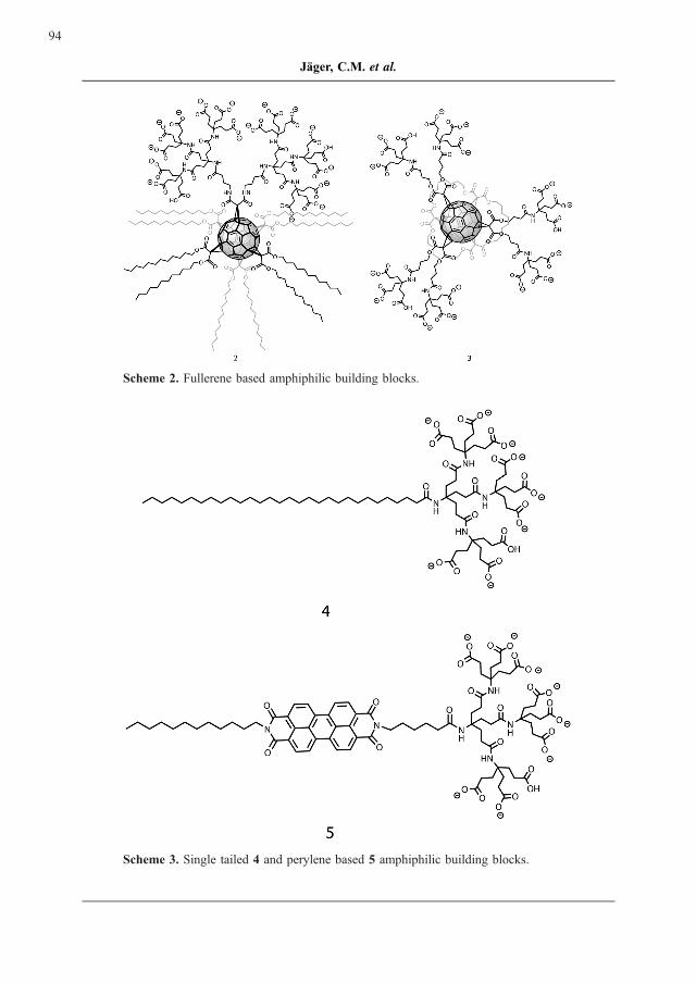
Scheme 2. Fullerene based amphiphilic building blocks.
Scheme 3. Single tailed 4 and perylene based 5 amphiphilic building blocks.
94
Jager, C.M. et al.
Structurally Persistent Micelles – cryo-TEM
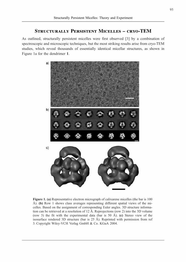
As outlined, structurally persistent micelles were first observed [3] by a combination of
spectroscopic and microscopic techniques, but the most striking results arise from cryo-TEM
studies, which reveal thousands of essentially identical micellar structures, as shown in
Figure 1a for the dendrimer 1.
Figure 1. (a) Representative electron micrograph of calixarene micelles (the bar is 100
A). (b) Row 1 shows class averages representing different spatial views of the mi-
celles. Based on the assignment of corresponding Euler angles. 3D structure informa-
tion can be retrieved at a resolution of 12 A. Reprojections (row 2) into the 3D volume
(row 3) the fit with the experimental data (bar is 50 A). (c) Stereo view of the
isosurface rendered 3D structure (bar is 25 A). Reprinted with permission from ref
3. Copyright Wiley-VCH Verlag GmbH & Co. KGaA 2004.
95
Structurally Persistent Micelles: Theory and Experiment
Because the micelles are oriented randomly, the TEM-picture contains views from literally
thousands of different angles. This ensemble of views can be used to calculate a 3D-
structure for the micelles [10 – 12], as shown in Figure 1c for the micelles depicted in
Figure 1.
The reconstruction shows high-density areas that were interpreted as corresponding to the
carboxylate head-groups of the dendrimer and a hollow core. The low density and high
mobility of the alkane chains of the dendrimer explains that the core of the micelle does not
show up in the TEM picture. However, the dimensions of the micelles led to the conclusion
that a relatively large concentration of water must be present in the hydrophobic core of the
micelles. The first MD simulations were therefore designed to test this hypothesis and to
investigate the structure of this water in a hydrophobic environment.
Molecular Dynamics Simulations – Heptameric Micelles
Finding a suitable starting geometry for MD simulations is always critical to their success. In
this case, the 3D reconstruction of the positions of the head-groups was used to construct a
putative complete micelle structure by adding the alkane chains and flooding the resulting
structure with water and adding sodium ions to neutralize the ensemble. The resulting
geometry was then first geometry-optimized and then equilibrated very slowly by perform-
ing successive MD simulations in which the geometrical restraints that held the micelle
together were removed very slowly and carefully. A micelle resulted that was stable for
100 ns simulation time. This probably indicates that it is a stable and observable entity as
unstable micelles dissociate within just a few nanoseconds in the simulations. The relaxation
and equilibration led to the expulsion of the water molecules from the hydrophobic core,
which became very ‘‘dry’’, and a concomitant shrinking of the micelle until it was approxi-
mately 5 A smaller than suggested by the cryo-TEM images [13]. This discrepancy is larger
than would normally be expected and raised the question as to exactly what the cryo-TEM
images were showing.
Unusually, staining with heavy-metal derivatives was not necessary in order to be able to
‘‘see’’ the micelles in the TEM images. TEM is usually considered to visualize differences in
density [14], so that we analyzed the density of our simulation box divided into small
voxels. The results revealed areas of higher than average density associated with the sodium
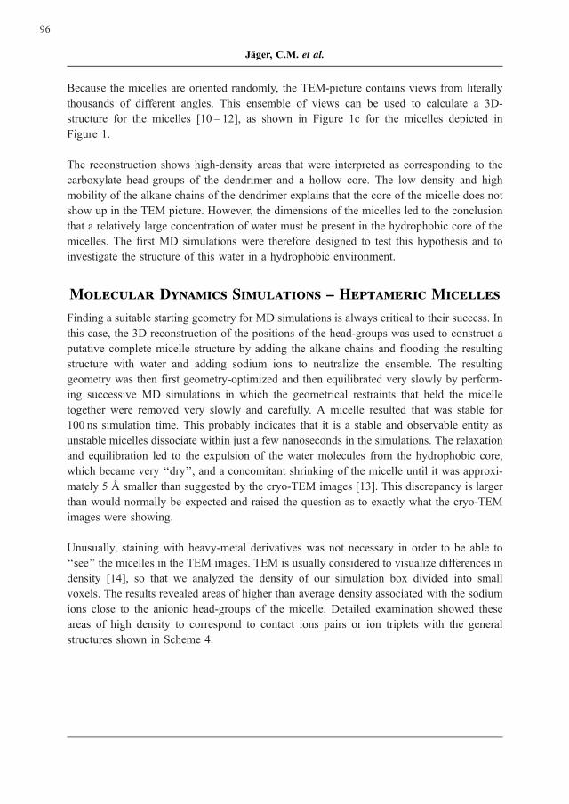
ions close to the anionic head-groups of the micelle. Detailed examination showed these
areas of high density to correspond to contact ions pairs or ion triplets with the general
structures shown in Scheme 4.
96
Jager, C.M. et al.
Scheme 4. Schematic structures of sodium carboxylate contact ion pairs (left) and
triplets (right). Note the importance of the waters (red) that form strongly hydrogen-
bonded bridges between waters (blue) coordinated directly to the sodium ion and
oxygen atoms of the carboxylates.
Since the simulations were carried out with quite simple force fields that might not repro-
duce the behaviour of ions in aqueous solution correctly, we tested their stability by using
snapshots from PM3 [15, 16] MD simulations of sodium formate in water as starting
structures for geometry optimizations using density-functional theory (DFT). These calcula-
tions confirmed that structures of the types shown in Figure 2 are both stable and persistent
in simulations and on geometry optimization.
This observation resolves the apparent difference between the stable micelle structures found
in the simulations and the 3D-reconstructions from the cryo-TEM images. The carboxylate
head-groups and their associated sodium ions together provide the high-density regions that
appear in the unstained cryo-TEM images. This observation has since been confirmed for
several systems in which the micelles are not observable in the TEM without staining if
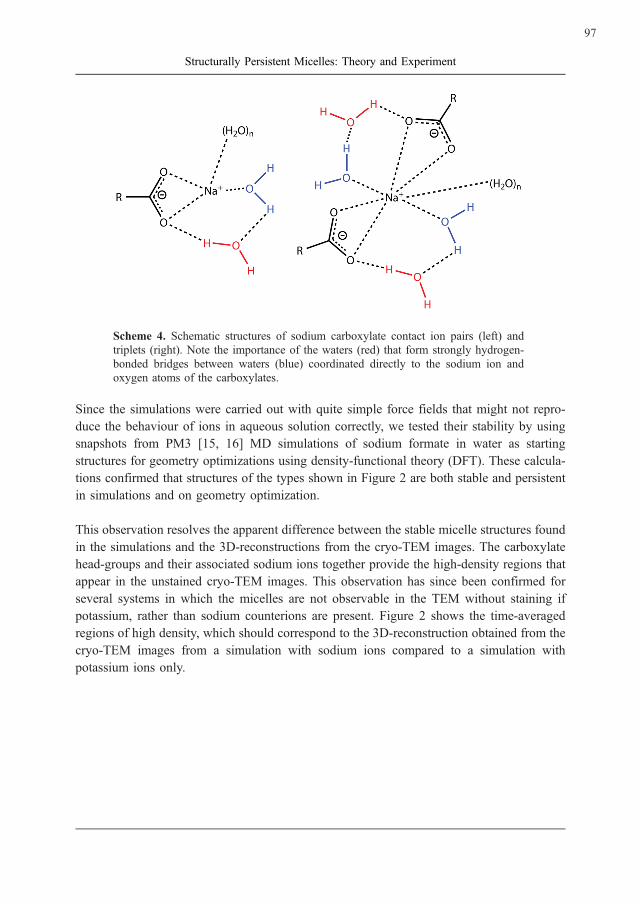
potassium, rather than sodium counterions are present. Figure 2 shows the time-averaged
regions of high density, which should correspond to the 3D-reconstruction obtained from the
cryo-TEM images from a simulation with sodium ions compared to a simulation with
potassium ions only.
97
Structurally Persistent Micelles: Theory and Experiment
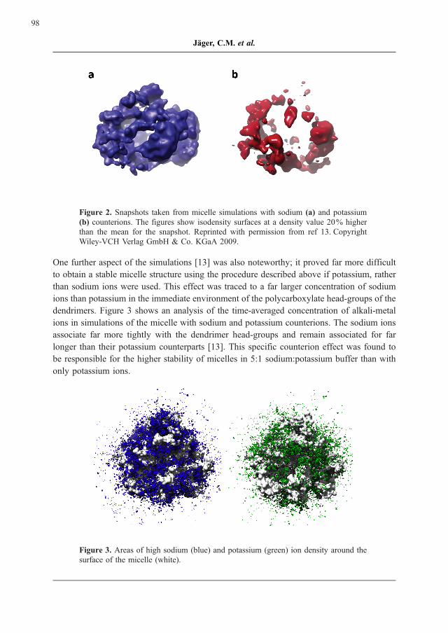
Figure 2. Snapshots taken from micelle simulations with sodium (a) and potassium
(b) counterions. The figures show isodensity surfaces at a density value 20% higher
than the mean for the snapshot. Reprinted with permission from ref 13. Copyright
Wiley-VCH Verlag GmbH & Co. KGaA 2009.
One further aspect of the simulations [13] was also noteworthy; it proved far more difficult
to obtain a stable micelle structure using the procedure described above if potassium, rather
than sodium ions were used. This effect was traced to a far larger concentration of sodium
ions than potassium in the immediate environment of the polycarboxylate head-groups of the
dendrimers. Figure 3 shows an analysis of the time-averaged concentration of alkali-metal
ions in simulations of the micelle with sodium and potassium counterions. The sodium ions
associate far more tightly with the dendrimer head-groups and remain associated for far
longer than their potassium counterparts [13]. This specific counterion effect was found to
be responsible for the higher stability of micelles in 5:1 sodium:potassium buffer than with
only potassium ions.
Figure 3. Areas of high sodium (blue) and potassium (green) ion density around the
surface of the micelle (white).
98
Jager, C.M. et al.
Specific Ion Effects and the Hofmeister Series
Franz Hofmeister was born in Prague in 1850 and died in Wurzburg in 1922 [17]. He
studied medicine in Prague and became Professor of Pharmacology there in 1885. After
Czech became the only language at the Charles University in Prague, he moved to Stras-
bourg in 1896, but was eventually forced to move to Wurzburg when Strasbourg became
French. He enjoyed a remarkable career and was the first to suggest that peptides and
proteins consist of amino-acid residues connected by amide bonds. This honour is often
accorded Emil Fischer, but Hofmeister spoke before Fischer at the conference in which both
announced their discovery. Hofmeister is best known for what is now known as the Hof-
meister series [18 – 21]. This series now describes the effects of ions on the solubility of
proteins in water, although Hofmeister never formulated it in terms of individual ions, but
rather for salts. Although phenomenological rationalizations for the Hofmeister series
abound [22 – 26], no really convincing microscopic explanation exists. Early ideas that ions
could provoke (or destroy) long-range order in water proved not to be correct [18, 27 – 29].
However, the effects described by the Hofmeister series clearly affect self-aggregation by
polyelectrolytes and may therefore even determine the shape, size and stability of structu-
rally persistent micelles.
This sensitivity of structurally persistent micelles to counterion effects makes them ideal
research objects for investigating Hofmeister-like effects on polyelectrolytes as the micelles
are uniformly structured and react strongly to changes in their ionic environment. These
sensitive but nonetheless well defined systems provide an unprecedented level of informa-
tion about specific ions effects in general and also about the factors that affect micelle
structure and stability.
Experimental (cryo-TEM) tests of the differences between sodium and potassium counter-
ions on the structures and stability of the micelles revealed strong effects. Replacing the
original 5:1 Na:K buffer with a pure potassium one at the same concentration resulted in
larger micelles than those observed originally. Remarkably, the original heptameric micelles
were obtained by titrating the solution with a five-fold excess of pure sodium buffer [13]. At
higher concentrations, the Na:K buffer gave the original heptameric micelles once more, but
the cryo-TEM images revealed fewer than at lower concentrations. At the same high con-
centration, a pure potassium buffer gave no micelles at all. Figure 4 shows the relevant cryo-
TEM images.
99
Structurally Persistent Micelles: Theory and Experiment
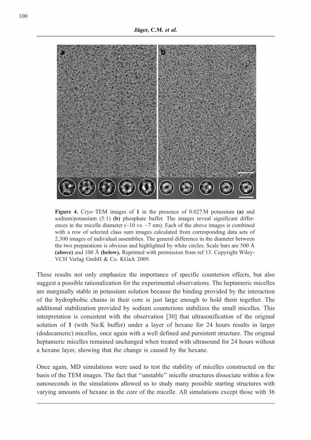
Figure 4. Cryo–TEM images of 1 in the presence of 0.027M potassium (a) and
sodium/potassium (5:1) (b) phosphate buffer. The images reveal significant differ-
ences in the micelle diameter (~10 vs. ~7 nm). Each of the above images is combined
with a row of selected class sum images calculated from corresponding data sets of
2,300 images of individual assemblies. The general difference in the diameter between
the two preparations is obvious and highlighted by white circles. Scale bars are 500 A
(above) and 100 A (below). Reprinted with permission from ref 13. Copyright Wiley-
VCH Verlag GmbH & Co. KGaA 2009.
These results not only emphasize the importance of specific counterion effects, but also
suggest a possible rationalization for the experimental observations. The heptameric micelles
are marginally stable in potassium solution because the binding provided by the interaction
of the hydrophobic chains in their core is just large enough to hold them together. The
additional stabilization provided by sodium counterions stabilizes the small micelles. This
interpretation is consistent with the observation [30] that ultrasonification of the original
solution of 1 (with Na:K buffer) under a layer of hexane for 24 hours results in larger
(dodecameric) micelles, once again with a well defined and persistent structure. The original
heptameric micelles remained unchanged when treated with ultrasound for 24 hours without
a hexane layer, showing that the change is caused by the hexane.
Once again, MD simulations were used to test the stability of micelles constructed on the
basis of the TEM images. The fact that ‘‘unstable’’ micelle structures dissociate within a few
nanoseconds in the simulations allowed us to study many possible starting structures with
varying amounts of hexane in the core of the micelle. All simulations except those with 36
100
Jager, C.M. et al.
hexane molecules led to fast dissociation of the micelles into smaller aggregates, whereas
that with 36 remained stable over 100 ns, both with sodium and potassium counterions [30].
The structure of the micelles in the simulations consisted of an equatorial ring of seven
dendrimers with two different caps of two and three, whereas the reconstructed cryo-TEM
3D structures suggest two equivalent caps, each consisting of three dendrimer monomers,
and a central ring of six dendrimers. This discrepancy is small and may either be caused by
force-field deficiencies or by the fact that the MD simulations sampled a slightly less stable
structure than that found in the experimental studies. However, the extremely well resolved
cryo-TEM images pointed to a further indication of the importance of the alkali-metal
counterions.
Attraction Between Polycarboxylates
The 3D-reconstructions of the cryo-TEM images suggest orientations for the polycarbox-
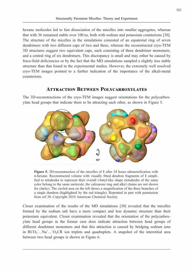
ylate head groups that indicate them to be attracting each other, as shown in Figure 5.
Figure 5. 3D-reconstruction of the micelles of 1 after 24 hours ultrasonification with
n-hexane. Reconstructed volume with visually fitted dendron fragments of 1 simpli-
fied to tetrahedra to represent their overall t-butyl-like shape (tetrahedra of the same
color belong to the same molecule, the calixarene ring and alkyl chains are not shown
for clarity). The circled area on the left shows a magnification of the three branches of
a single dendron (highlighted by the red triangle). Reprinted in part with permission
from ref 30. Copyright 2010 American Chemical Society.
Closer examination of the results of the MD simulations [30] revealed that the micelles
formed by the sodium salt have a more compact and less dynamic structure than their
potassium equivalent. Closer examination revealed that the orientation of the polycarbox-
ylate head groups in the former case does indicate attraction between head groups of
different dendrimer monomers and that this attraction is caused by bridging sodium ions
in RCO27...Na+...7O2CR ion triplets and quadruplets. A snapshot of the interstitial area
between two head groups is shown in Figure 6.
101
Structurally Persistent Micelles: Theory and Experiment
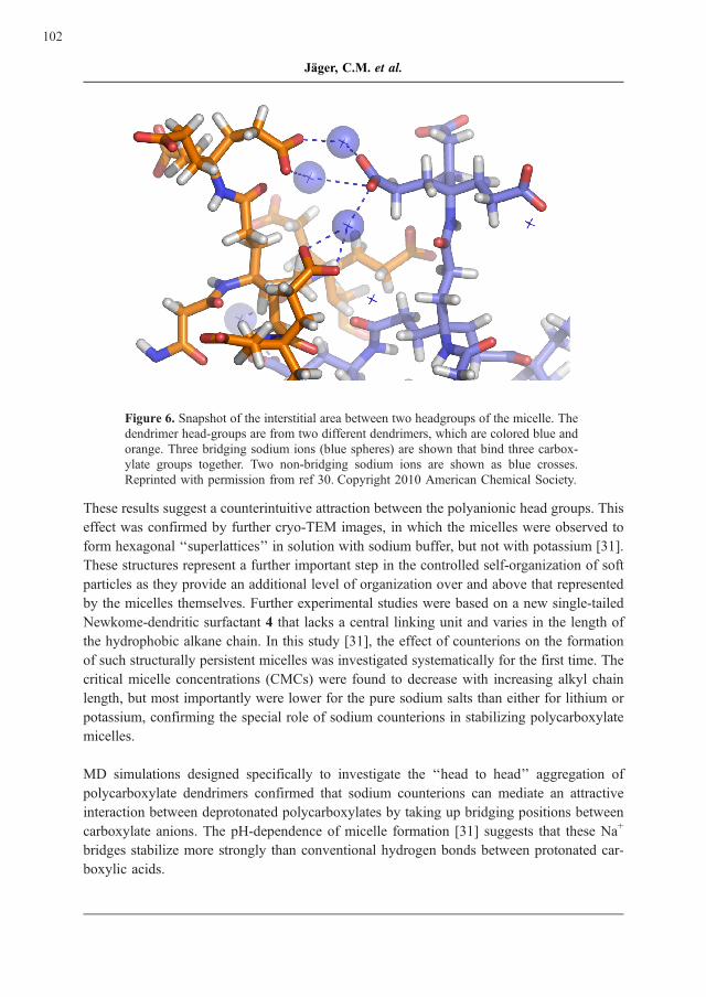
Figure 6. Snapshot of the interstitial area between two headgroups of the micelle. The
dendrimer head-groups are from two different dendrimers, which are colored blue and
orange. Three bridging sodium ions (blue spheres) are shown that bind three carbox-
ylate groups together. Two non-bridging sodium ions are shown as blue crosses.
Reprinted with permission from ref 30. Copyright 2010 American Chemical Society.
These results suggest a counterintuitive attraction between the polyanionic head groups. This
effect was confirmed by further cryo-TEM images, in which the micelles were observed to
form hexagonal ‘‘superlattices’’ in solution with sodium buffer, but not with potassium [31].
These structures represent a further important step in the controlled self-organization of soft
particles as they provide an additional level of organization over and above that represented
by the micelles themselves. Further experimental studies were based on a new single-tailed
Newkome-dendritic surfactant 4 that lacks a central linking unit and varies in the length of
the hydrophobic alkane chain. In this study [31], the effect of counterions on the formation
of such structurally persistent micelles was investigated systematically for the first time. The
critical micelle concentrations (CMCs) were found to decrease with increasing alkyl chain
length, but most importantly were lower for the pure sodium salts than either for lithium or
potassium, confirming the special role of sodium counterions in stabilizing polycarboxylate
micelles.
MD simulations designed specifically to investigate the ‘‘head to head’’ aggregation of
polycarboxylate dendrimers confirmed that sodium counterions can mediate an attractive
interaction between deprotonated polycarboxylates by taking up bridging positions between
carboxylate anions. The pH-dependence of micelle formation [31] suggests that these Na+
bridges stabilize more strongly than conventional hydrogen bonds between protonated car-
boxylic acids.
102
Jager, C.M. et al.
One remarkable feature of all these results is how well the simple force-field based simula-
tions are able to reproduce the quite subtle effects observed experimentally. There has been
considerable discussion of the lack of accuracy of force fields for metal ions, or more
accurately of combinations of force fields for metal ions and for water [32 – 34]. Surpris-
ingly, the interactions between ions of opposite charge seem to be far less critical than the
hydration of ions. Both direct MD simulations using the PM3 semi-empirical molecular
orbital Hamiltonian and subsequent geometry optimizations with density-functional theory
density reproduce the structures [31] observed in snapshots from the classical MD simula-
tions. The strongest argument for the reliability of the simulations, however, is that they have
been able to reproduce and in many cases even predict the unusual effects observed experi-
mentally.
Summary and Outlook
Structurally persistent micelles remain fascinating research objects, both from the point of
view of potential technological applications and because they reveal effects that are hidden
in more complex or less well defined systems. Above all, the combination of synthesis, cryo-
TEM and simulations has proven to be extraordinarily powerful and to lead to significant
progress. It is important in this respect that impulses for new research directions and specific
experiments or simulations may come from both experiment and simulation.
Acknowledgments
We are especially grateful for support from the Interdisciplinary Center for Molecular
Materials (ICMM) of the Universitat Erlangen-Nurnberg, from the Excellence Cluster ‘‘En-
gineering of Advanced Materials’’ (EAM), funded by the Deutsche Forschungsgemeinschaft
and a generous funding to C. B. (BO 1000/6 – 1).
References
[1] Shah, D.O. (Ed.) Micelles, Microemeulsions and Monolayers, Marcel Dekker, New
York, 1998.
[2] IUPAC. Compendium of Chemical Terminology, 2nd ed. (the ‘‘Gold Book’’). Com-
piled by McNaught, A.D. and Wilkinson, A. Blackwell Scientific Publications, Ox-
ford (1997). XML on-line corrected version: http://goldbook.iupac.org (2006) created
by Nic, M., Jirat, J., Kosata, B. updates compiled by Jenkins, A. ISBN 0 – 9678550 –
9-8.
doi: 10.1351/goldbook.
103
Structurally Persistent Micelles: Theory and Experiment
[3] Kellermann, M., Bauer, W., Hirsch, A., Schade, B., Ludwig, K., Bottcher, C. (2004)
The First Account of a Structurally Persistent Micelle. Angew. Chem. Int. Ed.
43:2959 – 2962.
doi: 10.1002/anie.200353510
[4] Burghardt, S., Hirsch, A., Schade, B., Ludwig, K., Bottcher, C. (2005) Switchable
Supramolecular Organization of Structurally Defined Micelles Based on an Amphi-
philic Fullerene. Angew. Chem. Int. Ed. 44:2976 – 2979.
doi: 10.1002/anie.200462465
[5] Schade, B., Ludwig, K., Bottcher, C., Hartnagel, U., Hirsch, A. (2007) Supramole-
cular Structure of 5-nm Spherical Micelles with D3 Symmetry Assembled from
Amphiphilic [3:3]-Hexakis Adducts. Angew. Chem. Int. Ed. 46:4393 – 4396.
[6] Hirsch, A. (2008) Amphiphilic architectures based on fullerene and calixarene plat-
forms: From buckysomes toshape-persistent micelles. Pure Appl. Chem. 80:571 –
587.
doi: 10.1351/pac200880030571
[7] Becherer, M., Schade, B., Bottcher, C., Hirsch, A. (2009) Supramolecular Assembly
of Self-Labeled Amphicalixarenes. Chem. Eur. J. 15:1637 – 1648.
doi: 10.1002/chem.200802008
[8] Schmidt, C.D., Bottcher, C., Hirsch, A. (2007) Synthesis and aggregation properties
of water-soluble Newkome-dendronized perylenetetracarboxdiimines. Eur. J. Org.
Chem. 5497 – 5505.
[9] Schmidt, C.D., Bottcher, C., Hirsch, A. (2009) Chiral Water-Soluble Perylenedii-
mides. Eur. J. Org. Chem. 5337 – 5349.
doi: 10.1002/ejoc.200900777
[10] van Heel, M., Harauz, G., Orlova, E.V., Schmidt, R., Schatz, M. (1996) A new
generation of the IMAGIC image processing system. J. Struct. Biol. 116:17 – 24.
doi: 10.1006/jsbi.1996.0004
[11] van Heel, M. (1987) Angular reconstitution: a posteriori assignment of projection
directions for 3D reconstruction. Ultramicroscopy 21:111 – 123.
doi: 10.1016/0304-3991(87)90078-7
[12] Orlova, E.V., Dube, P., Harris, J.R., Beckman, E., Zemlin, F., Markl, J., van Heel, M.
(1997) Structure of keyhole limpet hemocyanin type 1 (KLH1) at 15 A resolution by
electron cryomicroscopy and angular reconstitution. J. Mol. Biol. 271:417 – 437.
doi: 10.1006/jmbi.1997.1182
104
Jager, C.M. et al.
[13] Jager, C.M., Hirsch, A., Schade, B., Bottcher, C., Clark, T. (2009) Counterions
Control the Self-Assembly of Structurally Persistent Micelles; Theoretical Prediction
and Experimental Observation of Stabilization by Sodium Ions. Chem. Eur. J.
15:8586 – 8592.
doi: 10.1002/chem.200900885
[14] Williams, D.B., Carter, C.B. Transmission Electron Microscopy: A Textbook for
Materials Science, Plenum Press, New York, 1996.
[15] Stewart, J.J.P. (1989) Optimization of parameters for semiempirical methods I.
Method. J. Comp. Chem. 10:209 – 220.
doi: 10.1002/jcc.540100208
[16] Stewart, J.J.P. (1989) Optimization of parameters for semiempirical methods II.
Applications. J. Comp. Chem. 10:221 – 246.
doi: 10.1002/jcc.540100209
[17] Abernethy, J.L. (1967) Franz Hofmeister – the Impact of his Life and Research on
Chemistry. J. Chem. Ed. 44:177 – 180.
doi: 10.1021/ed044p177
[18] See, for instance Zhang, Y., Cremer, P.S. (2006) Interactions between macromole-
cules and ions: The Hofmeister series. Curr. Op. Chem. Biol. 10:658-663.
[19] Hofmeister, F. (1888) Zur Lehre von der Wirkung der Salze. Zweite Mittheilung
Arch. Exp. Pathol. Pharmakol. 24:247 – 260.
doi:10.1007/BF01918191
[20] Kunz, W., Henle, J., Ninham, B.W. (2004) Zur Lehre von der Wirkung der Salze
(About the science of the effect of salts): Franz Hofmeister’s historical papers. Curr.
Op. Colloid Interface Sci. 9:19 – 37.
doi: 10.1016/j.cocis.2004.05.005
[21] Kunz, W., LoNostro, P., Ninham, B.W. (2004) The Present State of Affairs with
Hofmeister Effects. Curr. Op. Colloid Interface Sci. 9:1 – 18.
doi: 10.1016/j.cocis.2004.05.004
[22] Collins, K.D, Washabaugh, M.W. (1985) The Hofmeister effect and the behaviour of
water at interfaces. Q. Rev. Biophys. 18:323 – 422.
doi: 10.1017/S0033583500005369
[23] Washabaugh, M.W., Collins, K.D. (1986) The systematic characterization by aqueous
column chromatography of solutes which affect protein stability. J. Biol. Chem.
261:12477 – 12485.
105
Structurally Persistent Micelles: Theory and Experiment
[24] Collins, K.D. (1995) Sticky ions in biological systems. Proc. Natl. Acad. Sci. U.S.A.
92:5553 – 5557.
doi: 10.1073/pnas.92.12.5553
[25] Neilson, G.W., Enderby, J.E. (1996) Aqueous Solutions and Neutron Scattering. J.
Phys. Chem. 100:1317 – 1322.
doi: 10.1021/jp951490y
[26] Enderby, J.E. (1995) Ion solvation via neutron scattering. Chem. Soc. Rev. 24:159 –
168.
doi: 10.1039/cs9952400159
[27] Collins, K.D., Neilson, G.W., Enderby, J.E. (2007) Ions in water: characterizing the
forces that control chemical processes and biological structure. Biophys. Chem.
128:95 – 104.
doi: 10.1016/j.bpc.2007.03.009
[28] Marcus, Y. (2009) Effect of ions on the structure of water: Structure making and
breaking. Chem. Rev. 109:1346 – 1370.
doi: 10.1021/cr8003828
[29] Collins, K.D. (2006) Ion hydration: Implications for cellular function, polyelectro-
lytes, and protein crystallization. Biophys. Chem. 119:271 – 281.
doi: 10.1016/j.bpc.2005.08.010
[30] Jager, C.M., Hirsch, A., Schade, B., Ludwig, K., Bottcher, C., Clark, T. (2010) Self-
Assembly of Structurally Persistent Micelles is Controlled by Specific Ion Effects
and Hydrophobic Guest. Langmuir 26:10460 – 10466.
[31] Rosenlehner, K., Schade, B., Bottcher, C., Jager, C.M., Clark, T., Hirsch A. (2010)
Sodium-Effect on the Self-Organization of Amphiphilic Carboxylates: Formation of
Structured Micelles and Superlattices. Chem. Eur. J. 16:9544 – 9554.
doi: 10.1002/chem.201001150
[32] Joung, I.S., Cheatham, III, T.E. (2008) Determination of alkali and halide monovalent
ion parameters for use in explicitly solvated biomolecular simulations. J. Phys.
Chem. 112:9020 – 9041.
doi: 10.1021/jp8001614
[33] Horinek, D., Mamatkulov, S.I., Netz, R.R., (2009) Rational design of ion force fields
based on thermodynamic solvation properties. J. Chem. Phys. 130:124507.
doi: 10.1063/1.3081142
[34] Hess, B., van der Vegt, N. (2009) Cation specific binding with protein surface
charges. Proc. Natl. Acad. Sci. U.S.A. 109:13296 – 13300.
doi: 10.1073/pnas.0902904106l
106
Jager, C.M. et al.