structure determination by x-ray crystallography978-1-4614-3954-7/1.pdfpalmer’s structure...
TRANSCRIPT
Structure Determination by X-rayCrystallography

Official contribution of the National Institute of Standards and Technology; not subject to copyright in the United States
[Reproduced by courtesy of N. I. S. T.]

Mark Ladd • Rex Palmer
StructureDetermination byX-ray Crystallography
Analysis by X-rays and Neutrons
Fifth Edition
Celebrating the Centenary of
X-ray Crystallography

Additional material to this book can be downloaded from http://extra.springer.com.
ISBN 978-1-4614-3956-1 ISBN 978-1-4614-3954-7 (eBook)DOI 10.1007/978-1-4614-3954-7Springer New York Heidelberg Dordrecht London
Library of Congress Control Number: 2012947362
# Springer Science+Business Media New York 1977, 1985, 1994, 2003, 2013
This work is subject to copyright. All rights are reserved by the Publisher, whether the whole or
part of the material is concerned, specifically the rights of translation, reprinting, reuse of
illustrations, recitation, broadcasting, reproduction on microfilms or in any other physical way,
and transmission or information storage and retrieval, electronic adaptation, computer software,
or by similar or dissimilar methodology now known or hereafter developed. Exempted from this
legal reservation are brief excerpts in connection with reviews or scholarly analysis or material
supplied specifically for the purpose of being entered and executed on a computer system, for
exclusive use by the purchaser of the work. Duplication of this publication or parts thereof is
permitted only under the provisions of the Copyright Law of the Publisher’s location, in its
current version, and permission for use must always be obtained from Springer. Permissions for
use may be obtained through RightsLink at the Copyright Clearance Center. Violations are liable
to prosecution under the respective Copyright Law.
The use of general descriptive names, registered names, trademarks, service marks, etc. in this
publication does not imply, even in the absence of a specific statement, that such names are
exempt from the relevant protective laws and regulations and therefore free for general use.
While the advice and information in this book are believed to be true and accurate at the date of
publication, neither the authors nor the editors nor the publisher can accept any legal
responsibility for any errors or omissions that may be made. The publisher makes no warranty,
express or implied, with respect to the material contained herein.
Printed on acid-free paper
Springer is part of Springer Science+Business Media (www.springer.com)
Rex PalmerReader Emeritus in Structural CrystallographyBirkbeck College, University of London,London, England
Visiting Professor in X-ray Crystallography
University of Greenwich, England
Senior Visiting Research Fellow
Christ Church University,
Canterbury, England
Mark LaddFormerly Head of Chemical PhysicsUniversity of SurreyGuildford, England

When you can measure what you are speaking about andexpress it in numbers, you know something about it; but whenyou cannot express it in numbers, your knowledge is of a meagreand unsatisfactory kind; it may be the beginning of knowledge,but you have scarcely in your thoughts advanced to the state ofscience, whatever the matter may be.
Lord Kelvin


To Valentia and Hilda


Foreword
I am privileged to write the Foreword to this fifth edition of Ladd and
Palmer’s Structure Determination by X-ray Crystallography, a textbook
that is now world renowned and that has helped educate two generations of
crystallographers in the theory and practice of modern crystallography,
myself included. Indeed, a well-worn first edition of this venerable text
remains on my shelves today, now somewhat battered and bruised from
passage through the hands of successive students who have learned the
fundamentals of crystallography from its pages.
This new fifth edition is especially timely, marking as it does a century
of discovery in which X-ray diffraction, and diffraction of other radiations,
has opened a window to the atomic world. From fundamental knowledge of
atomic interactions and chemical bonds in the simplest materials to the
atomic resolution analysis of the molecular machines of the cell, crystallo-
graphic science underpins much of our understanding of the world we live in
today. In recent years, advances in diffraction theory, automated technolo-
gies, and computational tools have helped move crystallography from a
specialist discipline to a standard laboratory tool across many fields of
science. In some fields, these advances have been so spectacularly successful
that the solution of the crystal structures of all but the most challenging
systems is now considered largely routine. At the same time, the develop-
ment of a new generation of high powered synchrotron, neutron and, most
recently, free electron laser facilities are pushing crystallographic science to
new frontiers, aiming to provide diffraction from single molecules, to locate
light atoms such as hydrogen in crystal structures, and to move beyond static
crystal structures towards time-resolved analyses of structural dynamics at
pico-second timescales.
For the interdisciplinary students of today seeking a thorough and
detailed understanding of the principles and methods of modern crystallog-
raphy, Ladd and Palmer remains as essential and relevant today as when it
first appeared some 35 years ago. Building from the fundamental concepts of
crystallography, through crystal symmetry to the mathematical formalism of
diffraction and on to the principle and practice of structure determination, the
text provides an excellent introduction to the techniques and applications of
crystallography, illustrated throughout by applications to real world pro-
blems. The fifth edition is expanded and enhanced with updated examples
and description of recent technical developments and achievements in X-ray
crystallography and benefits from a completely new chapter that describes
ix

the application of neutron crystallography in structural science. This is an
important addition. Neutrons are scattered by atomic nuclei and have a
magnetic moment. Hence, neutron diffraction can be used to determine
accurate atomic and magnetic structures of materials. With a new generation
of neutron sources and instruments now coming on-line, these properties will
be increasingly exploited in fundamental studies of new inorganic, organic,
and biological systems, of superconducting and magnetic materials, and for
structure-function analysis of hydrogen atoms in macromolecules.
Extending the scope of this classic text beyond the purely X-ray Crystal-
lography of its title to include diffraction of other radiations acknowledges
some of the new frontiers and ever-increasing impact of crystallographic
analysis in structural sciences. As has been the case for the last 35 years,
Ladd and Palmer is set to educate and equip the students of today to drive and
inspire the developments of tomorrow!
Neutron Sciences Directorate
Oak Ridge National Laboratory, TN, USA Dean A.A. Myles
x Foreword

Preface to the Fifth Edition
We were honoured to be asked by Springer, New York to prepare a fifth
edition of Structure Determination by X-ray Crystallography. First published
in 1977 under the Plenum imprint, this book has received wide acclaim in
both teaching and research in X-ray crystallography because of its extensive
and detailed coverage of all aspects of the subject.
As we prepare this new edition, we are entering the centenary of the
discovery of X-ray diffraction in 1912, the beginning of X-ray crystallogra-
phy as a science in its own right. Today, X-ray crystallography and the
complementary technique of neutron diffraction together provide the most
powerful tools for the investigation and elucidation of crystal and molecular
structures. X-ray and neutron crystallography may be described as the sci-
ence of the structure of materials, in the widest sense of the phrase, and their
ramifications are evident across a broad spectrum of scientific endeavour.
The power of computers and available software has unleashed an unprec-
edented ability to carry out with speed the complicated calculations involved
in crystal structure determination on a desktop PC. This is paralleled by the
availability of powerful X-ray and neutron sources and low temperature
devices for facilitating measurements at liquid nitrogen temperature or
lower, which provide ever higher precision in the determination of crystal
structures. However, a detailed knowledge of the theory underlying the
process of crystal structure determination is still required in order both to
ensure that the literature contains correct well-determined structures and to
understand the complexities introduced by features such as disorder and
twinning in crystals. There are many pitfalls in crystal structure determina-
tion to trap the unwary.
In this new edition, we have continued the approach that has been well
reviewed in its earlier editions. We have always kept in mind that students
meeting X-ray crystallography for the first time are encountering a new disci-
pline, and not merely extending the range of a subject already studied. In
consequence, we have chosen, for example, to discuss the geometry and sym-
metry of crystals in rather more detail than is found in other books on this
subject, for it is our experience that some of the difficulties that students meet in
introductory X-ray crystallography lie in their unfamiliarity with a three-
dimensional concept, whether they be final-year undergraduate or post-graduate
students in chemistry, biochemistry, materials science, geology, bioinformatics,
information technology, or physics. Both low molecular weight (small
molecules) and macromolecular methods (proteins) are covered in detail.
xi

As well as retaining and thoroughly revising the overall contents of the
earlier editions, we have added a significant chapter on neutron diffraction
studies, and sections introducing Molecular Modelling and Structure Predic-
tion. In order to maintain a workable size for the book, a number of elabora-
tions of mainly mathematical argument have been stored as Web Appendices
on the website http://extras.springer.com.
Although several novel methods have been added to the armoury of
crystal structure determination, we limit our discussion principally to Patter-
son interpretation, Direct Methods, Isomorphous and Molecular Replace-
ment and Powder Crystallography, and developments from them. The basic
problem remains the determination of the phases of X-ray reflections, and
this problem is addressed in these techniques discussed herein. In order to
simulate the actual process of structure determination, we are fortunate to be
able to include the XRAY program package prepared by Dr. Neil Bailey and
colleagues of the University of Sheffield, and we are grateful to him for
permission to use it in the present context. It has been modified (M.L.) for PC
operation and several enhancements made, including the presentation of
Fourier contour maps on the monitor. Although this package uses two-
dimensional data, much valuable insight into X-ray structure determination
can be gained, and a number of sets of X-ray data are included.
There are now numerous computer packages available for the many
aspects of crystallography that are in current use. We have referred to them
freely within the text, and they have been collected in an appendix together
with a reference to a source for each so that they become readily available to
the practising crystallographer. There are numerous references to each chap-
ter including website addresses for topics of crystallographic importance.
References among the text are given as “Sect. 1.2.3,” which refers to that
section in Chap. 1, or as “(3.4)” which refers to that equation in Chap. 3.
Each chapter contains a set of problems designed to assist the reader in the
understanding of the textual material, and detailed tutorial solutions are
provided. Some of these problems require computer assistance, and a set of
programs has been designed and included with the Web material and dated 1
January 2013 (Version 5.1). In this context, we are grateful to Dr. Jan Vissser
of the Technisch Physische Dienst, Delft, Professor Armel Le Bail of Labor-
atoire Fluorures, Universite du Main, LeMans, and Professor A L Spek of the
University of Utrecht for the continued incorporation of the programs ITO12,
ESPOIR, and LEPAGE, respectively, in the Program Suite for this book.
Finally we thank Springer Science + Business Media for inviting this edition
and bringing it to a state of completion.
University of Surrey
Guildford, England Mark Ladd
Birkbeck College
London, England, London Rex Palmer
xii Preface to the Fifth Edition

Disclaimer
Every effort has been made to ensure the correct functioning of the software
associated with this book. However, the reader planning to use the software
should note that, from the legal point of view, there is no warranty, expressed
or implied, that the programs are free from error or will prove suitable for a
particular application; by using the software the reader accepts full responsi-
bility for all the results produced, and the authors and publisher disclaim all
liability from any consequences arising from the use of the software. The
software should not be relied upon for solving a problem, the incorrect
solution of which could result in injury to a person or loss of property. If
you do use the programs in such a manner, it is at your own risk. The authors
and publisher disclaim all liability for direct or consequential damages
resulting from your use of the programs.
xiii


Table of Contents
Periodic Table . . . . . . . . . . . . . . . . . . . . . . . . . . . . . . . . . . . . . . . . . . . . . . . . . . . . . . . . . . ii
Physical Constants and Other Numerical Data . . . . . . . . . . . . . . . . . . . . . xxxi
Notation . . . . . . . . . . . . . . . . . . . . . . . . . . . . . . . . . . . . . . . . . . . . . . . . . . . . . . . . . . . . . . . . . .xxxiii
1 Crystal Morphology and Crystal Symmetry . . . . . . . . . . . . . . . . . . . . 1
1.1 Brief Historical Introduction . . . . . . . . . . . . . . . . . . . . . . . . . . . . . . . . . . 1
1.2 The Crystalline State . . . . . . . . . . . . . . . . . . . . . . . . . . . . . . . . . . . . . . . . . . 7
1.2.1 Crystallographic Reference Axes . . . . . . . . . . . . . . . . . . . . 7
1.2.2 Equation of a Plane . . . . . . . . . . . . . . . . . . . . . . . . . . . . . . . . . . . 7
1.2.3 Indices of Planes and the Law
of Rational Intercepts . . . . . . . . . . . . . . . . . . . . . . . . . . . . . . . . . 8
1.2.4 Axial Ratios . . . . . . . . . . . . . . . . . . . . . . . . . . . . . . . . . . . . . . . . . . . . 12
1.2.5 Zones . . . . . . . . . . . . . . . . . . . . . . . . . . . . . . . . . . . . . . . . . . . . . . . . . . . 12
1.3 Stereographic Projection: Brief Survey . . . . . . . . . . . . . . . . . . . . . . 15
1.4 External Symmetry of Crystals . . . . . . . . . . . . . . . . . . . . . . . . . . . . . . . 17
1.4.1 Two-Dimensional Point Groups . . . . . . . . . . . . . . . . . . . . . . 19
1.4.2 Three-Dimensional Point Groups . . . . . . . . . . . . . . . . . . . . 22
1.4.3 Quasicrystals, Buckyballs, and Icosahedral
Symmetry . . . . . . . . . . . . . . . . . . . . . . . . . . . . . . . . . . . . . . . . . . . . . . 32
1.5 Problems . . . . . . . . . . . . . . . . . . . . . . . . . . . . . . . . . . . . . . . . . . . . . . . . . . . . . . . 39
References and Bibliography . . . . . . . . . . . . . . . . . . . . . . . . . . . . . . . . . . . . . . . . 49
2 Lattices and Space-Group Theory . . . . . . . . . . . . . . . . . . . . . . . . . . . . . . . . 51
2.1 Introduction . . . . . . . . . . . . . . . . . . . . . . . . . . . . . . . . . . . . . . . . . . . . . . . . . . . . 51
2.2 Lattices . . . . . . . . . . . . . . . . . . . . . . . . . . . . . . . . . . . . . . . . . . . . . . . . . . . . . . . . . 51
2.2.1 Two-Dimensional Lattices . . . . . . . . . . . . . . . . . . . . . . . . . . . . 52
2.2.2 Choice of Unit Cell . . . . . . . . . . . . . . . . . . . . . . . . . . . . . . . . . . . 53
2.2.3 Three-Dimensional Lattices . . . . . . . . . . . . . . . . . . . . . . . . . . 54
2.3 Families of Planes and Interplanar Spacings . . . . . . . . . . . . . . . . . 62
2.4 Reciprocal Lattice: Geometrical Treatment . . . . . . . . . . . . . . . . . . 63
2.5 Unit-Cell Transformations . . . . . . . . . . . . . . . . . . . . . . . . . . . . . . . . . . . . 65
2.5.1 Bravais Unit-Cell Vectors . . . . . . . . . . . . . . . . . . . . . . . . . . . . 65
2.5.2 Directions (Zone Symbols) . . . . . . . . . . . . . . . . . . . . . . . . . . . 66
2.5.3 Coordinates of Sites in the Unit Cell . . . . . . . . . . . . . . . . . 67
2.5.4 Miller Indices . . . . . . . . . . . . . . . . . . . . . . . . . . . . . . . . . . . . . . . . . . 67
2.5.5 Reciprocal Unit-Cell Vectors . . . . . . . . . . . . . . . . . . . . . . . . . 68
2.6 Rotational Symmetries of Lattices . . . . . . . . . . . . . . . . . . . . . . . . . . . 71
xv

2.7 Space Groups . . . . . . . . . . . . . . . . . . . . . . . . . . . . . . . . . . . . . . . . . . . . . . . . . 72
2.7.1 Two-Dimensional Space Groups
(Plane Groups) . . . . . . . . . . . . . . . . . . . . . . . . . . . . . . . . . . . . . . 73
2.7.2 Plane Groups Related to 2mm . . . . . . . . . . . . . . . . . . . . . 79
2.7.3 Three-Dimensional Space Groups . . . . . . . . . . . . . . . . . 81
2.7.4 Screw Axes . . . . . . . . . . . . . . . . . . . . . . . . . . . . . . . . . . . . . . . . . . 84
2.7.5 Glide Planes . . . . . . . . . . . . . . . . . . . . . . . . . . . . . . . . . . . . . . . . . 87
2.7.6 Analysis of the Space-Group Symbol . . . . . . . . . . . . . 90
2.7.7 Orthorhombic Space Groups . . . . . . . . . . . . . . . . . . . . . . . 91
2.7.8 Relative Orientations of Symmetry
Elements in Space Groups . . . . . . . . . . . . . . . . . . . . . . . . . 93
2.7.9 Tetragonal and Hexagonal Space Groups . . . . . . . . . 95
2.8 Matrix Representation of Symmetry Operations . . . . . . . . . . . 98
2.8.1 Matrices in Point-Group Symmetry . . . . . . . . . . . . . . . 98
2.8.2 Matrices in Space-Group Symmetry . . . . . . . . . . . . . . . 100
2.9 Diffraction Symbols . . . . . . . . . . . . . . . . . . . . . . . . . . . . . . . . . . . . . . . . . . 101
2.10 Some Other Types of Symmetry . . . . . . . . . . . . . . . . . . . . . . . . . . . . 103
2.10.1 Black-White Symmetry . . . . . . . . . . . . . . . . . . . . . . . . . . . . 103
2.10.2 Color Symmetry . . . . . . . . . . . . . . . . . . . . . . . . . . . . . . . . . . . . 104
2.11 Problems . . . . . . . . . . . . . . . . . . . . . . . . . . . . . . . . . . . . . . . . . . . . . . . . . . . . . . 106
References . . . . . . . . . . . . . . . . . . . . . . . . . . . . . . . . . . . . . . . . . . . . . . . . . . . . . . . . . . . . 109
3 X-Rays and X-Ray Diffraction . . . . . . . . . . . . . . . . . . . . . . . . . . . . . . . . . . . . 111
3.1 Generation and Properties of X-Rays . . . . . . . . . . . . . . . . . . . . . . . 111
3.1.1 X-Rays and White Radiation . . . . . . . . . . . . . . . . . . . . . . . 111
3.1.2 Characteristic Radiation . . . . . . . . . . . . . . . . . . . . . . . . . . . . 113
3.1.3 Absorption of X-Rays . . . . . . . . . . . . . . . . . . . . . . . . . . . . . . 114
3.1.4 Monochromatic Radiation . . . . . . . . . . . . . . . . . . . . . . . . . . 116
3.1.5 Collimation . . . . . . . . . . . . . . . . . . . . . . . . . . . . . . . . . . . . . . . . . . 116
3.1.6 Synchrotron Sources . . . . . . . . . . . . . . . . . . . . . . . . . . . . . . . . 118
3.2 X-Ray Scattering . . . . . . . . . . . . . . . . . . . . . . . . . . . . . . . . . . . . . . . . . . . . . 121
3.2.1 Scattering by a Single Electron . . . . . . . . . . . . . . . . . . . . 122
3.2.2 Scattering by Two or More Electrons . . . . . . . . . . . . . 122
3.2.3 Waves and Wave Sums . . . . . . . . . . . . . . . . . . . . . . . . . . . . 123
3.2.4 Coherent and Incoherent Scattering . . . . . . . . . . . . . . . 127
3.2.5 Scattering by an Atom . . . . . . . . . . . . . . . . . . . . . . . . . . . . . . 128
3.3 Scattering by Regular Arrays of Atoms . . . . . . . . . . . . . . . . . . . . 130
3.3.1 Laue Equations . . . . . . . . . . . . . . . . . . . . . . . . . . . . . . . . . . . . . 130
3.3.2 Bragg Equation . . . . . . . . . . . . . . . . . . . . . . . . . . . . . . . . . . . . . 132
3.3.3 Equivalence of the Laue and Bragg
Equations . . . . . . . . . . . . . . . . . . . . . . . . . . . . . . . . . . . . . . . . . . . . 134
3.3.4 Further Analysis of the Path Difference . . . . . . . . . . . 135
3.4 Reciprocal Lattice: Analytical Treatment . . . . . . . . . . . . . . . . . . 135
3.4.1 Reciprocal Lattice Properties . . . . . . . . . . . . . . . . . . . . . . 137
3.4.2 Reciprocal Lattice and ReflectionCondition: Ewald Sphere . . . . . . . . . . . . . . . . . . . . . . . . . . . 138
3.5 Scattering by a Crystal Structure . . . . . . . . . . . . . . . . . . . . . . . . . . . . 139
3.5.1 Structure Factor Equation . . . . . . . . . . . . . . . . . . . . . . . . . . . 140
3.6 Using the Structure Factor Equation . . . . . . . . . . . . . . . . . . . . . . . . 140
3.6.1 Friedel’s Law . . . . . . . . . . . . . . . . . . . . . . . . . . . . . . . . . . . . . . . . . 140
3.6.2 Structure Factor for a Centrosymmetric Crystal . . . 141
xvi Table of Contents

3.7 Limiting Conditions and Systematic Absences . . . . . . . . . . . . . . 142
3.7.1 Body-Centered Unit Cell . . . . . . . . . . . . . . . . . . . . . . . . . . . . . 142
3.7.2 Screw Axes and Glide Planes . . . . . . . . . . . . . . . . . . . . . . . . 143
3.8 Practical Determination of Space Groups
from Diffraction Data . . . . . . . . . . . . . . . . . . . . . . . . . . . . . . . . . . . . . . . . . 152
3.8.1 Monoclinic Space Groups . . . . . . . . . . . . . . . . . . . . . . . . . . . . 153
3.8.2 Orthorhombic Space Groups . . . . . . . . . . . . . . . . . . . . . . . . . 154
3.8.3 Tetragonal Space Groups . . . . . . . . . . . . . . . . . . . . . . . . . . . . . 155
3.8.4 Hexagonal Space Groups . . . . . . . . . . . . . . . . . . . . . . . . . . . . . 155
3.9 Problems . . . . . . . . . . . . . . . . . . . . . . . . . . . . . . . . . . . . . . . . . . . . . . . . . . . . . . . 155
References . . . . . . . . . . . . . . . . . . . . . . . . . . . . . . . . . . . . . . . . . . . . . . . . . . . . . . . . . . . . 159
4 Intensities and Intensity Statistics . . . . . . . . . . . . . . . . . . . . . . . . . . . . . . . . 161
4.1 Intensity Expressions and Factors Affecting
Intensities . . . . . . . . . . . . . . . . . . . . . . . . . . . . . . . . . . . . . . . . . . . . . . . . . . . . . . 161
4.1.1 Polarization and Lorentz Factors . . . . . . . . . . . . . . . . . . . . . 162
4.1.2 Extinction . . . . . . . . . . . . . . . . . . . . . . . . . . . . . . . . . . . . . . . . . . . . . . 164
4.1.3 Absorption Measurement and Correction . . . . . . . . . . . . 165
4.1.4 Scaling . . . . . . . . . . . . . . . . . . . . . . . . . . . . . . . . . . . . . . . . . . . . . . . . . 167
4.1.5 Merging Equivalent Reflections . . . . . . . . . . . . . . . . . . . . . . 167
4.1.6 Practical Intensity Expression
and its Standard Deviation . . . . . . . . . . . . . . . . . . . . . . . . . . . . 168
4.1.7 Scale Factor for Fo . . . . . . . . . . . . . . . . . . . . . . . . . . . . . . . . . . . . 169
4.1.8 Thermal Vibrations and the Temperature
Factor . . . . . . . . . . . . . . . . . . . . . . . . . . . . . . . . . . . . . . . . . . . . . . . . . . 169
4.2 Intensity Statistics . . . . . . . . . . . . . . . . . . . . . . . . . . . . . . . . . . . . . . . . . . . . . 172
4.2.1 Determining Scale and Temperature
Factors . . . . . . . . . . . . . . . . . . . . . . . . . . . . . . . . . . . . . . . . . . . . . . . . . 172
4.2.2 Other Aspects of the Wilson Plot . . . . . . . . . . . . . . . . . . . . 175
4.2.3 Statistics of Reciprocal Space . . . . . . . . . . . . . . . . . . . . . . . . 175
4.2.4 Acentric and Centric Distributions
of Structure Factors . . . . . . . . . . . . . . . . . . . . . . . . . . . . . . . . . . . 177
4.2.5 Normalized Structure Factors . . . . . . . . . . . . . . . . . . . . . . . . 182
4.3 Problems . . . . . . . . . . . . . . . . . . . . . . . . . . . . . . . . . . . . . . . . . . . . . . . . . . . . . . . 185
References . . . . . . . . . . . . . . . . . . . . . . . . . . . . . . . . . . . . . . . . . . . . . . . . . . . . . . . . . . . . 186
5 Examination of Single Crystals: Optical and X-Ray
Diffraction Practice . . . . . . . . . . . . . . . . . . . . . . . . . . . . . . . . . . . . . . . . . . . . . . . . 187
5.1 Introduction . . . . . . . . . . . . . . . . . . . . . . . . . . . . . . . . . . . . . . . . . . . . . . . . . . . . 187
5.2 Crystal Growing . . . . . . . . . . . . . . . . . . . . . . . . . . . . . . . . . . . . . . . . . . . . . . . 187
5.2.1 Growing Crystals for X-Ray Diffraction . . . . . . . . . . . . 187
5.2.2 Crystallization from Solution . . . . . . . . . . . . . . . . . . . . . . . . . 188
5.2.3 Crystallization by Diffusion . . . . . . . . . . . . . . . . . . . . . . . . . . 188
5.2.4 Crystallization by Sublimation . . . . . . . . . . . . . . . . . . . . . . . 188
5.2.5 Other Issues . . . . . . . . . . . . . . . . . . . . . . . . . . . . . . . . . . . . . . . . . . . . 188
5.3 Optical Techniques . . . . . . . . . . . . . . . . . . . . . . . . . . . . . . . . . . . . . . . . . . . . 189
5.3.1 Polarized Light . . . . . . . . . . . . . . . . . . . . . . . . . . . . . . . . . . . . . . . . 189
5.3.2 Optical Classification of Crystals . . . . . . . . . . . . . . . . . . . . 190
5.3.3 Uniaxial Crystals . . . . . . . . . . . . . . . . . . . . . . . . . . . . . . . . . . . . . . 190
Table of Contents xvii

5.3.4 Birefringence . . . . . . . . . . . . . . . . . . . . . . . . . . . . . . . . . . . . . . . 192
5.3.5 Biaxial Crystals . . . . . . . . . . . . . . . . . . . . . . . . . . . . . . . . . . . . . 194
5.4 Single-Crystal X-Ray Diffraction Techniques:
Intensity Data Collection . . . . . . . . . . . . . . . . . . . . . . . . . . . . . . . . . . . . 197
5.4.1 Laue Method . . . . . . . . . . . . . . . . . . . . . . . . . . . . . . . . . . . . . . . . 197
5.4.2 Symmetry in Laue Photographs . . . . . . . . . . . . . . . . . . . 200
5.4.3 Laue Method and Synchrotron Radiation . . . . . . . . . 200
5.4.4 Oscillation Method . . . . . . . . . . . . . . . . . . . . . . . . . . . . . . . . . 205
5.5 Measurement of the Intensities of Diffraction Data . . . . . . . . 208
5.5.1 Single Counter or Serial Diffractometers . . . . . . . . . 209
5.6 Single-Crystal X-Ray Diffractometry . . . . . . . . . . . . . . . . . . . . . . . 209
5.6.1 Instrument Geometry . . . . . . . . . . . . . . . . . . . . . . . . . . . . . . . 209
5.6.2 Rotation of the Crystal into a
Diffracting Position . . . . . . . . . . . . . . . . . . . . . . . . . . . . . . . . . 210
5.6.3 Transformation from Miller Indices
to Diffractometer Angles . . . . . . . . . . . . . . . . . . . . . . . . . . . 211
5.6.4 Data Collection . . . . . . . . . . . . . . . . . . . . . . . . . . . . . . . . . . . . . 211
5.6.5 Scanning Over a Peak: o/yVersus o Scans . . . . . . . . . . . . . . . . . . . . . . . . . . . . . . . . . . . . . 212
5.7 Area Detectors (Position-Sensitive Detectors) . . . . . . . . . . . . . 213
5.7.1 Multiwire Proportional Counter . . . . . . . . . . . . . . . . . . . 213
5.7.2 FAST Area Detector (Enraf–Nonius
FAST) . . . . . . . . . . . . . . . . . . . . . . . . . . . . . . . . . . . . . . . . . . . . . . . 215
5.7.3 Image Plate . . . . . . . . . . . . . . . . . . . . . . . . . . . . . . . . . . . . . . . . . 215
5.7.4 Charge-Coupled Device Area Detectors . . . . . . . . . . 217
5.7.5 The Tiled CCD . . . . . . . . . . . . . . . . . . . . . . . . . . . . . . . . . . . . . 219
5.7.6 Charge-Coupled Device Including Tiled
CCD Versus Image Plate . . . . . . . . . . . . . . . . . . . . . . . . . . 219
5.7.7 Data Collection Strategies . . . . . . . . . . . . . . . . . . . . . . . . . 219
5.7.8 The CMOS Detector, Pilatus 1M Detector
System, and Continuous Rotation . . . . . . . . . . . . . . . . . 221
5.7.9 Data Processing Software . . . . . . . . . . . . . . . . . . . . . . . . . . 222
5.7.10 Detectors and Diffractometers . . . . . . . . . . . . . . . . . . . . . 222
5.7.11 Other Diffractometer Systems . . . . . . . . . . . . . . . . . . . . . 223
5.8 Monochromators . . . . . . . . . . . . . . . . . . . . . . . . . . . . . . . . . . . . . . . . . . . . . 223
5.8.1 Single-Type Crystal Monochromators . . . . . . . . . . . . 224
5.8.2 Double-Type Crystal Monochromators . . . . . . . . . . . 224
5.8.3 Monochromators for SynchrotronRadiation . . . . . . . . . . . . . . . . . . . . . . . . . . . . . . . . . . . . . . . . . . . . 225
5.9 Focusing Mirrors . . . . . . . . . . . . . . . . . . . . . . . . . . . . . . . . . . . . . . . . . . . . . 225
5.10 Twinning . . . . . . . . . . . . . . . . . . . . . . . . . . . . . . . . . . . . . . . . . . . . . . . . . . . . . . 226
5.10.1 Morphology of Twinning . . . . . . . . . . . . . . . . . . . . . . . . . . 226
5.10.2 Twinning and X-Ray Diffraction . . . . . . . . . . . . . . . . . . 228
5.11 Problems . . . . . . . . . . . . . . . . . . . . . . . . . . . . . . . . . . . . . . . . . . . . . . . . . . . . . . 229
References . . . . . . . . . . . . . . . . . . . . . . . . . . . . . . . . . . . . . . . . . . . . . . . . . . . . . . . . . . . . 232
xviii Table of Contents

6 Fourier Series and Fourier Transforms . . . . . . . . . . . . . . . . . . . . . . . . 235
6.1 Image Formation and Focusing . . . . . . . . . . . . . . . . . . . . . . . . . . . . 235
6.2 Fourier Series . . . . . . . . . . . . . . . . . . . . . . . . . . . . . . . . . . . . . . . . . . . . . . . . 236
6.2.1 Analysis of the Square Wave . . . . . . . . . . . . . . . . . . . . . . 238
6.2.2 Exponential Forms of Fourier Series . . . . . . . . . . . . . . 240
6.3 Fourier Series in X-Ray Crystallography . . . . . . . . . . . . . . . . . . 241
6.3.1 One-Dimensional Function . . . . . . . . . . . . . . . . . . . . . . . . 241
6.3.2 Two- and Three-Dimensional Functions . . . . . . . . . . 243
6.3.3 Units of Electron Density . . . . . . . . . . . . . . . . . . . . . . . . . . 245
6.4 Holes and Atoms . . . . . . . . . . . . . . . . . . . . . . . . . . . . . . . . . . . . . . . . . . . . 245
6.5 Generalized Fourier Transform . . . . . . . . . . . . . . . . . . . . . . . . . . . . 246
6.5.1 Fourier Transform of a Molecule . . . . . . . . . . . . . . . . . . 248
6.5.2 Fourier Transform of a Unit Cell . . . . . . . . . . . . . . . . . . 248
6.6 Practice with Transforms . . . . . . . . . . . . . . . . . . . . . . . . . . . . . . . . . . . 249
6.6.1 Optical Diffractometer . . . . . . . . . . . . . . . . . . . . . . . . . . . . . 249
6.6.2 Single Hole . . . . . . . . . . . . . . . . . . . . . . . . . . . . . . . . . . . . . . . . . . 249
6.6.3 Two or More Holes . . . . . . . . . . . . . . . . . . . . . . . . . . . . . . . . . 250
6.6.4 Change of Origin . . . . . . . . . . . . . . . . . . . . . . . . . . . . . . . . . . . 252
6.6.5 Systematic Absences . . . . . . . . . . . . . . . . . . . . . . . . . . . . . . . 252
6.6.6 Reconstruction of the Image . . . . . . . . . . . . . . . . . . . . . . . 252
6.6.7 Transforms and Inverse Transforms . . . . . . . . . . . . . . . 255
6.6.8 Delta Function . . . . . . . . . . . . . . . . . . . . . . . . . . . . . . . . . . . . . . 258
6.6.9 Weighted Reciprocal Lattice . . . . . . . . . . . . . . . . . . . . . . . 259
6.7 Some General Properties of Transforms . . . . . . . . . . . . . . . . . . . 261
6.8 Convolution . . . . . . . . . . . . . . . . . . . . . . . . . . . . . . . . . . . . . . . . . . . . . . . . . 261
6.8.1 Convolution and Diffraction . . . . . . . . . . . . . . . . . . . . . . . 261
6.8.2 Convolution Integral . . . . . . . . . . . . . . . . . . . . . . . . . . . . . . . 262
6.8.3 Convolution and Crystal Structure . . . . . . . . . . . . . . . . 264
6.9 Structure Solution in Brief . . . . . . . . . . . . . . . . . . . . . . . . . . . . . . . . . 266
6.9.1 Use of Heavy Atoms . . . . . . . . . . . . . . . . . . . . . . . . . . . . . . . 266
6.9.2 General Phase-Free Transform:
Patterson Function . . . . . . . . . . . . . . . . . . . . . . . . . . . . . . . . . . 267
6.9.3 Sign Relationships . . . . . . . . . . . . . . . . . . . . . . . . . . . . . . . . . . 268
6.10 Problems . . . . . . . . . . . . . . . . . . . . . . . . . . . . . . . . . . . . . . . . . . . . . . . . . . . . . 270
References . . . . . . . . . . . . . . . . . . . . . . . . . . . . . . . . . . . . . . . . . . . . . . . . . . . . . . . . . . . 272
7 Fourier Techniques in X-Ray Structure
Determination . . . . . . . . . . . . . . . . . . . . . . . . . . . . . . . . . . . . . . . . . . . . . . . . . . . . . . 273
7.1 Introduction . . . . . . . . . . . . . . . . . . . . . . . . . . . . . . . . . . . . . . . . . . . . . . . . . . 273
7.2 Analysis of the Unit-Cell Contents . . . . . . . . . . . . . . . . . . . . . . . . 273
7.2.1 Papaverine Hydrochloride,
C20H21NO4·HCl . . . . . . . . . . . . . . . . . . . . . . . . . . . . . . . . . . . . 274
7.2.2 Naphthalene, C10H8 . . . . . . . . . . . . . . . . . . . . . . . . . . . . . . . . 275
7.2.3 Molecular Symmetry . . . . . . . . . . . . . . . . . . . . . . . . . . . . . . . 275
7.2.4 Special Positions . . . . . . . . . . . . . . . . . . . . . . . . . . . . . . . . . . . . 276
7.2.5 Nickel Tungstate, NiWO4 . . . . . . . . . . . . . . . . . . . . . . . . . . 276
Table of Contents xix

7.3 Interpretation of Electron Density Distributions . . . . . . . . . . . . . 278
7.3.1 Peak Heights and Weights . . . . . . . . . . . . . . . . . . . . . . . . . . . . 279
7.3.2 Computation and Display of Electron
Density Distributions . . . . . . . . . . . . . . . . . . . . . . . . . . . . . . . . . . 279
7.3.3 Projections . . . . . . . . . . . . . . . . . . . . . . . . . . . . . . . . . . . . . . . . . . . . . 279
7.4 Methods of Solving the Phase Problem . . . . . . . . . . . . . . . . . . . . . . 281
7.4.1 Number of Reflections in the Data Set . . . . . . . . . . . . . . . 281
7.4.2 The Patterson Function . . . . . . . . . . . . . . . . . . . . . . . . . . . . . . . 282
7.4.3 Positions and Weights of Peaks
in the Patterson Function . . . . . . . . . . . . . . . . . . . . . . . . . . . . . 285
7.4.4 Sharpened Patterson Function . . . . . . . . . . . . . . . . . . . . . . . . 287
7.4.5 Symmetry of the Patterson Function
for a Crystal of Space Group Pm . . . . . . . . . . . . . . . . . . . . . 288
7.4.6 Vector Interactions in Other Space
Groups . . . . . . . . . . . . . . . . . . . . . . . . . . . . . . . . . . . . . . . . . . . . . . . . . 289
7.4.7 Examples of the Use of the Patterson Function
in Solving the Phase Problem . . . . . . . . . . . . . . . . . . . . . . . . 289
7.4.8 Determination of the Chlorine Atom Positions
in Papaverine Hydrochloride . . . . . . . . . . . . . . . . . . . . . . . . . 296
7.4.9 Determination of the Mercury Atom
Positions in KHg2 . . . . . . . . . . . . . . . . . . . . . . . . . . . . . . . . . . . . . 296
7.5 Heavy-Atom Method and Partial Fourier Synthesis . . . . . . . . . 301
7.5.1 Reliability Factor . . . . . . . . . . . . . . . . . . . . . . . . . . . . . . . . . . . . . . 303
7.5.2 Pseudosymmetry in Electron Density Maps . . . . . . . . . 308
7.5.3 Successive Fourier Refinement . . . . . . . . . . . . . . . . . . . . . . . 309
7.5.4 Difference-Fourier Synthesis . . . . . . . . . . . . . . . . . . . . . . . . . 309
7.5.5 Limitations of the Heavy-Atom Method . . . . . . . . . . . . . 310
7.5.6 Patterson Selection . . . . . . . . . . . . . . . . . . . . . . . . . . . . . . . . . . . . 310
7.5.7 Isomorphous Replacement . . . . . . . . . . . . . . . . . . . . . . . . . . . . 312
7.5.8 Further Details of the Isomorphous
Replacement Phasing Procedure . . . . . . . . . . . . . . . . . . . . . 319
7.6 Anomalous Scattering . . . . . . . . . . . . . . . . . . . . . . . . . . . . . . . . . . . . . . . . . 325
7.6.1 The Flack x Parameter . . . . . . . . . . . . . . . . . . . . . . . . . . . . . . . . 326
7.6.2 Effect of Anomalous Scattering
on the Symmetry of Diffraction Patterns . . . . . . . . . . . . 330
7.6.3 Form of the Structure Factor for a Structure
Composed of Heavy-Atom Anomalous
Scattering Species . . . . . . . . . . . . . . . . . . . . . . . . . . . . . . . . . . . . . 332
7.6.4 Phasing by Use of Anomalous Scattering . . . . . . . . . . . 334
7.6.5 Resolution of the Phase Problem for Proteins
Using Anomalous Scattering Measurements
(SIRAS Method) . . . . . . . . . . . . . . . . . . . . . . . . . . . . . . . . . . . . . . 335
7.6.6 Protein Phasing Using the Multiple-Wavelength
Anomalous Dispersion Technique (MAD) with
Synchrotron Radiation (SR) . . . . . . . . . . . . . . . . . . . . . . . . . . 337
xx Table of Contents

7.7 Charge flipping . . . . . . . . . . . . . . . . . . . . . . . . . . . . . . . . . . . . . . . . . . . . . . . . 338
7.8 Location of Hydrogen Atoms . . . . . . . . . . . . . . . . . . . . . . . . . . . . . . . . . 339
7.9 Problems . . . . . . . . . . . . . . . . . . . . . . . . . . . . . . . . . . . . . . . . . . . . . . . . . . . . . . . 340
References . . . . . . . . . . . . . . . . . . . . . . . . . . . . . . . . . . . . . . . . . . . . . . . . . . . . . . . . . . . . 347
8 Direct Methods and Refinement . . . . . . . . . . . . . . . . . . . . . . . . . . . . . . . . . . 351
8.1 Introduction . . . . . . . . . . . . . . . . . . . . . . . . . . . . . . . . . . . . . . . . . . . . . . . . . . . . 351
8.2 Direct Methods of Phase Determination . . . . . . . . . . . . . . . . . . . . . 351
8.2.1 Normalized Structure Factors . . . . . . . . . . . . . . . . . . . . . . . 351
8.2.2 Structure Invariants and Origin-Fixing
Reflections . . . . . . . . . . . . . . . . . . . . . . . . . . . . . . . . . . . . . . . . . . . . 352
8.2.3 Sign Determination: Centrosymmetric
Crystals . . . . . . . . . . . . . . . . . . . . . . . . . . . . . . . . . . . . . . . . . . . . . . . 355
8.2.4 Amplitude Symmetry and Phase Symmetry . . . . . . . 358
8.2.5P
2-Listing . . . . . . . . . . . . . . . . . . . . . . . . . . . . . . . . . . . . . . . . . . . 358
8.2.6 Symbolic-Addition Procedure: Example . . . . . . . . . . . 359
8.2.7 Calculation of E Maps . . . . . . . . . . . . . . . . . . . . . . . . . . . . . . . 360
8.2.8 Phase Determination: Non-centrosymmetric
Crystals . . . . . . . . . . . . . . . . . . . . . . . . . . . . . . . . . . . . . . . . . . . . . . . 361
8.2.9 Enantiomorph Selection . . . . . . . . . . . . . . . . . . . . . . . . . . . . . 367
8.2.10 Phase Determination in Space Group P21 . . . . . . . . . . 368
8.2.11 Advantages and Disadvantages
of Symbolic Addition . . . . . . . . . . . . . . . . . . . . . . . . . . . . . . . . 371
8.2.12 Signs of Trouble, and Past Remedies
When the Structure Failed to Solve . . . . . . . . . . . . . . . . . 372
8.2.13 Triplets, Quartets, and the SHELX
Program Strategy . . . . . . . . . . . . . . . . . . . . . . . . . . . . . . . . . . . . . 372
8.2.14 The SHELX Computer Program System . . . . . . . . . . . 374
8.2.15 The WinGX Program System . . . . . . . . . . . . . . . . . . . . . . . 375
8.2.16 Direct Methods in the Program SHELX-97
for Small Molecules . . . . . . . . . . . . . . . . . . . . . . . . . . . . . . . . . 375
8.2.17 Example of a SHELX-97 Structure Solution:
Crystal Code Name BW202W92(R) . . . . . . . . . . . . . . . . 377
8.3 Patterson Search Methods . . . . . . . . . . . . . . . . . . . . . . . . . . . . . . . . . . . . . 380
8.3.1 General Comments for Small Molecules
and Macromolecules . . . . . . . . . . . . . . . . . . . . . . . . . . . . . . . . . 381
8.3.2 Intramolecular Interatomic Vectors
and Molecular Orientation . . . . . . . . . . . . . . . . . . . . . . . . . . 382
8.3.3 Intermolecular Interatomic Vectors:
Translation Stage of MR . . . . . . . . . . . . . . . . . . . . . . . . . . . . 384
8.3.4 Crystal Packing and Refinement
of the Structure . . . . . . . . . . . . . . . . . . . . . . . . . . . . . . . . . . . . . . . 385
8.3.5 Patterson Search Methods for Small
Molecules . . . . . . . . . . . . . . . . . . . . . . . . . . . . . . . . . . . . . . . . . . . . . 386
8.3.6 The Program PATSEE . . . . . . . . . . . . . . . . . . . . . . . . . . . . . . . 387
8.3.7 Examples of Structure Solution
Using PATSEE . . . . . . . . . . . . . . . . . . . . . . . . . . . . . . . . . . . . . . . 388
8.3.8 Shake and Bake . . . . . . . . . . . . . . . . . . . . . . . . . . . . . . . . . . . . . . 399
Table of Contents xxi

8.4 Least-Squares Refinement . . . . . . . . . . . . . . . . . . . . . . . . . . . . . . . . . . . 400
8.4.1 Unit-Cell Dimensions . . . . . . . . . . . . . . . . . . . . . . . . . . . . . . 401
8.4.2 Least-Squares Parameters . . . . . . . . . . . . . . . . . . . . . . . . . . 401
8.4.3 Theory of Least-Squares Refinement
and Strategies to Use . . . . . . . . . . . . . . . . . . . . . . . . . . . . . . . 405
8.4.4 Least-Squares Refinement Against Fo2 . . . . . . . . . . . . 407
8.4.5 Constraints and Restraints . . . . . . . . . . . . . . . . . . . . . . . . . 408
8.5 Molecular Geometry . . . . . . . . . . . . . . . . . . . . . . . . . . . . . . . . . . . . . . . . . 408
8.5.1 Bond Lengths and Angles . . . . . . . . . . . . . . . . . . . . . . . . . 408
8.5.2 Torsion Angles . . . . . . . . . . . . . . . . . . . . . . . . . . . . . . . . . . . . . 411
8.5.3 Conformational Analysis . . . . . . . . . . . . . . . . . . . . . . . . . . 412
8.5.4 Mean Planes . . . . . . . . . . . . . . . . . . . . . . . . . . . . . . . . . . . . . . . . 414
8.6 Precision . . . . . . . . . . . . . . . . . . . . . . . . . . . . . . . . . . . . . . . . . . . . . . . . . . . . . . 415
8.7 Correctness of a Structure Analysis . . . . . . . . . . . . . . . . . . . . . . . . . 416
8.7.1 Databases . . . . . . . . . . . . . . . . . . . . . . . . . . . . . . . . . . . . . . . . . . . 417
8.8 Limitations of X-Ray Structure Analysis . . . . . . . . . . . . . . . . . . . 419
8.9 Disorder in Single Crystals . . . . . . . . . . . . . . . . . . . . . . . . . . . . . . . . . . 419
8.10 Computer Prediction of Crystal Structures . . . . . . . . . . . . . . . . . 422
8.10.1 Crystal Structure of 5-Azauracil . . . . . . . . . . . . . . . . . . . 422
8.10.2 Developments in Computer Crystal Structure
Prediction . . . . . . . . . . . . . . . . . . . . . . . . . . . . . . . . . . . . . . . . . . . 425
8.11 Blind Structure Prediction of the Flexible
Molecule 1-Benzyl-1H-Tetrazole . . . . . . . . . . . . . . . . . . . . . . . . . . . 426
8.12 Problems . . . . . . . . . . . . . . . . . . . . . . . . . . . . . . . . . . . . . . . . . . . . . . . . . . . . . . 433
References . . . . . . . . . . . . . . . . . . . . . . . . . . . . . . . . . . . . . . . . . . . . . . . . . . . . . . . . . . . . 435
9 Examples of Crystal Structure Determination . . . . . . . . . . . . . . . . . . 439
9.1 Introduction . . . . . . . . . . . . . . . . . . . . . . . . . . . . . . . . . . . . . . . . . . . . . . . . . . . 439
9.2 Crystal Structure of 2-Bromobenzo[b]Indeno[1,2-e] Pyran . . . . . . . . . . . . . . . . . . . . . . . . . . . . . . . . . . . . . . . . . . 439
9.2.1 Preliminary Physical and X-Ray
Measurements . . . . . . . . . . . . . . . . . . . . . . . . . . . . . . . . . . . . . . . 439
9.2.2 Intensity Measurement and Correction . . . . . . . . . . . . 444
9.2.3 Structure Analysis in the xz Projection . . . . . . . . . . . . 446
9.2.4 Three-Dimensional Structure
Determination . . . . . . . . . . . . . . . . . . . . . . . . . . . . . . . . . . . . . . . 447
9.2.5 Refinement . . . . . . . . . . . . . . . . . . . . . . . . . . . . . . . . . . . . . . . . . . 449
9.2.6 Molecular Geometry . . . . . . . . . . . . . . . . . . . . . . . . . . . . . . . 451
9.3 Crystal Structure of Potassium
2-Hydroxy-3,4-Dioxocyclobut-1-ene-1-Olate
Monohydrate (KHSQ) . . . . . . . . . . . . . . . . . . . . . . . . . . . . . . . . . . . . . . . 455
9.3.1 Preliminary X-Ray and Physical
Measurements . . . . . . . . . . . . . . . . . . . . . . . . . . . . . . . . . . . . . . . 455
9.3.2 Intensity Measurement and Correction . . . . . . . . . . . . 456
9.3.3P
2-Listing . . . . . . . . . . . . . . . . . . . . . . . . . . . . . . . . . . . . . . . . . . 456
9.3.4 Specifying the Origin . . . . . . . . . . . . . . . . . . . . . . . . . . . . . . . 457
9.3.5 Sign Determination . . . . . . . . . . . . . . . . . . . . . . . . . . . . . . . . . 458
xxii Table of Contents

9.3.6 The E Map . . . . . . . . . . . . . . . . . . . . . . . . . . . . . . . . . . . . . . . . . . 459
9.3.7 Completion and Refinement
of the Structure . . . . . . . . . . . . . . . . . . . . . . . . . . . . . . . . . . . . . 462
9.4 Crystal and Molecular Structure and Absolute
Configuration of 3b-Acetoxy-6,7-Epidithio-19-Norlanosta-5,7,9,11-Tetraene . . . . . . . . . . . . . . . . . . . . . . . . . . . . . . . . 465
9.4.1 Preparation and Preliminary Optical
and X-Ray Examinations . . . . . . . . . . . . . . . . . . . . . . . . . . 466
9.4.2 X-Ray Measurement of the Unit-Cell
Dimensions and Intensities . . . . . . . . . . . . . . . . . . . . . . . . 466
9.4.3 Structure Determination and Refinement . . . . . . . . . 468
9.4.4 Absolute Configuration . . . . . . . . . . . . . . . . . . . . . . . . . . . . . 468
9.5 Discussion of the Structure . . . . . . . . . . . . . . . . . . . . . . . . . . . . . . . . . . 468
9.6 Some Remarks on X-Ray Structure
Determination . . . . . . . . . . . . . . . . . . . . . . . . . . . . . . . . . . . . . . . . . . . . . . . . 470
9.7 Biomolecular Modeling: Bioinformatics . . . . . . . . . . . . . . . . . . . 471
9.8 Docking Oligomycin into ATP Synthase:
Ligand and Receptor . . . . . . . . . . . . . . . . . . . . . . . . . . . . . . . . . . . . . . . . . 471
9.8.1 Why Modeling Studies? . . . . . . . . . . . . . . . . . . . . . . . . . . . 471
9.9 X-Ray Structures and Absolute Configurations
of the Antibiotics Oligomycins A,B, and C:
Inhibitors of ATP Synthase . . . . . . . . . . . . . . . . . . . . . . . . . . . . . . . . . . 472
9.9.1 Summary . . . . . . . . . . . . . . . . . . . . . . . . . . . . . . . . . . . . . . . . . . . . 473
9.9.2 Background . . . . . . . . . . . . . . . . . . . . . . . . . . . . . . . . . . . . . . . . . 474
9.9.3 Experimental . . . . . . . . . . . . . . . . . . . . . . . . . . . . . . . . . . . . . . . . 474
9.9.4 Structure Determination and Refinement . . . . . . . . . 475
9.9.5 Results . . . . . . . . . . . . . . . . . . . . . . . . . . . . . . . . . . . . . . . . . . . . . . 475
9.9.6 Discussion . . . . . . . . . . . . . . . . . . . . . . . . . . . . . . . . . . . . . . . . . . 475
9.9.7 Conformational Variations
in the Macrocyclic Structures . . . . . . . . . . . . . . . . . . . . . 478
9.10 Structure of ATP Synthase (ATPase): The Receptor . . . . . . 480
9.11 Docking Oligomycin into ATPase . . . . . . . . . . . . . . . . . . . . . . . . . . 481
9.11.1 ATP Synthase FO Model . . . . . . . . . . . . . . . . . . . . . . . . . . 481
9.11.2 Homology Modeling . . . . . . . . . . . . . . . . . . . . . . . . . . . . . . . 482
9.11.3 Refining the Model: Energy Minimization . . . . . . . 482
9.11.4 Creation of a Pocket for Docking
Oligomycin into the ATP Synthase FO . . . . . . . . . . . 483
9.12 Problems . . . . . . . . . . . . . . . . . . . . . . . . . . . . . . . . . . . . . . . . . . . . . . . . . . . . . . 484
References . . . . . . . . . . . . . . . . . . . . . . . . . . . . . . . . . . . . . . . . . . . . . . . . . . . . . . . . . . . . 487
10 Proteins and Macromolecular X-Ray Analysis . . . . . . . . . . . . . . . . . 489
10.1 Introduction . . . . . . . . . . . . . . . . . . . . . . . . . . . . . . . . . . . . . . . . . . . . . . . . . . . 489
10.1.1 What Is a Protein? . . . . . . . . . . . . . . . . . . . . . . . . . . . . . . . . . . 489
10.2 Crystallization of Proteins and Complexes
for X-Ray Analysis . . . . . . . . . . . . . . . . . . . . . . . . . . . . . . . . . . . . . . . . . . . 491
10.2.1 Introduction . . . . . . . . . . . . . . . . . . . . . . . . . . . . . . . . . . . . . . . . . 491
10.2.2 Crystallization Conditions
for Macromolecules . . . . . . . . . . . . . . . . . . . . . . . . . . . . . . . . 492
Table of Contents xxiii

10.2.3 Properties of Protein Crystals . . . . . . . . . . . . . . . . . . . . 492
10.2.4 Crystallization of Proteins . . . . . . . . . . . . . . . . . . . . . . . . 492
10.2.5 Molecular Purity . . . . . . . . . . . . . . . . . . . . . . . . . . . . . . . . . . 493
10.2.6 Practical Considerations . . . . . . . . . . . . . . . . . . . . . . . . . . 493
10.2.7 Batch Crystallization . . . . . . . . . . . . . . . . . . . . . . . . . . . . . . 493
10.2.8 Microbatch Screening . . . . . . . . . . . . . . . . . . . . . . . . . . . . . 493
10.2.9 Vapor Diffusion Techniques . . . . . . . . . . . . . . . . . . . . . 494
10.2.10 Co-crystallization . . . . . . . . . . . . . . . . . . . . . . . . . . . . . . . . . 496
10.2.11 How to Improve the Crystals . . . . . . . . . . . . . . . . . . . . . 496
10.2.12 Heavy-Atom Derivatives for MIR . . . . . . . . . . . . . . . 497
10.2.13 Protein Complex Crystals with Small Molecules 498
10.3 Crystal Mounting for X-Ray Data Collection . . . . . . . . . . . . . . 499
10.3.1 Mounting at Room Temperature . . . . . . . . . . . . . . . . . 499
10.3.2 Cryo-Crystallography . . . . . . . . . . . . . . . . . . . . . . . . . . . . . 499
10.4 Macromolecular Crystallography . . . . . . . . . . . . . . . . . . . . . . . . . . . 501
10.4.1 Space Groups . . . . . . . . . . . . . . . . . . . . . . . . . . . . . . . . . . . . . . 501
10.4.2 X-Ray Diffraction from Macromolecular
Crystals . . . . . . . . . . . . . . . . . . . . . . . . . . . . . . . . . . . . . . . . . . . . 501
10.4.3 Recording X-Ray Diffraction
from Macromolecular Crystals . . . . . . . . . . . . . . . . . . . 503
10.4.4 Measurement of X-Ray Diffraction
from Macromolecular Crystals . . . . . . . . . . . . . . . . . . . 505
10.4.5 Problems with Data Collection
and Suggested Cures . . . . . . . . . . . . . . . . . . . . . . . . . . . . . . 508
10.4.6 Preliminary Structure Determination:
Unit Cell and Symmetry . . . . . . . . . . . . . . . . . . . . . . . . . . 509
10.4.7 Ricin Agglutinin . . . . . . . . . . . . . . . . . . . . . . . . . . . . . . . . . . . 509
10.5 Types of Fourier Synthesis for Protein Analysis . . . . . . . . . . . 512
10.5.1 Reconstruction of the Molecular Structure . . . . . . 512
10.5.2 Difference Electron Density . . . . . . . . . . . . . . . . . . . . . . 513
10.5.3 The 2FoðhklÞ�jFcðhklÞj Map . . . . . . . . . . . . . . . . . . . . . 514
10.6 Determination of the Phases for Protein Crystals . . . . . . . . . . 514
10.6.1 Introduction . . . . . . . . . . . . . . . . . . . . . . . . . . . . . . . . . . . . . . . . 514
10.6.2 Isomorphous Replacement (MIR) . . . . . . . . . . . . . . . . 514
10.6.3 Preparation and Screening
of Heavy-Atom Derivatives . . . . . . . . . . . . . . . . . . . . . . 515
10.6.4 Molecular Replacement (MR) . . . . . . . . . . . . . . . . . . . . 516
10.6.5 Example of a Self-Rotation Function:
Ricin Agglutinin . . . . . . . . . . . . . . . . . . . . . . . . . . . . . . . . . . . 520
10.6.6 Molecular Replacement in Practice . . . . . . . . . . . . . . 521
10.6.7 Application of the AmoRe Algorithms
to Ricin Agglutinin . . . . . . . . . . . . . . . . . . . . . . . . . . . . . . . . 525
10.7 SIRAS and MAD Phasing . . . . . . . . . . . . . . . . . . . . . . . . . . . . . . . . . . . 526
xxiv Table of Contents
10.8 Use of Phase Information and Density
Modification . . . . . . . . . . . . . . . . . . . . . . . . . . . . . . . . . . . . . . . . . . . . . . . . . 528
10.8.1 Properties of r(xyz) for Proteins . . . . . . . . . . . . . . . . 528
10.8.2 Programs for Density Modification . . . . . . . . . . . . . 528
10.8.3 Preparing to Refine the Structure . . . . . . . . . . . . . . . 529
10.9 Macromolecular Structure Refinement
and Solvent and Ligand Fitting . . . . . . . . . . . . . . . . . . . . . . . . . . . . 531
10.9.1 Refinement Techniques . . . . . . . . . . . . . . . . . . . . . . . . . . 531
10.9.2 Simulated Annealing . . . . . . . . . . . . . . . . . . . . . . . . . . . . 533
10.9.3 Least-Squares Refinement: Constrained,Restrained and Other Protocols . . . . . . . . . . . . . . . . . 534
10.10 Structure Validation: Final Checks . . . . . . . . . . . . . . . . . . . . . . . . 537
10.10.1 R-Factors . . . . . . . . . . . . . . . . . . . . . . . . . . . . . . . . . . . . . . . . . 537
10.10.2 Evaluation of Errors . . . . . . . . . . . . . . . . . . . . . . . . . . . . . 539
10.11 Geometry Validation: Final Checks . . . . . . . . . . . . . . . . . . . . . . . 539
10.11.1 Bond Lengths, Bond Angles,
Planarity, and Chirality . . . . . . . . . . . . . . . . . . . . . . . . . . 539
10.11.2 Conformation . . . . . . . . . . . . . . . . . . . . . . . . . . . . . . . . . . . . 540
10.12 Humidity Control and the Use of Cryoprotectants
in Protein Crystallography . . . . . . . . . . . . . . . . . . . . . . . . . . . . . . . . . 545
10.13 Problems . . . . . . . . . . . . . . . . . . . . . . . . . . . . . . . . . . . . . . . . . . . . . . . . . . . . . 545
References . . . . . . . . . . . . . . . . . . . . . . . . . . . . . . . . . . . . . . . . . . . . . . . . . . . . . . . . . . . . 546
11 Neutron Diffraction from Single Crystals . . . . . . . . . . . . . . . . . . . . . . . 549
11.1 Introduction . . . . . . . . . . . . . . . . . . . . . . . . . . . . . . . . . . . . . . . . . . . . . . . . . 549
11.1.1 Refinement of Hydrogen Atom Positions . . . . . . 550
11.2 Neutrons, Neutron Sources, and Data Collection . . . . . . . . . 551
11.2.1 Neutrons . . . . . . . . . . . . . . . . . . . . . . . . . . . . . . . . . . . . . . . . . . 551
11.2.2 Neutron Sources . . . . . . . . . . . . . . . . . . . . . . . . . . . . . . . . . 551
11.2.3 Neutron Data Collection . . . . . . . . . . . . . . . . . . . . . . . . . 551
11.2.4 Thermal Neutrons . . . . . . . . . . . . . . . . . . . . . . . . . . . . . . . . 553
11.3 Neutron Scattering . . . . . . . . . . . . . . . . . . . . . . . . . . . . . . . . . . . . . . . . . . 553
11.3.1 Neutron Scattering Lengths . . . . . . . . . . . . . . . . . . . . . 554
11.4 Experimental Neutron Diffraction Data Collection . . . . . . . 554
11.4.1 LADI-III and VIVALDI at ILL, Grenoble . . . . . 555
11.4.2 Oak Ridge National Laboratory
(ORNL) . . . . . . . . . . . . . . . . . . . . . . . . . . . . . . . . . . . . . . . . . . . 556
11.4.3 Other Neutron Sources . . . . . . . . . . . . . . . . . . . . . . . . . . 559
11.5 Deuteration and Perdeuteration . . . . . . . . . . . . . . . . . . . . . . . . . . . . 559
11.6 Examples of Structure Determination
by Neutron Crystallography . . . . . . . . . . . . . . . . . . . . . . . . . . . . . . . . 560
11.7 X-Ray and Neutron Structure of 1,8-(3,6,9-
Trioxaundecane-1,11-diyldioxy)-9,10-
dihydro-10-10 dimethylanthracene-9-ol . . . . . . . . . . . . . . . . . . . 560
11.7.1 Experimental . . . . . . . . . . . . . . . . . . . . . . . . . . . . . . . . . . . . . 561
11.7.2 Structure Analysis and Refinement . . . . . . . . . . . . . 564
11.7.3 Discussion of the Structure . . . . . . . . . . . . . . . . . . . . . . 565
11.7.4 Hydrogen Bonding . . . . . . . . . . . . . . . . . . . . . . . . . . . . . . . 566
11.8 The Pointless Program in CCP4 . . . . . . . . . . . . . . . . . . . . . . . . . . . 567
Table of Contents xxv
11.9 Determination of the Positions of the Deuterium
Atoms of the Bound Water Molecules in the Lectin
Protein Concanavalin A by Neutron Laue
Crystallography . . . . . . . . . . . . . . . . . . . . . . . . . . . . . . . . . . . . . . . . . . . . . 567
11.9.1 Introduction . . . . . . . . . . . . . . . . . . . . . . . . . . . . . . . . . . . . . . 567
11.9.2 Deuteration of the Concanavalin
A Crystals . . . . . . . . . . . . . . . . . . . . . . . . . . . . . . . . . . . . . . . . 568
11.9.3 Data Collection and Analysis . . . . . . . . . . . . . . . . . . . 568
11.9.4 X-Ray Model Refinement . . . . . . . . . . . . . . . . . . . . . . . 569
11.9.5 Neutron Structure Refinement . . . . . . . . . . . . . . . . . . 569
11.9.6 The Bound Water Structure . . . . . . . . . . . . . . . . . . . . . 570
11.9.7 The Metal Sites . . . . . . . . . . . . . . . . . . . . . . . . . . . . . . . . . . 570
11.9.8 The Saccharide Binding Site . . . . . . . . . . . . . . . . . . . . 571
11.9.9 Conclusion . . . . . . . . . . . . . . . . . . . . . . . . . . . . . . . . . . . . . . . 572
11.10 The Neutron Structure of the Formyl Peptide
Receptor Antagonist Cyclosporin H (CsH)
Unambiguously Determines the Solvent
and Hydrogen Bonding Structure for Crystal
Form II . . . . . . . . . . . . . . . . . . . . . . . . . . . . . . . . . . . . . . . . . . . . . . . . . . . . . . 574
11.10.1 Introduction . . . . . . . . . . . . . . . . . . . . . . . . . . . . . . . . . . . . . . 574
11.10.2 Experimental . . . . . . . . . . . . . . . . . . . . . . . . . . . . . . . . . . . . . 576
11.10.3 Structure Refinement . . . . . . . . . . . . . . . . . . . . . . . . . . . . 576
11.10.4 Description of the Neutron Structure
and Comparison with the X-Ray Structure . . . . . 579
11.10.5 Conclusion . . . . . . . . . . . . . . . . . . . . . . . . . . . . . . . . . . . . . . . 580
11.11 Problems . . . . . . . . . . . . . . . . . . . . . . . . . . . . . . . . . . . . . . . . . . . . . . . . . . . . . 582
References . . . . . . . . . . . . . . . . . . . . . . . . . . . . . . . . . . . . . . . . . . . . . . . . . . . . . . . . . . . . 583
12 Powder Diffraction . . . . . . . . . . . . . . . . . . . . . . . . . . . . . . . . . . . . . . . . . . . . . . . . . 585
12.1 Introduction . . . . . . . . . . . . . . . . . . . . . . . . . . . . . . . . . . . . . . . . . . . . . . . . . 585
12.1.1 Identification . . . . . . . . . . . . . . . . . . . . . . . . . . . . . . . . . . . . . 585
12.1.2 Crystallinity: Size and Strain Broadening . . . . . . 585
12.1.3 Unit-Cell Parameters . . . . . . . . . . . . . . . . . . . . . . . . . . . . 586
12.1.4 Expansion Properties . . . . . . . . . . . . . . . . . . . . . . . . . . . . 586
12.1.5 Phase Transitions and Alloy Systems . . . . . . . . . . 586
12.2 Crystal Structure Analysis with Powders . . . . . . . . . . . . . . . . . . 586
12.2.1 Crystal Structure Determination Scheme . . . . . . . 586
12.3 Basis of the Powder Method . . . . . . . . . . . . . . . . . . . . . . . . . . . . . . . 588
12.4 Data Collection . . . . . . . . . . . . . . . . . . . . . . . . . . . . . . . . . . . . . . . . . . . . . 590
12.4.1 Guinier-Type Cameras . . . . . . . . . . . . . . . . . . . . . . . . . . 590
12.4.2 Image Plate Camera . . . . . . . . . . . . . . . . . . . . . . . . . . . . . 592
12.4.3 Powder Diffractometers . . . . . . . . . . . . . . . . . . . . . . . . . 593
12.4.4 Diffractometry at a Neutron Source . . . . . . . . . . . . 594
12.5 Indexing Powder Patterns . . . . . . . . . . . . . . . . . . . . . . . . . . . . . . . . . . 598
12.5.1 General Indexing . . . . . . . . . . . . . . . . . . . . . . . . . . . . . . . . . 599
12.5.2 Reduced and Conventional Unit Cells . . . . . . . . . 601
12.5.3 Computer Indexing of the
Diffraction Pattern . . . . . . . . . . . . . . . . . . . . . . . . . . . . . . . 602
xxvi Table of Contents

12.6 Extracting Integrated Intensities from
a Powder Pattern . . . . . . . . . . . . . . . . . . . . . . . . . . . . . . . . . . . . . . . . . . . . 605
12.7 The Rietveld Procedure . . . . . . . . . . . . . . . . . . . . . . . . . . . . . . . . . . . . . 605
12.7.1 The Le Bail Method . . . . . . . . . . . . . . . . . . . . . . . . . . . . 607
12.7.2 The Pawley Method . . . . . . . . . . . . . . . . . . . . . . . . . . . . 608
12.8 Examples of Solved Structures . . . . . . . . . . . . . . . . . . . . . . . . . . . . . 608
12.8.1 Traditional Methods . . . . . . . . . . . . . . . . . . . . . . . . . . . . 609
12.8.2 SIR Program System . . . . . . . . . . . . . . . . . . . . . . . . . . . 611
12.8.3 EXPO Program System . . . . . . . . . . . . . . . . . . . . . . . . 612
12.9 Direct-Space Methods . . . . . . . . . . . . . . . . . . . . . . . . . . . . . . . . . . . . . . 613
12.9.1 Zeolites and the FOCUS Algorithm . . . . . . . . . . . 614
12.9.2 Zinc–Silicate Complex VIP-9 . . . . . . . . . . . . . . . . . 614
12.10 Monte Carlo Method . . . . . . . . . . . . . . . . . . . . . . . . . . . . . . . . . . . . . . . 617
12.10.1 Simulated Annealing . . . . . . . . . . . . . . . . . . . . . . . . . . . 621
12.11 ESPOIR Program System . . . . . . . . . . . . . . . . . . . . . . . . . . . . . . . . . . 621
12.12 Powder Diffraction with Proteins . . . . . . . . . . . . . . . . . . . . . . . . . . 623
12.12.1 T3R3 Zinc–Insulin Complex . . . . . . . . . . . . . . . . . . . 623
12.13 Maximum Entropy in Crystal Structure Analysis . . . . . . . . . 624
12.13.1 Most Probable Distribution . . . . . . . . . . . . . . . . . . . . 624
12.13.2 Electron Density Map . . . . . . . . . . . . . . . . . . . . . . . . . . 625
12.14 Log-Likelihood Gain in the Phase Problem . . . . . . . . . . . . . . . 626
12.14.1 Basis Set and Expansion of Reflections . . . . . . . 626
12.14.2 Log-Likelihood Gain . . . . . . . . . . . . . . . . . . . . . . . . . . . 627
12.14.3 Centroid Maps . . . . . . . . . . . . . . . . . . . . . . . . . . . . . . . . . . 627
12.15 Genetic Algorithms . . . . . . . . . . . . . . . . . . . . . . . . . . . . . . . . . . . . . . . . . 628
12.16 Energy Minimization Techniques . . . . . . . . . . . . . . . . . . . . . . . . . . 628
12.17 Concluding Remarks . . . . . . . . . . . . . . . . . . . . . . . . . . . . . . . . . . . . . . . 629
12.18 Problems . . . . . . . . . . . . . . . . . . . . . . . . . . . . . . . . . . . . . . . . . . . . . . . . . . . . . 630
References . . . . . . . . . . . . . . . . . . . . . . . . . . . . . . . . . . . . . . . . . . . . . . . . . . . . . . . . . . . . 632
13 Computer-Aided Crystallography . . . . . . . . . . . . . . . . . . . . . . . . . . . . . . . . 635
13.1 Introduction . . . . . . . . . . . . . . . . . . . . . . . . . . . . . . . . . . . . . . . . . . . . . . . . . 635
13.1.1 Collaborative Computational Projects . . . . . . . . . 635
13.1.2 Structure of the Web Program Packages . . . . . . 636
13.2 Derivation of Point Groups (EULR) . . . . . . . . . . . . . . . . . . . . . . . 636
13.3 Point-Group Recognition (SYMM) . . . . . . . . . . . . . . . . . . . . . . . . 637
13.4 Structure Determination Simulation (XRAY) . . . . . . . . . . . . . 640
13.4.1 Patterson Function . . . . . . . . . . . . . . . . . . . . . . . . . . . . . . 641
13.4.2 Superposition Function . . . . . . . . . . . . . . . . . . . . . . . . . 642
13.4.3 Structure Factor Calculation . . . . . . . . . . . . . . . . . . . 642
13.4.4 Least-Squares Refinement . . . . . . . . . . . . . . . . . . . . . 642
13.4.5 Electron Density Maps . . . . . . . . . . . . . . . . . . . . . . . . . 643
13.4.6 Direct Methods: Calculation of jEj Values . . . 643
13.4.7 Calculation of E Maps . . . . . . . . . . . . . . . . . . . . . . . . . 644
13.4.8 Bond Lengths and Bond Angles . . . . . . . . . . . . . . . 645
13.4.9 Scale and Temperature Factors
by Wilson’s Method . . . . . . . . . . . . . . . . . . . . . . . . . . . . 645
13.4.10 jEj Values Calculated fromthe Structure . . . . . . . . . . . . . . . . . . . . . . . . . . . . . . . . . . . . . 645
Table of Contents xxvii

13.5 Crystal Structure Analysis Problems . . . . . . . . . . . . . . . . . . . . . . . . 646
13.5.1 Ni o-Phenanthroline Complex (NIOP) . . . . . . . . . . . 647
13.5.2 2-Amino-4,6-dichloropyrimidine (CL1P) . . . . . . . 648
13.5.3 2-Amino-4-methyl-6-chloropyrimidine
(CL2P) . . . . . . . . . . . . . . . . . . . . . . . . . . . . . . . . . . . . . . . . . . . . . 648
13.5.4 m-Tolidine Dihydrochloride (MTOL) . . . . . . . . . . . 649
13.5.5 Nitroguanidine (NO2G) . . . . . . . . . . . . . . . . . . . . . . . . . . . 649
13.5.6 Bis(6-sulfanyloxy-1,3,5-triazin-2(1H)-one)
(BSTO) . . . . . . . . . . . . . . . . . . . . . . . . . . . . . . . . . . . . . . . . . . . . 650
13.5.7 2-S-methylthiouracil (SMTX and SMTY) . . . . . . . 650
13.6 General Crystal Structure and Other Programs . . . . . . . . . . . . . 650
13.6.1 One-Dimensional Fourier Summation
(FOUR1D) . . . . . . . . . . . . . . . . . . . . . . . . . . . . . . . . . . . . . . . . . 650
13.6.2 Two-Dimensional Fourier Summation
(FOUR2D) . . . . . . . . . . . . . . . . . . . . . . . . . . . . . . . . . . . . . . . . . 650
13.6.3 One-Dimensional Fourier Transform
(TRANS1) . . . . . . . . . . . . . . . . . . . . . . . . . . . . . . . . . . . . . . . . . 651
13.6.4 Reciprocal Unit Cell (RECIP) . . . . . . . . . . . . . . . . . . . . 651
13.6.5 Molecular Geometry (MOLGOM) . . . . . . . . . . . . . . . 651
13.6.6 Internal and Cartesian Coordinates
(INTXYZ) . . . . . . . . . . . . . . . . . . . . . . . . . . . . . . . . . . . . . . . . . 652
13.6.7 Linear Least Squares (LSLI) . . . . . . . . . . . . . . . . . . . . . 653
13.6.8 Matrix Operations (MATOPS) . . . . . . . . . . . . . . . . . . . 653
13.6.9 Q Values (QVALS) . . . . . . . . . . . . . . . . . . . . . . . . . . . . . . . 653
13.6.10 Le Page Unit-Cell Reduction
(LEPAGE) . . . . . . . . . . . . . . . . . . . . . . . . . . . . . . . . . . . . . . . . . 654
13.6.11 Zone symbols/Miller indices (ZONE) . . . . . . . . . . . 654
13.7 Automatic Powder Indexing: ITO12 . . . . . . . . . . . . . . . . . . . . . . . . 654
13.8 Automatic Powder Structure Solving: ESPOIR . . . . . . . . . . . . 655
13.8.1 Aragonite . . . . . . . . . . . . . . . . . . . . . . . . . . . . . . . . . . . . . . . . . . 655
13.8.2 a-Alumina (Corundum) . . . . . . . . . . . . . . . . . . . . . . . . . . . 656
13.9 Problems . . . . . . . . . . . . . . . . . . . . . . . . . . . . . . . . . . . . . . . . . . . . . . . . . . . . . . 658
References . . . . . . . . . . . . . . . . . . . . . . . . . . . . . . . . . . . . . . . . . . . . . . . . . . . . . . . . . . . . 658
Appendix A: Stereoviews and Crystal Models . . . . . . . . . . . . . . . . . . . . . . . 659
A.1 Stereoviews . . . . . . . . . . . . . . . . . . . . . . . . . . . . . . . . . . . . . . . . . . . . . . . . . . 659
A.2 Model of a Tetragonal Crystal . . . . . . . . . . . . . . . . . . . . . . . . . . . . . . 659
Appendix B: Schonflies’ Symmetry Notation . . . . . . . . . . . . . . . . . . . . . . . . . 663
B.1 Alternating Axis of Symmetry . . . . . . . . . . . . . . . . . . . . . . . . . . . . . . 663
B.2 Symmetry Notations . . . . . . . . . . . . . . . . . . . . . . . . . . . . . . . . . . . . . . . . . 663
Appendix C: Cartesian Coordinates . . . . . . . . . . . . . . . . . . . . . . . . . . . . . . . . . . . 665
C.1 Cartesian to Crystallographic Transformation
and Its Inverse . . . . . . . . . . . . . . . . . . . . . . . . . . . . . . . . . . . . . . . . . . . . . . . 665
Appendix D: Crystallographic Software . . . . . . . . . . . . . . . . . . . . . . . . . . . . . . 669
D.1 Single Crystal Suites . . . . . . . . . . . . . . . . . . . . . . . . . . . . . . . . . . . . . . . . 669
D.2 Single Crystal Structure Solution Programs . . . . . . . . . . . . . . . 670
xxviii Table of Contents
D.3 Single Crystal Twinning Software . . . . . . . . . . . . . . . . . . . . . . . . . . 670
D.4 Freestanding Structure Visualization Software . . . . . . . . . . . . 670
D.5 Powder Diffraction Data: Powder Indexing
Suites (Dedicated and Other) . . . . . . . . . . . . . . . . . . . . . . . . . . . . . . . 671
D.6 Powder Pattern Decomposition . . . . . . . . . . . . . . . . . . . . . . . . . . . . . 671
D.7 Structure Solution from Powder
Diffraction Data . . . . . . . . . . . . . . . . . . . . . . . . . . . . . . . . . . . . . . . . . . . . . . 671
D.8 Software for Macromolecular
Crystallography . . . . . . . . . . . . . . . . . . . . . . . . . . . . . . . . . . . . . . . . . . . . . . 672
D.8.1 Data Processing . . . . . . . . . . . . . . . . . . . . . . . . . . . . . . . . . . . 672
D.8.2 Fourier and Structure
Factor Calculations . . . . . . . . . . . . . . . . . . . . . . . . . . . . . . . . 672
D.8.3 Molecular Replacement . . . . . . . . . . . . . . . . . . . . . . . . . . . 672
D.8.4 Schematic Structure Plots . . . . . . . . . . . . . . . . . . . . . . . . . 673
D.8.5 Software for Packing, Molecular Geometry,
Validation and Deposition . . . . . . . . . . . . . . . . . . . . . . . . 673
D.8.6 Software for Graphics
and Model Building . . . . . . . . . . . . . . . . . . . . . . . . . . . . . . . 673
D.8.7 Software for Molecular Graphics
and Display . . . . . . . . . . . . . . . . . . . . . . . . . . . . . . . . . . . . . . . . 673
D.8.8 Software for Refinement . . . . . . . . . . . . . . . . . . . . . . . . . . 674
D.8.9 Software for Molecular Dynamics
and Energy Minimization . . . . . . . . . . . . . . . . . . . . . . . . . 674
D.8.10 Data Bases . . . . . . . . . . . . . . . . . . . . . . . . . . . . . . . . . . . . . . . . . 674
D.8.11 Synchrotron Web Page . . . . . . . . . . . . . . . . . . . . . . . . . . . . 675
D.9 Bioinformatics . . . . . . . . . . . . . . . . . . . . . . . . . . . . . . . . . . . . . . . . . . . . . . . 675
D.9.1 Molecular Modelling Software . . . . . . . . . . . . . . . . . . . 675
D.9.2 External Links . . . . . . . . . . . . . . . . . . . . . . . . . . . . . . . . . . . . . 676
D.9.3 Useful Homepages . . . . . . . . . . . . . . . . . . . . . . . . . . . . . . . . 677
Appendix E: Structure Invariants, Structure Seminvariants,
Origin and Enantiomorph Specifications . . . . . . . . . . . . . . . . . . . . . . . . . . . . . 679
E.1 Structure Invariants . . . . . . . . . . . . . . . . . . . . . . . . . . . . . . . . . . . . . . . . . . 679
E.2 Structure Seminvariants . . . . . . . . . . . . . . . . . . . . . . . . . . . . . . . . . . . . . 681
E.2.1 Difference Between Structure
Invariant and Structure Seminvariant . . . . . . . . . . . . 682
E.3 Origin Specification . . . . . . . . . . . . . . . . . . . . . . . . . . . . . . . . . . . . . . . . . . 682
E.4 Choice of Enantiomorph . . . . . . . . . . . . . . . . . . . . . . . . . . . . . . . . . . . . . 682
Tutorial Solutions . . . . . . . . . . . . . . . . . . . . . . . . . . . . . . . . . . . . . . . . . . . . . . . . . . . . . . . . 685
Index . . . . . . . . . . . . . . . . . . . . . . . . . . . . . . . . . . . . . . . . . . . . . . . . . . . . . . . . . . . . . . . . . . . . . . 737
Table of Contents xxix


Physical Constants and OtherNumerical Data
Atomic mass unit mu 1.6605 � 10�27 kg
Avogadro constant L 6.0221 � 1023 mol�1
Bohr radius for hydrogen a0 5.2918 � 10�11 m
Boltzmann constant k 1.3806 � 10�23 J K�1
Elementary charge e 1.6022 � 10�19 C
Permittivity of a vacuum e 8.8542 � 10�12 F m�1
Planck constant h 6.6261 � 10�34 J s
Rest mass of the electron me 9.1094 � 10�31 kg
Rest mass of the neutron mn 1.6749 � 10�27 kg
Rest mass of the proton mp 1.6726 � 10�27 kg
Speed of light in a vacuum c 2.9979 � 108 m s�1
Conversions
1 eV (electron-volt) ¼ 1.6022 � 10�19 J
1 A (Angstrom unit) ¼ 10�10 m ¼ 0.1 nm
Prefixes to Units
femto pico nano micro milli centi deci kilo mega giga
f p n m m c d k M G
10�15 10�12 10�9 10�5 10�3 10�2 10�1 103 106 109
xxxi
Projected Revision of SI Units
The year 1960 saw the publication of Le Systeme international d’unites (the SI)
as a rational and coherent system of units for scientific research and communi-
cation. A projected revised SI aims to eliminate certain infelicities in the current
system, particularly in relation to the kilogram, kelvin, mole, and ampere. The
standard kilogram, a Pt–Ir alloy, was adopted as a standard in 1889, but has very
slowly lost material over the intervening years. In the case of the kelvin, the
purity and isotopic composition of water need to be defined for a complete
specification of its triple point, which is used in fixing the kelvin.
The new SI scheme will define the values of certain constants exactly.
Thus, it begins with the speed of light (c), which was set exactly as
2.99792458 � 108 m s�1 in 1983. A re-definition of the other fundamental
SI units can then be projected. For example, the kilogram will be defined
such that the Planck constant (h) is exactly 6.6260693 � 10�34 J s, then the
kilogram will be fixed, since hn ¼ E ¼ mc2 and the metre and second have
defined values.
The metre is defined in terms of the speed of light, and the second as the
distance travelled by light in a vacuum in 1/(2.99792458 � 108) s. The
second was given originally as 1/(8.6400 � 104) of the mean solar day, but
in 1967 it was re-defined as the duration of 9.192631770 � 109 periods of the
radiation corresponding to the transition between two hyperfine levels in the
ground state of 133Cs at 0�K; these two units will be unaltered.
The mole hitherto based on the molar mass of 12C will be revised to that
mass of the isotope which makes the Avogadro constant exactly
6.0221415 � 1023 per mole. Changes have also been proposed for the
ampere, but the candela remains unaltered.
Notwithstanding the value of the SI, certain traditional units are still in
common use. Thus the Angstrom (1 A ¼ 10�10 m) remains a very conve-
nient unit in crystallography for quoting interatomic distances and wave-
lengths. Detailed accounts of the history, revisions, and proposed changes of the
fundamental units in the system may be found in the published literature1,2.
These changes in the fundamental units will not affect the numerical values
involved in the text of this book or in its set problems.
1 http://physics.nist.gov/cuu/Units/2Mills IM, Mohr PJ, Quinn TJ, Taylon BN, Williams ER (2011) Phil Trans Roy Soc. 369:3907ff
xxxii Physical Constants and Other Numerical Data

Notation
These notes provide a key to the main symbols and constants used throughout
the book. Inevitably, some symbols havemore than one use. This feature arises
partly from general usage in X-ray crystallography, and partly from a desire to
preserve a mnemonic character in the notation wherever possible. It is our
belief that, in context, no confusion will arise. Where several symbols are
closely linked, they are listed together under the first member of the set. Two
or more applications of one and the same symbol are separated by a semicolon.
A0(hkl) Components of the structure factor, measured along the real and
imaginary axes of an Argand diagramB0(hkl)
A(hkl) Components of the geometrical structure factor, measured along the real
and imaginary axes of an Argand diagramB(hkl)
A A-face-centred unit cell; absorption correction factor
A Angstrom unit
a, b, c Unit-cell edges parallel to the x, y, and z axes, respectively, of a crystal;intercepts made by the parametral plane on the x, y, and z axesrespectively; glide planes with translational components of a/2, b/2, and c/2, respectively
a, b, c Unit-cell edge vectors parallel to the x, y, and z axes, respectively
a*, b*, c* Reciprocal unit-cell edges associated with the x*, y*, and z* axes,respectively
a*, b*, c* Reciprocal unit-cell vectors associated with the x*, y*, and z* axes,respectively
B B-face-centred unit cell; overall isotropic temperature factor
Bj Isotropic temperature factor for the jth atom
C C-face-centred unit cell
C= Not constrained by symmetry to equal
c Speed of light; as a subscript: calculated, as in jFcjDm Experimentally measured crystal density
Dc Calculated crystal density
d Interplanar spacing
d(hkl) Interplanar spacing of the (hkl) family of planes
d* Distance in reciprocal space
d*(hkl) Distance from the chosen origin of the reciprocal lattice to the hklthreciprocal lattice point
Da Dalton; equivalent to mu
E Normalized structure factor (E value), including phase
jEj Amplitude of normalized structure factor, E (an “observed” value)
E, E(hkl) Normalized structure factor in centrosymmetric crystals (an “observed”
value)
Ec Normalized structure factor calculated from the atomic positions in the
unit cell
E Total energy of the hklth diffracted beam from one unit cell
e Electron charge
e, exp Exponential function
esd Estimated standard deviation
(continued)
Physical Constants and Other Numerical Data xxxiii
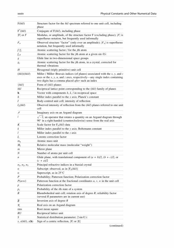
F(hkl) Structure factor for the hkl spectrum referred to one unit cell, including
phase
F*(hkl) Conjugate of F(hkl), including phase
jFj or F Modulus, or amplitude, of the structure factor F (excluding phase); jFj issuperfluous notation, but frequently used informally
Fo Observed structure “factor” (only ever an amplitude); jFoj is superfluousnotation, but frequently used informally
f / fj Atomic scattering factor / for the jth atom
fj,y Atomic scattering factor for the jth atom at a given sin y/lg Glide line in two-dimensional space groups
gj Atomic scattering factor for the jth atom, in a crystal, corrected for
thermal vibrations
H Hexagonal (triply primitive) unit cell
(hkl)/(hkil) Miller / Miller–Bravais indices (of planes) associated with the x, y, and zaxes or the x, y, u, and z axes, respectively—any single index containing
two digits has a comma placed after such an index
{hkl} Form of (hkl) planes
hkl Reciprocal lattice point corresponding to the (hkl) family of planes
h Vector with components h, k, l in reciprocal space
h Miller index parallel to the x axis; Planck’s constant
I Body-centred unit cell; intensity of reflection
Io(hkl) Observed intensity of reflection from the (hkl) planes referred to one unit
cell
i Imaginary axis on an Argand diagram
iffiffiffiffiffiffiffi�1
p; an operator that rotates a quantity on an Argand diagram through
90� in a right-handed (counterclockwise) sense from the real axis
K Scale factor for Fo(hkl) data
k Miller index parallel to the y axis; Boltzmann constant
l Miller index parallel to the z axis
L Lorentz correction factor
mu Atomic mass unit
Mr Relative molecular mass (molecular “weight”)
m Mirror plane
N Number of atoms per unit cell
n Glide plane, with translational component of (a + b)/2, (b + c)/2, or(c + a)/2
n1, n2, n3 Principal refractive indices in a biaxial crystal
o Subscript: observed, as in jFo(hkl)jo Superscript, as in 25�CP Probability; Patterson function; Polarization correction factor
P(uvw) Patterson function at the fractional coordinates u, v, w in the unit cell
p Polarization correction factor
pi Probability of the ith state of a system
R Rhombohedral unit cell; rotation axis of degree R; reliability factor
(several R parameters are in current use)
R Inversion axis of degree R
R Real axis on an Argand diagram
rms Root mean square
RU Reciprocal lattice unit
S Statistical distribution parameter; 2 sin y=ls, s(hkl), s(h) Sign of a centric reflection, jFj or jEj
(continued)
xxxiv Physical Constants and Other Numerical Data
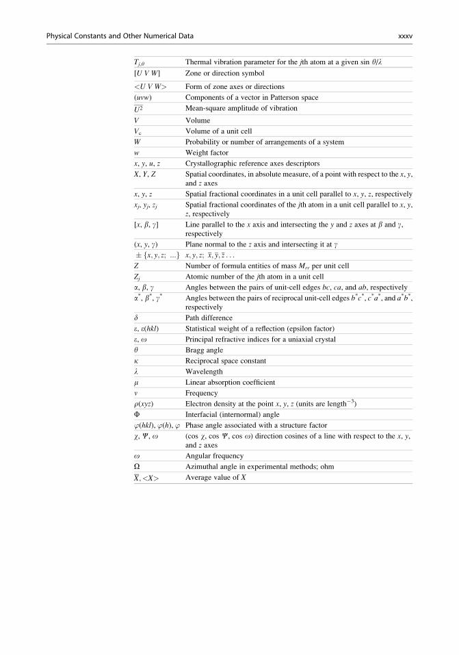
Tj,y Thermal vibration parameter for the jth atom at a given sin y/l[U V W] Zone or direction symbol
<U V W> Form of zone axes or directions
(uvw) Components of a vector in Patterson space
U2 Mean-square amplitude of vibration
V Volume
Vc Volume of a unit cell
W Probability or number of arrangements of a system
w Weight factor
x, y, u, z Crystallographic reference axes descriptors
X, Y, Z Spatial coordinates, in absolute measure, of a point with respect to the x, y,and z axes
x, y, z Spatial fractional coordinates in a unit cell parallel to x, y, z, respectively
xj, yj, zj Spatial fractional coordinates of the jth atom in a unit cell parallel to x, y,z, respectively
[x, b, g] Line parallel to the x axis and intersecting the y and z axes at b and g,respectively
(x, y, g) Plane normal to the z axis and intersecting it at g� fx; y; z; :::g x; y; z; x; y; z . . .
Z Number of formula entities of mass Mrr per unit cell
Zj Atomic number of the jth atom in a unit cell
a, b, g Angles between the pairs of unit-cell edges bc, ca, and ab, respectively
a*, b*, g* Angles between the pairs of reciprocal unit-cell edges b*c*, c*a*, and a*b*,respectively
d Path difference
e, e(hkl) Statistical weight of a reflection (epsilon factor)
e, o Principal refractive indices for a uniaxial crystal
y Bragg angle
k Reciprocal space constant
l Wavelength
m Linear absorption coefficient
n Frequency
r(xyz) Electron density at the point x, y, z (units are length�3)
F Interfacial (internormal) angle
’(hkl), ’(h), ’ Phase angle associated with a structure factor
w, C, o (cos w, cos C, cos o) direction cosines of a line with respect to the x, y,and z axes
o Angular frequency
O Azimuthal angle in experimental methods; ohm
X; <X> Average value of X
Physical Constants and Other Numerical Data xxxv