structure, development and reproduction of the red alga...
TRANSCRIPT
Proceedings of the Pakistan Academy of Sciences 50 (1):47–60 (2013) Pakistan Academy of SciencesCopyright © Pakistan Academy of SciencesISSN: 0377 - 2969 (print), 2306 - 1448 (online)
Research Article
————————————————Received, June 2012; Accepted, January 2013*Corresponding author: Zarina Ali; Email: [email protected]
Structure, Development and Reproduction of the Red Alga, Scinaia hatei from Pakistan
Mustafa Shameel1, S. Afaq-Husain1 and Zarina Ali2*
1Department of Botany, University of Karachi, Karachi-75270, Pakistan2Department of Botany, Federal Urdu University of Arts, Science & Technology,
Gulshan-e-Iqbal Campus, Karachi-75300, Pakistan
Abstract: The mucilaginous, marine red alga, Scinaia hatei Børgesen (Nemaliales), collected from various coastal areas of Karachi at the northern Arabian Sea, was investigated for its morphology, anatomy, growth and reproductive structures. This represents the first detailed study of this species. Scinaia hatei was compared with three other species, S. moniliformis subsp. pakistanensis Afaq-Husain et Shameel, S. saifullahii Afaq-Husain et Shameel and S. shameelii Afaq-Husain.
Keywords: Rhodophycota, Scinaia, taxonomy, morphology, anatomy, growth, development, reproduction
1. INTRODUCTION
Occurrence of the red alga, Scinaia hatei Børgesen was reported quite early from Karachi [1, 2] and other coastal areas of Pakistan [3-7]. Apart from a short taxonomic description [2], no detailed studies have been undertaken with respect to the taxonomy of this species. A few studies, however, focused on its antimicrobial activity [8-10] and phycochemistry [11-13]. Therefore, the present investigation was undertaken to examine the morphology, anatomy, growth and development of its various structures and reproductive structures. Our observations were compared with the descriptions of the other three Scinaia species from the coast of Pakistan [14-16] and a dichotomous key for the area is provided.
2. MATERIALS AND METHODS
The material was collected from different coastal areas of Karachi, Pakistan during February 1985 and September 2010; details are given in the section “Specimens Examined”. It was brought to the laboratory, thoroughly washed and preserved in 4 % formalin-seawater solution for further studies. Some of these specimens were used for the preparation
of herbarium sheets, which were deposited in the Seaweed Herbarium (KUH-SW), Phycology & Phycochemistry Lab (Room # 18), MAH Qadri Biological Research Centre, University of Karachi, Karachi, Pakistan. Morphology was examined either from fixed or freshly collected specimens while the study of their cellular structures was carried out from fixed material. The cross sections (C. S.) and longitudinal sections (L. S.) were prepared by free-hand cutting or by a rotary microtome after freezing the material. The material was stained in 1 % aniline blue and mounted in glycerin: acetic acid: distilled water (1: 1: 1, v/v) medium. Drawings were made with the help of a camera lucida.
Abbreviations used in the Figures: abrs = assimilatory branch system, ac = assimilatory cells, b = basal cell of carpogonial branch, c = carpogonium, cblt = coloured branchlets, cmd = central medulla, cor = cortex, cp = carpospores, cpm = carposporangium, h = hypogynous cell, hd = hypogynous daughter cell, m = monosporangium, omd = outer medulla, pf = pericarp filament, pfi = pericarp filament initial, r = rhizoidal filaments, ri = rhizoidal initial, spm = spermatangium, t = trichogyne, th = thickening, ti = trichogyne initial,

48 Mustafa Shameel et al
u = utricle.
3. RESULTS AND OBSERVATIONS
Scinaia hatei Børgesen 1931: 5
References: Børgesen 1931: 5, 1934: 32, 1938: 112, Anand 1943: 13, Salim 1965: 193, Krishnamurthy and Joshi 1970: 17, Shameel 1987: 514, 2000: 53, Shameel and Afaq-Husain 1987: 296, Shameel and Tanaka 1992: 45, Ormond and Banaimoon 1994: 118, Silva et al 1996: 115, Shameel et al 2000: 89 [1-7, 17-22].
3.1 Morphological Characters
Thalli up to 10 cm long, spreading radiallly, dull red to bright red, soft mucilaginous; holdfast discoid, 1-2 mm in diameter, giving rise usually to one rarely 3 axes; stipe slightly hard, cylindrico-conical, 4-6 mm long, less than 1 mm broad at the base and 1.5 mm below first dichotomy; branching dichotomous, very rarely trichotomous (Fig. 29), 6-8 times bifurcated; the segment between successive dichotomy cylindrico-terete, narrow below becoming broader and slightly compressed at the distal dichotomy, 3-20 mm long, 1-3 mm broad, smaller below longer in the distal region. The thallus is usually characterized by 1-3 deep constrictions in the distal part of branches, the constriction may be present at any place, either at the dichotomy or below or above it (Fig. 30-32); the apical segments more or less oblong or fusiform, broader above the constriction (more than 1.0 mm up to 1.5 mm) and then gradually tapering distally to a broad, truncate tip (Fig. 33); the subterminal segment cylindrical, 1.5 to less than 2.0 mm broad and of variable length up to 20 mm long, axis strand or mid-rib is not visible with naked eyes either in fresh, dried or formalin-preserved specimens.
3.2 Surface View
Two types of cells are observed when the thallus surface is examined under a microscope: (a) colourless usually large, polygonal cells (the utricles), 9-24 µm broad; (b) small, rounded cells with a darkly stained body in the center, 3-7 µm broad; present at places in between the above cells; 1-3 together, these are probably monosporangia or
spermatangia (Fig. 3).
3.3 Anatomical Features
The internal structure of the thallus is typical of the genus Scinaia. It is made up: (i) a 200-300 µm core of closely placed, thick and thin intertwining filaments, running longitudinally in the middle, throughout the length of the thallus; the filaments are 2-10 (-14) µm broad, sub-dichotomously branched, the cells elongated, cylindrical to fusiform (slightly broader in the middle) or become broader, up to 20 µm, at the distal end (Fig. 4c); (ii) a broad portion of loosely arranged filaments appear radiating from the axial core towards periphery, its width depending upon the thickness of the thallus; the filaments usually sub-dichotomously branched, thin, 1.5-2.5 µm broad, but as broad as 8 µm also present; and (iii) a 60-70 µm broad outer zone of cortex consisting of a loosely packed, colourless cells inside and closely packed coloured or pigmented cells (utricles) outside, which form the epidermis of the thallus (Fig. 1, 2).
In C. S. (Fig. 6, 7 & 34) the utricles appear oblong-rectangular to isodiametric, slightly flat above, longer than broad to as long as broad, 22-38 µm long ×17-29 µm broad, but cells as small as 13 × 7 µm were also observed, which may be the young utricles replacing the old ones. Below the utricles there is usually a single layer of dark coloured assimilatory cells, which may be round or conical, 6-20 µm broad or oblong-elongate, up to 24 µm long × 14 µm broad; the assimilatory cells also bear narrow coloured branchlets, which penetrate in between the colourless utricles and may bear spermatangia- or monosporangia-like bodies terminally (Fig. 7, 8, 34, 35). Rhizoidal branches are also produced in the assimilatory zone, which travel parallel to the surface usually below the assimilatory layer (Fig. 2, 16).
3.4 Growth of the Thallus
The microscopic examination of the growing point shows a small group of slender; sub-dichotomously branch free filaments in the center, consisting of very long cells not more than 1.0 µm broad, their apices are heavily loaded with cytoplasm and show slight bulging (Fig. 9). Their apical cells continuously
Morphology, Anatomy and Reproduction of Scinaia hatei 49
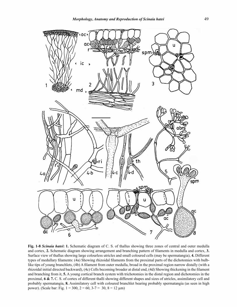
Fig. 1-8 Scinaia hatei: 1. Schematic diagram of C. S. of thallus showing three zones of central and outer medulla and cortex, 2. Schematic diagram showing arrangement and branching pattern of filaments in medulla and cortex, 3. Surface view of thallus showing large colourless utricles and small coloured cells (may be spermatangia), 4. Different types of medullary filaments: (4a) Showing rhizoidal filaments from the proximal parts of the dichotomies with bulb-like tips of young branchlets, (4b) A filament from outer medulla, broad in the proximal region narrow distally (with a rhizoidal initial directed backward), (4c) Cells becoming broader at distal end, (4d) Showing thickening in the filament and branching from it, 5. A young cortical branch system with trichotomies in the distal region and dichotomies in the proximal, 6 & 7. C. S. of cortex of different thalli showing different shapes and sizes of utricles, assimilatory cell and probably spermatangia, 8. Assimilatory cell with coloured branchlet bearing probably spermatangia (as seen in high power). (Scale bar: Fig. 1 = 300, 2 = 60, 3-7 = 30, 8 = 12 µm)

50 Mustafa Shameel et al
elongate and divide by cross wall, producing new cells proximally, increasing the length of the thallus, they are continued back as the axial filaments. The above filaments produce branch filaments sub-dichotomously, which diverge outward and grow in the direction of the thallus surface by repeated sub-dichotomous branching, which become repeatedly sub-trichotomous or quadrichotomous in distal region (Fig. 5).
3.5 Development of the Cortical System
The ultimate branches of the above filaments are 2-3 cells long; initially the cells are cylindrical, undifferentiated, 1-2 µm broad × 5-7 µm long, but the cells bearing them become up to 3 µm broad with dilations at their distal ends (Fig. 5, 10). The cells of the branch system further increase in size; and usually one branchlet out of a bunch, arising from the same supporting cell, cuts off a cell terminally which becomes much broader, round and slightly flat above, becoming narrow proximally, filled with dense cytoplasm emerging above the other branchlets of the branch (Fig. 11). On maturation it acquires a characteristic shape, about 5.5 µm broad (Fig. 14). These cells may be the monosporangia. The cells of the branchlets continue increasing in size, especially in breadth, and soon their terminal cells start differentiating into utricles by loosing their colour and enlarging to several times their original size (Fig. 12-15). These cells remain separate when young and start adhering to each other as they mature (Fig. 13, 15) and finally from the epidermal layer of the thallus (Fig. 6, 7, 34).
After differentiation of the epidermal cells, the inner cells of the branchlet differentiate into assimilatory cells. They become broad at distal end acquiring conical or rounded-cuniate shape and are densely filled with cytoplasm and plastids (Fig. 13, 15). The primary cells produce secondary assimilatory cells at distal end, which in turn produce new utricles (Fig. 15, 16). These cells become arranged in such a manner that they appear to form a single layer of cells below the utricles (Fig. 6). Some assimilatory cells also produce long, slender, coloured branchlets, which penetrate in between the utricles and later on may bear monosporangia or spermatangia terminally (Fig. 7, 8, 17, 34, 35).
The cortical branch system is easily observed in its entire form as in Fig. 5, after teasing out the tip of the thallus, but from mature thallus only assimilatory portion is separated and appears as in Fig. 17. The utricles are broken off due to adherence to each other; the stalk-like branchlets are commonly seen on such branches, bearing monosporangia and spermatangia which get detached during teasing of the tissues (Fig. 17). Some of the assimilatory cells also give rise to rhizoidal filaments, which travel below the assimilatory cells and may give rise to secondary assimilatory branches from the special thickenings, which appear in these filaments (Fig. 4d).
3.6 Development of the Outer Medulla
During development of the cortical system, the cells of the filaments, lying in between the assimilatory zone and axial core, simply increase in length several times of their original size, increasing the thickness of the thallus. They become broad and thick walled, their cytoplasmic contents get stretched in the form of fine thread, become colourless, giving rise to branches repeatedly in sub-dichotomous manner. The filaments remain separate from each other, forming the outer loose part of medulla. The filaments are up to 8 µm thick near the axial core, but reaching periphery their branches become 1.5-2.5 µm broad (Fig. 4b).
Besides sub-dichotomously branching from the distal ends of the cells, rhizoidal branches of more or less 2 µm breadths are also produced from any part of the cell in the mature thallus. It is usually observed that the basal cells of the dichotomy produce 1-2 (-3) rhizoidal branches which in turn also branch irregularly in the proximal region, their initials bear bulbous tips (Fig. 4a). It is also observed that one of the rhizoidal branches (proximal one) travels backward and joins the medulla; their initials have been observed directed backward towards axial core (Fig. 4b). Sometimes a cell of the filament broadens into a spindle-like thickening in the middle, from where new branches are produced (Fig. 4d).
3.7 Development of the Central Medulla
The cells lying proximal to the growing point in

Morphology, Anatomy and Reproduction of Scinaia hatei 51
Fig. 9-17 Scinaia hatei: 9. Filament system from growing point with thin long cell and swollen tips densely filed with cytoplasm, 10-17. Cortical filament system showing different stages of development of cortex: 10. Very young filament showing branching pattern, 11. Slightly older branch with broader cells and young monosporangia, 12. Young cortical branch showing carpogonial branch and start of differentiation of utricles, 13 & 14. Branches with slightly older utricles and start of differentiation of assimilatory cells, and coloured branchelets (14 showing mature monosporangia) 15. Branch showing close arrangement of utricles and young coloured branchlets, 16. A nearly mature branch with nearly mature utricles and the development of secondary assimilatory cells and a rhizoidal filament from the lower assimilatory cell, 17. A cortical branch from mature thallus (utricles broken away) showing a stalk-like coloured branchlet, whose apical cell/ cells broken away which might be monosporangia, spermantangia or vegetative cells. (Scale bar: Fig., 9-17 = 12 µm).

52 Mustafa Shameel et al
line with the axial filaments develop as described above to from the axial core or central medulla of the thallus. These cells elongate in the direction of the long axis and increase the length of the thallus. Many rhizoidal filaments are also produced in a similar pattern as described for the outer medulla, which travel along the axial filament or outward toward periphery.
3.8 Branching in the Filament
Any cell of a filament except the apical one puts forth a protuberance either at its distal end or from its proximal part which gets broaden. The protuberance rich in cytoplasm elongates and is cut off from the mother cell by a cross wall forming the filament initial, which then acts as the apical cell of the branch filament. The branch initial, when develops at the distal end of the mother cell, elongates in the direction of growth and the branching appears normal, dichotomous (Fig. 4b, 5). Lateral branching is initiated by the branch initial, which is produced on the proximal part of the mother cell and develops in any direction. Such branches usually develop on the proximal cells of the mother branches repeatedly so that a number of branches appear arising close to the parent filament (Fig. 4a).
3.9 Reproductive Structures
The thalli were probably monoecious. The following structures have been investigated in detail.
3.9.1 Spermatangia
Some rounded bodies in groups of 3 were commonly observed in surface view at corners of utricles, at small distances in greater frequency in the apical segments (Fig. 3). In C. S. these bodies appear broadly oblong, 7 × 5 µm in size, borne terminally on a coloured branchlet arising from the hypodermal cell (Fig. 7, 8, 34). These bodies may probably be spermatangia, each bearing a single spermatium, 5 × 3 µm in size.
3.9.2 Carpogonial Branch and Cystocarp
The carpogonial branches are 3-celled, borne on the distal portion of the 5th or 6th cell of a young cortical branch system, in the apical region of the thallus
(Fig. 12, 20, 23). The carpogonium is elongate, conical, broad below becoming narrow distally, 8.0-12.5 µm long × 3.5-4.5 µm broad; the middle cell is more or less as long as broad or slightly longer than broad, usually 6-8 µm long or broad; the basal cell is usually broader than long, about 3.5 µm long × 4.5 µm broad. The trichogyne is broad at the base (about 2.7 µm) and gradually tapers towards apex (Fig. 12, 23). Four-celled carpogonial branches without trichogyne and three-celled ones with little trichogyne have also been observed, which appear sterile (Fig. 18, 19).
After formation of the 3-celled carpogonial branch, either the trichogyne develops first, or the pericarp filaments are initiated first by the basal cell (Fig. 20, 24). Stages have been observed in between these two conditions, for example Fig. 21 exhibits an initial pericarp filament from the basal cell and initial trichogyne from the carpogonium, from which it appears that both might have been developed at the same time. In Fig. 23 the trichogyne is in a later stage of development than the pericarp filament in the basal cell, which shows that the trichogyne might have developed prior to the pericarp filament. The initials from basal cells develop into pericarp filament covering the middle cell product (Fig. 12, 25-27, 36, 37) and finally from the sterile covering (pericarp) of the cystocarp. The hypogynous cell gives rise to 3-4 daughter cells (Fig. 36, 37), arranged as two 1-celled and one 2-celled branches (Fig. 22, 27) or one 1-celled and 2-celled branches (Fig. 25, 26).
The division in the hypogynous cell usually occurs after the inception of pericarp filaments in the basal cell (Fig. 23, 24). But in Fig 22 there is only one pericarp filament initial and the trichogyne has just initiated, but 5 cells are present in the middle, showing that the hypogynous cell might have initiated division prior to that of basal cell. The development of gonimoblast initials is either from carpogonium or middle cell product, is not clear; it is obscured by the pericarp filaments developing from the basal cell (Fig. 12, 26).
3.9.3 Cystocarp
The cystocarps are present below the cortex, initially oblong becoming pyriform to round-cuniate on maturation, up to 340 µm broad. Their

Morphology, Anatomy and Reproduction of Scinaia hatei 53
Fig. 18-28 Scinaia hatei: 18. Four-celled sterile carpogonial branch without trichogyne, (19) Three-celled sterile carpogonial branch with a little trichogyne, 20. Trichogyne, 21. Carpogonial branch with trichogyne initial and a pericarp filament initial, 22. Carpogonial branch with trichogyne intial (4 daughter cells from hypogynous cell arranged as two 1-celled one 2-celled branches, and a pericarp flament initial from basal cell), 23. Carpogonial branch with developing trichogyne and a 2-celled pericarp filament, 24. Young carpogonial branch (trichogyne not initiated but 2 pericarp filaments initiated from basal cell), 25-27. Mature carpogonial branches with 3-4 hypogynous daughter cells arranged as one 1-celled and one 2-celled branches (25 & 26 show two 1-celled branches and one 2-celled branch in 27), 28. Gonimoblast filaments from different thalli bearing 2-3 carposporangia and liberated carpospores showing shape and size. (Scale bar: Fig., 18-28 = 12 µm).

54 Mustafa Shameel et al
Fig. 29-33 Scinaia hatei: 29-31. Thalli showing habit preparations on herbarium sheets (arrows showing trichtomy), 32. Thalli showing habit (in water), 33. Distal part of a branch showing constrictions and shape of apical segments (in water). (Scale bar: Fig., 29 = 35, 30 = 37.5, 31 = 0.56, 32 = 24, 33 = 50 mm).

Morphology, Anatomy and Reproduction of Scinaia hatei 55
Fig. 34-39 Scinaia hatei: 34. C. S. showing arrangement and shape of utricles and assimilatory cells (one assimilatory cell bearing coloured stalk-like branchlet with probably spermatangia), 35. C. S. showing monosporangium-like body on stalk-like coloured branchlet, 36. A very young cystocarp showing loose network of filaments, in the process of forming pericarp and hypogynous daughter cells (gonimoblast filaments not observed), 37. Carpogonial branch showing pericarp filaments from basal cell (hypogynous daughter cells in the middle and conical carpogonium above), 38. A mature gonimoblast, 39. Showing distal chains of carposporangia and liberated carpospores. (Scale bar: Fig., 34-35 = 30, 36 = 42, 37 = 12, 38 = 120, 39 = 22.5 µm).

56 Mustafa Shameel et al
neck is up to 100 µm long and 60 µm broad opening to the exterior through the ostiole, present on the surface of the thallus. The cystocarp encloses the carposporophyte or gonimoblast, which produces carpospores.
3.9.4 Gonimoblast
The gonimoblast is a broadly conical to cup-like compact body of sub-dichotomously branched filaments (Fig. 38). Their cells are up to 12 µm long × 4 µm broad. Two to three distal cells of the filaments increase in size and develop into carpogonia, oblong or oval, up to 17 µm long × 6-9 µm broad (Fig. 28, 39); each producing one carpospore, oblong 8-14 µm long × 5-6 µm broad.
3.9.5 Specimens Examined
Sandspit (Leg. S. Afaq-Husain 11-11-1985; M. Shameel 8-1-2008); Hawksbay (Leg. S. Afaq-Husain 7-11-1987, 21-3-1988, 9-12-1989; M. Shameel 4-2-2007); Buleji (Leg. M. Shameel 18-11-2009, 25-9-2010); Paradise Point (Leg. S. Afaq-Husain 23-12-1988, 14-11-1989; M. Shameel 27-12-2006); Naugaza Mazar (Leg. S. Afaq-Husain 24-4-1986); Cape Monze (Leg. S. Afaq-Husain 5-2-1985, 11-3-1986, 9-4-1989; M. Shameel 8-4-2005); Gadani (Leg. M. Shameel 12-1-1986).
3.9.6 Habitat
Usually sublittoral, but also grows on sand covered rocks near low tide mark, remaining submerged at low tides.
3.9.7 Type Locality
Part Okha, Gujarat, India.
3.9.8 Local Distribution
Karachi: Manora, Sandspit, Hawksbay, Buleji, Paradise Point, Naugaza Mazar and Cape Monze; Balochistan: Gadani.
3.9.9 Distribution in the Indian Ocean
India, Pakistan and Yemen.
4. DISCUSSION
Scinaia Bivona-Bernardi is a significant genus of
the family Galaxauraceae (order Nemaliales, class Nemaliophyceae, phylum Rhodophycota; fide [23]. Its four species have been recorded from the coast of Pakistan, which may be distinguished as follows: 1. Thallus cylindrical ........................................ .2 Thallus flat, compressed ...............S. shameelii 2. Main thallus constricted, utricles in
surface view polygonal.. ................................ 3 Main thallus not constricted, utricles in surface
view rosulate ................................ S. saifullahii 3. Thalli regularly constricted, appearing
moniliform (as string of beads) ............ S. moniliformmis subsp. pakistanensis Thalli 2-3 times irregularly constricted, not
moniliform ........................................... S. hatei
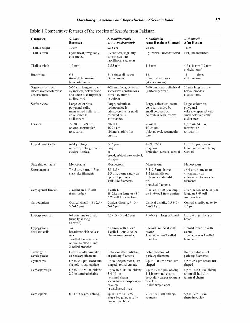
Apart from S. hatei the other three species have already been described earlier [14-16]. Their comparative features are presented in the Table 1.
Børgesen described Scinaia hatei using Indian material [17], nevertheless the species description remained incomplete and based mainly in morphological features [24, 25] until the present study. The present observations from Pakistani material correspond to the original description; the only difference found was the cystocarp size which is 340 µm in Pakistani material and up to 265 µm in the original Indian specimens.
The thalli of S. hatei are characterized by deep constrictions at irregular intervals and the segments which become broad distally (Table I). Scinaia bengalica Børgesen and S. carnosa Harvey have also been reported from India [18] and resemble S. hatei in height and cylindrical axes, but differ in having continuous, unconstricted thalli. Although constrictions are found in S. carnosa but these are considered to be accidental and do not develop as regular process of growth [18].
Other species which bear occasional to regularly constricted thalli are S. aborealis Huisman, S. articulata Setchell, S. salicornoides (Kützing) J. Agardh, S. tsingalensis Tseng, S. huismanii Vroom et I. A. Abbott and S. interrupta (de Candolle) Wynne. The present population differs from S. aborealis in its height which is about one-forth of the later besides the morphology, S. aborealis is
Morphology, Anatomy and Reproduction of Scinaia hatei 57
Table 1 Comparative features of the species of Scinaia from Pakistan.
Characters S. hateiBørgesen
S. moniliformmis subsp. pakistanensis
S. saifullahiiAfaq-Husain et Shameel
S. shameeliiAfaq-Husain
Thallus height 10 cm 22.5 cm 25 cm 11cmThallus form Cylindrical, irregularly
constrictedCylindrical, regularly constricted into moniliform segments
Cylindrical, unconstricted Flat, unconstricted
Thallus width 1-3 mm 2-5.5 mm 1-2 mm 4-5 (-6) mm (10 mm at dichotomy)
Branching 6-8times dichotomous (-trichotomous)
8-16 times di- to sub-dichotomous
14 times dichotomous (-trichotomous)
11 times dichotomous
Segments betweensuccessivedichotomies/constrictions
3-20 mm long, narrow, cylindrical, below broad and terete to compressed at distal end
4-26 mm long, between successive constrictions conico-cylindricalto oblong
3-60 mm long, cylindrical (uniformly broad)
20 mm long, narrow below, broadestat dichotomy
Surface view Large, colourless, polygonal cells, interspersed with small coloured cells at distances
Large, colourless, polygonal cells interspersed with small coloured cellsat distances
Large, colourless, round cells surrounded by small coloured orcolourless cells, rosette
Large, colourless, polygonalcells interspersed with small coloured cellsat distances
Utricles 22-38 × 17-29 µm,oblong, rectangular to squarish
30-38 × 16-21 µmoblong, slightly flat distally
20-41 × 10-24 µm,oblong, oval, rectangular-like
Up to 44-34 µm,rectangular to squarish
Hypodermal Cells 6-24 µm long or broad, oblong, round-cuniate, conical
5-15 µmlong orbroad, orbicular to conical,elongate
7-19 × 7-14long µm,orbicular- cuniate, conical
Up to 19 µm long or broad, orbicular, oblong, Conical
Sexuality of thalli Monoecious Monoecious Monoecious MonoecioussSpermatangia 7 × 5 µm, borne 1-3 on
stalk-like filaments3.5-4.5 ×2-3 µm, borne singly on up to 10 µm long stalk-like filaments
3-5×2-3 µm, borne 1-2 terminally on unbranched stalk-like orbranched filaments
5× 4 µm, borne up to 4 terminally on unbranched to branched filaments
Carpogonial Branch 3-celled on 5-6th cellfrom surface
3-celled,18-22.5µm long, on (5-) 6-7th cell from surface
3-celled, 18-25 µm long, on 5- 6th cell from surface
3 to 4-celled, up to 25 µm long, on 5-6th cell from surface
Carpogonium Conical distally, 8-12.5 × 3.5-4.5 µm
Conical distally, 9-10 × 3-4.5 µm
Conical distally, 7.5-9.0 × 3.0-5.5 µm
Conical distally, up to 10 × 6 µm
Hypogynous cell 6-8 µm long or broad (usually as long as broad)
3.5-5.5 × 3.5-4.5 µm 4.5-6.5 µm long or broad Up to 4.5 µm long or broad
Hypogynous daughter cells
3-4 broad roundish cells as one 1-celled + one 2-celled or two 1-celled + one 2-celled branches
3 narrow cells as one 1-celled + one 2-celled filamentous branches
3 broad, roundish cells as one 1-celled + one 2-celled branches
3 broad roundish cells as one 1-celled + one 2-celled branches
Trichogyne development
Before or after initiation of pericarp filaments
Before or after initiation of pericarp filaments
After initiation ofpericarp filaments
Before initiation of pericarp filaments
Cystocarps Up to 340 µm broad, urn-shaped, round-cuniate
Up to 320 µm broad, urn-shaped, round-cuniate
Up to 300 µm broad, urn-shaped
Up to 250 µm broad, urn-shaped
Carposporangia Up to 17 × 9 µm, oblong, 2-3 in terminal chains
Up to 16 × 10 µm, oblong, 3-4 (-5) in terminal chains, secondary carposporangia developin discharged ones
Up to 17 × 8 µm, oblong, 1-4 in terminal chains, secondary carposporangia develop in discharged ones
Up to 14 × 8 µm, oblong to roundish, 1-5 in terminal chains
Carpospores 8-14 × 5-6 µm, oblong up to 15 × 8.5- µm, shape irregular, usually longer than broad
7-14 × 6-7 µm oblong, roundish
Up to 12 × 7 µm, shape irregular

58 Mustafa Shameel et al
the only species with 4 hypogynous branches [26], It differs from S. articulata and S. salicornoides in the much larger size of its utricles, and from S. tsingalensis and S. interrupta in its larger branch diameter, S. interrupta has rhizoidal filaments on the cystocarp [25]. Reproductive structures of S. hatei have not been narrated in its type description nor worked out afterward.
The four species of Scinaia from Pakistan have several characters in common, which may prove of some significance as far as their affinities and evolution within the order Nemaliales is concerned:
1. The formation of utricles follows identical patterns of development in all the four species. Initially all the cells of the filaments at the growing tip are alike cylindrical, narrow and separate from each other (Fig. 9), gradually the apical cells of the lateral (deflected) branches become inflated, acquiring oblong-pyriform shape. At this stage the cortical branch system resembles with those found in the family Liagoraceae (Fig. 5, 12). During further development the inflated cells turn into utricles, adhering to each other, forming the continuous outer layer (epidermal layer) of the thallus, becoming a characteristic feature of the genus Scinaia.
2. All the four species contain 1-3 cells long, narrow and coloured branchlets in between the utricles in varying ratios, which are continuously produced by hypodermal cells. Their function appears to be three fold as mentioned below:
(a) The 1-celled or terminal cell of 2-celled branchlets gradually increase in size and turn into utricles probably to replace the injured or old ones. The 1-celled branchlets of different diameters tending to become utricles have been observed. In some cases terminal cell of a 2-celled branchlet is found tending to become a utricle.
(b) The coloured branchlets may produce monosporangia-like bodies terminally, which are visible in surface view as well as in C. S. or L. S. of cortex.
(c) The 2- to 3-celled branchlets become profusely branched and each daughter branch bears 1-4 spermatangia terminally.
3. The branching pattern in the filaments has not been given any importance in the previous works, although it may prove significant when comparing Scinaia with its related genera. The following types of branching are observed in the medullary filaments in all the four species.
(a) Normal branching appearing dichotomous (but subdichotomous and pseudodichotomous in origin) from distal end of cells of the filament (Fig. 5).
(b) Special thickening is developed in the filaments, from which branch filaments arise.
(c) several branch filaments arise repeatedly from the proximal part of basal cells of the filaments and their daughter filaments (Fig. 4a).
4. Carpogonial branch structure is also similar in all the four Pakistani species as well as in species described from other countries, e.g. S. cottonii Setchell [27], which is suspected to be a heterotypic synonym of S. latifrons Howe [28]. Carpogonial branch is usually 3-celled consisting of a distal carpogonium, a hypogynous cell and a basal cell. The basal cell produces filaments, which from the sterile covering (the pericarp) of the cystocarp. The hypogynous cell produces 3-4 daughter cells, usually large in size, in the form of 1-celled and 2-celled branches.
The carpogonium is described to produce gonimoblast initials after fertilization [26, 29]; but in the present case the development of initials is not observed either from carpogonium or the hypogynous daughter cells till later stage of development of the pericarp, which shows that the gonimoblast initials may not be distinguishable from pericarp filaments. Two species show variation from the usual, Scinaia shameelii Afaq-Husain also bears 4-celled carpogonial branches besides 3-celled ones, as is also reported in S. furcata [30]. Thus in this respect these species

Morphology, Anatomy and Reproduction of Scinaia hatei 59
show affinities with the members of Liagoraceae on one hand and with Tricleocarpa oblongata (Ellis et Solander) Huisman et Borowitzka as Galaxaura oblongata Ellis et Solander on the other, which also possess 4-celled carpogonial branches [31]. In S. moniliformis subsp. pakistanensis Afaq-Husain et Shameel the hypogynous daughter cells are narrow and filamentous, different in appearance from those found in other species of Scinaia.
The position of the carpogonial branch is also slightly debatable. In Scinaia the carpogonial branch is specialized accessory branch arising in addition to the vegetative filaments, whereas in Galaxaura Lamouroux and Tricleocarpa Huisman et Borowitzka the carpogonial branch arises in the position of a normal vegetative filament (replacing one of the dichotomies) [31, 32]. In the present species of Scinaia the carpogonial branches are found at the distal end of the bearing cell beside one vegetative branch; same position is shown by Zablackis ([30] p. 55, Fig. 12) or beside two branches but never beside 3 branches, although trichotomous branching of the vegetative filaments have been observed several times. Thus the end position of the carpogonial branch (a position from where a vegetative branch arises), the presence of only 1 or 2 vegetative branches beside the carpogonial branch and the absence of a carpogonial branch beside 3 vegetative branches may show that these carpogonial branches may not be of accessory origin and may have developed in place of one of the dichotomies or one of the trichotomies.
However, in S. hatei the position of carpogonial branches is clearly observed to be a little below the distal end of the bearing cell, a position appearing below the dichotomies (Fig. 20-22, 25) and here the carpogonial branches may be of accessory origin. In the present state of knowledge it is difficult to decide with certainty whether the carpogonial branches are accessory in origin or formed in place of vegetative branches, in the present species of Scinaia. Nevertheless, a possible explanation may be given as under. In the distal region the vegetative filaments are commonly trichotomous but proximal to it they are commonly dichotomous in all the four Pakistani species (Fig. 5), which probably shows that these proximal cells are also potentially capable of producing trichotomies but one branch
(the potential branch) remains suppressed or undeveloped and only two branches appear; the potential branch develops in the modified form of carpogonial branch, completing the trichotomous condition. This potential branch sometimes also develops into a vegetative branch, due to which trichtomous branching are also seen occasionally. One vegetative branch beside a carpogonial branch may be the result of suppression of one of the sister vegetative branches.
5. ACKNOWLEDGEMENTS
We are grateful to Prof. Dr. Anthony Chapman (Canada) and the anonymous referees for critical evaluation of the manuscript and for many valuable suggestions.
6. REFERENCES
1. Børgesen, F. Some marine algae from the northern part of the Arabian Sea with remarks on their geographical distribution. Kongelige Danske Videnskabernes Selskab, Biologiske Meddelelser 11: 1-72 (1934).
2. Anand, P.L. Marine Algae from Karachi. II. Rhodophyceae. Punjab University Botanical Publications, Lahore (1943).
3. Shameel, M. A. Preliminary survey of seaweeds from the coast of Lasbela, Pakistan. Botanica Marina 30: 511-515 (1987).
4. Shameel, M. Biodiversity of the seaweeds growing along Balochistan coast of the northern Arabian Sea. In: Proceedings of National O.N.R. Symposium in Arabian Sea as a Resource of Biological Diversity. Ahmad, V. U. (Ed.), HEJ Research Institute of Chemistry, Karachi University, Karachi, Pakistan, p. 45-64 (2000).
5. Shameel, M. & S. Afaq-Husain. Survey of algal flora from Lasbela Coast. In: Moderen Trends of Plant Sciences Research in Pakistan. Ilahi, I. & F. Hussain (Eds.), Proceedings of National Conference of Plant Scientists 3: 292-299 (1987).
6. Shameel, M. & J. Tanaka. A preliminary check-list of marine algae from the coast and inshore water of Pakistan. In: Cryptogamic Flora of Pakistan. Vol. 1. Nakaike, T. & S. Malik (Eds.), National Science Museum, Tokyo, Japan, p. 1-64 (1992).
7. Shameel, M., S. H. Khan & S. Afaq-Husain. Biodiversity of marine benthic algae along the coast of Balochistan, Pakistan. Pakistan Journal of Marine Biology 6: 69-100 (2000).
8. Usmanghani, K., M. Shameel, M. Sualeh, K. H. Khan & Z. A. Mahmood. Antibacterial and antifungal activities of marine algae from Karachi seashore of Pakistan. Fitoterapia 55: 73-77

60 Mustafa Shameel et al
(1984).9. Usmanghani, K. & M. Shameel. Studies on
the antimicrobial activity of certain seaweeds from Karachi coast. In: Prospects for Biosaline Research, Ahmad, R. & A. San Pietro (Eds.), Karachi University, p. 519-526 (1986).
10. Mandal, P., C. A. Pujol, M. J. Carlucci, K. Chattopadhyay, F. B. Damonte & B. Ray. Anti-herpatic activity of a sulfated xylomannan from Scinaia hatei. Phytochemistry 69: 2193-2199 (2008).
11. Valeem, E. E. & M. Shameel. Fatty acid composition of the class Ceramiophyceae (Rhodophyta) from north Arabian Sea. International Journal of Phycology and Phycochemistry 2: 141-148.
12. Hayee-Memon, A., M. Shameel & A. Zarina. Fatty acid composition of Scinaia hatei (Bonnemaisonales, Rhodophycota). International Journal of Phycology and Phycochemistry 6: 17-20 (2010).
13. Shameel, M. S. Afaq-Husain & A. Zarina. Phycochemical investigations on three species of the genus Scinaia Bivonia-Bernardi (Nemaliales, Rhodophycota) from the coast of Karachi (Pakistan). International Journal on Algae 13: 363-378 (2011).
14. Afaq-Husain, S. Structure and reproduction of a new alga, Scinaia shameelii (Rhodophyta) from Pakistan. Candollea 51: 445-459 (1996).
15. Afaq-Husain, S. & M. Shameel. Structure, development and reproduction of a new species Scinaia saifullahii (Bonnemaisoniales, Rhodophyta) from the north Arabian Sea. Pakistan Journal of Scientific and Industrial Research 40: 104-113 (1997).
16. Afaq-Husain, S. & M. Shameel. Structure and reproduction of Scinaia moniliformis pakistanensis var. nov. (Nemaliales, Rhodophyta). Pakistan Journal of Botany 33: 53-68 (2001).
17. Børgesen, F. Some Indian Rhodophyceae especially from the presidency of Bombay. Bulletin of Miscellaneous Information, Royal Botanical Garden, Kew 1931: 1-24 (1931).
18. Børgesen, F. Two species of Scinaia from South India. Botaniska Notiser 1938: 183-189 (1938).
19. Salim, K. M. The distribution of marine algae along Karachi coast. Botanica Marina 8: 183-198 (1965).
20. Krishnamurty, V. & H. V. Joshi. A Check-List of
Indian Marine Algae. Central Salt and Marine Chemicals Research Institute, Bhavnagar (1970).
21. Ormond, R. F. G. & S. A. Banaimoon. Ecology of intertidal macroalgal assemblages on the Hadramout coast of Southern Yemen, an area of seasonal upwelling. Marine Ecology Progress Series 105: 105-120 (1994).
22. Silva, P.C., P.W. Basson & R.L. Moe. Catalogue of the benthic marine algae of the Indian Ocean. University of California Publications Botany 79: 1-1259 (1996).
23. Shameel, M. Change of divisional nomenclature in Shameelian classification of algae. International Journal of Phycology Phycochemistry 4: 225-232 (2008).
24. Vroom, P. S. & I. A. Abbott. Scinaia huismanii sp. nov. (Nemaliales, Rhodophyta): an addition to the exploration of the marine algae of the northwestern Hawaiian Island. Phycologia 43: 445-454 (2004).
25. León-Cisneros, K., R. Riosmena-Rodriguez, A. I. Neto & G. Hernández-Carmona. The red algal genus Scinaia (Nemaliales, Rhodophyta) on the Gulf of California, Mexico: a taxonomic account. Phycologia 48: 186-210 (2009).
26. Huisman, J. M. The red algal genus Scinaia (Galaxauraceae, Nemaliales) from Australia. Phycologia 25: 271-296 (1986).
27. Kajimura, M. The morphology of Scinaia cottonii Setchell (Galaxauraceae, Rhodophyta). Botanica Marina 38: 535-541 (1995).
28. León-Cisneros, K. & R. Riosmena-Rodriguez. Morphometric of Scinaia latifrons (Nemaliales, Rhodophyta) in the southwestern Gulf of California, Mexico. Algae 20: 31-36 (2005).
29. Huisman, J. M. The Scinaia assemblage (Galaxauraceae, Rhodophyta): a re-appraisal. Phycologia 24: 403-418 (1985).
30. Zablackis, E. The red algae Scinaia furcata sp. nov. (Galaxauraceae, Nemaliales) from Hawaii. Phycologia 26: 53-58 (1987).
31. Magruder, W. H. Reproduction and life history of the red alga Galaxaura oblongata (Nemaliales, Galaxauraceae). Journal of Phycology 20: 402-409 (1984).
32. Huisman, J. M. & M. A. Borowitzka. The revision of the Australian species of Galaxaura (Rhodophyta, Galaxauraceae) with a description of Tricleocarpa gen. nov. Phycologia 29: 150-172 (1990).