structure, function, and evolution of phosphoglycerate...
TRANSCRIPT

Review
Structure, function, and evolution of phosphoglyceratemutases: comparison with fructose-2,6-bisphosphatase, acid
phosphatase, and alkaline phosphatase
Mark J. Jedrzejas*
Department of Microbiology, University of Alabama at Birmingham, 933 19th Street South, CHSB-19 room 545,Birmingham, AL 35-294-2041, USA
Contents
1. Introduction . . . . . . . . . . . . . . . . . . . . . . . . . . . . . . . . . . . . . . . . . . . . . . . . . . . . . . . . . . 264
2. Cofactor dependent Saccharomyces cerevisiae phosphoglycerate mutase . . . . . . . . . . . . . . . . 2652.1. Structural aspects of Saccharomyces cerevisiae phosphoglycerate mutase . . . . . . . . . . 2652.2. Mechanism of catalysis of the S. cerevisiae dPGM . . . . . . . . . . . . . . . . . . . . . . . . . . 270
2.3. Structural comparison of dPGM with other enzymes . . . . . . . . . . . . . . . . . . . . . . . . 2702.3.1. Structural comparison of S. cerevisiae dPGM with 6-phosphofructo-2-kinase/
fructose-2,6-bisphosphatase . . . . . . . . . . . . . . . . . . . . . . . . . . . . . . . . . . . . 271
2.3.2. Structural comparison of S. cerevisiae dPGM with acid phosphatase . . . . . . . 2722.3.3. Overall comparison of active sites of dPGM, Fru26P2ase, and AcPase . . . . . 273
3. Bacillus stearothermophilus cofactor independent phosphoglycerate mutase. . . . . . . . . . . . . . 277
3.1. Structure of B. stearothermophilus phosphoglycerate mutase . . . . . . . . . . . . . . . . . . . 2773.2. Catalytic mechanism for B. stearothermophilus iPGM and its comparison to alkaline
phosphatase catalysis . . . . . . . . . . . . . . . . . . . . . . . . . . . . . . . . . . . . . . . . . . . . . . . 278
Progress in Biophysics & Molecular Biology 73 (2000) 263±287
0079-6107/00/$ - see front matter 7 2000 Elsevier Science Ltd. All rights reserved.PII: S0079-6107(00)00007-9
www.elsevier.com/locate/pbiomolbio
* Tel.: +1-205-975-7627; fax: +1-205-975-5424.E-mail address: [email protected] (M.J. Jedrzejas).
Abbreviations: AcPase, acid phosphatase; AlPase, alkaline phosphatase; ATP, adenosine 5 '-triphosphate; 13PG,1,3-diphosphoglyceric acid; 23PG, 2,3-diphosphoglyceric acid; Fru26P2, fructose-2,6-biphosphate; Fru26P2ase, fruc-
tose-2,6-biphosphatase; 6PFru-2-K/Fru26P2ase, 6-phosphofructo-2-kinase/fructose-2,6-bisphosphatase; 2PGA, 2-phosphoglyceric acid; 3PGA, 3-phosphoglyceric acid; PGM, phosphoglycerate mutase; bPGM, bisphosphoglyceratemutase; dPGM, 23PGA dependent PGM; iPGM, 23PGA independent PGM.
4. Functional properties and comparison of the S. cerevisiae dPGM with the B. stearothermophilus
iPGM enzyme . . . . . . . . . . . . . . . . . . . . . . . . . . . . . . . . . . . . . . . . . . . . . . . . . . . . . . . . . 280
5. Conclusions . . . . . . . . . . . . . . . . . . . . . . . . . . . . . . . . . . . . . . . . . . . . . . . . . . . . . . . . . . 282
Acknowledgements . . . . . . . . . . . . . . . . . . . . . . . . . . . . . . . . . . . . . . . . . . . . . . . . . . . . . . . . . . 282
References . . . . . . . . . . . . . . . . . . . . . . . . . . . . . . . . . . . . . . . . . . . . . . . . . . . . . . . . . . . . . . . . 283
1. Introduction
Phosphoglycerate mutase (PGM) enzymes catalyze the isomerization of phosphoglyceratesubstrates, a process essential for the metabolism of glucose and/or 2,3-phosphoglycerate(23PGA) in nearly all organisms (Fothergill-Gilmore and Watson, 1989). At least twoknown distinct classes of PGM enzymes were identi®ed and one of them catalyzes theinterconversion of 3-phosphoglycerate (3PGA) and 2-phosphoglycerate (2PGA). Thisenzyme is known as monophosphoglycerate mutase (mPGM) (EC 5.4.2.1) (Meyerhof andKiessling, 1935) in contrast to a bisphosphoglycerate mutase (bPGM) (EC 5.4.2.4/EC3.1.3.13) which catalyzes primarily the interconversion of 1,3-phosphoglycerate (13PGA)and 23PGA (Forthergill-Gilmore and Watson, 1989). The second enzyme is a member ofanother class of PGMs and this group of enzymes also has the ability to carry out thereaction catalyzed by mPGMs as well as the break down of 23PGA to 3PGA (Rapportand Luebring, 1950). This form of the PGM enzymes was isolated from erythrocytes ofmany vertebrates (Forthergill-Gilmore and Watson, 1989). The mPGM enzymes aredivided into two main groups, the ones dependent on 23PGA as a cofactor (dPGM) andthe ones independent of 23PGA (iPGM).The dPGM enzymes are commonly identi®ed among all vertebrates, fungi such as
Saccharomyces cerevisiae, and in some bacterial organisms such as Haemophilus in¯uenzaewhereas the iPGM enzymes are speci®c for all plants, selected invertebrates, algae, and somebacteria such as E. coli or B. subtilis (Fothergill-Gilmore and Watson, 1989; Fraser et al., 1999;Singh and Setlow, 1979). Some bacterial organisms with larger genomes like E. coli orB. subtilis, however, have both types of mPGM, dPGM and iPGM, but only one form ispredominantly active (Fraser et al., 1999; Singh and Setlow, 1979). There is no sequencesimilarity between the iPGMs, and dPGMs or bPGM enzymes (Singh and Setlow, 1979;Watabe and Freese, 1979; Fothergill-Gilmore and Watson, 1989; Fothergill-Gilmore andMichels, 1993; Leyva-Vazquez and Setlow, 1994; Grana et al., 1992, 1995). The dPGMenzymes have been identi®ed in monomeric, dimeric or tetrameric forms having a molecularweight of approximately 27 kDa per monomer (Fig. 1). Sequence analysis of all knowndPGMs showed that these enzymes are similar across the species. They also exhibit signi®cantsequence and functional similarity to the bPGM enzymes which is consistent with the datashowing that dPGMs can perform the same reactions as the bPGMs but at very di�erent rates(Rose, 1980). The overall percent homology among dPGMs/bPGMs analyzed in Fig. 1 is 36%with the identity between individual pairs varying from 49% to as high as 98%.
M.J. Jedrzejas / Progress in Biophysics & Molecular Biology 73 (2000) 263±287264

In contrast to dPGMs, the iPGM enzymes are monomers and are signi®cantly larger thandPGMs or bPGMs, with a molecular weight of 050 kDa per monomer (Chander et al., 1999).The sequence similarity among iPGMs, even from di�erent kingdoms, is very high suggestingtheir structural and functional similarity as well as a common evolutionary ancestor (Grana etal., 1995). They also have been identi®ed to have some sequence similarities with alkalinephosphatases but only over a limited part of the active site residues responsible for metalbinding (Galperin et al., 1998; Chander et al., 1999; Jedrzejas et al., 2000a; Jedrzejas andSetlow, 2000). The studies of the catalytic properties of the iPGM enzymes were less advancedrelative to the dPGMs primarily due to a lack of their structural information (Blattler andKnowles, 1980; Breathnach and Knowles, 1977; Britton et al., 1971; Gatehouse and Knowles,1977; Leadlay et al., 1977). Recently, however, the ®rst structure of the iPGM enzyme fromBacillus stearothermophilus has been obtained (Jedrzejas et al., 2000a). Using this structuralinformation, together with structure-guided site-directed mutagenesis, the mechanism ofcatalysis of this enzyme has also been elucidated and was shown to involve a phosphoserineintermediate with a two-step catalytic process involving phosphatase and phosphotransferaseactivity (Jedrzejas et al., 2000a). In addition, this enzyme was clearly linked in its properties tothe E. coli alkaline phosphatase (AlPase) and other members of the alkaline phosphatasefamily of enzymes although only in the active site area responsible for the phosphatase activityor metal-binding residues, and not the phosphotransferase activity (Galperin et al., 1998;Jedrzejas and Setlow, 2000). Although both iPGM and AlPase are binuclear metalloenzymes(Dismukes, 1996), the E. coli and B. stearothermophilus iPGMs are manganese dependent(Fraser et al., 1999; Chander et al., 1998; Singh and Setlow, 1978) whereas the E. coli AlPase iszinc dependent (Jedrzejas et al., 2000a; Jedrzejas and Setlow, 2000; Coleman, 1992; Kim andWycko�, 1991; Ried and Wilson, 1976; Applebury et al., 1970; Petitclerc et al., 1970). It is alsovery likely that all iPGMs are Mn2+- dependent, though more evidence is needed to supportthis conclusion. For a review of the iPGM structure and catalysis as well as its comparison tothe alkaline phosphatase family of enzymes see a recent review by Jedrzejas and Setlow (2000).Studies of the catalysis of dPGM enzymes (Jones et al., 1978) advanced signi®cantly sooner
than those of iPGMs due to the availability of the three-dimensional structure of this enzymefrom Saccharomyces cerevisiae (Campbell et al., 1974). This structure was recently obtained atan increased resolution of X-ray di�raction, 2.3 AÊ , by Ridgen et al. (1998) followed by thestructure of the protein complexed with two sulfate ions (Ridgen et al., 1999) and, ®nally, withthe 3PGA substrate-enzyme complex at 1.7 AÊ resolution of di�raction (Crowhurst et al., 1999).
2. Cofactor dependent Saccharomyces cerevisiae phosphoglycerate mutase
2.1. Structural aspects of Saccharomyces cerevisiae phosphoglycerate mutase
The yeast enzyme has a tetrameric form similar to other dPGM enzymes which are alsotetramers or sometimes dimers or monomers. The structure of this tetrameric dPGM enzymehas an arrangement of a dimer of dimers with 246 protein residues, 27 kDa, per monomer.Typical of glycolytic enzymes, every monomer has an a/b fold with a three layer sandwichconsisting of a largely parallel b-sheet core ¯anked on both sides by a-helices (Fig. 2(a))
M.J. Jedrzejas / Progress in Biophysics & Molecular Biology 73 (2000) 263±287 265
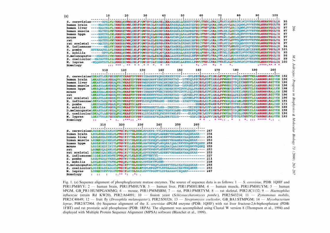
Fig. 1. (a) Sequence alignment of phosphoglycerate mutase enzymes. The source of sequence data is as follows: 1 Ð S. cerevisiae, PDB: 1QHF and
PIR1:PMBYY; 2 Ð human brain, PIR1:PMHUYB; 3 Ð human liver, PIR1:PMHUBM; 4 Ð human muscle, PIR1:PMHUYM; 5 Ð humanbPGM, GB_PR1:HUMPGAMMG; 6 Ð mouse, PIR1:PMMSBM; 7 Ð rat, PIR1:PMRTYM; 8 Ð rat skeletal, PIR2:JC1132; 9 Ð Haemophilusin¯uenzae (strain Rd KW20), PIR2:A64091; 10 Ð ®ssion yeast (Schizosaccharomyces pombe ), PIR2:S43214; 11 Ð Zymomonas mobilis,
PIR2:C40649; 12 Ð fruit ¯y (Drosophila melanogaster ), PIR2:S50326; 13 Ð Streptomyces coelicolor, GB_BA1:STMPGM; 14 Ð Mycobacteriumleprae, PIR2:S72904. (b) Sequence alignment of the S. cerevisiae dPGM enzyme (PDB: 1QHF) with rat liver fructose-2,6-biphosphatase (PDB:1FBT) and rat prostatic acid phosphatase (PDB: 1RPA). The alignment was accomplished using Clustal W version 8 (Thompson et al., 1994) anddisplayed with Multiple Protein Sequence Alignment (MPSA) software (Blanchet et al., 1999).
M.J.Jedrzeja
s/Progress
inBiophysics
&Molecu
larBiology73(2000)263±287
266
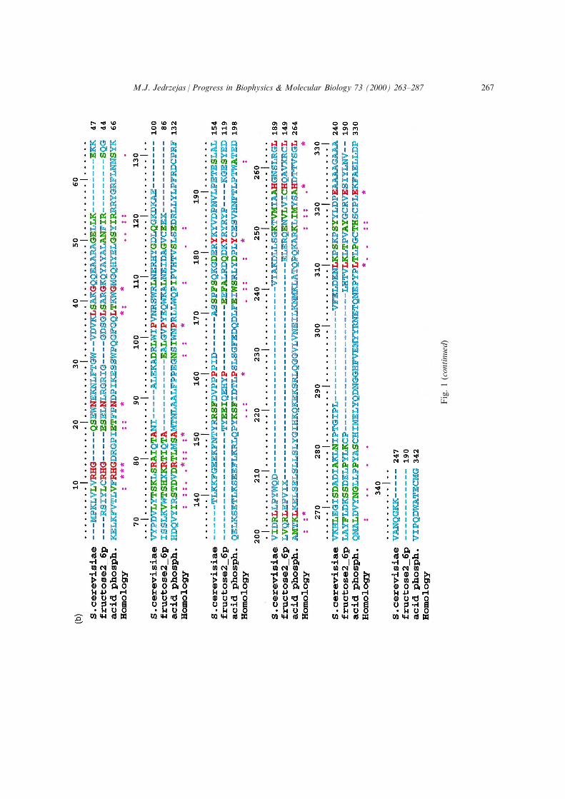
Fig.1(continued)
M.J. Jedrzejas / Progress in Biophysics & Molecular Biology 73 (2000) 263±287 267

(Campbell et al., 1974; Ridgen et al., 1998). However, every monomer is built from twodomains with the larger domain having a nucleotide-binding fold that was thought not to haveany physiological relevance (Campbell et al., 1974; Winn et al., 1981). However, chargedligands other than nucleotides, such as the phosphoglycerates (23PGA, 3PGA, 2PGA), canlikely bind in this location (Bazan and Fletterrick, 1990). Not surprisingly, the active site ofthis dPGM was located in this area by Winn et al. (1981). There is one active site permonomer located in a crevice at the C-terminal end of the b-sheet and this active site utilizesresidues only from this monomer. The active site has, among other amino acids, two histidineresidues, His8 and His181, with the His8 involved in forming a phosphohistidine±enzymeintermediate as a part of the catalytic cycle of this dPGM enzyme (Fig 2(b)) (Winn et al., 1981;Fothergill-Gilmore and Watson, 1989; Rose 1971, 1980; Nairn et al., 1995). The precisedetermination of all residues involved in the activity is still sketchy at this time due to the¯exibility of at least part of the residues playing the catalytic role. Speci®cally, the C-terminalpart containing 14 residues of the enzyme has a large degree of ¯exibility and its presence wasnot observed in any of the structures reported to date, except for the recent complex structurewith 3PGA substrate where a part of this tail was located (Crowhurst et al., 1999). Based onthe later studies, this part of the enzyme is seen to be crucial for activity as it partially coversthe active site area, possible only during the phosphate transfer between the 2 and 3 positionsof the glycerate during the catalytic process. This C-terminal tail interacts with the substratewhich stabilizes the so called `cap or lid' formation (Crowhurst et al., 1999). Therefore, theaccess to the active site is regulated by the conformation of the C-terminal tail of the enzyme.Site-directed mutagenesis of the very similar human bisphosphoglycerate mutase performed onthe amino acid in this region of the enzyme showed the reduction of the enzyme's catalyticactivity (Garel et al., 1989). Also, the proteolytic removal of these residues caused signi®cantdecrease of activity (Sasaki et al., 1966; Price et al., 1985). Based on the complex structures ofthe enzyme with 3PGA, residues other than His8 and His181 that interact with the 3PGAsubstrate and, therefore, get implicated in the activity, are as follows: Arg7, Ser11, Asn14,Arg59 (Fig 2(b), Table 1) (Crowhurst et al., 1999). However, other structural data availableshow two sulfate ions in the yeast dPDM's active site that seem to mimic the positions of thetwo phosphate ions of the 23PGA cofactor/substrate and/or the reaction intermediate of thisenzyme (Ridgen et al., 1999). These two sulfate ions interact with additional residues of theenzyme: Thr20, Arg87, Tyr89, Arg113, Arg114, and Asn183 (Fig 2(b), Table 1). Based on thelimited structural data of this enzyme complexes, the active site of the yeast dPGM, de®ned asall residues interacting with the substrate/product or the cofactor, includes all 12 abovementioned residues (Table 1). The His181 residue does not interact with 3PGA or any of thesulfate ions. Based on the sequence comparison of sequence data available for the dPGMs aswell as for the bPGM enzymes, all these residues except Thr20 are strictly conserved whichreinforces the importance of these residues for catalysis. Other residues in the position of thenonconserved Thr20 are, however, a very similar nature and include either Ser or Cys(Fig. 1(a)). As one might expect, the residues important for catalysis should be conserved inevolution. This de®nitely is the case for the iPGM enzymes known to date, where all 15identi®ed active site residues are strictly conserved even for organisms across di�erentkingdoms (Jedrzejas et al., 2000a).
M.J. Jedrzejas / Progress in Biophysics & Molecular Biology 73 (2000) 263±287268

Fig. 2. (a) General structure of S. cerevisiae dPGM. The structure of the yeast dPGM is based on its X-raystructure (Crowhurst et al., 1999) (PDB: 1QHF). (b) Active site residues of S. cerevisiae phosphoglycerate mutase.The interactions of the substrate/product with the protein are shown as determined by Crowhurst et al. (1999)
(PDB: 1QHF).
M.J. Jedrzejas / Progress in Biophysics & Molecular Biology 73 (2000) 263±287 269

2.2. Mechanism of catalysis of the S. cerevisiae dPGM
The catalytic mechanism of this enzyme was thought to involve two histidine residues, His8and His181, and proceed through a phosphohistidine±enzyme intermediate utilizing His8(Rose, 1971, 1980; Han and Rose, 1979; Nairn et al., 1995). The role of His8 and theformation of the intermediate utilizing this residue have been well established. However, therole of the conserved His181 residue in all known dPGMs and bPGMs is still not clear, but itwas originally thought to act as a proton donor/acceptor (acid/base catalyst) during thesuspected catalysis through the phospho±His8 intermediate and/or the suggested hydrolysis ofthe phosphate group (Rose, 1980). The important role of this residue has been con®rmed bysite-directed mutagenesis (White and Fothergill-Gilmore, 1992; White et al., 1993a) which forthe yeast enzyme leads to activity below 5% of the level of the wild-type enzyme. Mutations ofHis181 or equivalent residues in dPGMs from other organisms such as Schizosaccharomycespombe (Nairn et al., 1996) or human erythrocyte bPGM lead to similar loss of activity (Garelet al., 1993). Such mutations are, however, associated with structural alterations which in thecase of the yeast enzyme lead to the disruption of its tetrameric structure (White et al., 1993b).Therefore, the unambiguous assignment of the role of this residue in catalysis is stillquestionable.The catalytic mechanism is initiated by 3PGA (2PGA) binding to the His8 phosphorylated
enzyme's active site (phosphorylated by the 23PGA cofactor), followed by the transfer of thephosphate group from His8 to the C2 (C3) carbon atom of 3PGA (2PGA) to create a reactionintermediate, 23PGA. This substrate phosphorylation part of the reaction is followed by thereorientation of the 23PGA intermediate in the active site to bring the other phosphate groupinto the proximity of the now available His8 residue. The transfer of this phosphate to thishistidine follows in order to regenerate the phospho-enzyme for further catalysis. The 2PGA(3PGA) product then dissociates from the enzyme's active site making it available for thebinding of another substrate molecule (Fig 2(b)) (Ridgen et al., 1999).The mechanism for the other reaction of interconversion of 13PGA and 23PGA have also
been proposed by Ridgen et al. (1999). Similar to the main reaction catalyzed by dPGMs, itinvolves binding of 13PGA to the unphosphorylated enzyme, followed by the transfer of the1phosphate group to His8 to form a phospho-enzyme intermediate and a 3PGA intermediate.The reorientation of the 3PGA and the transfer of the phosphate to its O2 position winds upthe reaction. This review will, however, focus on the main reaction catalyzed by mPGMs(dPGMs and iPGMs) which is the interconversion of 3- and 2PGA.
2.3. Structural comparison of dPGM with other enzymes
The structure of the yeast dPGM enzyme has been found to be similar in sequence and in itsthree-dimensional structure to two unexpected phosphatase enzymes, fructose-2,6-bisphosphatase (Bazan et al., 1989; Lee et al., 1996; Hasemann et al., 1996) and acidphosphatase (Schneider et al., 1993; LaCount et al., 1998). The sequence similarity of the yeastdPGM (PDB: 1QHF) and the rat liver Fru26P2ase (PDB: 1FBT) is 46% whereas the rat acidphosphatase (AcPase) (PDB: 1RPA) is a bit lower, 35%. Their structures were aligned pairwisewith the yeast dPGM enzyme using only the Ca coordinates utilizing the program O (Jones et
M.J. Jedrzejas / Progress in Biophysics & Molecular Biology 73 (2000) 263±287270

al., 1991). There were nine structure-based alignment segments between the yeast dPGM andFru26P2ase containing 160 residues that aligned with an r.m.s. deviation of 1.63 AÊ , whereassimilar structure-based alignment between the dPGM and the rat AcPase produced sixsegments containing 95 residues and an r.m.s. deviation of 1.68 AÊ . The structure-basedalignments agreed exactly with the sequence alignment of these enzymes performed usingClustal W (Thompson et al., 1994) (Fig. 1(b)). However, only ®ve residues, Arg7, His8, Asn14,Arg59, and His181, out of all twelve residues (Fig. 1(b), Table 1) implicated in the activity ofthe yeast dPGM enzyme were conserved in this alignment. These ®ve conserved residues arealso conserved among all other PGMs, dPGMs and bPGMs, analyzed in Fig 1(a).
2.3.1. Structural comparison of S. cerevisiae dPGM with 6-phosphofructo-2-kinase/fructose-2,6-bisphosphatase
The Fru26P2ase composes only one domain of a bifunctional enzyme 6-phosphofructo-2-kinase/fructose-2,6-bisphosphatase (6PFru-2-K/Fru26P2ase) (EC 2.7.1.105/3.1.3.46) (Lively et
Table 1Selected essential interactions in the active site of the yeast dPGM enzyme
dPGM residue Atom type Substrate analog or protein residue Atom type Distance (AÊ )a
Arg7 NH2 3PGA O1 3.47
NE 3PGA O3 3.49His8 ND1 3PGA O3 3.64
NE2 3PGA O2P 3.83
Gly9 O His181 ND1 2.84Ser11 N 3PGA O1P 3.31
N Sulfate1 O3 3.14
OG Sulfate1 O4 2.90Asn14 N 3PGA O4P 3.81
ND2 3PGA O2 3.60Thr20 OG1 Sulfate1 O4 2.68
Arg59 NE 3PGA O2P 2.78Glu86 ?? Water O ??Arg87 NH2 Sulfate2 O2 3.14
Tyr89 OH Sulfate2 O1 2.83Arg113 NE Sulfate2 O1 2.90
NH2 Sulfate2 O4 2.93
Arg114 NH1 Sulfate2 O2 2.92NH2 Sulfate2 O4 3.14
His181 NE2 His8 NE2 2.79
Asn183 ND2 Sulfate2 O1 3.23
a Data based on Crowhurst et al. (1999) for the 3PGA substrate, and on Ridgen et al. (1999) for the complex withsulfate ions as well on the structures from the Protein Data Bank (PDB) accession numbers 1QHF and 5PGM, re-
spectively.
M.J. Jedrzejas / Progress in Biophysics & Molecular Biology 73 (2000) 263±287 271

al., 1988) which catalyzes the degradation of fructose-2,6-biphosphate (Fru26P2). The kinasedomain catalyzes the synthesis of Fru26P2. Each of the reactions is performed by di�erentactive sites of the enzyme located in its two di�erent domains. The regulation of the levels ofFru26P2 is essential for the regulation of glycolysis versus gluconeogenesis through activationor inhibition of 6-phosphofructo-kinase or fructose-1,6-bisphosphatase, two rate-limitingenzymes in sugar metabolism (Bazan et al., 1989; Bazan and Fletterrick, 1990). Such regulationof levels of Fru26P2 leads to the control of the energy storage and levels of blood glucose(Pilkis et al., 1995). Several isozymes of the enzyme have been identi®ed including the enzymeisolated from liver, skeletal muscle, heart, testis, and brain (Darville et al., 1989; Tsuchiya andUyeda, 1994; Sakata et al., 1991; Ventura et al., 1992). The structure of the dimeric rat liverFru26P2ase domain was obtained (Lee et al., 1996) followed by the structure of the wholeenzyme from the rat testis; also dimeric in nature (Hasemann et al., 1996). Based on thedimeric structure of the whole enzyme, the phosphatase and the kinase domains of the enzymeare well separated and are clearly independent of one another (Hasemann et al., 1996). Thekinase domain has an a/b type fold with a central six-stranded b-sheet surrounded by seven a-helices. A nucleotide binding fold is located at the C-terminal end of the ®rst b-strand and thisis where the kinase active site is located. Both structures of the phosphatase domain of thewhole rat testis enzyme (Hasemann et al., 1996) and the truncated one from rat liver (Lee etal., 1996) are very similar to one another. This phosphatase domain is also similar in structureand sequence to the yeast dPGM enzyme (Fig. 1(b)). Therefore, here we will focus ourdiscussion on the rat liver enzyme's structure of the phosphatase domain alone.The dimeric phosphatase domain, like other glycolytic enzymes, has an a/b-fold (Sternberg
et al., 1981) with the nucleotide-binding topology and the core six-stranded b-sheet surroundedby a-helices on both sides (Lee et al., 1996). The active site of the phosphatase domain is at theC-terminal end of the four-stranded parallel b-sheet. Above the active site there are two longloops that extend away from the core of the enzyme. The area between these loops leads to afunnel-like active site. The active site includes two key histidine residues, His7 and His141,which correspond to the yeast dPGM's His8 and His181 (Bazan et al., 1989; Campbell et al.,1974). A phosphate group has been located in this site in close proximity to the above histidineresidues, 4.2 AÊ away from ND1 His7 and 4.1 AÊ away from ND1 His141 and the phosphorusatom (Lee et al., 1996). Another residue interacting with the phosphorus atom of thephosphate group is OE1 of Glu76, 3.6 AÊ away followed by the guanidinium groups of Arg6(3.7 AÊ away) and Arg56 (4.0 AÊ away) (Fig. 3). His7 was also implicated in the formation ofthe phosphohistidine±enzyme intermediate (Tauler et al., 1990).
2.3.2. Structural comparison of S. cerevisiae dPGM with acid phosphatase
The acid phosphatases, on the other hand, catalyze the hydrolysis of phosphate monoestersand at times a phosphotransfer of phosphate between phosphoester and alcohols. The enzymecatalysis progresses optimally at acidic conditions (Vincent et al., 1992; van Etten, 1982;Bodansky, 1972). These enzymes are found among animals as well as plants (Vincent et al.,1992). In contrast, the totally structurally di�erent alkaline phosphatases (Sowadsky et al.,1985; Kim and Wycko�, 1991) work optimally at alkaline conditions (Coleman, 1992; Gettinset al., 1985; Coleman and Gettins, 1983; Gettins and Coleman, 1983). The acid phosphatases,
M.J. Jedrzejas / Progress in Biophysics & Molecular Biology 73 (2000) 263±287272

unlike the alkaline phosphatases, do not utilize metal ions in their catalysis. They insteadutilize histidine to form an enzyme±phosphohistidine intermediate which is essential for theircatalysis (van Etten, 1982). In contrast, alkaline phosphatases utilize a phospho-serine enzymeintermediate for their catalysis and have a binuclear Zn(II) active site (Coleman, 1992;Coleman et al., 1983, 1997) and are related to the iPGM enzymes (Galperin et al., 1995;Jedrzejas et al., 2000a; Jedrzejas and Setlow, 2000).The structure of rat prostatic acid phosphatase, which is also dimeric in nature, was
determined by Schneider et al. (1993) followed by the structure of the human prostatic enzymedetermined by LaCount et al. (1998). The structure of the enzyme comprises two domains, ana/b type domain and a smaller a-helical domain (Schneider et al., 1993). The a/b domain has acore of seven-stranded mixed b-sheets ¯anked on both sides by a-helices. The a-domain formsa ¯at disk-like cap above the other domain of the enzyme. The active site is located in anopen, easily accessible cleft between the two domains at the C-terminal end of the parallel b-strands of the a/b domain. The residues conserved among acid phosphatases that are in theactive site area are implicated in the activity of this enzyme and are as follows: Arg11, His12,Arg15, Arg79, His257, and Asp258 (Fig. 4) (Schneider et al., 1993). His12 has been proposedto form a phosphohistidine±enzyme intermediate (Ostanin et al., 1992). This residue is in closeproximity to several Arg residues: Arg11, Arg15, Arg79 and His257, which create a positivelycharged pocket suggestive of phosphate binding. His12 and His257 correspond to similarresidues in the dPGM enzyme, His8 and His181, respectively (Fig. 1(b)). All of these residues,except Arg15, are conserved in sequence and structure among all three enzymes discussed hereand are conserved among all dPGM enzymes' sequences (Fig. 1(a) and (b).
2.3.3. Overall comparison of active sites of dPGM, Fru26P2ase, and AcPase
Based on an analysis of structural data and mutagenesis results together with the sequenceanalysis of all known dPGM enzymes, 14 residues were implicated in the catalysis carried outby dPGMs (Table 1). Out of these 14 residues only six are uniformly conserved amongFru26P2ases and AcPase enzymes: Arg7, His8, Glu9, Asn14, Arg59, and His181 (Figs. 1(a)and (b), 3 and 4). The precise mechanism of the dPGM catalytic process is still largelyspeculative. However, a crucial component, a histidine residue that accepts a phosphate groupduring the reaction, has been unequivocally identi®ed (Rose, 1971; Nairn et al., 1995). Anotherhistidine residue, His181, was con®rmed as being very important for catalysis but its precisefunction is still speculative (White and Fothergill-Gilmore, 1992; White et al., 1993a).Originally, this histidine was suggested to perform an acid/base type of function based on thedonation or extraction of a proton during the formation of phosphohistidine intermediate(donation of H for neutralization of the O2 (O3) oxygen of the substrate's glycerate moiety) orduring the hydrolysis of the phosphohistidine intermediate (extraction of H from a watermolecule hydrolyzes the His8±phosphate bond) (Rose, 1980). This function could also beperformed, as recently suggested by Ridgen et al. (1999), by a water molecule polarized byGlu86. This scenario is consistent with studies of the E86Q mutant of the yeast enzyme, whichretains less than 5% of the wild-type enzyme activity (Ridgen et al., 1999). Surprisingly, Glu86is not conserved in the human or in the rat prostatic AcPases. It is conserved, however, in therat liver and testis Fru26P2ase (Glu76) (Figs. 1(b), 3 and 4) (Lee et al., 1996; Hasemann et al.,
M.J. Jedrzejas / Progress in Biophysics & Molecular Biology 73 (2000) 263±287 273

1996). An inspection of the prostatic AcPase enzyme structure, however, shows Asp258 in a
nearby location. This residue could interact with the proposed catalytic water molecule and
facilitate the mechanism as would a Glu residue. The precise mechanism leading to the
formation and subsequent hydrolysis of the enzyme-phosphate complex cannot be
unquestionably delineated at this time. This is largely due to the complexity of the mechanism
and the ¯exible nature of at least part of the residues in the active site including the C-terminal
tail capping this site. The structural information on sulfate (Ridgen et al., 1999) and especially
on the 3PGA substrate binding in the active site (Crowhurst et al., 1999) clari®ed signi®cantly
the proposed catalytic mechanism of this enzyme. The structure of these complexes de®nitely
narrowed down the possible mechanism involved in the catalysis and pointed to some new
residues or to the assignment of di�erent functions for some of them. For example, for the
yeast dPGM the proposed function of His181 as a proton donor/acceptor was later replaced
by a proposed functional water molecule and a close-by Glu86 residue (Ridgen et al., 1999).
However, more structural information on substrate/product binding in the active site would
help identify all residues involved in the catalysis and contribute to the determination of their
precise role. Such information will likely be obtained in the future.
Fig. 3. The residues in the active site area of the S. cerevisiae dPGM (PDB: 1QHF) aligned with the rat liverfructose-2,6-bisphosphatase (Lee et al., 1996) (PDB: 1FBT). Among other conserved residues, both catalytic
histidines, His8/His7 and His181/His141, are conserved.
M.J. Jedrzejas / Progress in Biophysics & Molecular Biology 73 (2000) 263±287274

Based on the sequence analysis (Fig. 1(b)) and the overall structure similarities as well as the
similarities of the active sites of the three enzymes, 23PGA-dependent monophosphoglycerate
mutase, fructose-2,6-bisphosphatase, and acid phosphatase are very similar in their properties.
The catalytic mechanism of these enzymes seems to follow the same paradigm and consists of
two main steps: phosphorylation of a histidine residue to create an enzyme-phosphate
intermediate and subsequent hydrolysis of this phosphate to transfer this group to the
substrate or to release an inorganic phosphate group. The ®rst possibility corresponds to the
function of dPGM and AcPase enzymes and has two aspects, a phosphatase and a
phosphotransferase activity, the second to the Fru26P2ase which has only the phosphatase part
of the reaction. As stated above, the AcPase enzymes have the ability to phosphorylate
themselves (phosphatase activity), to release this phosphate group by the hydrolysis of the
phosphate monoester linkage to the enzyme (part of the phosphatase activity), or to transfer
the phosphate to other chemical groups such as alcohols (phosphotransferase ability). All three
enzymes have the ability, however, to perform all these reactions albeit with very di�erent
catalytic rates and all these reactions involve the same phosphohistidine intermediate (Tauler et
al., 1987).
Fig. 4. Active site residues of S. cerevisiae dPGM aligned with those of the rat prostatic acid phosphatase(Schneider et al., 1993) (PDB: 2RPA). Both active site histidine residues, His8/His12 and His181/His257, are also
conserved.
M.J. Jedrzejas / Progress in Biophysics & Molecular Biology 73 (2000) 263±287 275

For dPGM, Fru26P2ase, and AcPase the presence of the phosphohistidine intermediate hasbeen con®rmed and localized, in each case, to one His residue: His8 for the yeast dPGM, His7for the human Fru26P2ase, and His12 for the rat prostatic AcPase enzyme. Moreover, for allthese enzymes other histidine residues, His181 for the yeast dPGM, His141 for Fru26P2ase,and His257 for AcPase, were identi®ed to be very important for catalysis. Mutation of thishistidine leads to very signi®cantly compromised enzymes. Both histidines are conserved in thesequence analysis shown in Fig. 1(a) and (b) as well as in the three dimensional structuresshown in Figs. 3 and 4. Similar to the yeast dPGM, all residues involved in the activity relatedto each of these above enzymes have not, however, been clearly identi®ed, in part due to theexpected ¯exibility of these active sites and the immediate adjacent areas of the structure. Inaddition, even though the general aspects of the catalytic mechanism of these enzymes havebeen known for many years, the exact mechanisms and involved residues are still unknown.Signi®cant hints were provided by the structural information of complexes of these enzymeswith substrate or substrate mimicking chemicals such as the recent structural data on the yeastdPGM with 3PGA or sulfate residues in its active sites (Ridgen et al., 1999; Crowhurst et al.,1999).
The functional and structural similarities of all three enzymes analyzed above provide strongevidence for their close evolutionary similarity and, furthermore, show that they areevolutionarily related (see also Bazan et al., 1989). These enzymes catalyzing similar reactiontypes are likely to have descended from a common ancestral protein and acquired their uniquefeatures through divergent evolution (Fothergill-Gilmore, 1987; Rossmann, 1981). Thedivergent sequences and di�erent preferred substrates suggest that the progenitor of all thesedi�erent enzymes possibly had the a�nity for a negatively charged molecule such as a smallmolecule or an ion, with a nucleotide-like binding fold built from b-sheets and a-helices. Thede®nitively de®ned active site possibly developed later with one or two catalytic groups like thehistidine residues, His8 and His181 (dPGM numbering scheme) (Bazan and Fletterrick, 1990).During the course of evolution, the initial/progenitor enzyme developed di�erences in its activesite and its adjacent areas, loops or other structures, evident from the comparison of thestructures of the three enzymes discussed above, and based on surface features that probablyled to the specialization of function and the emergence of three di�erent albeit similar familiesof enzymes and dPGMs together with the closely related bPGMs, Fru26P2ases, and AcPases(Fig. 1(a) and (b)) (Bazan and Fletterrick, 1990; Tauler et al., 1987). The relationship betweenthese enzymes is not obvious from their primary structures. However, the comparison of theirthree-dimensional structures ®nally revealed their similarities. As is often the case, the tertiarystructure seems to be more conserved than the primary structure (Chothia et al., 1987;Richardson, 1981).
As mentioned earlier, the dPGM and bPGM enzymes have the ability to catalyze the samereactions but at signi®cantly di�erent rates. dPGM catalyzes the 23PGA-dependentinterconversion of 2- and 3PGA, and bPGM catalyzes the interconversion of 13PGA and23PGA (Fothergill-Gilmore and Watson, 1989). In addition to performing similar catalyticfunctions, signi®cant sequence homology of these two group of enzymes supports the notionthat these two groups of enzymes are isoenzymes. The sequence similarities among the dPGMsare, however, higher than their sequence similarity with the bPGM enzyme (Fig. 1(a)). This is
M.J. Jedrzejas / Progress in Biophysics & Molecular Biology 73 (2000) 263±287276

suggestive of gene divergence being responsible for the creation of bPGMs followed later bythe evolutionary divergence among dPGMs (Fothergill-Gilmore, 1989).It seems, however, that the other type of PGMs, the 23PGA independent mPGM (iPGM), is
not evolutionarily related to any of the other PGMs, or to the other enzymes related todPGM/bPGM, Fru26P2ases or AcPases (Fothergill-Gilmore and Watson, 1989). Having inmind that the iPGMs are approximately twice as large as the dPGM/bPGM enzymes, it ispossible that the development of the iPGM enzymes might have been a gene-doubling processas has been observed for a number of other enzymes (Lawrence and Trayer, 1984; Poorman etal., 1984). The three-dimensional structural information about the iPGM enzyme from Bacillusstearothermophilus, representative of all iPGM enzymes, have only recently been madeavailable (Jedrzejas et al., 2000a). The structure of this iPGM has two distinct domains whicheither individually or together do not structurally align in any way with the yeast dPGMenzyme structure. Sequence comparison of both types of PGMs, combined with their lack ofany three-dimensional structural alignment/similarity, of these two enzymes clearly shows thatthese enzymes did not evolve through the gene-doubling process. Signi®cant structuralhomology has been, however, identi®ed between the iPGM enzyme and the E. coli alkalinephosphatase (AlPase) (Jedrzejas and Setlow, 2000; Jedrzejas et al., 2000a). The distribution ofthese two types of PGMs in di�erent organisms, dPGMs speci®c to vertebrates and iPGMspeci®c to plants (see Section 1) (Carreras et al., 1982; Price et al., 1985), suggests that bothtypes of genes were present early in evolution. Animals lost the iPGM gene prior to theradiation of vertebrates whereas plants probably lost the dPGM gene when the early formsdiverged from the primitive unicellular organisms (Fothergill-Gilmore and Watson, 1989).Some eubacteria with larger genomes such as E. coli or B. subtilis, however, have both
dPGM and iPGM genes and both of these genes are expressed at somewhat similar levels(Fraser et al., 1999; Kunst et al., 1997). Also, in E. coli both of these enzymes, dPGM andiPGM, were active albeit at somewhat di�erent rates (Fraser et al., 1999). In general thedPGM enzyme appears to be more active in E. coli. In their evolution, these selectedunicellular eubacterial organisms somehow managed to retain both genes.
3. Bacillus stearothermophilus cofactor independent phosphoglycerate mutase
3.1. Structure of B. stearothermophilus phosphoglycerate mutase
The structure of the ®rst iPGM enzyme has just recently been published by Jedrzejas et al.(2000a). The B. stearothermophilus iPGM enzyme consists of 511 residues (57 kDa) and itsactive form structure contains two Mn(II) ions (Chander et al., 1999). It is a monomer whichcontains two distinctly separated domains of approximately equal size which interact with oneanother through an extended surface area contact (Fig. 5(a)). There is a narrow cleft betweenthese domains which is where the active site of the enzyme is located. This crystal structurealso includes a 3PGA substrate as well as a catalytic, ordered water molecule bound in theactive site of the enzyme (Fig. 5(b)). Both domains have a central b-sheet surrounded by a-helices on the outside. The active site contains residues from both domains. However, residuesinteracting with the Mn(II) ions and the phosphate group of 3PGA originate from one
M.J. Jedrzejas / Progress in Biophysics & Molecular Biology 73 (2000) 263±287 277
domain, A, whereas residues interacting with the glycerate part of the substrate originate fromthe other domain, B.
3.2. Catalytic mechanism for B. stearothermophilus iPGM and its comparison to alkalinephosphatase catalysis
The precise mechanism of catalysis of this iPGM was proposed based on the complexstructure with 3PGA and Mn(II), structure-guided mutant studies, sequence and structurecomparison with the E. coli AlPase (Jedrzejas et al., 2000a, 2000b), and earlier biochemicalstudies (Smith et al., 1988; Leadlay et al., 1977). Another structure was recently obtained forthe iPGM complex with the other substrate/product, 2PGA, which closes the catalytic loop ofa substrate to a product transformation or vice versa (Jedrzejas et al., 2000b). The catalyticmechanism from 3- to 2PGA isomerisation involves phosphorylation of the Ser62 residue by3PGA (phosphatase activity) followed by the reorientation of the remaining glycerate of 3PGAand the transfer of the phosphate to the O2 oxygen of this repositioned glycerate(phosphotransferase activity). The active site water molecule hydrolyzes the interaction of the2PGA product with Mn(II) ion and frees up the active site for the next round of catalysis.Both Mn(II) ions are inherently involved in this catalytic process (Dismukes, 1996) throughtheir interactions with active site residues and the substrates/products as well as the orderedactive site water molecule.For spore forming bacteria, such as B. stearothermophilus, a Mn(II) requirement leads to a
physiologically relevant pH sensitivity of the enzyme (Kuhn et al., 1991; Kuhn et al., 1993,1995). A pH of 8 is optimal for enzyme activity whereas activity is essentially lost at pH06. Inspores the pH is as low as 6.5 in comparison to the one in the vegetative cells which is 08.0(Chander et al., 1998; Swerdlow et al., 1981; Singh and Setlow, 1979). Therefore, during thesporulation process pH drops down to 06.5 and iPGM becomes inactive allowing for 3PGAdeposits that can be later utilized as the main source of energy required for the germinatingspore when pH rises back to 08. The new vegetative cell utilizes the 3PGA deposits in theglycolysis pathway to form ATP necessary for other biochemical reactions (Magil et al., 1994,1996; Setlow and Kornberg, 1970). For a detailed description of the iPGM mechanism pleaserefer to Jedrzejas et al. (2000a, 2000b) as well as to Jedrzejas and Setlow (2000).The information shown above allowed the assignment of the phosphatase activity of the
enzyme to domain A and domain B to the phosphotransferase activity. The phosphatasedomain of iPGM has been shown to have a general fold similar to E. coli AlPase (Sowadski etal., 1985). Furthermore, the structural positioning of all metal-binding residues in the activesites for both enzymes is also essentially identical (Fig. 6). However, there is no similaritybetween the phosphotransferase domain of iPGM and the AlPase.A detailed comparison of both enzyme types, iPGMs and AlPases, has also been provided
based on their sequences and three-dimensional structures, by Jedrzejas and Setlow (2000), andbased on a sequence in an earlier report by Galperin et al. (1998). Both the phosphatasedomain of iPGM and the AlPase enzyme were clearly identi®ed to be structurally andfunctionally related to one another. Both are binuclear metalloenzymes (Dismukes, 1996;Johnson and Price, 1988), the B. stearothermophilus being Mn(II)-dependent whereas E. coliAlPase is Zn(II)-dependent (Applebury et al., 1970; Anderson et al., 1975; Applebury and
M.J. Jedrzejas / Progress in Biophysics & Molecular Biology 73 (2000) 263±287278

Coleman, 1969; Bosron et al., 1975, 1977), and their similar catalytic mechanisms utilize a
phosphoserine±enzyme intermediate. This mechanism is unlike the behavior of dPGMs/
Fru26P2ases/AcPases which utilize a phosphohistidine±enzyme intermediate for their catalysis.
As noted earlier, iPGMs are speci®c to plants as well as some other organisms/cells (see
Section 1) whereas AlPases have been identi®ed in animal, yeast, and bacterial cells but, not so
far, in plants (Galperin et al., 1998). Considering the structural, functional, and mechanistic
similarities of both iPGMs and AlPases (similar to the dPGMs/Fru26P2ases/AcPases enzymes),
it is possible that these two groups of proteins (iPGMs and AlPases) are evolutionarily related
and have evolved from a common ancestor enzyme; di�erent from the dPGM related ancestral
enzyme discussed earlier. Furthermore, similarities have already been identi®ed (Galperin et al.,
Fig. 5. (a) General structure of B. stearothermophilus iPGM enzyme. Both the phosphatase and thephosphatransferase domains are labeled along with the active site containing 3PGA substrate/product (Jedrzejas etal., 2000a). (b) Active site residues color coded by the domain: phosphatase domain B Ð green, phosphotransferase
domain A Ð red. Two Mn(II) ions as well as the catalytic water and 3PGA are also shown.
M.J. Jedrzejas / Progress in Biophysics & Molecular Biology 73 (2000) 263±287 279

1998) between all known iPGMs and AlPases among archeal proteins such as Methanococcusjannaschii MJ1612 and MJ0010 (Koonin et al., 1997) and paralogous proteins inMethanobacterium thermoautotrophicum (Smith et al., 1997) or in Archaeoglobus fulgidus(Klenk et al., 1997).
4. Functional properties and comparison of the S. cerevisiae dPGM with theB. stearothermophilus iPGM enzyme
Both enzyme groups, dPGMs and iPGMs, perform their catalysis through a phosphoenzymeintermediate. For the cofactor dependent dPGM enzyme, the enzyme is probablyphosphorylated by the 23PGA cofactor on His8, a process required for the enzyme activity.The functional, active enzyme form is its phosphorylated enzyme. For the cofactor independentiPGM enzyme, the substrate itself phosphorylates the enzyme not on a histidine but on a Ser62residue. The native and active iPGM enzyme is not a phospho±enzyme. Although the dPGMenzymes are not metalloenzymes, all iPGM enzymes are metalloenzymes and most likely all
Fig. 5 (continued)
M.J. Jedrzejas / Progress in Biophysics & Molecular Biology 73 (2000) 263±287280

utilize manganese ions as do the B. stearothermophilus and E. coli enzymes (Jedrzejas andSetlow, 2000). However, the substitution of Mn(II) for other metals, especially Zn(II), cannotbe totally excluded at this time. The pH sensitivity of iPGMs from spore-forming bacteria isphysiologically relevant but for other bacterial organisms such as E. coli it is not. Therefore, itseems likely that all bacterial iPGM enzymes evolved from a gene in a common bacterialancestor having the ability to create spores which later in the evolutionary process some ofthese organisms lost.For both enzymes, after the phosphorylation step a signi®cant reaction intermediate
reorientation must occur to either remove a phosphate from the reaction intermediate fordPGM or to transfer the phosphate to the appropriate place in the glycerate for iPGM. Noneof the reactions of iPGM catalysis, except for the ®nal release of the product, involve ahydrolysis step due to the metalloenzyme nature of iPGM, and the ability of Mn(II) toneutralize charge through its interactions (Christianson and Cox, 1999; Christianson, 1997).For dPGM, however, two extra hydrolysis steps are likely required: hydrolysis of the phospho±enzyme bond and the hydrolysis of the reaction intermediate±phosphate bond. Implicated in
Fig. 6. Active site alignment of the phosphatase domain of B. stearothermophilus iPGM and E. coli AlPase. BothMn(II) ions, catalytic water and 3PGA of iPGM and two Zn(II) ions of AlPase are also shown.
M.J. Jedrzejas / Progress in Biophysics & Molecular Biology 73 (2000) 263±287 281

this process was the role of the acid/base hydrolysis of His181 or activation of a watermolecule by Glu86. Since the mutants of either His181 or Glu86 still retain some enzymeactivity, it is possible that both mechanisms (acid/bone hydrolysis and water activation) canperform this necessary function for catalysis. The dPGM enzyme as well as bPGM have anadditional ability to interconvert 13PGA and 23PGA as well as synthesize the 3PGA from23PGA. The iPGM enzyme is limited in this aspect as it only interconverts 3- and 2PGA.
5. Conclusions
Clearly, both dPGM and bPGM enzymes are evolutionarily related. This relation is visiblein both their primary and tertiary structures. Their catalysis proceeds via a phosphohistidine±enzyme intermediate and also involves the reorientation of their substrates in the process.These two groups of related enzymes show a moderate to low sequence homology toFru26P2ases and to AcPases but the comparison of the tertiary structures of all these enzymesmore clearly shows their structural and evolutionary similarity. All of these enzymesphosphorylate a histidine residue, a process essential for their catalytic activity which requirestwo main components, phosphatase and phosphotransferase. Some of these enzymes specializein just one component, the phosphatase reaction, but all have the ability to perform both ofthem albeit at di�erent rates. All of these enzymes are evolutionarily related to a commonancestral enzyme and they probably acquired their specialized traits through divergentevolution, although gene duplication can not be excluded. The divergence of the proteinsurface, the gain of loops, as well as changes in the areas adjacent to the active site presumablyresulted in the catalytic di�erences leading to di�erent enzymes: PGMs, Fru26P2ases, andAcPases.The iPGM enzymes, on the other hand, are not related to any of the other PGM group of
enzymes, both in sequence and in three-dimensional structure, but are related to the family ofalkaline phosphatases. This relationship is hardly evident from the primary structures ofiPGMs and AlPases (unlike the relationship for dPGMs/bPGMs/Fru26P2ases/AcPases)whereas clearly evident from the comparison of the tertiary structures, predominantly for theactive site residues responsible for metal binding. In terms of evolution, iPGMs and AlPasesprobably evolved from a common predecessor, an archeal protein. The specialization of thefunctional properties of the current enzymes probably evolved similarly to the dPGM familythrough changes of the active site environment and adjacent loop areas, as well as throughdivergence of protein surfaces.
Acknowledgements
The author would like to acknowledge the review of the manuscript by Dr. Elise A. Sudbeckand the helpful discussions with Dr. Peter Setlow.
M.J. Jedrzejas / Progress in Biophysics & Molecular Biology 73 (2000) 263±287282
References
Anderson, R.A., Bosron, W.F., Kenned, F.S., Vallee, B.L., 1975. Role of magnesium in Escherichia coli alkaline
phosphatase. Proc. Natl. Acad. Sci. USA 72, 2989±2993.
Applebury, M.L., Coleman, J.E., 1969. Escherichia coli Co(II) alkaline phsophatase. J. Biol. Chem. 244, 709±718.
Applebury, M.L., Johnson, B.P., Coleman, J.E., 1970. Phosphate binding to alkaline phosphatase. Metal ion
dependence. J. Biol. Chem. 245, 4968±4976.
Bazan, J.F., Fletterrick, R.J., 1990. In: Pilkis, S.J. (Ed.), Fructose 2,6-Bisphosphate. CRC Press, Boca Raton, FL,
pp. 125±171.
Bazan, J.F., Fletterrick, R.J., Pilkis, S.J., 1989. Evolution of a bifunctional enzyme: 6-phosphofructo-2-kinase/
fructose-2,6-bisphosphatase. Proc. Natl. Acad. Sci., USA 86.
Blanchet, C., Geourjon, C., Deleage, G., 1999. Multiple Protein Sequence Analysis. IBCP, CNRS UPR 412, L.on,
France.
Blattler, A.W., Knowles, J.R., 1980. Phosphoglycerate mutases: Stereochemical course of the phosphoryl group
transfers catalyzed by the cofactor-dependent enzyme from rabbit muscle and the cofactor-independent enzyme
from wheat germ. Biochemistry 19, 738±743.
Bodansky, O., 1972. Acid phosphatase. Adv. Clin. Chem. 15, 43±147.
Bosron, W.F., Anderson, R.A., Falk, M.C., Kennedy, F.S., Vallee, B.L., 1977. E�ect of magnesium on the
properties of zinc alkaline phosphatase. Biochemistry 16, 610±614.
Bosron, W.F., Kennedy, F.S., Vallee, B.L., 1975. Zinc and magnesium content of alkaline phosphatase from
Escherichia coli. Biochemistry 14, 2275±2282.
Breathnach, R., Knowles, J.R., 1977. Phosphoglycerate mutase from wheat germ: studies with 18O-labeled substrate,
investigations of the phosphatase and phosphoryl transfer activities, and evidence for a phosphoryl-enzyme
intermediate. Biochemistry 16 (14), 3054±3060.
Britton, H.G., Carreras, J., Grisolia, S., 1971. Mechanism of action of 2,3-diphosphoglycerate-independent
phosphoglycerate mutase. Biochemistry 11, 4522±4533.
Campbell, J.W., Watson, H.C., Hodgson, G.I., 1974. Structure of yeast phosphoglycerate mutase. Nature 250, 301±
303.
Carreras, J., Mezquita, J., Bosch, J., Bartrons, R., Pons, G., 1982. Phylogeny, ontogeny of the phosphoglycerate
mutases-IV. Distribution of glycerate-2,3-P2 dependent and independent phosphoglycerate mutases in algae,
fungi, plants and animals. Comp. Biochem. Physiol. [B] 71, 591±597.
Chander, M., Setlow, B., Setlow, P., 1998. The enzymatic activity of phosphoglycerate mutase from gram-positive
endospore-forming bacteria requires Mn2+ and is pH sensitive. Can. J. Microbiol. 44, 759±767.
Chander, M., Setlow, P., Lamani, E., Jedrzejas, M.J., 1999. Structural studies on a 2,3-diphosphoglycerate
independent phosphoglycerate mutase from Bacillus stearothermophilus. J. Struct. Biol. 126, 156±165.
Chothia, C., Lesk, A.M., 1987. The evolution of protein structures. Cold Spring Harb. Symp. Quant. Biol. 52, 399±
405.
Christianson, D.W., Cox, J.D., 1999. Catalysis by metal-activated hydroxide in zinc and manganese metalloenzymes.
Annu. Rev. Biochem. 68, 35±57.
Christianson, D.W., 1997. Structural chemistry and biology of manganese metalloenzymes. Prog. Biophys. Molec.
Biol. 67, 217±252.
Coleman, J.E., 1987. In: Torriani-Gorini, A., Rothman, F.G., Silver, S., Wright, A., Yagil, E. (Eds.), Phosphate
Metabolism and Cellular Regulation in Microorganisms. Am. Soc. Microbiol, Washington, DC, pp. 127±138.
Coleman, J.E., 1992. Structure and mechanism of alkaline phosphatase. Annu. Rev. Biophys. Biomol. Struc. 21,
441±483.
Coleman, J.E., Gettins, P., 1983. Alkaline phosphatase, solution structure, and mechanism. Adv. Enzymol. Relat.
Areas Mol. Biol. 55, 381±452.
Coleman, J.E., Nakamura, K., Chlebowski, J.F., 1983. 65Zn(II), 115Cd(II), 60Co(II), and Mg(II) binding to alkaline
phosphatase of Escherichia coli. Structural and functional e�ects. J. Biol. Chem. 258, 386±395.
Crowhurst, G.S., Dalby, A.R., Isupov, M.N., Campbell, J.W., Littlechild, J.A., 1999. Structure of a
phosphoglycerate mutase: 3-phosphoglyceric acid complex at 1.7 AÊ . Acta Crystallog. Sect. D. 55, 1822±1826.
M.J. Jedrzejas / Progress in Biophysics & Molecular Biology 73 (2000) 263±287 283

Darville, M.I., Crepin, K.M., Hue, L., Rousseau, G.G., 1989. 5 ' ¯anking sequence and structure of a gene encoding
rat 6-phosphofructo-2-kinase/fructose-2,6-bisphosphatase. Proc. Natl. Acad. Sci., USA 86, 6543±6547.
Dismukes, G.C., 1996. Manganese enzymes with binuclear active sites. Chem. Rev. 96, 2909±2926.
Fothergill-Gilmore, L.A., 1987. Evolution in glycolysis. Biochem. Soc. Trans. 15, 993±995.
Fothergill-Gilmore, L.A., Michels, P.A.M., 1993. Evolution of glycolysis. Progr. Biophys. Mol. Biol. 59, 105±235.
Fothergill-Gilmore, L.A., Watson, H.C., 1989. The phosphoglycerate mutases. Adv. Enzymol. 62, 227±313.
Fraser, H.I., Kratskhelia, M., White, M.F., 1999. The two analogous phosphoglycerate mutases of Escherichia coli.
FEBS Lett. 455, 344±348.
Galperin, M.Y., Bairoch, A., Koonin, E.V., 1998. A superfamily of metalloenzymes uni®es phosphopentomutase
and cofactor-independent phosphoglycerate mutase with alkaline phosphatases and sulfatases. Prot. Science 7,
1829±1838.
Garel, M.C., Joulin, V., Le Boulch, P., Calvin, M.C., Prehu, M.O., Arous, N., Longin, R., Rosa, R., Rosa, J.,
Cohen-Solal, M., 1989. Human bisphosphoglycerate mutase. Expression in Escherichia coli and use of site-
directed mutagenesis in the evaluation of the role of the carboxyl-terminal region in the enzymatic mechanism. J.
Biol. Chem. 264, 18966±18972.
Garel, M.C., Lemarchandel, V., Calvin, M.C., Arous, N., Craescu, C.T., Prehu, M.O., Rosa, J., Rosa, R., 1993.
Amino acid residues involved in the catalytic site of human erythrocyte bisphosphoglycerate mutase. Functional
consequences of substitutions of His187 and Arg89. Eur. J. Biochem. 213, 493±500.
Gatehouse, J.A., Knowles, J.R., 1977. Phosphoglycerate mutase from wheat germ: studies with isotopically labeled
3-phospho-D-glycerates showing that the catalyzed reaction is intramolecular. Biochemistry 16, 3045±3050.
Gettins, P., Coleman, J.E., 1983. 31P nuclear magnetic resonance of phosphoenzyme intermediates of alkaline
phosphatase. J. Biol. Chem. 258, 408±416.
Gettins, P., Metzler, M., Coleman, J.E., 1985. Alkaline phosphatase. 31P NMR probes of the mechanism. J. Biol.
Chem. 260, 2875±2883.
Grana, X., Lecea, L., El-Maghrabi, M.R., Urena, J.M., Caellas, C., Carreras, J., Puigdomenech, P., Pilkis, S.J.,
Climent, F., 1992. Cloning and sequencing of a cDNA encoding 2,3-bisphosphoglycerate-independent
phosphoglycerate mutase from maize. J. Biol. Chem. 267, 12797±12803.
Grana, X., Perez De La Ossa, P., Broceno, C., Stocker, M., Garriage, J., Puigdomenech, P., Climent, F., 1995. 2,3-
Bisphosphoglycerate-independent phosphoglycerate mutase is conserved among di�erent phylogenetic kingdoms.
Comp. Biochem. Physiol. 112B, 287±293.
Han, C-H., Rose, Z.B., 1979. Active site phosphohistidine peptides from red cell bisphosphoglycerate synthase and
yeast phosphoglycerate mutase. J. Biol. Chem. 254, 8836±8839.
Hasemann, C.A., Istvan, E.S., Uyeda, K., Deisenhofer, J., 1996. The crystal structure of the bifunctional enzyme 6-
phosphofructo-2-kinase/fructose-2,6-bisphosphatase reveals distinct domain homologies. Structure 4, 1017±1029.
Jedrzejas, M.J., Chander, M., Setlow, P., Krishnasamy, G., 2000a. Crystal structure and mechanism of action of
2,3-diphosphoglycerate independent phosphoglycerate mutase from Bacillus stearothermophilus. EMBO J. 19,
1419±1431.
Jedrzejas, M.J., Chander, M., Setlow, P., Krishnasamy, G., 2000b. Mechanism of catalysis of the cofactor
independent phosphoglycerate mutase from Bacillus stearothermophilus: crystal structure of the complex with 2-
phosphoglycerate. J. Biol. Chem., in press.
Jedrzejas, M.J., Setlow, P., 2000. Comparison of the binuclear metalloenzymes diphosphoglycerate-independent
phosphoglycerate mutase and alkaline phosphatase: their mechanism of catalysis via a phosphoserine
intermediate. Chem. Rev., submitted.
Johnson, M., Price, N.C., 1988. Do metal ions promote the re-activation of the 2,3-bisphosphoglycerate-independent
phosphoglycerate mutases? Biochem. J. 252, 111±117.
Jones, S.R., Kidman, L.A., Knowles, J.R., 1978. Stereochemistry of phosphoryl group transfer using a chiral [16O,17O, 18O] stereochemical course of alkaline phosphotase. Nature 275, 564±565.
Jones, T.A., Zou, J-T., Cowan, S.W., Kjeldgaard, M., 1991. Improved methods for building protein models in
electron density maps and the location of errors in these maps. Acta Cryst. A47, 110±119.
Kim, E.E., Wycko�, H.W., 1991. Reaction mechanism of alkaline phosphatase based on crystal structures. J. Mol.
Biol. 218, 449±464.
Klenk, H.P., Clayton, R.A., Tomb, J.F., White, O., Nelson, K.E., Ketchum, K.A., Dodson, R.J., Gwinn, M.,
M.J. Jedrzejas / Progress in Biophysics & Molecular Biology 73 (2000) 263±287284
Hickey, E.K., Peterson, J.D., Richardson, D.L., Kerlavage, A.R., Graham, D.E., Kyrpides, N.C., Fleischmann,
R.D., Quackenbush, J., Lee, N.H., Sutton, G.G., Gill, S., Kirkness, E.F., Dougherty, B.A., McKenn, K.,
Adams, M.D., Loftus, B., Venter, J.C., et al., 1997. The complete genome sequence of the hyperthermophilic,
sulphate-reducing archaeon Archaeoglobus fulgidus. Nature 390, 364±370.
Koonin, E.V., Mushegian, A.R., Galperin, M.Y., Walker, D.R., 1997. Comparison of archaeal and bacterial
genomes: computer analysis of protein sequences predicts novel functions and suggests a chimeric origin for the
archaea. Mol. Microbiol. 25, 619±637.
Kuhn, N.J., Setlow, B., Setlow, P., 1993. Manganese(II) activation of 3-phosphoglycerate mutase of Bacillus
megaterium: pH-sensitive interconversion of active and inactive forms. Arch. Biochem. Biophys. 306, 342±349.
Kuhn, N.J., Setlow, B., Setlow, P., Cammack, R., Williams, R., 1995. Cooperative manganese(II) activation of 3-
phosphoglycerate mutase of Bacillus megaterium: a biological pH-sensing mechanism in bacterial spore formation
and germination. Arch. Biochem. Biophys. 319, 35±42.
Kunst, F., Ogasawara, N., Moszer, I., Albertini, A.M., Alloni, G., Azevedo, V., Bertero, M.G., Bessieres, P.,
Bolotin, A., Borchert, S., Borriss, R., Boursier, L., Brans, A., Braun, M., Brignell, S.C., Bron, S., Brouillet, S.,
Bruschi, C.V., Caldwell, B., Capuano, V., Carter, N.M., Choi, S.K., Codani, J.J., Connerton, I.F., Danchin, A.,
et al., 1997. The complete genome sequence of the gram-positive bacterium Bacillus subtilis. Nature 390, 249±256.
LaCount, M.W., Handy, G., Lebioda, L., 1998. Structural origins of L(+)-tartrate inhibition of human prostatic
acid phosphatase. J. Biol. Chem. 273, 30406±30409.
Lawrence, GM., Trayer, I.P., 1984. Hexokinase isoenzymes: antigenic cross-reactivities and amino acid
compositional relatedness. Comp. Biochem. Physiol. B 79, 233±238.
Leadlay, P.F., Breathnach, R., Gatehouse, J.A., Johnson, P.E., Knowles, J.R., 1977. Phosphoglycerate mutase from
wheat germ: studies with isotopically labeled 3-phospho-D-glycerates showing that the catalyzed reaction is
intramolecular. Appendix: phosphoglycerate mutase from wheat germ: isolation crystallization, and properties.
Biochemistry 16, 3050±3053.
Lee, Y.H., Ogata, C., P¯ugrath, J.W., Levitt, D.G., Sarma, R., Banaszak, L.J., Pilkis, S.J., 1996. Crystal structure
of the rat liver fructose-2,6-bisphosphatase based on selenomethionine multiwavelength anomalous dispersion
phases. Biochemistry 35, 6010±6019.
Leyva-Vazquez, M.-A., Setlow, P., 1994. Cloning and nucleotide sequence of the genes encoding triosephosphate
isomerase, phosphoglycerate mutase and enolase from Bacillus subtilis. J. Bacteriol. 176, 3903±3910.
Lively, M.O., El-Maghrabi, M.R., Pilkis, J., D'Angelo, G., Colosia, A.D., Ciavola, J.A., Fraser, B.A., Pilkis, S.J.,
1988. Complete amino acid sequence of rat liver 6-phosphofructo-2-kinase/fructose-2,6-bisphosphatase. J. Biol.
Chem. 263, 839±849.
Magil, N.G., Cowan, A.E., Koppel, D.E., Setlow, P., 1994. The internal pH of the forespore compartment of
Bacillus megaterium decreases by about 1 pH unit during sporulation. J. Bacteriol. 178, 2252±2258.
Magil, N.G., Cowan, A.E., Leyva-Vazquez, M.-A., Brown, M., Koppel, D.E., Setlow, P., 1996. Analysis of the
relationship between the decrease in pH and accumulation of 3-phosphoglyceric acid in developing forespore of
Bacillus species. J. Bacteriol. 178, 2204±2210.
Meyerhof, O., Kiessling, W., 1935. Biochem. Z. 276, 239±253.
Nairn, J., Krell, T., Coggins, J.R., Pitt, A.R., Fothergill-Gilmore, L.A., Walter, R., Price, N.C., 1995. The use of
mass spectrometry to examine the formation and hydrolysis of the phosphorylated form of phosphoglycerate
mutase. FEBS Lett. 359, 192±194.
Nairn, J., Price, N.C., Kelly, S.M., Rigden, D., Fothergill-Gilmore, L.A., Krell, T., 1996. Phosphoglycerate mutase
from Schizosaccharomyces pombe: development of an expression system and characterisation of three histidine
mutants of the enzyme. Biochim. Biophys. Acta 296, 69±75.
Ostanin, K., Harms, E.H., Stevis, P.E., Kuciel, R., Zhou, M.M., van Etten, R.L., 1992. Overexpression, site-
directed mutagenesis, and mechanism of Escherichia coli acid phosphatase. J. Biol. Chem. 267, 22830±22836.
Petitclerc, C., Lazdunski, C., Chappelet, D., Moulin, A., Lazdunski, M., 1970. The functional properties of the
Zn2(plus)-and Co2(plus)-alkaline phosphatases of Escherichia coli. Labelling of the active site with
pyrophosphate, complex formation with arsenate, and reinvestigation of the role of the zinc atoms. Eur. J.
Biochem. 14, 301±308.
Poorman, R.A., Randolph, A., Kemp, R.G., Heinrikson, R.L., 1984. Evolution of phosphofructokinase Ð gene
duplication and creation of new e�ector sites. Nature 309, 467±469.
M.J. Jedrzejas / Progress in Biophysics & Molecular Biology 73 (2000) 263±287 285

Price, N.C., Duncan, D., McAlister, J.W., 1985. Inactivation of rabbit muscle phosphoglycerate mutase by limited
proteolysis with thermolysin. Biochem. J. 229, 167±171.
Rapport, S., Luebring, J., 1950. J. Biol. Chem. 183, 507±516.
Richardson, J.S., 1981. The anatomy and taxonomy of protein structure. Adv. Protein Chem. 34, 167±339.
Ridgen, D.J., Walter, R.A, Phillips, S.E.V., Fothergill-Gilmore, L.A., 1999. Sulphate ions observed in the 2.12 AÊ
structure of a new crystal form of S. cerevisiae phosphoglycerate mutase provide insights into understanding the
catalytic mechanism. J. Mol. Biol. 286, 1507±1517.
Ridgen, D.J., Alexeev, D., Phillips, S.E.V., Fothergill-Gilmore, L.A., 1998. The 2.3 AÊ X-ray crystal structure of S.
cerevisiae phosphoglycerate mutase. J. Mol. Biol. 276, 449±459.
Ried, T.W., Wilson, I.W., 1976. E. coli alkaline phosphatase. In: Boyer, P.D (Ed.), Enzymes, 4. Academic Press,
New York, pp. 373±416.
Rose, Z.B., 1971. The phosphorylation of yeast phosphoglycerate mutase. Arch. Biochem. Biophys. 146, 359±360.
Rose, Z.B., 1980. The enzymology of 2,3-bisphosphoglycerate. Adv. Enzymol. Relat. Areas Mol. Biol. 51, 211±253.
Rossmann, M.G., 1981. Evolution of glycolytic enzymes. Phil. Trans. R. Soc. Lond. B 293, 191±203.
Sakata, J., Abe, Y., Uyeda, K., 1991. Molecular cloning of the DNA and expression and characterization of rat
testes fructose-6-phosphate,2-kinase:fructose-2,6-bisphosphatase. J. Biol. Chem. 266, 15764±15770.
Sasaki, R., Sugimoto, R., Chiba, H., 1966. Yeast phosphoglyceric acid mutase-modifying enzyme. Arch. Biochem.
Biophys. 115, 53±61.
Schneider, G., Lindqvist, Y., Vihko, P., 1993. Three-dimensional structure of rat acid phosphatase. EMBO J. 12,
2609±2615.
Setlow, P., Kornberg, A., 1970. Biochemical studies of bacterial sporulation and germination. XXIII. Energy
metabolism in early stages of germination of Bacillus megaterium spores. J. Biol. Chem. 245, 3637±3644.
Singh, R.P., Setlow, P., 1978. Phosphoglycerate mutase in developing forespores of Bacillus megaterium may be
regulated by the intrasporal level of free manganous ion. Biochem. Biophys. Res. Commun. 82, 1±5.
Singh, R.P., Setlow, P., 1979. Puri®cation and properties of phosphoglycerate phosphomutase from spores and cells
of Bacillus megaterium. J. Bacteriol. 137, 1024±1027.
Smith, D.R., Doucette-Stamm, L.A., Deloughery, C., Lee, H., Dubois, J., Aldredge, T., Bashirzadeh, R., Blakely,
D., Cook, R., Gilbert, K., Harrison, D., Hoang, L., Keagle, P., Lumm, W., Pothier, B., Qiu, D., Spadafora, R.,
Vicaire, R., Wang, Y., Wierzbowski, J., Gibson, R., Jiwani, N., Caruso, A., Bush, D., Reeve, J.N., et al., 1997.
Complete genome sequence of Methanobacterium thermoautotrophicum deltaH: functional analysis and
comparative genomics. J. Bacteriol. 179, 7135±7155.
Smith, G.C., Mc. Williams, A.D., Hans, L.F., 1988. Wheat germ phosphoglycerate mutase. Evidence for a
metalloenzyme. Biochem. Biophys. Res. Commun. 136, 336±340.
Sowadski, J.M., Handschumacher, M.D., Murthy, M.H.K., Foster, B.A., Wycko�, H.W., 1985. Re®ned structure of
alkaline phosphatase from Escherichia coli at 2.8 AÊ resolution. J. Mol. Biol. 186, 417±433.
Sternberg, M.J.E., Cohen, F.E., Taylor, W.R., Feldmann, R.J., 1981. Analysis and prediction of structural motifs in
the glycolytic enzymes. Phil. Trans. R. Soc. Lond. B 293, 177±189.
Swerdlow, B.M., Setlow, B., Setlow, P., 1981. Levels of H+ and other monovalent cations in dormant and
germinated spores of Bacillus megaterium. J. Bacteriol. 148, 20±29.
Tauler, A., Lin, K., Pilkis, S.J., 1990. Hepatic 6-phosphofructo-2-kinase/fructose-2,6-bisphosphatase. Use of site-
directed mutagenesis to evaluate the roles of His-258 and His-392 in catalysis. J. Biol. Chem. 265, 15617±15622.
Tauler, A., el-Maghrabi, M.R., Pilkis, S.J., 1987. Functional homology of 6-phosphofructo-2-kinase/fructose-2,6-
bisphosphatase, phosphoglycerate mutase, and 2,3-bisphosphoglycerate mutase. J. Biol. Chem. 262, 16808±16815.
Thompson, J.D., Higgins, D.G., Gibson, T.J., 1994. CLUSTAL W: improving the sensitivity of progressive multiple
sequence alignment through sequence weighting, position-speci®c gap penalties and weight matrix choice. Nucleic
Acids Res. 22, 4673±4680.
Tsuchiya, Y., Uyeda, K., 1994. Bovine heart fructose 6-P,2-kinase:fructose 2,6-bisphosphatase mRNA and gene
structure. Arch. Biochem. Biophys. 310, 467±474.
Ventura, F., Rosa, J.L., Ambrosio, S., Pilkis, S.J., Bartrons, R., 1992. Bovine brain 6-phosphofructo-2-kinase/
fructose-2,6-bisphosphatase. Evidence for a neural-speci®c isozyme. J. Biol. Chem. 267, 17939±17943.
Vincent, J.B., Crowder, M.W., Averill, B.A., 1992. Hydrolysis of phosphate monoesters: a biological problem with
multiple chemical solutions. Trends Biochem. Sci. 17, 105±110.
M.J. Jedrzejas / Progress in Biophysics & Molecular Biology 73 (2000) 263±287286

Watabe, K., Freese, E., 1979. Puri®cation and properties of the manganese-dependent phosphoglycerate mutase ofBacillus subtilis. J. Bacteriol. 137, 773±778.
White, M.F., Fothergill-Gilmore, L.A., 1992. Development of a mutagenesis, expression and puri®cation system foryeast phosphoglycerate mutase. Investigation of the role of active-site His181. Eur. J. Biochem. 207, 709±714.
White, M.F., Fothergill-Gilmore, L.A., Kelly, S.M., Price, N.C., 1993a. Substitution of His-181 by alanine in yeast
phosphoglycerate mutase leads to cofactor-induced dissociation of the tetrameric structure. Biochem. J. 291, 479±483.
White, M.F., Fothergill-Gilmore, L.A., Kelly, S.M., Price, N.C., 1993b. Dissociation of the tetrameric
phosphoglycerate mutase from yeast by a mutation in the subunit contact region. Biochem. J. 295, 743±748.Winn, S.I., Watson, H.C., Harkins, R.N., Fothergill, L.A., 1981. Structure and activity of phosphoglycerate mutase.
Philos. Trans. R. Soc. Lond. Ser. B 293, 121±130.
M.J. Jedrzejas / Progress in Biophysics & Molecular Biology 73 (2000) 263±287 287