structure-function studies of the rna polymerase ii...
TRANSCRIPT

research papers
112 doi:10.1107/S0907444908039875 Acta Cryst. (2009). D65, 112–120
Acta Crystallographica Section D
BiologicalCrystallography
ISSN 0907-4449
Structure–function studies of the RNA polymerase IIelongation complex
Florian Brueckner, Karim-Jean
Armache, Alan Cheung, Gerke E.
Damsma, Hubert Kettenberger,
Elisabeth Lehmann, Jasmin
Sydow and Patrick Cramer*
Gene Center Munich and Center for Integrated
Protein Science CIPSM, Department of
Chemistry and Biochemistry, Ludwig-
Maximilians-Universitat Munchen,
81377 Munich, Germany
Correspondence e-mail:
RNA polymerase II (Pol II) is the eukaryotic enzyme that is
responsible for transcribing all protein-coding genes into
messenger RNA (mRNA). The mRNA-transcription cycle can
be divided into three stages: initiation, elongation and
termination. During elongation, Pol II moves along a DNA
template and synthesizes a complementary RNA chain in a
processive manner. X-ray structural analysis has proved to be
a potent tool for elucidating the mechanism of Pol II
elongation. Crystallographic snapshots of different functional
states of the Pol II elongation complex (EC) have elucidated
mechanistic details of nucleotide addition and Pol II trans-
location. Further structural studies in combination with in
vitro transcription experiments led to a mechanistic under-
standing of various additional features of the EC, including its
inhibition by the fungal toxin �-amanitin, the tunability of the
active site by the elongation factor TFIIS, the recognition of
DNA lesions and the use of RNA as a template.
Received 11 September 2008
Accepted 26 November 2008
1. Crystallography of the RNA polymerase II elongationcomplex
Crystallographic studies of Pol II from Saccharomyces cere-
visiae were initiated in the Kornberg laboratory using the core
enzyme, which consists of ten different protein subunits and
has a total molecular weight of 469 kDa. Initial crystals (Fu et
al., 1999) could be dehydrated using a soaking procedure,
which shrank the unit cell and improved the diffraction from 6
to 3 A resolution (Cramer et al., 2000). Phase information was
obtained from multiple heavy-atom derivatives, including a
six-Ta-atom cluster (Cramer et al., 2000). Subsequently, a
refined atomic model of the free core enzyme was obtained at
2.8 A resolution (Cramer et al., 2001) and as a tailed template
elongation complex (EC) that revealed the DNA–RNA
hybrid at 3.3 A resolution (Gnatt et al., 2001).
Crystals of the complete 12-subunit Pol II (molecular
weight 514 kDa) including the two additional subunits Rpb4
and Rpb7 were subsequently obtained but displayed a high
solvent content of 80% and only diffracted to around 4 A
resolution. This resulted in a backbone model of the complete
enzyme (Armache et al., 2003; Bushnell & Kornberg, 2003). In
order to obtain an atomic model of the complete Pol II, atomic
models of the core Pol II (at 2.8 A resolution) and of the
additional heterodimeric subcomplex Rpb4/7 (at 2.3 A reso-
lution) were combined and refined against the diffraction data
obtained from a complete Pol II crystal at 3.8 A resolution
(Armache et al., 2005). Further attempts were made to
improve the diffraction of complete Pol II crystals, unfortu-
nately without success. These included a search for a different
crystal form, removal of the unstructured C-terminal tail of
the largest Pol II subunit, cross-linking of the crystals with
glutaraldehyde, controlled dehydration of the crystals,
freezing in liquid ethane and crystal annealing. However, the
resolution limit has recently been extended to 3.4 A
(Brueckner & Cramer, 2008) by optimizing the crystallization
conditions and using the highly sensitive PILATUS 6M pixel
detector, which has an increased signal-to-noise ratio
(Broennimann et al., 2006). The electron density was further
improved by an improved processing and refinement strategy,
which included the use of XSCALE with zero-dose extra-
polation to compensate for radiation damage (Diederichs et
al., 2003) and CNS v.1.2 (Brunger, 2007) with a bulk-solvent
parameter grid search for refinement and map calculation
(Brueckner & Cramer, 2008). More recently, a data set
extending to 3.0 A resolution has been collected and the
electron density was further enhanced by zonal scaling
(Vassylyev, Vassylyeva, Perederina et al., 2007, and unpub-
lished results).
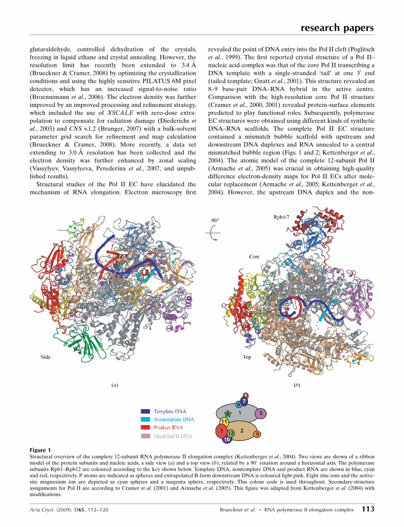
Structural studies of the Pol II EC have elucidated the
mechanism of RNA elongation. Electron microscopy first
revealed the point of DNA entry into the Pol II cleft (Poglitsch
et al., 1999). The first reported crystal structure of a Pol II–
nucleic acid complex was that of the core Pol II transcribing a
DNA template with a single-stranded ‘tail’ at one 30 end
(tailed template; Gnatt et al., 2001). This structure revealed an
8–9 base-pair DNA–RNA hybrid in the active centre.
Comparison with the high-resolution core Pol II structure
(Cramer et al., 2000, 2001) revealed protein-surface elements
predicted to play functional roles. Subsequently, polymerase
EC structures were obtained using different kinds of synthetic
DNA–RNA scaffolds. The complete Pol II EC structure
contained a mismatch bubble scaffold with upstream and
downstream DNA duplexes and RNA annealed to a central
mismatched bubble region (Figs. 1 and 2; Kettenberger et al.,
2004). The atomic model of the complete 12-subunit Pol II
(Armache et al., 2005) was crucial in obtaining high-quality
difference electron-density maps for Pol II ECs after mole-
cular replacement (Armache et al., 2005; Kettenberger et al.,
2004). However, the upstream DNA duplex and the non-
research papers
Acta Cryst. (2009). D65, 112–120 Brueckner et al. � RNA polymerase II elongation complex 113
Figure 1Structural overview of the complete 12-subunit RNA polymerase II elongation complex (Kettenberger et al., 2004). Two views are shown of a ribbonmodel of the protein subunits and nucleic acids, a side view (a) and a top view (b), related by a 90� rotation around a horizontal axis. The polymerasesubunits Rpb1–Rpb12 are coloured according to the key shown below. Template DNA, nontemplate DNA and product RNA are shown in blue, cyanand red, respectively. P atoms are indicated as spheres and extrapolated B-form downstream DNA is coloured light pink. Eight zinc ions and the active-site magnesium ion are depicted as cyan spheres and a magenta sphere, respectively. This colour code is used throughout. Secondary-structureassignments for Pol II are according to Cramer et al. (2001) and Armache et al. (2005). This figure was adapted from Kettenberger et al. (2004) withmodifications.
template strand in the bubble region were disordered in the
crystal structure. Reduced scaffolds lacking these disordered
parts (‘minimal nucleic acid scaffolds’) were used to determine
structures of the core Pol II EC (Westover et al., 2004a,b). The
synthetic scaffold EC structures revealed the exact location of
the downstream DNA and several nucleotides upstream of the
hybrid (Figs. 1 and 2). Mechanisms were suggested for how Pol
II unwinds downstream DNA and how it separates the RNA
product from the DNA template at the end of the hybrid. In
both cases, Pol II-induced distortion of the nucleic acid
duplexes and steric hindrance by Pol II surface loops seem to
play important roles.
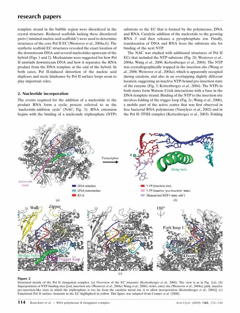
2. Nucleotide incorporation
The events required for the addition of a nucleotide to the
product RNA form a cyclic process referred to as the
‘nucleotide-addition cycle’ (NAC; Fig. 3). RNA extension
begins with the binding of a nucleoside triphosphate (NTP)
substrate to the EC that is formed by the polymerase, DNA
and RNA. Catalytic addition of the nucleotide to the growing
RNA 30 end then releases a pyrophosphate ion. Finally,
translocation of DNA and RNA frees the substrate site for
binding of the next NTP.
The NAC was studied with additional structures of Pol II
ECs that included the NTP substrate (Fig. 2b; Westover et al.,
2004a; Wang et al., 2006; Kettenberger et al., 2004). The NTP
was crystallographically trapped in the insertion site (Wang et
al., 2006; Westover et al., 2004a), which is apparently occupied
during catalysis, and also in an overlapping slightly different
location, suggesting an inactive NTP-bound pre-insertion state
of the enzyme (Fig. 3; Kettenberger et al., 2004). The NTPs in
both states form Watson–Crick interactions with a base in the
DNA-template strand. Binding of the NTP to the insertion site
involves folding of the trigger loop (Fig. 2c; Wang et al., 2006),
a mobile part of the active centre that was first observed in
free bacterial RNA polymerase (Vassylyev et al., 2002) and in
the Pol II–TFIIS complex (Kettenberger et al., 2003). Folding
research papers
114 Brueckner et al. � RNA polymerase II elongation complex Acta Cryst. (2009). D65, 112–120
Figure 2Structural details of the Pol II elongation complex. (a) Overview of the EC structure (Kettenberger et al., 2004). The view is as in Fig. 1(a). (b)Superposition of NTP-binding sites [red, insertion site (Westover et al., 2004a; Wang et al., 2006); violet, entry site (Westover et al., 2004a); pink, inactivepre-insertion-like state in which the triphosphate is too far from the catalytic metal ion A to allow incorporation (Kettenberger et al., 2004)]. (c)Functional Pol II surface elements in the EC highlighted in yellow. This figure was adapted from Cramer et al. (2008).

of the trigger loop closes the active site and may be involved in
selection of the correct NTP (Fig. 3). The NTP-complex
structures revealed contacts of the nucleotide with the poly-
merase, which explain the discrimination of ribonucleotides
against deoxyribonucleotides, and provided insights into the
selection of the nucleotide complementary to the templating
DNA base.
Catalytic nucleotide incorporation apparently follows the
two-metal-ion mechanism suggested for all polymerases
(Steitz, 1998). The Pol II active site contains a persistently
bound metal ion (metal A) and a second mobile metal ion
(metal B) (Cramer et al., 2001). Metal A is held in place by
three invariant aspartate side chains and binds the RNA 30 end
(Cramer et al., 2001), whereas metal B binds the NTP
triphosphate moiety (Westover et al., 2004a).
Recent studies of functional complexes of the bacterial
RNA polymerase revealed the close conservation of the EC
structure (Vassylyev, Vassylyeva, Perederina et al., 2007) and
provided additional insights into nucleotide incorporation
(Vassylyev, Vassylyeva, Zhang et al., 2007). As for Pol II, NTP
binding to the insertion site can induce folding of the trigger
loop. However, in the presence of the antibiotic streptolydigin
the NTP binds in the inactive pre-insertion state, in which the
triphosphate and metal B are too far from metal A to permit
catalysis. This finding supported a two-step mechanism of
nucleotide incorporation (Fig. 3; Vassylyev, Vassylyeva, Zhang
et al., 2007; Kettenberger et al., 2004). The NTP first binds in
the inactive state to an open active-centre conformation.
Complete folding of the trigger loop then leads to closure of
the active centre, delivery of the NTP to the insertion site and
catalysis. An alternative model for nucleotide addition
involves binding of the NTP to a putative entry site in the
pore, in which the nucleotide base is oriented away from the
DNA template, and possible rotation of the NTP around
metal ion B directly into the insertion site (Westover et al.,
2004a).
After nucleotide incorporation, the substrate-binding site is
occupied by the 30 end of the product RNA and the EC adopts
the pretranslocation state. Pol II translocates by a one-base-
pair step in order to free the substrate-binding site for the next
round of incorporation and thereby reaches the post-trans-
location state (Fig. 4a). The Brownian ratchet model of
translocation assumes that Brownian motion gives rise to
oscillation of the EC between pre-translocation and post-
translocation states, establishing the translocation equilibrium.
Substrate NTPs can only bind in the post-translocation state
and would act like the pawl of a ratchet. X-ray structural
evidence for the existence of the translocation equilibrium was
recently obtained with an EC labelled with 5-bromouracil in
the template strand (Figs. 4a and 4c; Svetlov & Nudler, 2008;
Brueckner & Cramer, 2008). Soaking the crystals with the
preserved translocation equilibrium with the inhibitor
�-amanitin resulted in the structure of the �-amanitin-
inhibited EC at 3.4 A resolution, which was suggested to
represent a translocation intermediate (Fig. 4b). In this
putative intermediate the DNA–RNA hybrid adopts a post-
translocation state (Fig. 4d), whereas the state of the down-
stream DNA is intermediary between pre-translocation and
post-translocation. The template base entering the active
centre (the templating base) was found in a new ‘pre-
templating’ position above the central bridge helix (Fig. 4e).
Two Pol II elements, the trigger loop and the bridge helix,
were observed in new conformations, suggesting their
involvement in facilitating translocation. �-Amanitin appar-
ently traps the trigger loop and bridge helix in these confor-
mations with direct and indirect contacts, thereby inhibiting
nucleotide incorporation and translocation. An independent
study also revealed the direct contact between the trigger loop
and �-amanitin and additionally showed a role of the trigger
loop in substrate selection and fidelity (Kaplan et al., 2008).
3. Obstacles during elongation
During active transcription, Pol II must overcome intrinsic
DNA-arrest sites, which are generally rich in A�T base pairs
and pose a natural obstacle to transcription. At such sites,
Pol II moves backwards along DNA and RNA, resulting in
extrusion of the RNA 30 end through the polymerase pore
research papers
Acta Cryst. (2009). D65, 112–120 Brueckner et al. � RNA polymerase II elongation complex 115
Figure 3Schematic representation of the extended model for the nucleotide-addition cycle (NAC). The vertical dashed line indicates register +1. Thesteps where �-amanitin interferes with the NAC are indicated. For details,refer to the text. This figure was adapted from Brueckner & Cramer(2008) with modifications.

beneath the active site and transcriptional arrest. The RNA-
cleavage stimulatory factor TFIIS can rescue an arrested
polymerase by creating a new RNA 30 end at the active site
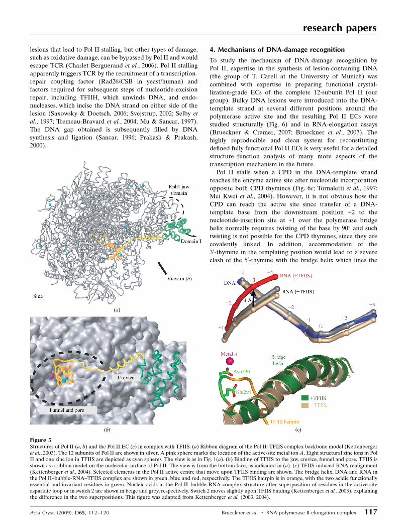
from which transcription can resume. The mechanism of TFIIS
function was elucidated from the structures of Pol II and a
Pol II EC in complex with TFIIS (Fig. 5; Kettenberger et al.,
2003, 2004). TFIIS inserts a hairpin into the polymerase pore
and complements the active site with acidic residues, changes
the enzyme conformation and repositions the RNA transcript
(Kettenberger et al., 2003, 2004). These studies supported the
idea that the Pol II active site is tunable, as it can catalyze
different reactions, including RNA synthesis and RNA
cleavage (Kettenberger et al., 2003; Sosunov et al., 2003).
Other obstacles to transcription are bulky lesions in the
DNA-template strand, e.g. the UV-light-induced cyclobutane
pyrimidine dimer (CPD), or intrastrand cross-links induced by
the anticancer drug cisplatin (Fig. 6). Bulky DNA lesions can
block transcription and replication and lead to mutations that
can cause cancer (Mitchell et al., 2003). Cells can eliminate
bulky DNA lesions slowly by genome-wide nucleotide-
excision repair (NER). However, for rapid and efficient repair
cells use an NER subpathway referred to as transcription-
coupled DNA repair (TCR). TCR specifically removes lesions
such as CPDs from the DNA strand transcribed by Pol II
(Saxowsky & Doetsch, 2006). It is thought that only those
lesions that can stably stall Pol II trigger TCR. CPDs are bulky
research papers
116 Brueckner et al. � RNA polymerase II elongation complex Acta Cryst. (2009). D65, 112–120
Figure 4Structure of the �-amanitin-inhibited Pol II elongation complex. (a) Pre-translocation and post-translocation states of the EC. The nucleic acid scaffoldused is depicted schematically with respect to the active-site metal ion A (magenta). The colour key is used throughout. (b) Overview of the �-amanitin-inhibited Pol II EC structure. The view is as in Fig. 1(a). �-Amanitin (stick model), nucleic acids (base in pre-templating position as a stick model), metalA, the bridge helix and the trigger loop (Leu1081 as a stick model) are highlighted using the colour key in (a). Part of the protein is omitted for clarity.(c, d) Bromine anomalous difference Fourier maps (pink net) of the free EC (c) and the �-amanitin-inhibited EC (d). Br atoms are depicted as yellowspheres and their positions are indicated. The view is rotated by 90� around a vertical axis compared with (b). (e) The +1 DNA-template base adopts apre-templating position. The initial unbiased Fo � Fc difference map for the nucleic acids is shown around the +1 position and is contoured at 2.5�. The+1 base in the pre-templating site is highlighted in violet. The view is rotated by 90� around a horizontal axis compared with (b). This figure was adaptedfrom Brueckner & Cramer (2008).
lesions that lead to Pol II stalling, but other types of damage,
such as oxidative damage, can be bypassed by Pol II and would
escape TCR (Charlet-Berguerand et al., 2006). Pol II stalling
apparently triggers TCR by the recruitment of a transcription-
repair coupling factor (Rad26/CSB in yeast/human) and
factors required for subsequent steps of nucleotide-excision
repair, including TFIIH, which unwinds DNA, and endo-
nucleases, which incise the DNA strand on either side of the
lesion (Saxowsky & Doetsch, 2006; Svejstrup, 2002; Selby et
al., 1997; Tremeau-Bravard et al., 2004; Mu & Sancar, 1997).
The DNA gap obtained is subsequently filled by DNA
synthesis and ligation (Sancar, 1996; Prakash & Prakash,
2000).
research papers
Acta Cryst. (2009). D65, 112–120 Brueckner et al. � RNA polymerase II elongation complex 117
4. Mechanisms of DNA-damage recognition
To study the mechanism of DNA-damage recognition by
Pol II, expertise in the synthesis of lesion-containing DNA
(the group of T. Carell at the University of Munich) was
combined with expertise in preparing functional crystal-
lization-grade ECs of the complete 12-subunit Pol II (our
group). Bulky DNA lesions were introduced into the DNA-
template strand at several different positions around the
polymerase active site and the resulting Pol II ECs were
studied structurally (Fig. 6) and in RNA-elongation assays
(Brueckner & Cramer, 2007; Brueckner et al., 2007). The
highly reproducible and clean system for reconstituting
defined fully functional Pol II ECs is very useful for a detailed
structure–function analysis of many more aspects of the
transcription mechanism in the future.
Pol II stalls when a CPD in the DNA-template strand
reaches the enzyme active site after nucleotide incorporation
opposite both CPD thymines (Fig. 6c; Tornaletti et al., 1997;
Mei Kwei et al., 2004). However, it is not obvious how the
CPD can reach the active site since transfer of a DNA-
template base from the downstream position +2 to the
nucleotide-insertion site at +1 over the polymerase bridge
helix normally requires twisting of the base by 90� and such
twisting is not possible for the CPD thymines, since they are
covalently linked. In addition, accommodation of the
30-thymine in the templating position would lead to a severe
clash of the 50-thymine with the bridge helix which lines the
Figure 5Structures of Pol II (a, b) and the Pol II EC (c) in complex with TFIIS. (a) Ribbon diagram of the Pol II–TFIIS complex backbone model (Kettenbergeret al., 2003). The 12 subunits of Pol II are shown in silver. A pink sphere marks the location of the active-site metal ion A. Eight structural zinc ions in PolII and one zinc ion in TFIIS are depicted as cyan spheres. The view is as in Fig. 1(a). (b) Binding of TFIIS to the jaw, crevice, funnel and pore. TFIIS isshown as a ribbon model on the molecular surface of Pol II. The view is from the bottom face, as indicated in (a). (c) TFIIS-induced RNA realignment(Kettenberger et al., 2004). Selected elements in the Pol II active centre that move upon TFIIS binding are shown. The bridge helix, DNA and RNA inthe Pol II–bubble-RNA–TFIIS complex are shown in green, blue and red, respectively. The TFIIS hairpin is in orange, with the two acidic functionallyessential and invariant residues in green. Nucleic acids in the Pol II–bubble-RNA complex structure after superposition of residues in the active-siteaspartate loop or in switch 2 are shown in beige and grey, respectively. Switch 2 moves slightly upon TFIIS binding (Kettenberger et al., 2003), explainingthe difference in the two superpositions. This figure was adapted from Kettenberger et al. (2003, 2004).
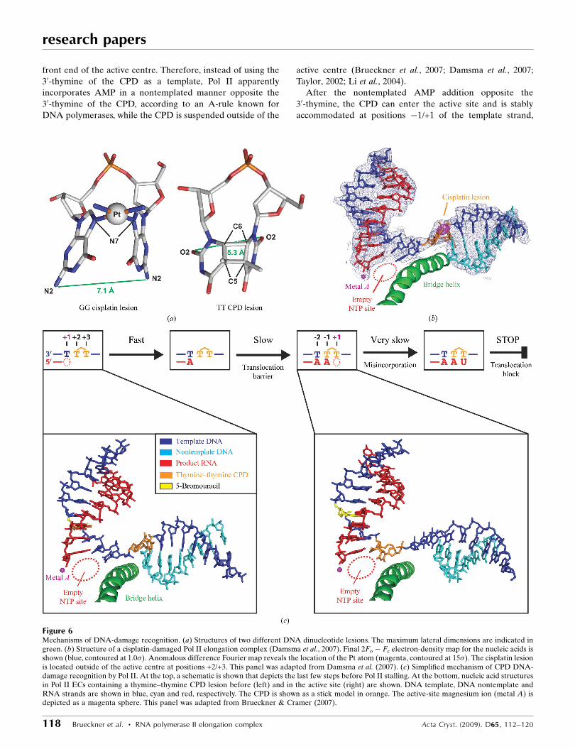
front end of the active centre. Therefore, instead of using the
30-thymine of the CPD as a template, Pol II apparently
incorporates AMP in a nontemplated manner opposite the
30-thymine of the CPD, according to an A-rule known for
DNA polymerases, while the CPD is suspended outside of the
active centre (Brueckner et al., 2007; Damsma et al., 2007;
Taylor, 2002; Li et al., 2004).
After the nontemplated AMP addition opposite the
30-thymine, the CPD can enter the active site and is stably
accommodated at positions �1/+1 of the template strand,
research papers
118 Brueckner et al. � RNA polymerase II elongation complex Acta Cryst. (2009). D65, 112–120
Figure 6Mechanisms of DNA-damage recognition. (a) Structures of two different DNA dinucleotide lesions. The maximum lateral dimensions are indicated ingreen. (b) Structure of a cisplatin-damaged Pol II elongation complex (Damsma et al., 2007). Final 2Fo � Fc electron-density map for the nucleic acids isshown (blue, contoured at 1.0�). Anomalous difference Fourier map reveals the location of the Pt atom (magenta, contoured at 15�). The cisplatin lesionis located outside of the active centre at positions +2/+3. This panel was adapted from Damsma et al. (2007). (c) Simplified mechanism of CPD DNA-damage recognition by Pol II. At the top, a schematic is shown that depicts the last few steps before Pol II stalling. At the bottom, nucleic acid structuresin Pol II ECs containing a thymine–thymine CPD lesion before (left) and in the active site (right) are shown. DNA template, DNA nontemplate andRNA strands are shown in blue, cyan and red, respectively. The CPD is shown as a stick model in orange. The active-site magnesium ion (metal A) isdepicted as a magenta sphere. This panel was adapted from Brueckner & Cramer (2007).
forming a Watson–Crick base pair between the 30-thymine and
the adenine at the 30 end of the product RNA (Fig. 6c). Now
only UMP can be incorporated opposite the 50-thymine
(Brueckner et al., 2007; Mei Kwei et al., 2004). The UMP
misincorporation is very slow and is the rate-limiting step in
reaching the stalled state (Brueckner et al., 2007). Specific
UMP misincorporation may arise from the unusual position of
the CPD 50-thymine, which adopts a wobble position with
respect to the base in the undamaged complex (Brueckner et
al., 2007). The wobbled 50-thymine can form two hydrogen
bonds to UTP, but not to other NTPs. Pol II stalls because
translocation of the CPD 50-thymine–uracil mismatch base
pair from position +1 to position �1 is strongly disfavoured.
This translocation event would move the damage-containing
mismatch into the �1 position of the DNA–RNA hybrid,
resulting in a distortion that is likely to destabilize the EC
(Kireeva et al., 2000). Replacement of the misincorporated
UMP by AMP in an artificial scaffold enables CPD bypass
(Brueckner et al., 2007). Thus, Pol II stalling requires CPD-
directed misincorporation and distortions arising from the
CPD alone are insufficient to cause Pol II stalling. Indeed, a
T�U mismatch base pair alone was sufficient to stall the vast
majority of Pol II complexes (Brueckner et al., 2007). In
contrast, DNA polymerases can correctly incorporate adenine
opposite both CPD thymines and, depending on the type of
polymerase, this can lead to stalling or lesion bypass (Li et al.,
2004; Ling et al., 2003).
The anticancer drug cisplatin [cis-diamminedichloro-
platinum(II)] forms 1,2-d(GpG) DNA intrastrand cross-links
(cisplatin lesions) that stall Pol II and trigger transcription-
coupled DNA repair (Wang & Lippard, 2005; Kartalou &
Essigmann, 2001; Corda et al., 1991, 1993; Tornaletti et al.,
2003; Jung & Lippard, 2006). Whereas in the CPD lesion two
neighbouring thymine bases are covalently linked with a
cyclobutane ring including the C5 and C6 atoms, in a cisplatin
lesion the Pt atom coordinates the N7 atoms of two adjacent
guanines in a DNA strand (Fig. 6a). The cisplatin lesion can be
stably accommodated in a Pol II EC at position +2/+3 of the
template strand, but translocation to position +1/+2 is dis-
favoured (Fig. 6b; Damsma et al., 2007); these are both also the
case for the CPD lesion. There is strong evidence that adenine
is incorporated in a nontemplated fashion opposite the
cisplatin 30-guanine, as proposed for the CPD lesion. However,
unlike the CPD lesion, the cisplatin lesion cannot be stably
accommodated in the active site (positions �1/+1). There are
two possible causes. Firstly, the cisplatin lesion is a more bulky
dinucleotide lesion than the CPD lesion. The maximum lateral
dimension is 7.2 A (N2–N2 distance), compared with 5.3 A
(O2–O2 distance) for the CPD lesion (Fig. 6a). Modelling
suggested that a conformational change of the bridge helix
would be required to accommodate the lesion in the active
site. Secondly, a G�A mismatch base pair would be formed at
position �1 in contrast to a stabilizing T�A base pair in the
case of the CPD lesion.
In conclusion, the mechanism of recognition by transcribing
Pol II is different for the two dinucleotide lesions. At a
cisplatin lesion, Pol II stalls because the lesion cannot be
delivered to the active site, whereas it stalls at a CPD lesion
after delivery to the active site and specific UMP misincor-
poration opposite the 50-thymine. Bypass of the CPD lesion is
only possible by artificially replacing the resulting T�U
mismatch by a T�A match. Remarkably, bypass of the cisplatin
lesion is also possible, but only by artificially providing a
starting transcript that extends at least up to the 30-guanine. In
this case, bypass is even possible in presence of a G�A
mismatch with the 30-guanine of the cisplatin dimer.
5. RNA as a template for Pol II
Although Pol II generally uses DNA as a template, there is
also evidence that Pol II can use RNA templates. Recent
structures have shown that an RNA template–product duplex
can bind to the site normally occupied by the DNA–RNA
hybrid and provided the structural basis for the phenomenon
of RNA-dependent RNA synthesis by Pol II (Lehmann et al.,
2007). Complementary in vitro enzyme assays revealed that
the RNA-dependent RNA polymerase (RdRP) activity
resides in the site used during transcription, but is slower and
less processive than the DNA-dependent activity. The RdRP
activity of Pol II provides a missing link in molecular evolu-
tion, because it suggests that Pol II evolved from an ancient
replicase that duplicated RNA genomes. There is compelling
evidence that the ancient RdRP activity of Pol II is still rele-
vant for the replication of the RNA genome of the hepatitis �virus (HDV) and it may also be used in certain cellular
processes as many organisms lack dedicated single-subunit
RdRPs.
6. Conclusion
Combining X-ray crystallographic analysis of Pol II ECs with
in vitro transcription experiments allowed exploration of the
basic mechanisms of transcription elongation, including the
nucleotide-addition cycle, and additional features such as the
mechanism of TFIIS function, DNA-damage recognition and
RNA-templated RNA synthesis. Further aspects of tran-
scription elongation still await further characterization using
the available system. Further investigations could focus on the
regulation by additional protein factors or RNA molecules,
transcriptional mutagenesis and fidelity and the effect of other
kinds of DNA lesions, e.g. oxidative lesions, to name a few.
The authors wish to thank Stefan Benkert, Claudia Buchen,
Dirk Kostrewa and other members of the Cramer laboratory
for help. Special thanks to Dmitry G. Vassylyev for discussions
and help. FB was supported by the Nanosystems Initiative
Munich (NIM) and the graduate program Protein Dynamics in
Health and Disease of the Elitenetzwerk Bayern (ENB). GED
was supported by the ENB. PC was supported by the Deutsche
Forschungsgemeinschaft, the Sonderforschungsbereich SFB646,
the Transregio 5 Chromatin, the EU Research Grant Network
3D Repertoire, the Nanosystems Initiative Munich NIM and
the Fonds der Chemischen Industrie. Part of this work was
research papers
Acta Cryst. (2009). D65, 112–120 Brueckner et al. � RNA polymerase II elongation complex 119
performed at the Swiss Light Source (SLS) at the Paul
Scherrer Institut, Villigen, Switzerland.
References
Armache, K.-J., Kettenberger, H. & Cramer, P. (2003). Proc. NatlAcad. Sci. USA, 100, 6964–6968.
Armache, K.-J., Mitterweger, S., Meinhart, A. & Cramer, P. (2005). J.Biol. Chem. 280, 7131–7134.
Broennimann, Ch., Eikenberry, E. F., Henrich, B., Horisberger, R.,Huelsen, G., Pohl, E., Schmitt, B., Schulze-Briese, C., Suzuki, M.,Tomizaki, T., Toyokawa, H. & Wagner, A. (2006). J. SynchrotronRad. 13, 120–130.
Brueckner, F. & Cramer, P. (2007). FEBS Lett. 581, 2757–2760.Brueckner, F. & Cramer, P. (2008). Nature Struct. Mol. Biol. 15,
811–818.Brueckner, F., Hennecke, U., Carell, T. & Cramer, P. (2007). Science,
315, 859–862.Brunger, A. T. (2007). Nature Protoc. 2, 2728–2733.Bushnell, D. A. & Kornberg, R. D. (2003). Proc. Natl Acad. Sci. USA,
100, 6969–6972.Charlet-Berguerand, N., Feuerhahn, S., Kong, S. E., Ziserman, H.,
Conaway, J. W., Conaway, R. & Egly, J. M. (2006). EMBO J. 25,5481–5491.
Corda, Y., Job, C., Anin, M. F., Leng, M. & Job, D. (1991).Biochemistry, 30, 222–230.
Corda, Y., Job, C., Anin, M. F., Leng, M. & Job, D. (1993).Biochemistry, 32, 8582–8588.
Cramer, P. et al. (2008). Annu. Rev. Biophys. 37, 337–352.Cramer, P., Bushnell, D. A., Fu, J., Gnatt, A. L., Maier-Davis, B.,
Thompson, N. E., Burgess, R. R., Edwards, A. M., David, P. R. &Kornberg, R. D. (2000). Science, 288, 640–649.
Cramer, P., Bushnell, D. A. & Kornberg, R. D. (2001). Science, 292,1863–1876.
Damsma, G. E., Alt, A., Brueckner, F., Carell, T. & Cramer, P. (2007).Nature Struct. Mol. Biol. 14, 1127–1133.
Diederichs, K., McSweeney, S. & Ravelli, R. B. G. (2003). Acta Cryst.D59, 903–909.
Fu, J., Gnatt, A. L., Bushnell, D. A., Jensen, G. J., Thompson, N. E.,Burgess, R. R., David, P. R. & Kornberg, R. D. (1999). Cell, 98,799–810.
Gnatt, A. L., Cramer, P., Fu, J., Bushnell, D. A. & Kornberg, R. D.(2001). Science, 292, 1876–1882.
Jung, Y. & Lippard, S. J. (2006). J. Biol. Chem. 281, 1361–1370.Kaplan, C. D., Larsson, K. M. & Kornberg, R. D. (2008). Mol. Cell, 30,
547–556.Kartalou, M. & Essigmann, J. M. (2001). Mutat. Res. 478, 1–21.Kettenberger, H., Armache, K.-J. & Cramer, P. (2003). Cell, 114,
347–357.Kettenberger, H., Armache, K.-J. & Cramer, P. (2004). Mol. Cell, 16,
955–965.
Kireeva, M. L., Komissarova, N., Waugh, D. S. & Kashlev, M. (2000).J. Biol. Chem. 275, 6530–6536.
Lehmann, E., Brueckner, F. & Cramer, P. (2007). Nature (London),450, 445–449.
Li, Y., Dutta, S., Doublie, S., Bdour, H. M., Taylor, J. S. & Ellenberger,T. (2004). Nature Struct. Mol. Biol. 11, 784–790.
Ling, H., Boudsocq, F., Plosky, B. S., Woodgate, R. & Yang, W. (2003).Nature (London), 424, 1083–1087.
Mei Kwei, J. S., Kuraoka, I., Horibata, K., Ubukata, M., Kobatake, E.,Iwai, S., Handa, H. & Tanaka, K. (2004). Biochem. Biophys. Res.Commun. 320, 1133–1138.
Mitchell, J. R., Hoeijmakers, J. H. & Niedernhofer, L. J. (2003). Curr.Opin. Cell Biol. 15, 232–240.
Mu, D. & Sancar, A. (1997). J. Biol. Chem. 272, 7570–7573.Poglitsch, C. L., Meredith, G. D., Gnatt, A. L., Jensen, G. J., Chang,
W. H., Fu, J. & Kornberg, R. D. (1999). Cell, 98, 791–798.Prakash, S. & Prakash, L. (2000). Mutat. Res. 451, 13–24.Sancar, A. (1996). Annu. Rev. Biochem. 65, 43–81.Saxowsky, T. T. & Doetsch, P. W. (2006). Chem. Rev. 106, 474–
488.Selby, C. P., Drapkin, R., Reinberg, D. & Sancar, A. (1997). Nucleic
Acids Res. 25, 787–793.Sosunov, V., Sosunova, E., Mustaev, A., Bass, I., Nikiforov, V. &
Goldfarb, A. (2003). EMBO J. 22, 2234–2244.Steitz, T. A. (1998). Nature (London), 391, 231–232.Svejstrup, J. Q. (2002). Nature Rev. Mol. Cell Biol. 3, 21–29.Svetlov, V. & Nudler, E. (2008). Nature Struct. Mol. Biol. 15, 777–
779.Taylor, J. S. (2002). Mutat. Res. 510, 55–70.Tornaletti, S., Donahue, B. A., Reines, D. & Hanawalt, P. C. (1997). J.
Biol Chem. 272, 31719–31724.Tornaletti, S., Patrick, S. M., Turchi, J. J. & Hanawalt, P. C. (2003). J.
Biol Chem. 278, 35791–35797.Tremeau-Bravard, A., Riedl, T., Egly, J. M. & Dahmus, M. E. (2004).
J. Biol Chem. 279, 7751–7759.Vassylyev, D. G., Sekine, S., Laptenko, O., Lee, J., Vassylyeva, M. N.,
Borukhov, S. & Yokoyama, S. (2002). Nature (London), 417,712–719.
Vassylyev, D. G., Vassylyeva, M. N., Perederina, A., Tahirov, T. H. &Artsimovitch, I. (2007). Nature (London), 448, 157–162.
Vassylyev, D. G., Vassylyeva, M. N., Zhang, J., Palangat, M.,Artsimovitch, I. & Landick, R. (2007). Nature (London), 448,163–168.
Wang, D., Bushnell, D. A., Westover, K. D., Kaplan, C. D. &Kornberg, R. D. (2006). Cell, 127, 941–954.
Wang, D. & Lippard, S. J. (2005). Nature Rev. Drug Discov. 4,307–320.
Westover, K. D., Bushnell, D. A. & Kornberg, R. D. (2004a). Cell, 119,481–489.
Westover, K. D., Bushnell, D. A. & Kornberg, R. D. (2004b). Science,303, 1014–1016.
research papers
120 Brueckner et al. � RNA polymerase II elongation complex Acta Cryst. (2009). D65, 112–120