structure of human prostasin, a target …merck.com prostasin (also called cap1) is an extracellular...
TRANSCRIPT

1
STRUCTURE OF HUMAN PROSTASIN, A TARGET FOR THE REGULATION OF HYPERTENSION.
Keith W. Rickert, Paul Kelley, Noel J. Byrne, Ronald E. Diehl, Dawn L. Hall, Allison M. Montalvo, John C. Reid, Jennifer M. Shipman, Bradley W. Thomas, Sanjeev K. Munshi,
Paul L. Darke, and Hua-Poo Su Global Structural Biology, Merck Research Laboratories, West Point, PA 19486
Running title: Structure of Prostasin Address correspondence to Keith W. Rickert, WP26-354, P.O Box 4, West Point PA 19486. Phone 215-652-5323 Fax: 215-993-0026; Email: [email protected] or Hua-Poo Su,
WP14-1101, P. O. Box 4, West Point PA 19486. Phone 215-652-7347. Email: [email protected]
Prostasin (also called CAP1) is an extracellular serine protease implicated in the modulation of fluid and electrolyte regulation via proteolysis of the epithelial sodium channel. Several disease states, particularly hypertension, can be affected by modulation of epithelial sodium channel activity. Thus, understanding the biochemical function of prostasin and developing specific agents to inhibit its activity could have a significant impact on a widespread disease. We report the expression of the prostasin proenzyme in E. coli as insoluble inclusion bodies, refolding, and activation via proteolytic removal of the N-terminal propeptide. The refolded and activated enzyme was shown to be pure and monomeric, with kinetic characteristics very similar to prostasin expressed from eukaryotic systems. Active prostasin was crystallized and the structure determined to 1.45Å resolution. These apo-protein crystals were soaked with nafamostat, allowing the structure of the inhibited acyl-enzyme intermediate structure to be determined to 2.0Å resolution. Comparison of the inhibited and apoprotein forms of prostasin suggest a mechanism of regulation through stabilization of a loop which interferes with substrate recognition.
Prostasin (also known as CAP1) is a serine protease originally isolated from seminal fluid as a secreted protein (1). It is widely expressed in mammalian epithelial tissue as a 40 kDa glycophosphatidylinositol (GPI)-anchored protein(1-3). Prostasin is implicated in the regulation of sodium and fluid levels via proteolysis of the epithelial sodium channel (ENaC) γ subunit (4-9).
ENaC performs an essential function in several
epithelial tissues, including the colon, kidney, and lung (5,7,9,10). In these tissues, sodium is primarily transported across the apical membrane via ENaC, and fluid transport across the membrane is highly responsive to sodium concentration. Inappropriate function of ENaC leads to misregulation of blood pressure in humans (11,12). The same ENaC mutations which lead to hypertension in humans give rise to defects in lung fluid clearance in mice (11), suggesting that proper ENaC function may also be important in other diseases where sodium and fluid homeostasis is severely disrupted, for example cystic fibrosis and diarrhea (13,14).
While ENaC activity can be regulated via transcription and translation, for example in response to the hormone aldosterone (11), proteolysis of ENaC can lead to channel activation (9), and inhibition of this process might provide a direct and specific method to modulate ENaC activity for therapeutic benefit. Cleavage of an inhibitory peptide in the ENaC γ subunit by prostasin has been shown to stimulate sodium transport by 2-3 fold in cell-based experiments (4,5,7-10). Other proteases implicated to activate ENaC in model systems include furin, mCAP-2, mCAP-3, and TMSP-1 (5,8-11). Some of these proteases require activation by yet other proteases (15,16), so clarity regarding which enzymes may represent appropriate targets for reducing ENaC activity has yet to be achieved. Highly selective small-molecule inhibitors of proteases thought to modulate ENaC will aid in understanding the roles of different proteases in channel function, and may also provide a therapeutic benefit for hypertension and other diseases, e.g., cystic fibrosis (12-14).
http://www.jbc.org/cgi/doi/10.1074/jbc.M805262200The latest version is at JBC Papers in Press. Published on October 14, 2008 as Manuscript M805262200
Copyright 2008 by The American Society for Biochemistry and Molecular Biology, Inc.
by guest on July 17, 2018http://w
ww
.jbc.org/D
ownloaded from

2
Although there are numerous agents for the treatment of hypertension, many individuals require multiple medications to achieve adequate control (17), and the condition continues to exact a high cost to society and so it seems that additional modes of treatment remain desirable.
Prostasin belongs to the classical serine protease family, with homology to trypsin, chymotrypsin, and kallikrein, and has a trypsin-like substrate specificity (1,2,18). In common with these, enzyme activation of prostasin occurs via cleavage of the pro-protein to produce a light chain and a heavy chain that are disulfide-linked. Prostasin has some features unusual in serine proteases, such as a high degree of sensitivity to monovalent and divalent cations which may relate to prostasin’s role in ENaC regulation (18). However, to date, there have been no reports of potent and selective small molecule inhibitors of prostasin. High resolution structural studies have been used to significant advantage in the design of selective protease inhibitors (19-22). Availability of the three dimensional structure of prostasin could offer a significant advantage in the design of prostasin inhibitors.
Previous studies have shown that active prostasin can be expressed in a recombinant baculovirus system and purified to homogeneity, enabling more extensive biochemical studies (18). We report the expression of prostasin in E. coli, and the refolding, proteolytic activation, and purification to yield pure and fully active enzyme. Expression in E. coli is novel and can allow for rapid generation of site-directed mutants to allow the biochemical determination of structure-function relationships. We describe the high resolution crystal structure of the protease domain of this enzyme, which may provide valuable information in the design or optimization of specific small-molecule inhibitors.
Experimental Procedures
Construction of prostasin variants. The native sequence (accession number: NM_002773) and all expression constructs are illustrated in Figure 1. In all constructs, the C-terminal GPI anchor domain and preceding linker were replaced with a His6 tag to aid in purification. Cysteines 154 and 203 were not expected to be involved in intradomain disulfide bonds and were mutated to
serine and alanine (18). In the case of baculovirus variant 40, the N-linked glycosylation site was also removed via site-directed mutagenesis. For baculovirus expression constructs, the native signal sequence and propeptide were replaced with an insect cell signal sequence (melittin, GP64, or GP67) to generate the native N-terminus of the mature protein, as the propeptide is not required for activity (18). Bacterial expression constructs contained the original native propeptide sequence at the N-terminus, but with the addition of an enterokinase recognition sequence to allow specific cleavage to generate the active enzyme. Detailed methods for the expression of prostasin in both baculovirus and E. coli are provided in the Supplementary Experimental Procedures.
Refolding and purification of prostasin from E. coli. Prostasin variants 26 and 28 (Figure 1) were solubilized from inclusion bodies in 8 M urea with 0.1 M Tris/HCl, pH 8.0, and 2 mM DTT after extensive washing. Solubilized protein was bound to Ni-NTA Superflow resin (Qiagen) and eluted with 0.3 M imidazole. The urea-solubilized material was then refolded in a buffer containing 1 M L-arginine, 0.1 M Tris/HCl, 5 mM reduced glutathione, and 0.5 mM oxidized glutathione, pH 8.0, at a final concentration of 10 mg/L. Following diafiltration, protein was purified via Ni(II) affinity and anion exchange chromatography. Eluted fractions were evaluated via SDS-PAGE and mass spectrometry (MS), and fractions in which prostasin was detected by MS were then pooled for further processing. Prostasin zymogen was converted to the active form by addition of enterokinase (EKMax, Invitrogen) at a final concentration of 2 U/mL (7.5 U/mg prostasin) in the presence of 0.5 mM reduced glutathione. The resultant cleavage reaction was maintained at 4ºC for 48 hours and monitored by MS and SDS-PAGE. Following completion of cleavage as determined by MS, the reaction was incubated at 4ºC overnight after the addition of 1 mM oxidized glutathione. Active prostasin was purified from enterokinase and misfolded prostasin via Ni(II) affinity and anion exchange chromatography. Fractions were selected based on activity, then pooled to give the final purified prostasin for further characterization and crystallography. Further details of the purification are found in the Supplementary Experimental Procedures.
by guest on July 17, 2018http://w
ww
.jbc.org/D
ownloaded from

3
Enzyme assay. Enzyme assays were performed at ambient temperature (22-24ºC) in 0.1 M Tris/HCl pH 9 with 0.1% CHAPS using a peptide substrate, Ac-KHYR-AMC (Anaspec) at 50 μM (18). Data were collected using a BMGLabTech Fluostar Optima reader, with excitation and emission filter wavelengths of 340 and 450 nm, respectively. Measurements of KM were made by varying the concentration of substrate from 10-250 μM, while enzyme concentration was held constant at 19 nM. Absolute quantitation of product was determined by comparison to an independent AMC standard at concentrations from 2 nM to 1.7 μM.
Size Exclusion Chromatography (SEC). Analytical SEC measurements were made using a Superdex 200 5/150 column (GE Healthcare), equilibrated in 50 mM potassium phosphate, pH 6.8, and 0.3 M NaCl, at a flow rate of 0.15 mL/min with injections of 0.5-5 μg of protein. Retention times were converted to apparent molecular masses using mixed protein standards (thyroglobulin, gamma-globulin, ovalbumin, and myoglobin) (Bio-Rad).
Mass spectrometry (MS). Whole-protein mass measurements were made by binding samples (0.1-2 μg) to a reverse-phase protein trap column (Michrom), where they were desalted by washing with 2% acetonitrile, 0.01% trifluoroacetic acid (TFA) and eluted with a solution of 64% acetonitrile, 0.01% TFA into an electrospray mass spectrometer (LTQ, Thermo). The resultant spectra were deconvoluted using Promass (Novatia) to yield the whole protein mass. For free cysteine determination, samples were treated with 50 mM iodoacetamide at room temperature for 30 minutes prior to analysis. For active site determination, prostasin was incubated with 5 μM nafamostat mesylate (BioMol International) for 5 minutes in 50 mM Tris/HCl pH 8.5, with a final NaCl concentration of 0.2 M or less, prior to mass spectroscopic analysis (23).
Protein crystallization. Prostasin variant 26, comprising human prostasin residues 45-285 with the mutations C154S and C203A, a C-terminal thrombin-cleavage site and His6 tag (Figure 1b), and variant 28, comprising human prostasin residues 45-289 with the same mutations and C-terminus, were concentrated to a final concentration of 12-14 mg/mL as determined by
Bradford assay (Bio-Rad) in 50 mM Tris/HCl, pH 8.5, 0.1 M NaCl. Crystals of both variants were obtained by the vapor diffusion method. Initial crystals were obtained in hanging drops containing 1 μL of protein, mixed with 1 μL of reservoir solution consisting of 100 mM Bis-Tris pH 6.3 and 30% polyethylene glycol (PEG) 3350. Diffraction quality crystals formed as a result of streak seeding into hanging drops containing 1 μL of protein, mixed with 1 μL of reservoir solution consisting of 100 mM Bis-Tris pH 5.8 and 21-26% polyethylene glycol (PEG) 3350. Crystals were cryoprotected for data collection in a solution containing 100 mM Bis-Tris pH 6.5, 20% PEG 3350, 100 mM NaCl and 20% ethylene glycol.
Protein structure determination. In-house diffraction data were collected with Rigaku X-ray sources using R-Axis image plate detectors. Data were processed and scaled with HKL2000 (24). Initial phases were obtained by molecular replacement with Phaser (25), using the model of human plasma kallikrein (PDB code: 2ANW) (26). The models for variant 28, apoprotein and nafamostat-inhibited acyl-enzyme intermediate, were built and improved with iterative cycles of manual rebuilding in Coot and MIFit followed by refinement with Refmac (27,28). There was sufficient density to build the model continuously from residues Ile 45 to Gln 289, which encodes all of the native, mature protein residues. Nafamostat was introduced by soaking crystals of apoprotein in a buffer containing 1 mM nafamostat, 100 mM Bis-Tris pH 5.8, 100 mM NaCl, and 30% PEG 3350. Structural alignments were performed using LSQMAN (29). Figures were generated using PyMOL (30). Model geometry was verified using Procheck (31).
Results and Discussion
Expression of prostasin variants. Initially, prostasin variants were expressed by secretion from insect cells, as used by Shipway et al. to examine the catalytic properties of prostasin (18). While the expression levels for the glycosylated prostasin variants 801 and 35 (Figure 1b) were acceptable (0.4-1.0 mg/L), and the purified proteins were well behaved in solution and could be readily concentrated to 20 mg/mL, they did not form crystals using a broad screen of conditions. Prostasin variant 40, which lacks the N-linked
by guest on July 17, 2018http://w
ww
.jbc.org/D
ownloaded from

4
glycosylation site, expressed to much lower levels in this system (<0.1 mg/L).
Expression in E. coli was examined in two strains: BL21(DE3) and Origami-2. In BL21(DE3), prostasin variants 26 and 28 expressed at 50-100 mg/L as insoluble inclusion bodies. While Origami-2 cells possess an oxidizing intracellular environment sometimes helpful in disulfide formation, as needed for prostasin, expression of prostasin variants 26 and 28 in these cells gave mainly insoluble protein at 1-5 mg/L. Accordingly, prostasin variants 26 and 28 were expressed in BL21(DE3) cells and refolded in vitro.
Refolding of prostasin. The primary challenge to devising a refolding protocol for prostasin was evaluating the product for the correctness of its fold. While enzymatic activity would normally be the preferred measure of success, prostasin was expressed in the inactive zymogen form to ensure generation of a native N-terminus. Enterokinase, the enzyme used to cleave the propeptide, was active in the prostasin assay, making interpretation of activity assays from mixtures containing both enzymes problematic. While the amount of soluble protein captured on Ni(II) columns was of some guidance (Table 1, conditions 1-3), the majority of the protein in these mixtures seemed to be misfolded aggregates. The best tool in these circumstances to judge the quality of refolded zymogen was MS following reverse-phase chromatography. Misfolded proteins are adsorbed much more strongly to the reverse-phase surface than well-folded proteins (32,33), while MS ensures accurate identification of the protein species eluted from the column. Purification of the solubilized protein on Ni(II) affinity media was performed to simplify the mass spectrum observed after refolding, making the analysis more consistent and semi-quantitative. Using these tools, small-scale refolding experiments were carried out to find optimal conditions, detailed in Table 1. In scaled-up refolds, although the yields of soluble protein after refolding and the first chromatographic step were quite reasonable, the pool still contained substantial amounts of misfolded protein. Prostasin zymogen was heterogeneous by both anion exchange chromatography (Supplementary Figure 1) and analytical SEC, which showed a mixture of monomer with an apparent size of 37 kDa, dimer
at ~ 70 kDa, and larger species (Figure 2b, dashed line).
Cleavage of the zymogen with enterokinase was carried out in the presence of reduced glutathione to allow for disulfide shuffling, as additional MS experiments suggested that the zymogen had not completely achieved the native disulfide configuration (Supplementary Table 1), similar to the behavior observed for trypsinogen (34). Cleavage was rapid and completely specific when monitored by MS, while SDS-PAGE continued to show that some of the protein was resistant to cleavage which we hypothesized was misfolded.
Following cleavage, prostasin was purified on
two additional columns. The Ni(II) affinity column was used primarily to remove enterokinase, after which prostasin could be tracked and quantified via activity assay. The final anion exchange step proved to be the most important in removing misfolded species. Active prostasin eluted as the first species from the column, at a considerably lower ionic strength than either the zymogen or aggregated species (Figure 2a), and activity coincided only with this first peak (Figure 2a, dashed line). In contrast to the zymogen, MS of the fractions from the anion exchange column showed that >95% of the MS-active species coincided with the first peak. Analytical SEC and SDS-PAGE of later fractions from the anion-exchange column showed that these contained prostasin in an inactive, aggregated state (data not shown). While both variants 26 and 28 could be refolded to yield active protein, the final yield of active protein was only 1-2% since the bulk of the protein formed inactive misfolded species and was removed on the final chromatographic step.
Characterization of the final refolded protein. Refolded prostasin was of high purity as shown in the gel (Figure 3). SEC is often used to profile refolded protein as it readily distinguishes lower-order species from the aggregates that result from misfolding. As seen in Figure 2b, purified refolded prostasin eluted as a single peak, corresponding to a molecular mass of 24 kDa, identical to that seen for prostasin variants 801 and 35 (Supplementary Table 2). Given that the cleaved propeptide has a molecular weight of 2 kDa, the difference between monomeric zymogen which elutes from SEC with an apparent mass of
by guest on July 17, 2018http://w
ww
.jbc.org/D
ownloaded from

5
37 kDa (actual MW 30 kDa) and the active protein (actual MW 28 kDa) is remarkable. The active protein is more compact in terms of hydrodynamic radius, which may be related to its tendency to adopt a native disulfide configuration (34).
A more demanding measure of the quality of the refolded protein is a comparison of its kinetic constants with that of similar variants folded in eukaryotic cells. Initial reaction rates were measured across a range of substrate concentrations, and an example of the resulting data is shown in Figure 4 along with the nonlinear curve fit used to obtain KM and kcat. As seen in Table 2, the kcat and kcat /KM for the refolded prostasin variants 26 and 28 are extremely similar to those seen for the analogous variant 40 expressed in insect cells, and only slightly lower than seen for the glycosylated variants 801 and 35. While glycosylation may be important for efficient expression of this protein in eukaryotes, it has no significant effect on enzymatic function in our assays.
Mass spectrometry can offer insights into protein covalent structure complementary to those seen via other analytical measures. Refolded prostasin 26 and 28 give clean mass spectra with only one species seen after deconvolution and excellent correspondence to the predicted mass as seen in Table 3 and Supplementary Figure 2. Treatment of either variant 26 or 28 with iodoacetamide showed no changes in the observed molecular mass (Table 3), indicating that all 8 cysteine residues are forming intramolecular disulfides. MS can also be used as an active site titrant. Treatment of either variant with nafamostat prior to mass measurement shows a complete shift of the mass observed to a species at +161 Da from the original mass, indicating that all of the protein observed via MS is capable of conversion to the expected acyl-enzyme intermediate.
The structure of Prostasin. The crystal structure of the protease domain of
prostasin is structurally similar to other serine proteases, consisting of two β barrel-like subdomains with four conserved disulfide bonds (Figure 5). The two subdomains are separated by a cleft that contains the active site. In prostasin, the catalytic triad is formed by His85, Asp134, and Ser238 (Figure 5a), corresponding to chymotrypsin residues H57, D102, and S195. The
proximity of the hydroxyl group of Ser238 to the imidazole ring of His85, and the hydrogen bond between Oδ of Asp134 and Nδ of His85 suggest that the catalytic mechanism observed in other serine proteases is conserved in prostasin.
In comparison with other serine proteases with known structures, the core of the domain is highly conserved, whereas the loops are less conserved (Figure 5b). The sequence identity between the protease domain of prostasin and those of chymotrypsin, trypsin, elastase, and kallikrein are 37%, 38%, 32%, and 39%, respectively. The RMSD (Å) between the Cα atoms of prostasin and chymotrypsin, trypsin, elastase, and kallikrein are 1.057, 1.084, 1.060, and 0.979, respectively. The four disulfide bonds in prostasin are conserved in the other serine proteases. Prostasin Cys154, which was mutated to Ser to aid crystallization, is in the same position as Cys122 in chymotrypsin that forms a disulfide bond with Cys1 in the N-terminal light chain. Analogous to Cys1 of chymotrypsin, Cys37 is present in the N-terminal propeptide fragment of prostasin. A second cysteine, Cys203, was mutated to Ala, also to aid crystallization. This cysteine is not conserved among serine proteases. Prostasin has a total of 12 cysteines, so Cys203 may form a sixth disulfide bond with Cys306 in the C-terminal region. Since the C- terminal region is GPI anchored to the cell, it is possible that formation of a sixth disulfide bond may orient the active site towards the surface of the cell to facilitate the co-localization of the catalytic site to its substrate.
Despite the overall similarity to other serine proteases, a comparison of the prostasin structure with other known protease structures of high sequence identity reveals differences in residues that occupy key substrate specificity-determining positions. The closest known structures were chosen by a Blast search of the PDB database using prostasin as the query sequence (35), using an E-score cutoff of E-45. Using the structure of chymotrypsin complexed to bovine pancreatic trypsin inhibitor (PDB code 1T7C) (36) as a model for substrate binding, 22 residues of prostasin appear to form the peptide binding cleft. Of the residues that form the peptide binding cleft, five residues appear to be poorly conserved: Glu129, Gly130, Pro214, His215, and Gln235. From the model, Glu129 appears to interact with the substrate at the P4 position; this is consistent with
by guest on July 17, 2018http://w
ww
.jbc.org/D
ownloaded from

6
previous data that suggests a prostasin preference for basic residues in the P4 position (18). Gly 130, Pro 214, and His215 appear to line the S3 subsite, with the carbonyl oxygens of Gly130 and Pro214 oriented towards the substrate. This subsite can accomodate a long residue but the carbonyl oxygens may restrict the charge in the P3 position. This would support a P3 preference towards histidine, lysine or arginine which was found by library screening (18). Gln235 appears to be adjacent to the P1' and P2' positions but it is difficult to determine if it affects specificity. (37). By using only a sequence alignment, the protease hepsin is identical to prostasin in four of the five poorly conserved residues suggesting that hepsin might have a similar substrate specificity as prostasin. However, a structural comparison shows that the loops that contain these residues are different in lengths and would not form the same interactions with substrate.
Adjacent to the catalytic triad in serine proteases, there is a pocket which binds and confers specificity for the P1 position of substrates. In the structure of the prostasin apoprotein, this pocket is occluded by a loop containing residues 258-262 (Figure 6a). A visual inspection of over 1200 serine protease structures reveals that blockage of the S1 site is not unique to prostasin but has been found in other serine proteases such as prostate kallikrein (PDB code:1GVZ) (38), granzyme K (PDB code: 1MZA) (39), and α1-Tryptase (PDB codes: 1LTO and 2F9N) (40,41) and either Na+ free or mutant forms of thrombin (PDB codes: 2AFQ, 1RD3, and 1TQ0) (42-44). All of these enzymes are described as having reduced catalytic activity; in the case of thrombin, enzyme forms which show the occluded pocket are severely catalytically impaired compared to non-occluded enzyme forms (42-44), presumably because this loop position interferes with substrate binding. To better understand how prostasin recognizes substrate and to facilitate structure-based inhibitor design, the structure of prostasin was solved bound to the inhibitor nafamostat (23). Nafamostat mesilate acts as a slow substrate of many serine proteases: it reacts to form an acyl-enzyme intermediate, with a covalent bond between the catalytic serine and the resulting 4-guanidinobenzoic acid, followed by a slower deacylation step (23). The structure of the acyl-enzyme intermediate of prostasin shows that the
loop shifts to reveal the S1 site (Figure 6a). Movement of the loop is tethered on the ends by Trp258 and the disulfide bonded Cys262 (chymotrypsin residues W215 and C220). Upon inhibitor binding, the Cα of Asp260 (chymotrypsin S217) shifts by 5.4Å. This accompanies a rotation of the side chain away from the active site, shifting the carboxylate oxygens by ~ 10Å. In the apoprotein structure of prostasin, Asp260 makes hydrogen bonds to a network of water molecules adjacent to the catalytic serine.
For chymotrypsin-like serine proteases, the specificity for the P1 position of the substrate is primarily determined by a residue at the bottom of the S1 pocket (analogous to Ser189 in chymotrypsin). In prostasin, the equivalent residue is Asp232, which is conserved in proteases characteristic of the trypsin-family. This aspartate confers specificity towards lysine and arginine in the P1 position of substrates. In the structure of the 4-guanidinobenzoate ester adduct of prostasin the terminal guanidinium group of the inhibitor forms hydrogen bonds with both carboxylate oxygens of Asp232 (Figure 6b). Additional hydrogen bonds to the inhibitor are provided by the carbonyl oxygens of Ala233, Asp260, and Arg267. As shown in Figure 6c, the electron density surrounding nafamostat is clearly defined, suggesting that the rotational flexibility of the guanidinium group is constrained by multiple hydrogen bonds. Shipway et al. have extensively characterized prostasin substrate specificity (18). They have found that prostasin prefers an arginine or lysine in the P1 position. The structure of the acyl-enzyme intermediate suggests that an arginine may be preferred over lysine in the P1 position. The guanidinium moiety of nafamostat makes numerous hydrogen bonds to prostasin, and the arginine Nε may similarly hydrogen bond to the carbonyl oxygen of Asp260.
Shipway et al. also have found that metal ions regulate prostasin activity, and divalent cations show more potent inhibition (18). Based on the movement of the loop from comparisons between the prostasin structures in this study, one may speculate that a mechanism for the previously described divalent cation-mediated regulation may be to affect the energetic favorability for the loop conformation in which the loop moves to block or expose the S1 pocket.
by guest on July 17, 2018http://w
ww
.jbc.org/D
ownloaded from

7
The prostasin structures presented here allow a detailed understanding of substrate specificity that was not available by just sequence alignment. Substrates can be modeled to determine key interactions that determine specificity as well as providing a basis for designing inhibitors. The presence of the blocked S1 subsite in the apoprotein structure reveals a potential mechanism for regulating prostasin activity. The structure of prostasin opens up the opportunity to develop specific inhibitors and chemical probes to further understand and regulate its role in controlling sodium channels in normal and disease states.
by guest on July 17, 2018http://w
ww
.jbc.org/D
ownloaded from

8
REFERENCES 1. Yu, J. X., Chao, L., and Chao, J. (1994) J. Biol. Chem. 269, 18843-18848 2. Yu, J. X., Chao, L., and Chao, J. (1995) J. Biol. Chem. 270, 13483-13489 3. Chen, L. M., Skinner, M. L., Kauffman, S. W., Chao, J., Chao, L., Thaler, C. D., and Chai, K. X.
(2001) J. Biol. Chem. 276, 21434-21442 4. Tong, Z., Illek, B., Bhagwandin, V. J., Verghese, G. M., and Caughey, G. H. (2004) Am. J.
Physiol. Lung Cell Mol. Physiol. 287, L928-935 5. Bruns, J. B., Carattino, M. D., Sheng, S., Maarouf, A. B., Weisz, O. A., Pilewski, J. M., Hughey,
R. P., and Kleyman, T. R. (2007) J. Biol. Chem. 282, 6153-6160 6. Andreasen, D., Vuagniaux, G., Fowler-Jaeger, N., Hummler, E., and Rossier, B. C. (2006) J. Am.
Soc. Nephrol. 17, 968-976 7. Vuagniaux, G., Vallet, V., Jaeger, N. F., Pfister, C., Bens, M., Farman, N., Courtois-Coutry, N.,
Vandewalle, A., Rossier, B. C., and Hummler, E. (2000) J. Am. Soc. Nephrol. 11, 828-834 8. Vuagniaux, G., Vallet, V., Jaeger, N. F., Hummler, E., and Rossier, B. C. (2002) J. Gen. Physiol.
120, 191-201 9. Vallet, V., Chraibi, A., Gaeggeler, H. P., Horisberger, J. D., and Rossier, B. C. (1997) Nature 389,
607-610 10. Adachi, M., Kitamura, K., Miyoshi, T., Narikiyo, T., Iwashita, K., Shiraishi, N., Nonoguchi, H.,
and Tomita, K. (2001) J. Am. Soc. Nephrol. 12, 1114-1121 11. Garty, H., and Palmer, L. G. (1997) Physiol. Rev. 77, 359-396 12. Snyder, P. M. (2005) Endocrinology 146, 5079-5085 13. Donaldson, S. H., and Boucher, R. C. (2007) Chest 132, 1631-1636 14. Greig, E. R., Boot-Handford, R. P., Mani, V., and Sandle, G. I. (2004) J. Pathol. 204, 84-92 15. List, K., Hobson, J. P., Molinolo, A., and Bugge, T. H. (2007) J. Cell Physiol. 213, 237-245 16. Netzel-Arnett, S., Currie, B. M., Szabo, R., Lin, C. Y., Chen, L. M., Chai, K. X., Antalis, T. M.,
Bugge, T. H., and List, K. (2006) J. Biol. Chem. 281, 32941-32945 17. Chobanian, A. V., Bakris, G. L., Black, H. R., Cushman, W. C., Green, L. A., Izzo, J. L., Jr., Jones,
D. W., Materson, B. J., Oparil, S., Wright, J. T., Jr., and Roccella, E. J. (2003) JAMA 289, 2560-2572
18. Shipway, A., Danahay, H., Williams, J. A., Tully, D. C., Backes, B. J., and Harris, J. L. (2004) Biochem. Biophys. Res. Commun. 324, 953-963
19. Congreve, M., Murray, C. W., and Blundell, T. L. (2005) Drug Discov. Today 10, 895-907 20. Abbenante, G., and Fairlie, D. P. (2005) Med. Chem. 1, 71-104 21. Leung, D., Abbenante, G., and Fairlie, D. P. (2000) J. Med. Chem. 43, 305-341 22. Williams, S. P., Kuyper, L. F., and Pearce, K. H. (2005) Curr. Opin. Chem. Biol. 9, 371-380 23. Ramjee, M. K., Henderson, I. M., McLoughlin, S. B., and Padova, A. (2000) Thromb. Res. 98,
559-569 24. Otwinowski, Z., and Minor, W. (1997) Methods Enzymol. 276, 307-326 25. McCoy, A. J., Grosse-Kunstleve, R. W., Storoni, L. C., and Read, R. J. (2005) Acta Crystallogr. D
Biol. Crystallogr. 61, 458-464 26. Tang, J., Yu, C. L., Williams, S. R., Springman, E., Jeffery, D., Sprengeler, P. A., Estevez, A.,
Sampang, J., Shrader, W., Spencer, J., Young, W., McGrath, M., and Katz, B. A. (2005) J. Biol. Chem. 280, 41077-41089
27. Emsley, P., and Cowtan, K. (2004) Acta Crystallogr. D Biol. Crystallogr. 60, 2126-2132 28. Murshudov, G. N., Vagin, A. A., and Dodson, E. J. (1997) Acta Crystallogr. D Biol. Crystallogr.
53, 240-255 29. Kleywegt, G. J., and Read, R. J. (1997) Structure 5, 1557-1569 30. DeLano, W. L. The Pymol Molecular Graphics System (2002) Delano Scientific, Palo Alto, CA,
USA 31. Laskowski, R. A., Moss, D. S., and Thornton, J. M. (1993) J. Mol. Biol. 231, 1049-1067 32. Hearn, M. T. W. (2002) in HPLC of Biological Macromolecules (Gooding, K. M., and Regnier, F.
E., eds), Marcel Dekker, Inc., New York
by guest on July 17, 2018http://w
ww
.jbc.org/D
ownloaded from

9
33. Corran, P. H. (1989) in HPLC of Macromolecules: a Practical Approach (Oliver, R. W. A., ed), IRL Press, Oxford
34. al-Obeidi, A. M., and Light, A. (1988) J. Biol. Chem. 263, 8642-8645 35. Altschul, S. F., Madden, T. L., Schaffer, A. A., Zhang, J., Zhang, Z., Miller, W., and Lipman, D. J.
(1997) Nucleic Acids Res. 25, 3389-3402 36. Czapinska, H., Helland, R., Smalas, A. O., and Otlewski, J. (2004) J. Mol. Biol. 344, 1005-1020 37. Herter, S., Piper, D. E., Aaron, W., Gabriele, T., Cutler, G., Cao, P., Bhatt, A. S., Choe, Y., Craik,
C. S., Walker, N., Meininger, D., Hoey, T., and Austin, R. J. (2005) Biochem. J. 390, 125-136 38. Carvalho, A. L., Sanz, L., Barettino, D., Romero, A., Calvete, J. J., and Romao, M. J. (2002) J.
Mol. Biol. 322, 325-337 39. Hink-Schauer, C., Estebanez-Perpina, E., Wilharm, E., Fuentes-Prior, P., Klinkert, W., Bode, W.,
and Jenne, D. E. (2002) J. Biol. Chem. 277, 50923-50933 40. Rohr, K. B., Selwood, T., Marquardt, U., Huber, R., Schechter, N. M., Bode, W., and Than, M. E.
(2006) J. Mol. Biol. 357, 195-209 41. Marquardt, U., Zettl, F., Huber, R., Bode, W., and Sommerhoff, C. (2002) J. Mol. Biol. 321, 491-
502 42. Pineda, A. O., Chen, Z. W., Caccia, S., Cantwell, A. M., Savvides, S. N., Waksman, G., Mathews,
F. S., and Di Cera, E. (2004) J. Biol. Chem. 279, 39824-39828 43. Johnson, D. J., Adams, T. E., Li, W., and Huntington, J. A. (2005) Biochem. J. 392, 21-28 44. Carter, W. J., Myles, T., Gibbs, C. S., Leung, L. L., and Huntington, J. A. (2004) J. Biol. Chem.
279, 26387-26394 45. Tsukada, H., and Blow, D. M. (1985) J. Mol. Biol. 184, 703-711 46. Fehlhammer, H., and Bode, W. (1975) J. Mol. Biol. 98, 683-692 47. Meyer, E., Cole, G., Radhakrishnan, R., and Epp, O. (1988) Acta Crystallogr. B 44 ( Pt 1), 26-38
FOOTNOTES
The abbreviations used are: CAP1, channel activating protease-1; ENaC, epithelial sodium channel; GPI, glycosylphosphatidylinositol; TMSP-1, transmembrane serine protease 1; MS, mass spectrometry; SEC, size-exclusion chromatography; AMC, 7-amino-4-methylcoumarin; TFA, trifluoroacetic acid, RFU, relative fluorescent unit; PEG, polyethylene glycol; RMSD, root mean square deviation; IPTG, isopropylthiogalactopyranoside; PBS, phosphate-buffered saline. The atomic coordinates and structure factors for the apoprotein and inhibited forms of prostasin have been deposited in the Protein Data Bank (PDB codes: 3DFJ and 3DFL, respectively), Research Collaboratory for Structural Bioinformatics, Rutgers University, New Brunswick, NJ (http://www.rcsb.org/). While this manuscript was under revision, an article describing inhibitor-bound crystals of prostasin became available electronically at doi:10.1016/j.bmcl.2008.08.029 (PDB codes 3E16 and 3E0P).
FIGURE LEGENDS Figure 1. A) Map of the prostasin domain structure, for the natural human sequence (top) and proteins expressed in insect cells (middle) and bacteria (bottom), showing the signal sequence, propeptide, protease domain, and C-terminal region. B) Alignment of prostasin variants. Residues in red text represent signal sequences cleaved during expression. The variant labeled as human is the full wild-type human sequence, while variant 1 is recombinant mouse prostasin as purchased from R&D Systems. Residues in blue text represent the propeptide sequence, either cleaved during expression (mouse) or in vitro (variants 26,28). Residues in orange text were removed before evaluation of kinetic values. Residues highlighted in red represent N-linked
by guest on July 17, 2018http://w
ww
.jbc.org/D
ownloaded from

10
glycosylation sites. Residues highlighted in green represent introduced site-directed mutations, as described in the text. Figure 2. Chromatographic traces of refolded prostasin variant 28. A) Anion-exchange chromatography of prostasin following activation. The peak corresponding to active, monomeric prostasin is marked "monomer". Superimposed with a dashed line are the results of activity assays of the fractions from this column. B) Size exclusion chromatography of the zymogen (dashed line) and activated prostasin (solid line). Molecular weights in kDa of the standards used are shown on the chart at their elution positions. Figure 3. Coomassie-stained reduced prostasin 28 examined by 14% Tris-glycine SDS-PAGE. Lane 1: Molecular weight markers. Lane 2: Washed and urea solubilized inclusion bodies of prostasin. Lane 3: Ni(II)-purified, urea solubilized prostasin proenzyme. Lanes 4 and 5: Pure, active prostasin at 1.3 and 5 μg load. [Lanes 1, 2, and 3 are from the same gel but intermediate lanes were omitted; Lanes 4 and 5 are from a different gel which was aligned by the molecular weight markers.] Figure 4. Kinetics of hydrolysis of Ac-KHYR-amc by prostasin 28. Rates at each substrate concentration were determined from a linear fit of 8 time points taken over 12 minutes. The plotted points represent an average of 3 independent determinations. The line shown represents a nonlinear fit of the Michaelis-Menten equation. Figure 5 A) Stereoview of the ribbon diagram depicting the prostasin protease domain. The structure is colored by secondary structural elements with β strands in cyan, α helices in orange, and loops in pink. The structure is oriented to show the two β sheet subunits which form the protease domain. The four disulfide bonds are shown in yellow. The catalytic triad, made of His85, Asp 134, and Ser238, are shown as sticks. B) Cα traces of overlays of the protease domains of prostasin, chymotrypsin, trypsin, elastase, and kallikrein. The structures-based alignment are shown with prostasin in blue, chymotrypsin in red (PDB code: 4CHA) (45), trypsin in pink (PDB code: 2PTN) (46), elastase in orange (PDB code: 3EST) (47), and kallikrein in brown (PDB code: 2ANW) (26). The disulfide bonds are shown in yellow. The core of the domain is highly conserved in the proximity of the active site. As a representative, the catalytic triad of prostasin is shown. Figure 6 A) Cα traces of the prostasin apoprotein and nafamostat-inhibited structures showing the shift of the loop over the S1 substrate site. The apoprotein form is shown in dark grey with the loop shown in red. The nafamostat-inhibited structure is shown in light grey with the loop shown in purple. Asp260 is shown in stick representation. The Cα atom of Asp260 shifts by 5.4Å upon binding to nafamostat. The sidechain of Asp260, which coordinates a network of hydrogen bonded water molecules near the catalytic serine, swings away from the active site upon binding. The catalytic residues are shown for reference. The surface of the nafamostat-bound form of prostasin is illustrated, indicating the location of the S1 site as a cavity near the center. B) Hydrogen bonds made by bound nafamostat. The terminal nitrogens of the covalently attached 4-guanidinobenzoic acid form hydrogen bonds with the carboxyl oxygens of Asp232. Additional hydrogen bonds are formed with the carbonyl oxygens of Ala233 and Arg267. The carbonyl oxygen of Asp260 also makes an additional hydrogen bond with the guanidinium group.
by guest on July 17, 2018http://w
ww
.jbc.org/D
ownloaded from

11
C) The electron density maps from the nafamostat-inhibited acyl-enzyme intermediate crystal. Shown in green is the difference density (Fo-Fc) map contoured at 3σ, calculated in the absence of bound 4-guanidinobenzoic acid. The final electron density map (2Fo-Fc), contoured at 1σ, is shown in blue. The density for the guanidinium group is well defined, illustrating the conformational constraints resulting from the hydrogen bonds made to the protein.
by guest on July 17, 2018http://w
ww
.jbc.org/D
ownloaded from

12
TABLES Table 1: Results of Refolding Conditions
Condition Buffer Additive None 2 M Urea 0.4 M L-
arginine 0.4 M L-arginine
0.4 M L-arginine
1.0 M L-arginine
Protein purified prior to refold
No Yes
Protein concentration in refold (mg/L)
10 10 10 10 5 10
Refold Results 1st column yield (mg/L)
<0.1 0.1 1 1.5 2 4
MS signal n.d. n.d. n.d. + ++ +++ Final yield (mg/L) n.d. 0.01 0.04 0.05 0.05 0.15*
Refolding results for prostasin variant 28. Yields are expressed as total protein (determined by Bradford assay) per L of refold buffer, either from pooled fractions after the first chromatographic step or for the final purified protein. Mass spectroscopic signal was assessed on a qualitative basis for different conditions, each with a nominal 1 μg protein injection. All conditions used a buffer containing 0.1 M Tris/HCl, pH 8.0, 5 mM reduced glutathione, and 0.5 mM oxidized glutathione. Similar results were observed for variant 26. * Denotes the conditions used in subsequent studies. n.d. – not determined. Table 2: Kinetic Constants for Prostasin Variants.
Variant Host Cell Glycosy-lated KM (μM) kcat
(s-1) kcat/KM
(s-1M-1) x103 1 Mouse Y 49.3 ± 8.0 0.27 ± 0.04 5.4 ± 0.03
801 Insect (Sf21) Y 33.5 ± 2.6 0.23 ± 0.01 7.0 ± 0.85 35 Insect (Sf21) Y 43.0 ± 3.5 0.17 ± 0.01 3.9 ± 0.05 40 Insect (Sf21) N 40.4 ± 2.4 0.13 ± 0.02 3.1 ± 0.19 28 Bacterial (E. Coli) N 34.3 ± 1.8 0.14 ± 0.01 3.9 ± 0.13 26 Bacterial (E. Coli) N 38.8 ± 2.0 0.12 ± 0.01 3.2 ± 0.31
Rate constants are measured for the hydrolysis of Ac-KHYR-amc at room temperature. Mouse prostasin was used as purchased from R&D systems. Variants 801, 35, and 40 were expressed in insect cells and purified to homogeneity as described in the Supplementary Experimental Procedures.
by guest on July 17, 2018http://w
ww
.jbc.org/D
ownloaded from

13
Table 3: Mass Spectroscopic Results for Refolded and Purified Prostasin Variants.
Prostasin Variant: 28 26
Theoretical Mass (Da): 28,351 27,879 Observed Mass 28,350 27,879 Observed Mass + Iodoacetamide 28,350 27,879 Observed Mass + Nafamostat 28,510 28,040 Table 4: Crystallographic Statistics Unbound
Apoprotein
Nafamostat-inhibited
complex
Resolution (Å) 50-1.45(1.50-1.45) 50-2.0 (2.07-2.0)
Completeness (%) 99.3(96.9) 99.9 (99.8)
Average redundancy 4.2(3.4) 4.3(4.3)
Rsym (%) 5.3 (33.0) 8.1 (33.7)
<I/σI> 21.8(3.6) 17.6 (4.0)
Dimensions of P212121 cell
a (Å) 53.0 53.5
b (Å) 58.4 54.8
c (Å) 85.4 83.3
α=β=γ (°) 90 90
RMS Deviations
Bond lengths (Å) 0.006 0.008
Bond angles (o) 1.036 1.164
No. of protein atoms (non-hydrogen) 1885 1871
No. waters and ions 408 270
Mean B-value of all atoms 17.35 18.04
No. refl. total/ no. refl. free set 42240/2253 16236/870
Rwork/Rfree (%) 19.0/21.3 17.3/22.5
Values in parenthesis are for the highest resolution bin.
by guest on July 17, 2018http://w
ww
.jbc.org/D
ownloaded from
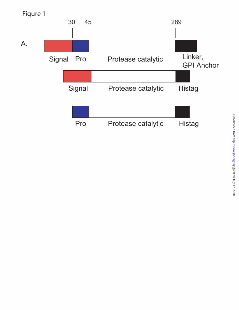
Figure 1
Signal Pro Protease catalytic Linker,
GPI Anchor
Protease catalyticPro Histag
A.
Signal Protease catalytic Histag
30 45 289
by guest on July 17, 2018http://w
ww
.jbc.org/D
ownloaded from

B. HUMAN MAQKGVLGPGQLGAVAILLYLGLLRSGTGAEGAEAPCGVAPQARITGGSSAVAGQWPWQV 60 V1 (MOUSE) -----------------------------ADGTEASCGAVIQPRITGGGSAKPGQWPWQV 31 V801 ------------------------MVSAIVLYVLLAAAAHSAFAITGGSSAVAGQWPWQV 36 V35 -----------------------MKFLVNVALVFMVVYISYIYAITGGSSAVAGQWPWQV 37 V40 ----------------------MPMLSAIVLYVLLAAAAHSAFAITGGSSAVAGQWPWQV 38 V26 -----------------------MAEGAEAPCGVAPQARDDDDKITGGSSAVAGQWPWQV 37 V28 -----------------------MAEGAEAPCGVAPQARDDDDKITGGSSAVAGQWPWQV 37 HUMAN SITYEGVHVCGGSLVSEQWVLSAAHCFPSEHHKEAYEVKLGAHQLDSYSEDAKVSTLKDI 120 V1 (MOUSE) SITYDGNHVCGGSLVSNKWVVSAAHCFPREHSREAYEVKLGAHQLDSYSNDTVVHTVAQI 91 V801 SITYEGVHVCGGSLVSEQWVLSAAHCFPSEHHKEAYEVKLGAHQLDSYSEDAKVSTLKDI 96 V35 SITYEGVHVCGGSLVSEQWVLSAAHCFPSEHHKEAYEVKLGAHQLDSYSEDAKVSTLKDI 97 V40 SITYEGVHVCGGSLVSEQWVLSAAHCFPSEHHKEAYEVKLGAHQLDSYSEDAKVSTLKDI 98 V26 SITYEGVHVCGGSLVSEQWVLSAAHCFPSEHHKEAYEVKLGAHQLDSYSEDAKVSTLKDI 97 V28 SITYEGVHVCGGSLVSEQWVLSAAHCFPSEHHKEAYEVKLGAHQLDSYSEDAKVSTLKDI 97 HUMAN IPHPSYLQEGSQGDIALLQLSRPITFSRYIRPICLPAANASFPNGLHCTVTGWGHVAPSV 180 V1 (MOUSE) ITHSSYREEGSQGDIAFIRLSSPVTFSRYIRPICLPAANASFPNGLHCTVTGWGHVAPSV 151 V801 IPHPSYLQEGSQGDIALLQLSRPITFSRYIRPICLPAANASFPNGLHCTVTGWGHVAPSV 156 V35 IPHPSYLQEGSQGDIALLQLSRPITFSRYIRPISLPAANASFPNGLHCTVTGWGHVAPSV 157 V40 IPHPSYLQEGSQGDIALLQLSRPITFSRYIRPISLPAASASFPNGLHCTVTGWGHVAPSV 158 V26 IPHPSYLQEGSQGDIALLQLSRPITFSRYIRPISLPAANASFPNGLHCTVTGWGHVAPSV 157 V28 IPHPSYLQEGSQGDIALLQLSRPITFSRYIRPISLPAANASFPNGLHCTVTGWGHVAPSV 157 HUMAN SLLTPKPLQQLEVPLISRETCNCLYNIDAKPEEPHFVQEDMVCAGYVEGGKDACQGDSGG 240 V1 (MOUSE) SLQTPRPLQQLEVPLISRETCSCLYNINAVPEEPHTIQQDMLCAGYVKGGKDACQGDSGG 211 V801 SLLTPKPLQQLEVPLISRETCNCLYNIDAKPEEPHFVQEDMVCAGYVEGGKDACQGDSGG 216 V35 SLLTPKPLQQLEVPLISRETCNALYNIDAKPEEPHFVQEDMVCAGYVEGGKDACQGDSGG 217 V40 SLLTPKPLQQLEVPLISRETCNALYNIDAKPEEPHFVQEDMVCAGYVEGGKDACQGDSGG 218 V26 SLLTPKPLQQLEVPLISRETCNALYNIDAKPEEPHFVQEDMVCAGYVEGGKDACQGDSGG 217 V28 SLLTPKPLQQLEVPLISRETCNALYNIDAKPEEPHFVQEDMVCAGYVEGGKDACQGDSGG 217 HUMAN PLSCPVEGLWYLTGIVSWGDACGARNRPGVYTLASSYASWIQSKVTELQPRVVPQTQESQ 300 V1 (MOUSE) PLSCPMEGIWYLAGIVSWGDACGAPNRPGVYTLTSTYASWIHHHVAELQ----------- 260 V801 PLSCPVEGLWYLTGIVSWGDACGARNRPGVYTLASSYASWIQSKVTELQPRVVPQTQESQ 276 V35 PLSCPVEGLWYLTGIVSWGDACGARNRPGVYTLASSYASWIQSKVTELQ----------- 266 V40 PLSCPVEGLWYLTGIVSWGDACGARNRPGVYTLASSYASWIQSKV--------------- 263 V26 PLSCPVEGLWYLTGIVSWGDACGARNRPGVYTLASSYASWIQSKV--------------- 262 V28 PLSCPVEGLWYLTGIVSWGDACGARNRPGVYTLASSYASWIQSKVTELQ----------- 266 HUMAN PDSNLCGSHLAFSSAPAQGLLRPILFLPLGLALGLLSPWLSEH- 343 V1 (MOUSE) ----------------------------------HHHHHHHHHH 270 V801 PDSNLCGSHLAFSSAPAQ---------------GLLRSHHHHHH 305 V35 --------------------------LVPRGSGSGS--HHHHHH 282 V40 --------------------------LVPRGSGSGS--HHHHHH 279 V26 --------------------------LVPRGSGSGSLEHHHHHH 280 V28 --------------------------LVPRGSGSGSLEHHHHHH 284
by guest on July 17, 2018http://w
ww
.jbc.org/D
ownloaded from

Vol (mL)
0 10 20 30 40 50 60 70
mAU
(280
nm
)
0
100
200
300
400
Act
ivity
(RFU
/min
/μL)
0
200
400
600
800
1000
1200MonomerA
Time (min)
0 5 10 15 20
AU (2
80 n
m)
0.00
0.01
0.02
0.03
0.04
0.05
B670 155 43 17 1.5
Figure 2
by guest on July 17, 2018http://w
ww
.jbc.org/D
ownloaded from


Substrate concentration (μM)
0 20 40 60 80 100 120 140
Rat
e (R
FU/m
in)
0
200
400
600
800
1000
1200
1400
1600
Figure 4
by guest on July 17, 2018http://w
ww
.jbc.org/D
ownloaded from


Paul L. Darke and Hua-Poo SuMontalvo, John C. Reid, Jennifer M. Shipman, Bradley W. Thomas, Sanjeev K. Munshi, Keith W. Rickert, Paul Kelley, Noel J. Byrne, Ronald E. Diehl, Dawn L. Hall, Allison M.
Structure of human prostasin, a target for the regulation of hypertension
published online October 14, 2008J. Biol. Chem.
10.1074/jbc.M805262200Access the most updated version of this article at doi:
Alerts:
When a correction for this article is posted•
When this article is cited•
to choose from all of JBC's e-mail alertsClick here
Supplemental material:
http://www.jbc.org/content/suppl/2008/10/17/M805262200.DC1
by guest on July 17, 2018http://w
ww
.jbc.org/D
ownloaded from