structure of the hepatitis e virus-like particle suggests ... · structure of the hepatitis e...
TRANSCRIPT
Structure of the hepatitis E virus-like particlesuggests mechanisms for virus assemblyand receptor bindingTom S. Y. Guua,1, Zheng Liub,1, Qiaozhen Yea, Douglas A. Mataa, Kunpeng Lic, Changcheng Yinb, Jingqiang Zhangc,2,and Yizhi Jane Taoa,2
aDepartment of Biochemistry and Cell Biology, Rice University, Houston, TX 77005; bDepartment of Biophysics, Health Science Centre, Peking University,Beijing, China 100191; and cState Key Laboratory for Biocontrol, Sun Yat-Sen University, Guangzhou, China 510275
Edited by Michael G. Rossmann, Purdue University, West Lafayette, IN, and approved June 15, 2009 (received for review May 1, 2009)
Hepatitis E virus (HEV), a small, non-enveloped RNA virus in the familyHepeviridae, is associated with endemic and epidemic acute viralhepatitis in developing countries. Our 3.5-Å structure of a HEV-likeparticle (VLP) shows that each capsid protein contains 3 linear do-mains that form distinct structural elements: S, the continuous capsid;P1, 3-fold protrusions; and P2, 2-fold spikes. The S domain adopts ajelly-roll fold commonly observed in small RNA viruses. The P1 and P2domains both adopt �-barrel folds. Each domain possesses a potentialpolysaccharide-binding site that may function in cell-receptor bind-ing. Sugar binding to P1 at the capsid protein interface may lead tocapsid disassembly and cell entry. Structural modeling indicates thatnative T � 3 capsid contains flat dimers, with less curvature than thoseof T � 1 VLP. Our findings significantly advance the understanding ofHEV molecular biology and have application to the development ofvaccines and antiviral medications.
capsid � HEV
V iral hepatitis is principally caused by 5 distinct viruses namedhepatitis A–E. Despite their similar names, the 5 viruses are
unrelated, and they have totally different genome structures withdistinct replication mechanisms. Hepatitis E virus (HEV) isresponsible for endemic hepatitis as well as sporadic epidemicsof acute, enterically transmitted hepatitis in the developingworld, including parts of Asia, the Middle East, Africa, andMexico (1, 2). HEV accounts for more than 50% of acute viralhepatitides in young adults in these regions, with a case fatalityof 1–2% in regular patients and up to 20% in pregnant women.
Given the lack of a robust cell culture system, and because HEVis not closely related to any other well-characterized virus, little isknown about the molecular biology of HEV or its strategy forreplication (1). HEV is a small, non-enveloped virus with a 7.2 kb,positive-sense RNA genome. Its genomic RNA is polyadenylatedand contains 3 ORFs. Located near the 5�-end, ORF1 encodes anon-structural polyprotein with multiple functional domains, in-cluding those for methyltransferase, protease, helicase, and poly-merase. The viral capsid protein (CP) is encoded by ORF2 near the3�-end. ORF3, which partially overlaps with the other 2 ORFs,codes for an immunogenic protein of unknown function. HEV wasoriginally classified in the Caliciviridae family because of its struc-tural similarity to other caliciviruses; however, it is now the solemember of the Hepeviridae family. The genomic RNA of HEVexhibits several distinct features compared to the genomic RNA ofcaliciviruses, including a methylated cap at the 5�-end and an ORF1with functional domains arranged in a different order (1, 3).
Previous studies of HEV assembly have primarily focused on theoverexpression of viral proteins. The ORF2 capsid protein, HEV-CP, contains a total of 660 amino acid residues. At the HEV-CP Nterminus is a signal peptide followed by an arginine-rich domainthat potentially play a role in viral RNA encapsidation duringassembly (3, 4). HEV-CP contains 3 putative N-glycosylation sites(5), but the biological relevance of such potential modifications isunclear (6, 7). The receptor binding site has been mapped to the
second half of the polypeptide chain, although the cell receptor forHEV has not yet been identified (8). Expression of the entire ORF2in insect cells results in proteolytic removal of the first 111 and thelast 52 residues (9), yielding a 55-kDa protein capable of self-assembly into virus-like particles (VLPs) (10). The size of theHEV-CP in infectious virions is unknown. HEV-CP is a key antigenthat stimulates the host immune response, and 6 antigenic domainshave been identified (11). One neutralization site has been mappedto the polypeptide region between amino acids 452 and 617 (12).
The structure of the infectious HEV particle has only beenanalyzed by immunoelectron microscopy (1). A 22-Å resolutioncryo-EM reconstruction has been obtained for the HEV VLP (10).The VLP displays T � 1 symmetry with a diameter of 270 Å, smallerthan the native HEV particle, which displays T � 3 symmetry withan estimated diameter of 350–400 Å (10). The surface of the VLPis dominated by 30 dimeric protrusions, and each capsid subunitappears to have 2 domains (10). HEV VLP possesses dominantantigenic activity similar to that of authentic HEV particles, and istherefore a promising candidate for use in vaccine development. Atruncated ORF2 polypeptide is currently undergoing clinical trialsas a vaccine candidate (13).
Here we report the crystal structure of HEV VLP determined to3.5-Å resolution. Each HEV-CP contains 3 linear domains, S(118–313), P1 (314–453), and P2 (454–end), the final 2 of which arelinked by a long, flexible hinge linker. The S domain forms acontinuous capsid shell that is reinforced by 3-fold protrusionsformed by P1 and 2-fold spikes formed by P2. It adopts the jelly-roll�-barrel fold that is most closely related to plant T � 3 viruses. P1and P2 contain compact, 6-stranded �-barrels that resemble the�-barrel domain of phage sialidase and the receptor-binding do-main of calicivirus, respectively, both of which are capable ofpolysaccharide binding. The highly exposed P2 domain likely playsan important role in antigenicity determination and virus neutral-ization. Structural modeling shows that the assembly of the nativeT � 3 capsid requires flat capsid protein dimers with less curvaturesthan those found in the T � 1 VLP, suggesting that additional Nterminal sequences may be involved in particle size regulation.
Results and DiscussionBiochemical Characterization of the HEV ORF2 Protein. For crystal-lization, HEV ORF2112–608 was overexpressed in Sf21 insect cells
Author contributions: Q.Y., C.Y., J.Z., and Y.J.T. designed research; T.S.Y.G., Z.L., Q.Y.,D.A.M., K.L., J.Z., and Y.J.T. performed research; J.Z. contributed new reagents/analytictools; T.S.Y.G., Z.L., Q.Y., and Y.J.T. analyzed data; and T.S.Y.G., Q.Y., D.A.M., and Y.J.T.wrote the paper.
The authors declare no conflict of interest.
This article is a PNAS Direct Submission.
Data deposition: The atomic coordinates and structure factors have been deposited in theProtein Data Bank, www.pdb.org (PDB ID 3HAG).
1T.S.Y.G. and Z.L. contributed equally to this work.
2To whom correspondence may be addressed. E-mail: [email protected] or [email protected].
This article contains supporting information online at www.pnas.org/cgi/content/full/0904848106/DCSupplemental.
12992–12997 � PNAS � August 4, 2009 � vol. 106 � no. 31 www.pnas.org�cgi�doi�10.1073�pnas.0904848106
and purified to near homogeneity by chromatography. Accord-ing to size-exclusion chromatography, the ORF2112–608 protein,hereon referred to as HEV-CP, was purified as dimers with anexpected molecular weight of approximately 107 kDa (Fig. S1).Higher molecular weight peaks corresponding to VLPs were notobserved, consistent with an earlier report that the overexpres-sion of ORF2112–608 in Sf9 cells generates soluble proteins only(10). VLPs were found in the cell media only when Tn5 insectcells were used and infected cells were harvested after prolongedincubation (10). For cryo-electron microscopy, HEV-CP wasoverexpressed in Tn5 cells, and the VLPs were purified bysucrose gradient ultracentrifugation.
Structure Determination. HEV-CP was crystallized in the spacegroup P63 with a � 241.1 Å and c � 519.9 Å (Table S1). Unit celldimensions of this magnitude are unusually large for a 54-kDaprotein. Self-rotation function and crystal packing considerationfurther confirmed that the HEV-CP protein had assembled into aVLP during crystallization, possibly stimulated by the low pH andhigh ionic strength of the crystallization solution. The structure ofthe HEV VLP was then determined by phase extension and 20-foldnon-crystallographic symmetry (NCS) averaging using a 14-Åcryo-EM reconstruction of the VLP as a phasing model.
Our initial map was of excellent quality with continuous main-chain density and defined side-chain density that allowed thepolypeptide chain to be traced without ambiguity (Fig. S2). The sidechain features became even more prominent after structure factorsharpening with a negative B factor. Our final model contains 468out of a total of 497 residues present in the ORF2112–608 construct.The missing residues are the first 6, the last 3, and several internalloop regions (148–149, 357–360, 483–488, 574–576, and 589–593).There are 2 cysteines in the ORF2368–606 sequence, but neither isinvolved in intra- or inter-molecular disulfide bonding.
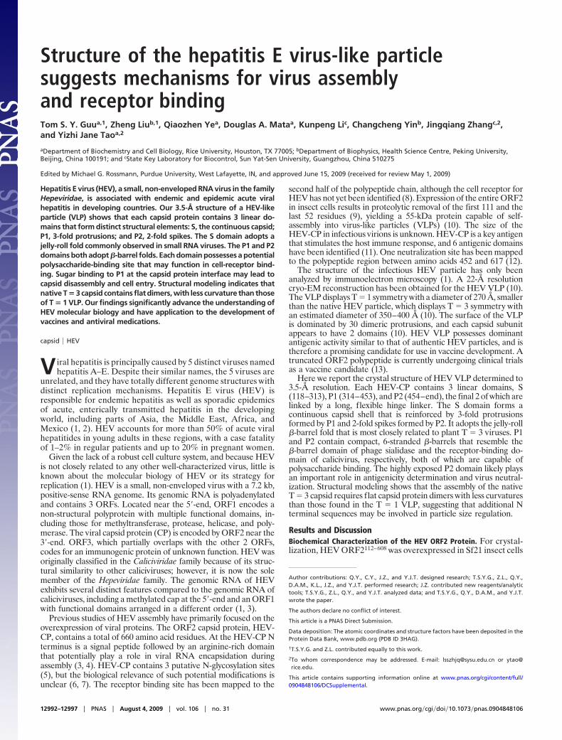
The 3.5-Å structure of the HEV VLP fits well into the 14-Å
cryo-EM map (Fig. 1 A and B), indicating that there is virtually nodifference in the structure of VLPs assembled during crystallizationand those found in cell media. The crystal structure shows an emptycapsid shell with an inner diameter of approximately 125 Å and anouter diameter of approximately 270 Å. The most prominentstructural feature is a total of 30 dimeric spikes situated on theicosahedral 2-fold symmetry axes. These dimeric spikes are approx-imately 30 Å tall. Capsid protrusions are also observed at icosa-hedral 3-fold symmetry axes. These trimeric protrusions formisolated units that do not interact with each other. Broad depres-sions are observed near icosahedral 5-fold symmetry axes.
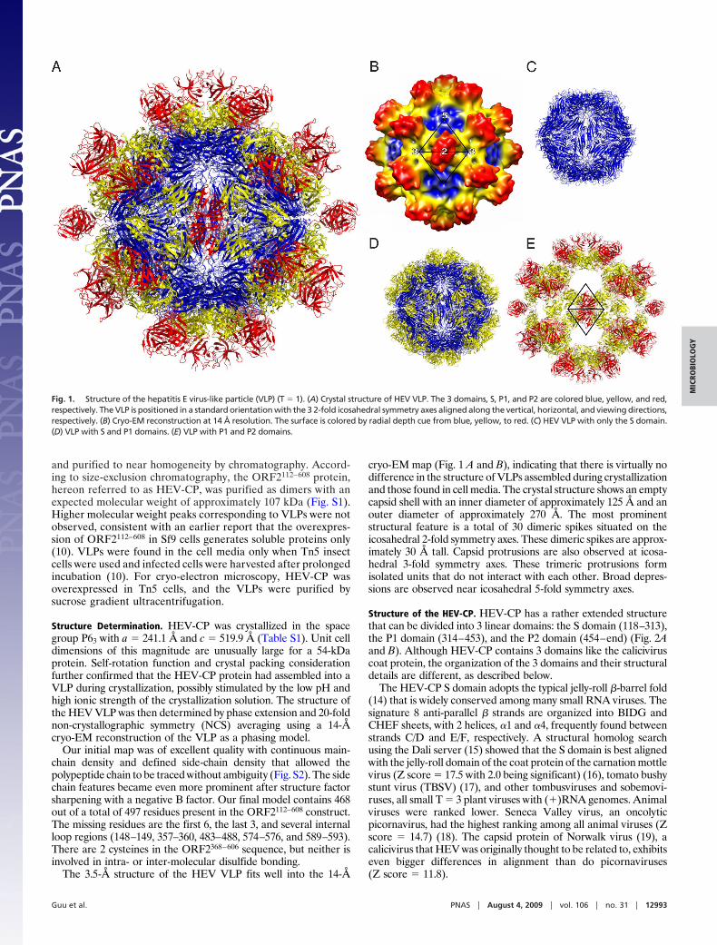
Structure of the HEV-CP. HEV-CP has a rather extended structurethat can be divided into 3 linear domains: the S domain (118–313),the P1 domain (314–453), and the P2 domain (454–end) (Fig. 2Aand B). Although HEV-CP contains 3 domains like the caliciviruscoat protein, the organization of the 3 domains and their structuraldetails are different, as described below.
The HEV-CP S domain adopts the typical jelly-roll �-barrel fold(14) that is widely conserved among many small RNA viruses. Thesignature 8 anti-parallel � strands are organized into BIDG andCHEF sheets, with 2 helices, �1 and �4, frequently found betweenstrands C/D and E/F, respectively. A structural homolog searchusing the Dali server (15) showed that the S domain is best alignedwith the jelly-roll domain of the coat protein of the carnation mottlevirus (Z score � 17.5 with 2.0 being significant) (16), tomato bushystunt virus (TBSV) (17), and other tombusviruses and sobemovi-ruses, all small T � 3 plant viruses with (�)RNA genomes. Animalviruses were ranked lower. Seneca Valley virus, an oncolyticpicornavirus, had the highest ranking among all animal viruses (Zscore � 14.7) (18). The capsid protein of Norwalk virus (19), acalicivirus that HEV was originally thought to be related to, exhibitseven bigger differences in alignment than do picornaviruses(Z score � 11.8).
Fig. 1. Structure of the hepatitis E virus-like particle (VLP) (T � 1). (A) Crystal structure of HEV VLP. The 3 domains, S, P1, and P2 are colored blue, yellow, and red,respectively. The VLP is positioned in a standard orientation with the 3 2-fold icosahedral symmetry axes aligned along the vertical, horizontal, and viewing directions,respectively. (B) Cryo-EM reconstruction at 14 Å resolution. The surface is colored by radial depth cue from blue, yellow, to red. (C) HEV VLP with only the S domain.(D) VLP with S and P1 domains. (E) VLP with P1 and P2 domains.
Guu et al. PNAS � August 4, 2009 � vol. 106 � no. 31 � 12993
MIC
ROBI
OLO
GY
The HEV-CP P1 domain has the appearance of a squashed�-barrel consisting of 6 anti-parallel �-strands. The 2 ends of thebarrel are flanked by several �-helices (Fig. 2 A and C). One sideof the barrel extensively interacts with the S domain through �B,�C, and loops CD, EF, and GH. The structure of the HEV-CP P1domain is related to that of the P2 domain of the calicivirus coatprotein (Z score � 2.8) (20). Other top structural homologs are thehuman UPF1 human helicase core (Z score � 5.1, 1st) (21), the�-barrel domain of endo-alpha-sialidase (22), the tRNA-bindingdomain of the translation elongation factor Tu (23), and thereceptor-binding domain of the avian reovirus fiber �C (24). Ofthese top homologs, the endosialidase of bacteriophage K1F is mostinteresting due to its ability to bind sialic acid molecules, which arewidely distributed in animal tissues and bacteria (22) (Fig. 2C). Bystructural superposition, the potential sialic acid binding site ofHEV-CP P1 is mapped to a helix-turn-helix motif (376–391)located at one end of the �-barrel (Fig. 2C).
The HEV-CP P2 domain forms the dimeric spike on the surfaceof the capsid (Figs. 1 and 2D). The overall fold of P2 is similar tothat of P1, except for a large insertion (from 504–533) between �20and �22 from the central �-barrel. This 30-amino acid insertion,comprised of 3 � strands and 1 � helix, mediates the interactionbetween the surface spike and the nearby 3-fold protrusion, thushelping support the spike. On top of the surface spike are 3 highlyexposed large loop insertions (482–490, 550–566, and 583–593) thatmay play an important role in antigenicity determination. Super-imposition of P2 and P1 by Dali server yields a Z score of 2.1. P2is also homologous to the receptor binding domain of norovirus inthe Caliciviridae family (Z score � 2.0) (25). Noroviruses useblood-group trisaccharides as cell receptors. Superimposing the 2structures brought the trisaccharide to the top of the HEV surfacespike between loops 550–566 and 583–593. Interestingly, thispotential sugar binding site in P2 is structurally equivalent to thepotential sialic acid-binding site in the P1 domain when the 2domain structures are aligned together.
The P1 and P2 domains are connected by a long linker and donot interact directly. Structural flexibility in the linker is importantbecause it allows the P2 domain to dimerize properly in thedifferent types of dimers encountered in the native T � 3 capsid
(see discussion below). Flexible hinge regions are also identified inthe coat proteins of many T � 3 viruses, including TBSV andcalicivirus, where dimers appear to be the assembly unit. Closeinspection of the HEV linker sequence 445NQHEQDRPTPS-PAPSRPF462 indicates that it is rich in proline and thus a poorsubstrate for proteases in general. This explains our observationthat protease treatment of the purified protein (with trypsin andchymotrypsin at up to a 1:1 mass ratio for 15 min at 20 °C) did notresult in significant degradation. Evolutionary pressure broughtabout by the enteric transmission route of HEV may have selectedfor a capsid protein with sequence and structural features that makethe virus highly resistant to proteases.
Residues 118–131 at the N terminus of the HEV-CP form theN-terminal arm (Fig. 2 A and B). The arm makes a sharp turn atthe beginning of �B, forming an extended loop that interacts witha 2-fold related and then a 3-fold related molecule nearby. In TBSV,the N-terminal R segment has an important role in regulatingparticle size (14). It wedges between 2 capsid protein subunits,thereby creating flat dimers on icosahedral 2-fold axes. The N-terminal arm of HEV-CP is away from the 2-fold dimer interfaceand adopts a different conformation compared to the R segment ofTBSV.
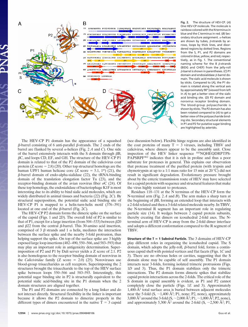
Structure of the T � 1 Subviral Particle. The 3 domains of HEV-CPplay different roles in organizing the icosahedral capsid. The Sdomain, which adopts the jelly-roll, �-barrel fold, forms a contin-uous capsid shell with pointed vertices and flat facets (Figs. 1C and3). There are no obvious holes or cavities, suggesting that the Sdomain alone may be capable of self assembly. The P1 domaininteracts near 3-folds, forming isolated trimeric protrusions (Figs.1D and 3). Thus, the P1 domain stabilizes only the trimericinteractions. The P2 domain forms dimeric spikes that stabilizecapsid protein interactions across the 2-folds. The critical role of theS domain in capsid assembly is evident, as P1 and P2 cannotcompletely close the particle (Figs. 1E and 3). Approximately1,400-Å2 total surface area is buried between adjacent moleculesnear the 5-fold (S, �1,400 Å2; P1, none; P2, none), approximately3,000 Å2 around the 3-fold (S, �2,000 Å2; P1, �1,000 Å2; P2, none),and approximately 5,300 Å2 around the 2-fold (S, �2,500 Å2; P1,
Fig. 2. The structure of HEV-CP. (A)One HEV-CP molecule. The molecule israinbow colored with the N terminus inblue and the C terminus in red. (B) Sec-ondary structure assignment. �-helicesare shown by tubes, �-strands by ar-rows, loops by thick lines, and disor-dered regions by dotted lines. Regionsfrom the S, P1, and P2 domains arecoloredinblue,yellow,andred, respec-tively, as in Fig. 1. The conventionalnaming scheme for the 8 �-strands(BIDG and CHEF) from the jelly-roll�-barrel is shown in parentheses. (C) P1domain and endosialidase �-barrel do-main. The sialic acid molecule is shownby sticks. Compared to (A), the P1 do-main is rotated along the vertical axisby approximately 90° [viewed from leftin A] to get a better view of the sialicacid binding site. (D) P2 domain andnorovirus receptor binding domain.The blood-group polysaccharide isshownbysticks.TheP2domainhasalsobeen rotated compared to (A) to get abetter view of the polysaccharide bind-ing site. Secondary structural elementsin P1 and P2 for putative sugar bindingare highlighted by asterisks.
12994 � www.pnas.org�cgi�doi�10.1073�pnas.0904848106 Guu et al.
�200 Å2; P2, �2,600 Å2). Based on the buried surface areas, the5-fold interaction is the weakest, and the 2-fold interaction is moststable.
The P2 domain alone is capable of dimerization (26). Dimeriza-tion of the P2 domain is mediated by an extended loop (550–566)and 3 �-strands from the central �-barrel (�18, �24, and �27). The4 structural elements provide a flat interface that is largely hydro-phobic in nature. Previous mutagenesis identified a cluster of 6hydrophobic residues critical for dimeric interactions: A597, V598,A599, L601, and A602 (26). Another study found that the deletionof residues 585–610 led to reduced oligomerization and aberrantfolding of the protein (27). The VLP structure shows that residues594–600 form one of the �-strands (�27) at the dimer interface.Amino acid substitutions within this �-strand are likely to affecteither the folding or properties of the interface, thus resulting in thedisruption of the tight packing between the 2 �-sheets.
The VLP structure allows us to evaluate the potential physio-logical relevance of the 3 potential N-glycosylation sites (5). N137is partially hidden near the inner surface of the capsid shell andN310 is completely buried, suggesting that glycosylation at eithersite is unlikely. N562 is exposed to solvent at the very top of thesurface spike and could potentially be subjected to glycosylation inthe ER. The inner surface of the capsid shell is covered with a largenumber of basic amino acid side chains (R128, R133, R186, R189,R193, and R195, 6 from each subunit), remarkably different fromdsRNA viruses in which a large number of negatively chargedresidues on the inner surface are used to facilitate the movement ofdsRNA genome during particle-associated transcription [for anexample, see (28)]. These arginine side chains from HEV-CPpresumably help to neutralize the negative charges of the genomicRNA. Around the 5-fold axes is a ring of 5 tyrosine residues (Y288)that are hydrogen bonded to 5 serine residues (S200), which are alsopositioned around the 5-fold axes, but closer to the particle interior.
We speculate that the dissociation of the VLP at alkaline pH mayhave been caused by de-protonation of the tyrosine side chain,resulting in the destabilization of the 5-fold interaction.
Given that a potential sugar binding site is found in both theHEV-CP P1 and P2 domains, it is important to determine which ofthe 2 domains functions in cell receptor binding. An earlier studyshowed that the ORF2368–606 protein was able to bind and penetratedifferent cell lines susceptible to HEV and to inhibit HEV infection(8). The ORF2368–606 protein, however, contains both potentialreceptor binding sites. The potential sugar binding sequence fromthe P1 domain, 376ADTLLGGLPTELISSA391, is strictly conservedamong all 4 HEV genotypes, suggesting that it has an importantfunctional role in cell receptor binding. In contrast, loops 550–566and 583–593, the potential sugar binding site of the P2 domain,contain 3 and 4 hypervariable amino acid sites, respectively, indi-cating that these regions are instead likely to mediate antibodyrecognition and immune escape. Moreover, the putative sugarbinding motif in the P1 domain (376–391) forms a hidden pocketat the interface between 2 HEV-CP molecules around the 3-fold.Therefore, receptor binding to P1 may potentially lead to thedestabilization of the HEV-CP trimer, resulting in conformationalchanges that eventually lead to membrane penetration and genomerelease into the infected cell. Further mutagenesis studies targetingthese 2 potential sugar-binding sites will determine which domainfunctions in host-cell binding and virus internalization.
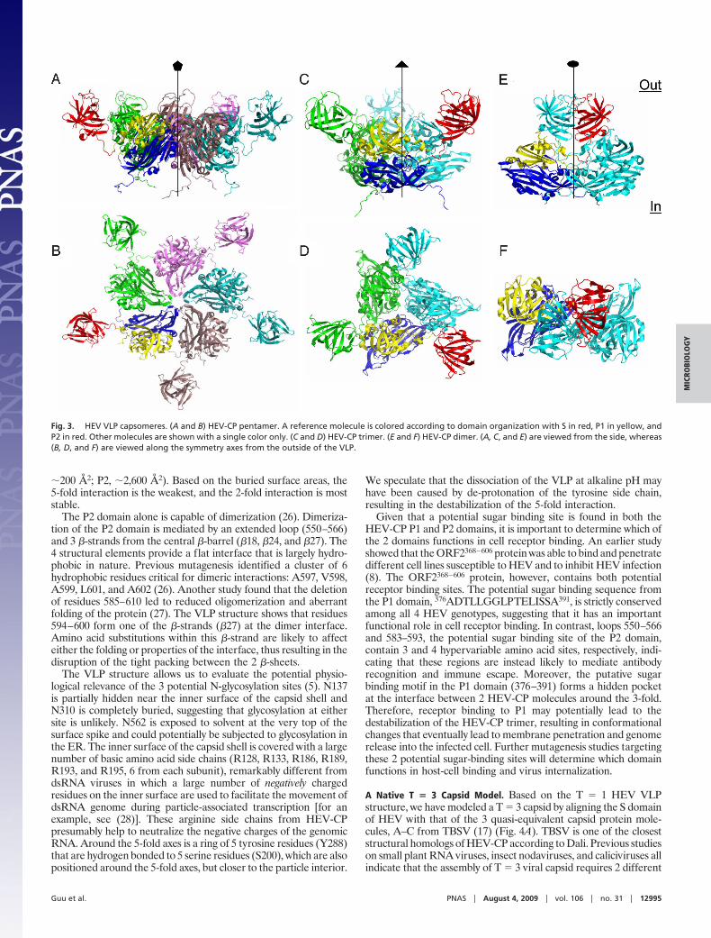
A Native T � 3 Capsid Model. Based on the T � 1 HEV VLPstructure, we have modeled a T � 3 capsid by aligning the S domainof HEV with that of the 3 quasi-equivalent capsid protein mole-cules, A–C from TBSV (17) (Fig. 4A). TBSV is one of the closeststructural homologs of HEV-CP according to Dali. Previous studieson small plant RNA viruses, insect nodaviruses, and caliciviruses allindicate that the assembly of T � 3 viral capsid requires 2 different
Fig. 3. HEV VLP capsomeres. (A and B) HEV-CP pentamer. A reference molecule is colored according to domain organization with S in red, P1 in yellow, andP2 in red. Other molecules are shown with a single color only. (C and D) HEV-CP trimer. (E and F) HEV-CP dimer. (A, C, and E) are viewed from the side, whereas(B, D, and F) are viewed along the symmetry axes from the outside of the VLP.
Guu et al. PNAS � August 4, 2009 � vol. 106 � no. 31 � 12995
MIC
ROBI
OLO
GY
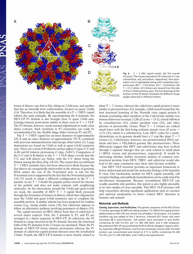
forms of dimers: one that is flat, sitting on 2-fold axes, and anotherthat has an inwardly bent conformation, located on quasi 2-folds(14). Therefore, it is likely that the assembly of a T � 3 HEV capsidfollows the same principle. By superimposing the S domains, theHEV-CP P1 domain is also brought close to quasi 3-fold axes,forming trimeric protrusions similar to those seen in T � 1 VLP.The P2 domain, however, needs manual adjustments to make closedimer contacts. Such variations in P2 orientation can easily beaccommodated by the flexible hinge linker between P1 and P2.
The T � 3 HEV capsid has an inner diameter of approximately220 Å and an outer diameter of approximately 370 Å, consistentwith previous immunoelectron microscopy observation (1). Largedepressions are found on 5-fold as well as quasi 6-fold symmetryaxes. There are a total of 90 dimeric surface spikes (2 types, C-C andA-B) and 60 trimeric protrusions (1 type, A-B-C). Comparison ofthe C-C and A-B dimers to the T � 1 VLP dimer reveals that theC-C and A-B dimers are flatter, with the C-C dimer being theflattest among the three (Fig. 4 B–D). The reason that recombinantT � 3 HEV particles have not been observed is likely because theflat dimers are energetically unfavorable in the absence of genomicRNA and/or the rest of the N-terminal arm. A role for theN-terminal arm is supported by the fact that the N-terminal peptide118–131 needs to adopt a different configuration in the T � 3particle: in our T � 3 model the peptide points toward the interiorof the particle and does not make contacts with neighboringmolecules. As the interactions around the 5-fold and quasi 6-foldare weak, the assembly of HEV T � 3 capsid likely proceedsthrough the formation of trimers of dimers, which then oligomerizenear the 5-fold and quasi 6-fold symmetry axes to complete theassembly process. A similar scheme has been proposed for tombusviruses (e.g., turnip crinkle virus) (29), but calicivirus appears tofollow an alternative pathway involving pentamers of dimers (19).
The capsid structure of HEV differs from that of calicivirus inseveral major respects. First, the 3 domains S, P1, and P2 arearranged in a linear sequence in HEV-CP. In calicivirus, the P2domain is a large insertion in the P1 domain. Second, the structuralfolds of the P1 domains in the 2 viruses are unrelated. Third, the P1domain of HEV-CP forms trimeric protrusions whereas the P1domain of calicivirus capsid protein interacts near the icosahedral2-folds. Fourth, the HEV-CP S domain is more closely related to
plant T � 3 viruses, whereas the calicivirus capsid protein is moresimilar to picornaviruses. For example, a Dali search found that thebest structural homolog of the Norwalk virus capsid protein Sdomain (excluding other members of the Caliciviridae family) washuman rhinovirus serotype 2 (30) (Z score � 15.5), closely followedby coxsackievirus (31), cricket paralysis virus (32), and otherpicorna or picorna-like viruses. Plant T � 3 viruses are rankedmuch lower with the first being sesbania mosaic virus (Z score �12.5) (33), which is a sobemovirus. Last, HEV codes for a meth-yltransferase so its genome should have a 5�-cap like plant T � 3viruses (1). Caliciviruses, however, use protein-primed RNA syn-thesis and have a VPg-linked genome like picornaviruses. Thesedifferences suggest that HEV and caliciviruses may have evolvedthrough 2 separate lineages that are each related to small plant(�)RNA viruses and picornaviruses, respectively. It would beinteresting whether further structural analysis of common non-structural proteins from HEV, TBSV, and calicivirus would alsolead to the same conclusion once these data become available.
Our HEV VLP structure provides an important framework tobetter understand and model the molecular biology of the hepatitisE virus. Our mechanistic models for HEV capsid assembly, cellreceptor binding, and antibody neutralization can be easily tested bysite-directed mutagenesis. Because recombinant HEV-CP canreadily assemble into particles, this system is also highly amenableto in vitro studies of virus assembly. This HEV VLP structure willhelp researchers develop significant applications such as vaccinesand antiviral medications to better control HEV infection inhumans and animals.
Materials and MethodsCloning, Expression, and Purification. The genetic sequence of the HEV (ChinaT1 Isolate, genotype 4) ORF2 encoding residues 112–608 of the capsid protein(abbreviated as HEV-CP) was cloned into pFastBac1 (Invitrogen). A 6-residuehistidine tag was added to the C terminus. Infected Sf21 insect cells wereharvested 48 h post-infection. Cell pellets were sonicated in lysis buffercontaining 50 mM Tris-HCl, pH 7.5, 300 mM NaCl, and 5 mM imidazole.Recombinant HEV-CP was purified by Ni-NTA affinity (Qiagen), heparin affin-ity, Superdex-200 gel filtration, and Q anion exchange columns (GE). Purifiedprotein was concentrated and stored at 4 °C in buffer containing 50 mMTris-HCl, pH 7.5, 200 mM NaCl, and 5 mM �-mercaptoethanol.
Fig. 4. T � 3 HEV capsid model. (A) The overallstructure. The 3 quasi-equivalent CP molecules A–C arecolored blue, red, and yellow, respectively. One asym-metric unit is highlighted along with icosahedral sym-metry axes. (B) T � 1 VLP dimer. (C) T � 3 A-B dimer. (D)T � 3 C-C dimer. All 3 dimers are viewed from the sideof their 2-fold symmetry axes. The line drawings at thebottom of the CP dimers illustrate the different hingeangles observed in different dimers.
12996 � www.pnas.org�cgi�doi�10.1073�pnas.0904848106 Guu et al.
Cryo-EM and Image Reconstruction. The VLPs for cryoEM were obtained byexpressing the HEV-ORF2112–660 protein (HE-JF4 isolate, genotype 4) in Tn5 insectcells as previously described (10). Gradient-purified VLP samples were diluted toabout2.5mg/mLwithPBS (pH7.2)andplunge-frozenonQuantifoilgrids. Imageswere collected on film using a JEM2011 electron microscope at 200 kV and50,000� magnification. Defocus values of approximately 1.0 and 3.0 �m wereused. Micrographs were digitized at 1.27 Å/pixel. Data processing was carried outusing the IMIRS package (34). Particle orientations were first determined byFourier common line and subsequently refined by projection-matching. Imagedefocus values were estimated using the Ctfit from the EMAN package (35). Atotal of 1,051 particle images were used to reconstruct the final map. Theeffective resolution was estimated to be 14-Å resolution (FSCC � 0.5).
Crystallization, Data Collection, and Data Processing. Crystals of HEV-CP wereobtained using vapor diffusion. The hanging drop consisted of equal volumes ofprotein (10 mg/mL) and well solution (0.1 M sodium cacodylate, pH 6.5, and 1.5M lithium sulfate). Hexagonal-shaped crystals appeared after 2 weeks of incu-bation at 20 °C and grew to full size (0.4 � 0.4 � 0.1 mm3) after 2 months.Microseeding and the addition of the detergent n-tetradecyl-b-D-maltosideaccelerated crystal growth and improved crystal quality. Diffraction images werecollected (oscillation angle � 0.5°) from frozen crystals presoaked in cryo-protectant made of well solution with 25% glycerol. Diffraction data wereprocessed using the program HKL2000 (36). HEV-CP crystals had the space groupof P63 with a � 241.1 Å and c � 519.9 Å. Our final data set was obtained from 2crystals indexed in the same handedness (Table S1).
Structure Determination. Self-rotation function using the program GLRF (37)showed peaks consistent with icosahedral symmetry. Packing considerationfurther suggested that there were 2 VLPs in each unit cell, with 1/3 of theparticle in each asymmetric unit. The icosahedral 3-fold symmetry axis of theVLP was aligned with the crystallographic 63 symmetry axis. Of the 2 possiblecrystal packing arrangements with VLP centered at (0, 0, 0) or (1/3, 2/3, 0), the
former gave better crystal contact considering that HEV VLP has a diameter ofonly approximately 270 Å. Of the 6 parameters describing VLP position andorientation, the only unknown was the rotation angle of the VLP around thecrystallographic 63 symmetry axis. Using diffraction data from 4.5- to 3.5-Åresolution, self-rotation function determined that 1 icosahedral 5-fold axiswas at � � 49.20 °, � � 37.38 °.
A 14-Å cryoEM reconstruction, flattened to a binary map with densities cor-responding to the capsid shell and solvent regions set to 1 and 0, respectively, wasused for initial phasing. EM map magnification and the contour level for mapflattening were optimized by maximizing correlation between the phasingmodel and the diffraction data. Using 20-fold NCS averaging, phases weregradually (one reciprocal lattice interval per step) extended to 3.5 Å using theprograms RAVE (38) and CCP4 (39), with 10 cycles of averaging at each phaseextension step. To optimize NCS matrices, the same phase extension procedurewas repeated by manually adjusting the icosahedral 5-fold axis with � varyingfrom48.90° to49.50°at0.05° intervals. Theobservationof right-handed �-helicesconfirmed the correct handedness of our map. The final map was sharpenedusing B � �150 Å2 (Fig. S2). Atomic models were built in O (40) and subsequentrefinements were performed using CNS (41) with 20-fold NCS constraints. TheRamachandran plot calculated for the final model showed 82% of non-Glyresidues in the most-favored regions with none in disallowed regions based onmain-chain dihedral angles.
Ribbon diagrams and C� traces were prepared using Molscript (42), PyMOL(W.L. Delano, http://www.pymol.org), and Chimera (43).
ACKNOWLEDGMENTS. We thank B.V.V. Prasad, J. Pan, R.R. Reed, and C. Ke forvaluable discussions, and the staff at CHESS and APS for assistance with datacollection. This work was supported by the National Institutes of Health (Y.J.T.),the National Natural Scientific Foundation of China (J.Z.), the Major State BasicResearch Development Program of China (C.Y.), the Major State Science andTechnology Project of China (C.Y.), the Welch Foundation (Y.J.T.), the HamillFoundation (Y.J.T.), and by the Kresge Science Initiative Endowment Fund at RiceUniversity.
1. Emerson UE, Purcell RH (2007) in Fields Virology, eds Knipe,D, Howley P (LippincottWilliams & Wilkins, Philadelphia), Vol 1, pp 3047–3058.
2. Chandra V, Taneja S, Kalia M, Jameel S (2008) Molecular biology and pathogenesis ofhepatitis E virus. J Biosci 33:451–464.
3. Tam AW, et al. (1991) Hepatitis E virus (HEV): Molecular cloning and sequencing of thefull-length viral genome. Virology 185:120–131.
4. Surjit M, Jameel S, Lal SK (2004) The ORF2 protein of hepatitis E virus binds the 5� regionof viral RNA. J Virol 78:320–328.
5. Zafrullah M, Ozdener MH, Kumar R, Panda SK, Jameel S (1999) Mutational analysis ofglycosylation, membrane translocation, and cell surface expression of the hepatitis Evirus ORF2 protein. J Virol 73:4074–4082.
6. Graff J, et al. (2005) In vitro and in vivo mutational analysis of the 3�-terminal regionsof hepatitis e virus genomes and replicons. J Virol 79:1017–1026.
7. Torresi J, Li F, Locarnini SA, Anderson DA (1999) Only the non-glycosylated fraction ofhepatitis E virus capsid (open reading frame 2) protein is stable in mammalian cells.J Gen Virol 80:1185–1188.
8. He S, et al. (2008) Putative receptor-binding sites of hepatitis E virus. J Gen Virol89:245–249.
9. Zhang Y, McAtee P, Yarbough PO, Tam AW, Fuerst T (1997) Expression, characteriza-tion, and immunoreactivities of a soluble hepatitis E virus putative capsid proteinspecies expressed in insect cells. Clin Diagn Lab Immunol 4:423–428.
10. Xing L, et al. (1999) Recombinant hepatitis E capsid protein self-assemblesinto a dual-domain T � 1 particle presenting native virus epitopes. Virology265:35– 45.
11. Khudyakov YE, et al. (1999) Antigenic domains of the open reading frame 2-encodedprotein of hepatitis E virus. J Clin Microbiol 37:2863–2871.
12. Meng J, et al. (2001) Identification and characterization of the neutralizationepitope(s) of the hepatitis E virus. Virology 288:203–211.
13. Shrestha MP, et al. (2007) Safety and efficacy of a recombinant hepatitis E vaccine.N Engl J Med 356:895–903.
14. Harrison SC (2007) in Fields Virology, eds Knipe D, Howley P (Lippincott Williams &Wilkins, Philadelphia), Vol 1, pp 59–98.
15. Holm L, Kaariainen S, Rosenstrom P, Schenkel A (2008) Searching protein structuredatabases with DaliLite v 3. Bioinformatics 24:2780–2781.
16. Morgunova E, et al. (1994) The atomic structure of Carnation Mottle Virus capsidprotein. FEBS Lett 338:267–271.
17. Olson AJ, Bricogne G, Harrison SC (1983) Structure of tomato busy stunt virus IV: Thevirus particle at 29 A resolution. J Mol Biol 171:61–93.
18. Venkataraman S, et al. (2008) Structure of Seneca Valley Virus-001: An oncolyticpicornavirus representing a new genus. Structure 16:1555–1561.
19. Prasad BV, et al. (1999) X-ray crystallographic structure of the Norwalk virus capsid.Science 286:287–290.
20. Chen R, Neill JD, Estes MK, Prasad BV (2006) X-ray structure of a native calicivirus:structural insights into antigenic diversity and host specificity. Proc Natl Acad Sci USA103:8048–8053.
21. Cheng Z, Muhlrad D, Lim MK, Parker R, Song H (2007) Structural and functional insightsinto the human Upf1 helicase core. EMBO J 26:253–264.
22. Stummeyer K, Dickmanns A, Muhlenhoff M, Gerardy-Schahn R, Ficner R (2005) Crystalstructure of the polysialic acid-degrading endosialidase of bacteriophage K1F. NatStruct Mol Biol 12:90–96.
23. Andersen GR, Thirup S, Spremulli LL, Nyborg J (2000) High resolution crystal structureof bovine mitochondrial EF-Tu in complex with GDP. J Mol Biol 297:421–436.
24. Guardado-Calvo P, et al. (2008) Crystal structure of the avian reovirus inner capsidprotein sigmaA. J Virol 82:11208–11216.
25. Cao S, et al. (2007) Structural basis for the recognition of blood group trisaccharides bynorovirus. J Virol 81:5949–5957.
26. Li TC, et al. (2005) Essential elements of the capsid protein for self-assembly into emptyvirus-like particles of hepatitis E virus. J Virol 79:12999–13006.
27. Xiaofang L, Zafrullah M, Ahmad F, Jameel S (2001) A C-Terminal Hydrophobic Regionis Required for Homo-Oligomerization of the Hepatitis E Virus Capsid (ORF2) Protein.J Biomed Biotechnol 1:122–128.
28. Pan J, et al. (2009) Atomic structure reveals the unique capsid organization of a dsRNAvirus. Proc Natl Acad Sci USA 106:4225–4230.
29. Sorger PK, Stockley PG, Harrison SC (1986) Structure and assembly of turnip crinkle virusII Mechanism of reassembly in vitro. J Mol Biol 191:639–658.
30. Verdaguer N, Blaas D, Fita I (2000) Structure of human rhinovirus serotype 2 (HRV2). JMol Biol 300:1179–1194.
31. Muckelbauer JK, et al. (1995) The structure of coxsackievirus B3 at 35 A resolution.Structure 3:653–667.
32. Tate J, et al. (1999) The crystal structure of cricket paralysis virus: the first view of a newvirus family. Nat Struct Biol 6:765–774.
33. Bhuvaneshwari M, et al. (1995) Structure of sesbania mosaic virus at 3 A resolution.Structure 3:1021–1030.
34. Liu H, et al. (2008) Symmetry-adapted spherical harmonics method for high-resolution3D single-particle reconstructions. J Struct Biol 161:64–73.
35. Ludtke SJ, Baldwin PR, Chiu W (1999) EMAN: Semiautomated software for high-resolution single-particle reconstructions. J Struct Biol 128:82–97.
36. Otwinowski Z, Minor W (1997) in Methods in Enzymology, eds Carter CW, Sweet RM(Academic, New York), Vol 276, pp 307–326.
37. Tong L, Rossmann MG (1997) Rotation function calculations with GLRF program.Methods Enzymol 276:594–611.
38. Jones TA (1992) in Molecular Replacement, eds Dodson EJ, Gover S, Wolf W (SERCDaresbury Laboratory, Warrington), pp 91–105.
39. CCP4 (Collaborative Computational Project Number 4) (1994) The CCP4 suite: Programsfor protein crystallography. Acta Crystallogr D Biol Crystallogr 50:760–763.
40. Jones TA, Zou JY, Cowan SW, Kjeldgaard M (1991) Improved methods for buildingprotein models in electron density maps and the location of errors in these models.Acta Crystallogr A 47:110–119.
41. Brunger AT, et al. (1998) Crystallography & NMR system: A new software suite formacromolecular structure determination. Acta Crystallogr D Biol Crystallogr 54:905–921.
42. Kraulis PJ (1991) MOLSCRIPT: A program to produce both detailed and schematic plotsof protein structures. J Appl Crystallogr 24:946–950.
43. Pettersen EF, et al. (2004) UCSF Chimera–a visualization system for exploratory researchand analysis. J Comput Chem 25:1605–1612.
Guu et al. PNAS � August 4, 2009 � vol. 106 � no. 31 � 12997
MIC
ROBI
OLO
GY