structure of the mammalian nos regulator dimethylarginine dimethylaminohydrolase: a basis for the...
TRANSCRIPT
Structure 14, 901–911, May 2006 ª2006 Elsevier Ltd All rights reserved DOI 10.1016/j.str.2006.03.006
Structure of the Mammalian NOS RegulatorDimethylarginine Dimethylaminohydrolase:A Basis for the Design of Specific Inhibitors
Daniel Frey,1 Oliver Braun,1 Christophe Briand,1
Milan Vasak,1 and Markus G. Grutter1,*1Department of BiochemistryUniversity of ZurichWinterthurerstrasse 190CH-8057 ZurichSwitzerland
Summary
Dimethylarginine dimethylaminohydrolase (DDAH) isinvolved in the regulation of nitric oxide synthase
(NOS) by metabolizing the free endogenous argininederivatives N u-methyl-L-arginine (MMA) and N u,N u-di-
methyl-L-arginine (ADMA), which are competitive in-hibitors of NOS. Here, we present high-resolution crys-
tal structures of DDAH isoform 1 (DDAH-1) isolatedfrom bovine brain in complex with different inhibitors,
including S-nitroso-L-homocysteine and Zn2+, a regu-lator of this mammalian enzyme. The structure of
DDAH-1 consists of a propeller-like fold similar to otherarginine-modifying enzymes and a flexible loop, which
adopts different conformations and acts as a lid at theentrance of the active site. The orientation and interac-
tion mode of inhibitors in the active site give insightinto the regulation and the molecular mechanism of
the enzyme. The presented structures provide a basisfor the structure-based development of specific
DDAH-1 inhibitors that might be useful in the therapeu-tic treatment of NOS dysfunction-related diseases.
Introduction
Nitric oxide (NO) is an important signaling and effectormolecule involved in pathophysiological and physiolog-ical processes such as neurotransmission, regulation ofvascular tone, and bacterial defense (Colasanti andSuzuki, 2000). NO is produced by three homologousnitric oxide synthases (NOS) converting L-arginine toL-citrulline: endothelial NOS (eNOS) and neuronal NOS(nNOS), which are predominantly regulated by cytosolicCa2+, which, in turn, modulates protein-protein interac-tion between calmodulin and NOS (Bredt and Snyder,1994; Kone et al., 2003), and the inducible NOS (iNOS).High toxicity, free diffusibility, and potent chemical reac-tivity of the free radical NO require strict, tight control ofthe three isoforms of NOS to enable efficient signalingand to prevent simultaneous damage to other cellularcomponents (Rassaf et al., 2004). For example, severalcardiovascular risk factors like hypertension, smoking,diabetes mellitus, and vascular inflammation are relatedto the dysfunction of NOS (Vallance and Leiper, 2004).
It is well established that NO synthesis can be com-petitively inhibited by the endogenously methylated ar-ginine derivatives Nu-methyl-L-arginine (monomethy-
*Correspondence: [email protected]
lated L-arginine, MMA) and Nu,Nu-dimethyl-L-arginine(asymmetric dimethylated L-arginine, ADMA). MMAand ADMA are competitive endogenous inhibitors ofall three isoforms of NOS, while Nu,Nu�-dimethyl-L-argi-nine (symmetric dimethylated L-arginine, SDMA) doesnot inhibit NOS (Leiper and Vallance, 1999; Tran et al.,2003). Methylated arginines appear in proteins that aremodified posttranslationally (Paik and Kim, 1971) or asfree amino acids in various mammalian tissues (Uenoet al., 1992). The enzyme Nu,Nu-dimethyl-L-arginine di-methylaminohydrolase (DDAH, EC 3.5.3.18) convertsMMA and ADMA to L-citrulline and mono- or dimethyl-amine, respectively, thus regulating NOS activity (Mac-Allister et al., 1996). This enzyme does not degrade thenon-NOS inhibitor SDMA.
Mammalian DDAH exists in two isoforms—DDAH-1,which colocalizes mainly with nNOS, and DDAH-2,which is predominantly expressed in tissues containingeNOS—suggesting an isoform-dependent regulation ofNOS (Tran et al., 2000). Overexpression of DDAH acti-vates NOS (Dayoub et al., 2003), whereas inhibition ofDDAH leads to accumulation of ADMA, causing de-creased NOS activity and NO levels (Cooke, 2004). In-creased levels of MMA and ADMA in plasma and urinehave been implicated in diseases linked with low levelsof NO, such as uremia, chronic heart failure, atheroscle-rosis, and hyperhomocysteinemia (Tsao and Cooke,1998). In contrast, in diseases such as septic shock, mi-graine, inflammation, and neurodegenerative disorders,excess NOS activity or NO production is involved (Val-lance and Leiper, 2002). Attenuation of NO productionby inhibiting DDAH might therefore be a valuable ap-proach to fight these diseases.
Besides the known endogenous DDAH inhibitorsL-citrulline, Zn2+, L-homocysteine, and S-nitroso-L-homocysteine, only a few designed inhibitors that areeither not selective for DDAH or lack DDAH isoformspecificity are currently known (Rossiter et al., 2005;Stone et al., 2005; Vallance et al., 2005). Although thecrystal structure of a bacterial DDAH is known (Mur-ray-Rust et al., 2001), modeling of a mammalian enzymestructure was deemed difficult because of low sequencesimilarity (Rossiter et al., 2005).
Here, we present various high-resolution crystalstructures of the mammalian DDAH-1 from bovine brain,which include: (1) structures of the unliganded enzymein two different crystal forms, (2) structures of the Zn2+
bound enzyme at two different pH values, and (3) struc-tures of the enzyme in complex with the reactionproduct L-citrulline, the inhibitor L-homocysteine, andthe endogenous suicide inhibitor S-nitroso-L-homocys-teine. Based on the DDAH-1 structure, a high-quality ho-mology model of the bovine enzyme isoform DDAH-2could be built that reveals differences in the bindingpocket between both isoforms.
The structures presented here can serve as an idealbasis for the design of inhibitors that could have apotential pharmaceutical use in diseases in which over-production of NO is involved (Vallance and Leiper,2002).

Structure902Structure902
Table 1. X-Ray Data and Refinement Statistics
Zn2+ (pH 9.0) Zn2+ (pH 6.3) Ia IIa Citb Hcyc HcyNOd
Space group P212121 P212121 P21 P21 P21 P21 P21
Wavelength (A) 1.0000 0.8550 0.9793 0.9793 1.0001 0.9793 0.8998
Cell dimensions
a (A) 44.38 44.40 40.96 42.84 40.7 44.7 40.66
b (A) 74.63 76.49 79.61 81.02 80.1 79.4 79.91
c (A) 81.40 81.49 44.87 44.68 44.9 70.6 44.89
b (º) 90 90 108.5 100.6 108.08 104.8 108.58
Resolution (A) 1.6 2.0 1.7 1.7 1.2 1.8 1.08
Completeness (%)e 98.4 (96.0) 99.2 (96.3) 96.2 (93.3) 99.3 (93.6) 98.0 (94.3) 98.1 (90.7) 98.6 (96.0)
Number of unique reflections 35,849 19,299 28,708 32,693 91,611 48,415 114,012
Multiplicity 7.24 5.08 2.62 3.45 2.98 3.25 3.65
I/s(I)e 10.4 (4.5) 10.8 (4.0) 12.0 (4.8) 16.9 (4.8) 10.8 (2.0) 9.5 (2.2) 15.7 (4.1)
Rsyme 7.6 (49.7) 6.8 (44.2) 8.7 (29.3) 6.0 (19.8) 7.0 (33.4) 12.6 (37.6) 7.7 (33.9)
Processing program XDS Denzo Denzo Denzo Denzo Denzo Denzo
Resolution range (A) 20–1.6 40–2.0 30–1.7 30–1.7 20–1.2 30–1.8 20–1.08
Number of reflections in
working and test set
35,844/1,182 19,241/935 27,225/1,392 31,033/1,622 81,946/2,580 45,805/2,422 112,848/1,126
Rcryst (%) 20.3 22.9 18.8 19.4 12.2 17.6 11.1
Rfree (%) 23.8 25.9 21.9 21.7 16.8 21.2 14.0
Water moleculesf 333 98 336 247 393 (5) 656 482
Rmsd
Bond lengths (A) 0.004 0.006 0.004 0.004 0.013g 0.007 0.016g
Bond angles (º) 1.3 1.2 1.4 1.3 0.030g 1.4 0.034g
Mean B value (A2) 24.0 29.6 16.6 21.0 15.0 14.0 13.3
a Crystal forms I and II of Zn2+ free DDAH-1.b–d Complex structures with L-citrulline (Cit), L-homocysteine (Hcy), and S-nitroso-L-homocysteine (HcyNO), respectively.e Values in parentheses are for the highest-resolution shell.f Numbers in parentheses indicate the number of half waters.g Rmsd values in A for the refinements with SHELX.
Results
Overall StructureWe determined crystal structures of ligand-free DDAH-1in two different crystal forms, of DDAH-1 with boundZn2+ at pH 6.3 and pH 9.0, and of DDAH-1 in complexwith L-citrulline, L-homocysteine, and S-nitroso-L-ho-mocysteine at high resolution (between 1.08–2.0 A; seeTable 1). The overall fold of the enzyme consists of fiverepeats of a bbab motif (Figure 1A) with a root-mean-square deviation (rmsd) of 0.19–0.50 A for all Ca atomsbetween the different structures. The CATH Databaseclassifies the protein as a five-stranded propeller be-longing to the homologous superfamily of L-arginine/glycine amidinotransferase (3.75.10.10) (Orengo et al.,1997).
As a result of the modular architecture of DDAH-1,there is a channel in the center of the molecule that isclosed in the middle by a salt bridge between Glu77and Lys174 that, in turn, forms the bottom of the activesite (Figure 1B). One side of the channel represents a wa-ter-filled pore delineated by the first b strand of each ofthe five propeller blades. The other side of the channel isthe active site cleft whose outermost boundaries are de-fined by short loop regions and a helical structures. Aflexible loop region formed by amino acids 25–36 closesthe active site upon binding of the substrate (Figures 1Aand 2).
Structure of the Unliganded Enzyme andConformational Changes in the Lid Region
Three different conformations for a surface stretch rep-resenting amino acids 25–36, here referred to as the lid
region, were observed in the various structures (Fig-ure 2). A closed form was observed in five structuresof the liganded enzyme, in the Zn2+-containing structureat pH 9.0, and in one unliganded enzyme structure (crys-tal form I, see Table 1). Crystal packing in crystal form Icontaining the unliganded enzyme prevents the lid re-gion from adopting an open conformation. The openconformation was observed in crystal form II of the unli-ganded enzyme. Here, the lid region forms an a helix re-orienting Leu29 12 A away from the active site and leav-ing the active site entrance open and accessible for thesubstrate to enter and bind. An alternative open confor-mation was observed in the Zn2+-containing structure atpH 6.3, indicating high flexibility of the lid region in theopen conformation. The high flexibility of the lid regionis also reflected by high crystallographic B factors: theaverage B factor value for amino acids 25–36 is 50 A2,compared to an overall value of 21 A2 (Figure 3). Theclosed lid conformation allows for the formation of anadditional hydrogen bond between the amino group ofthe ligand and the main chain carbonyl of Leu29. Thisadditional interaction enables the rearrangement fromthe a-helical (open) to the loop (closed) conformationof the lid. In the latter case, the isobutyl side chain ofLeu29 blocks the entrance of the active site (Figure 2).
Structures of Zn2+ Bound to the Active Site
The protein purified from bovine brain contained oneZn2+ ion with an occupancy of w95% and an apparentdissociation constant of w4 nM as previously described(Knipp et al., 2001). This protein was crystallized at pH6.3 and pH 9.0. The conformation of the lid loop is differ-ent in the two structures. In the crystal form grown at pH

Crystal Structure of Mammalian DDAH903
Figure 1. Overall Structure of DDAH-1
(A) Stereoview of the overall structure of
DDAH-1 (ribbon depiction) in complex with
L-citrulline (space filled). The bbab blades of
the propeller are colored green, magenta,
cyan, orange, and blue from the N to the C ter-
minus. The connecting loops and helices be-
tween the blades are colored gray. Residues
25–36 form a lid shown in red. L-citrulline is
shown with carbon atoms in yellow, nitrogen
in blue, and oxygen in red. All figures were
generated with the program PyMOL (DeLano,
2002).
(B) Stereoview of a cross-section through
DDAH-1. The DDAH-1 L-citrulline complex
was used for the surface representation. Wa-
ter molecules were excluded for the calcula-
tion of the surface. Water molecules in the
water channel and in the pore are shown as
red spheres, and L-citrulline is shown as
a stick model. The two residues, Glu77 and
Lys174, shielding the active site from the
water-filled pore are shown in green, and
the lid is shown in red.
6.3, the lid region adopts a second open conformation(Figure 2) that is fixed by water molecules mediatingcrystal contacts. At pH 9.0, the lid is in a closed confor-mation that is induced by a glycine molecule present inthe crystallization buffer acting as a ligand.
The observed distances and angles between zinc andthe coordinating atoms at both pH values are summa-rized in Tables 2 and 3. In both structures, the Zn2+ isbound to the enzyme in a distorted tetrahedral environ-
Figure 2. Conformations of the Lid Region
The three different conformations of the lid region of DDAH-1 are
shown. The open conformation (crystal form II), the closed confor-
mation (crystal form I), and the Zn2+ bound structure at pH 6.3 are
shown in blue, red, and green, respectively (Table 1). Residues 24–
37 for all three structures are represented as ribbons, and the side
chain Leu29 is shown as a stick model. The surface is calculated
by using the structure of crystal form II without water molecules.
L-citrulline is shown as a stick model, and the active site cysteine
is marked by a yellow surface. The distance between the Cg atom
of Leu29 in the open and closed conformation is 12 A. N and C
mark the N- and C-terminal end of the lid, respectively.
ment coordinated by Cys273 and three water molecules.At pH 6.3, two water molecules are hydrogen bonded tothe side chains of Asp78 and Glu77. The third water mol-ecule is not stabilized by additional protein ligands(Figure 4A and Tables 2 and 3).
At pH 9.0, the Zn2+ is still in a tetrahedral environment;however, the position compared to the structure at pH6.3 is slightly changed (Figure 4B and Tables 2 and 3).At pH 9.0, the side chain of His172 is deprotonated andthe salt bridge to Asp126 is weakened, allowing His172to adopt two different conformations in the same struc-ture (Figure 4B). The first conformation is equivalent to
Figure 3. Comparison of the Temperature Factors of Crystal Forms I
and II
B factors (A2) of each residue were averaged and plotted against the
residue number. Overall B factors of crystal forms I and II were 17
and 21 A2, respectively. A mean B value of 50 A2 in the open confor-
mation (crystal form II) indicates the high mobility of the lid region. In
the closed conformation (crystal form I), the mean B value is only
18 A2 for these residues, indicating that the lid region is more rigid.

Structure904
that seen at pH 6.3. Cys273 and three water ligands,which are hydrogen bonded to Asp78, Glu77, andHis172, coordinate the Zn2+ ion. In the second conforma-tion, the imidazole group coordinates the Zn2+ directly:the distal nitrogen of the side chain is now in the positionas the water ligand observed in the first conformation.
L-Citrulline, L-Homocysteine, and S-Nitroso-L-
Homocysteine Bound to DDAH-1Crystals of the closed lid conformation were obtainedwith L-citrulline, the product of the enzymatic reactionbound in the active site of DDAH-1 (Figure 5A). Differenceelectron density maps revealed clear density in the ac-tive site for the ligand, although citrulline was not in-cluded in the first round of model building (Figure 5A).Its Ca-carboxyl group forms three salt bridges, two withthe guanidino group of Arg144 of DDAH-1, and one withthe guanidino group of Arg97. Two hydrogen bonds be-tween the Ca-amino group of the ligand and the mainchain carbonyls of Val267 and Leu29 of the enzyme aswell as a salt bridge to the side chain of Asp72 further sta-bilize L-citrulline in the active site. In addition, side chainsof Asp78 and Glu77 form hydrogen bonds to the ureidomoiety of L-citrulline and position the ligand in the activesite. The hydrophobic moiety (Cb–Cd) is shielded byPhe75 on one side and Leu171 on the other side. Sg ofCys273 points away from L-citrulline, indicating a sterichindrance between the protein and the ligand. The activesite water occupies the same position as it did in thestructure of the bacterial enzyme (Murray-Rust et al.,2001). It forms a hydrogen bond with the side chain hy-droxyl group of Ser175 and the proximal nitrogen of theimidazole ring of His172. The active site water moleculeis linked to a series of three water molecules in a waterchannel leading to the surface of the enzyme (Figure1B). This water channel allows for the replenishment ofwater molecules that participate in the hydrolysis ofMMA and ADMA.
L-homocysteine has been identified recently as a riskfactor for cardiovascular diseases (Lentz et al., 2003). Anumber of investigations report on elevated levels ofADMA and a decreased production of NO in hyperho-
Table 2. Distances between Zinc and the Coordinating Atoms
Crystal Structure Zn-S(p)a Zn-N(p)a Zn-O(w)a
pH 6.3b 2.19 — 1.84, 1.92, 2.37
pH 9.0c 2.27 2.38 2.08, 2.03, 1.81
Mean value of
observed distances
(Alberts et al., 1998)
2.21 6 0.13 2.07 6 0.09 2.12 6 0.15
a (p) or (w) indicate protein or water ligands, respectively.b Coordinate error 6 0.23.c Coordinate error 6 0.1 A (Cruickshank, 1999).
mocysteinemia (Boger et al., 2000; Selley, 2003; Stuhlin-ger et al., 2003). Recently, homocysteine was reportedto directly inhibit DDAH in vitro. From their data, the au-thors proposed the formation of a disulfide bridge be-tween homocysteine and DDAH (Stuhlinger et al., 2001).
Our structure in the presence of L-homocysteineclearly indicates that L-homocysteine binds in the activesite pocket in the same orientation as L-citrulline (Fig-ure 5B), and that it binds with a closed lid conformation.However, even in the absence of reducing agents, noelectron density of a formed disulfide bond was foundbetween the two sulfur atoms. Therefore, our results donot support the formation of a disulfide bond proposedby Stuhlinger et al. (2001).
Knipp et al. (2005) showed that upon reaction ofDDAH-1 with S-nitroso-L-homocysteine a so far unob-served covalent product, N-thiosulfoximide (Cys273:S-NH-S(O):homocysteine), exists in the active site. Theproposed reaction proceeds through a sulfiliminosulfo-nium ion (Cys273:S-N]S+-homocysteine 4 Cys273:S+]N-S-homocysteine). Isotope labeling experimentsrevealed that the oxygen atom from a water molecule isincorporated, reacting exclusively with the sulfur atomof homocysteine (Knipp et al., 2005). In the crystal struc-ture, N-thiosulfoximide is clearly seen in the differenceFourier electron density map (Figure 5C). Interestingly,refinement of the structure revealed two isomers of N-thiosulfoximide: Cys273:S-NH-S(O):homocysteine andCys273:S(O)-NH-S:homocysteine. Since the specific at-tack of a water molecule on the sulfur atom of homocys-teine has already been shown (Knipp et al., 2005), the for-mation of both isomers in the crystal structure is, to ourknowledge, new, indicating a subsequent slow rear-rangement previously not observed.
N-thiosulfoximide is stabilized by several salt bridgesand hydrogen bonds (Figure 6). Similar to what occurs inthe other DDAH-1 complex structures, Arg144 andArg97 stabilize the Ca-carboxyl group via salt bridges,whereas Leu29, Val267, and Asp72 stabilize the Ca-amino group. As was already observed in the DDAH-1L-citrulline complex, the lid region adopts a closed con-formation. The nitrogen atom of N-thiosulfoximide formsa hydrogen bond to Asp78 in both isomers. However,the hydrogen bond between the thionyl oxygen andthe proximal nitrogen atom of the imidazole group ofHis172 is only present in the isomer with the oxygenatom bound to the homocysteine sulfur atom. On theother hand, a hydrogen bond to the main chain nitrogenof Leu270 stabilizes the isomer with the oxygen atom lo-cated at the Cys273 sulfur atom.
The nitrogen atom of N-thiosulfoximide occupiesa similar position as N3 of L-citrulline, and there is a slightshift of 0.6 A toward Cys273 due to longer S-N and C-Sbonds and the missing repulsion of the free side chain ofCys273 as seen in the DDAH-1 L-citrulline complex. As
Table 3. Angles between Zinc and the Coordinating Ligands
Crystal Structure S-Zn-N N-Zn-O S-Zn-O O-Zn-O
pH 6.3 — — 112.0, 107.6, 100.1 102.5, 109.3, 125.0
pH 9.0 126.7 99.6, 105.6 113.9, 114.8, 110.0 114.0, 95.6, 107.3
Mean value of observed
angles (Alberts et al., 1998)
112 6 4 110 6 9 105 6 7 111 6 9

Crystal Structure of Mammalian DDAH905
Figure 4. Zn2+ Binding Site
(A) 2Fo 2 Fc Fourier map of the Zn2+ binding
site at pH 6.3. The active site residues
Cys273, His172, Glu77, and Asp78 and resi-
due Asp268 are shown as sticks. The bound
Zn2+ ion is shown in gray, and the bound wa-
ter molecules are shown in red. Metal ion li-
gand interactions are represented as dotted
lines, and their corresponding lengths are in-
dicated in A.
(B) 2Fo 2 Fc Fourier map of the Zn2+ binding
site at pH 9.0. Interactions of the metal ion
in the first and second coordination sphere
are shown. The Zn2+ atom is shown in gray,
and water molecules are shown in red. The
second conformation of His172 and the alter-
nating water molecule are shown in gray and
purple, respectively.
structural information on N-thiosulfoximides are notavailable in the Cambridge Structural Database (Allen,2002), bond lengths were compared to similar com-pounds. Bond lengths of 1.81, 1.62, 1.63, 1.5, and 1.81 Afor Cb to Sg, Sg to N, N to Sd, Sd to O, and Sd to Cg(Cys273:S-NH-S(O):Hcy), respectively, agree well withmodeled data for oxidized methionine (Schuttelkopfand van Aalten, 2004) and small-molecule X-ray datafor sulfoximines (Cambridge Structural Database [Allen,2002]). As seen for small molecules, the S-N bondlengths in the N-thiosulfoximide isomers are shorter(1.62/1.63 6 0.03 A and 1.59/1.62 6 0.03 A respectively[Cruickshank, 1999]) than expected for a single bondS-N (1.7 A) indicating partial double bond character. Re-markably, superposition of the active site residues of theL-citrulline and N-thiosulfoximide structures onlyshowed a difference in the newly formed S-N bond ofthe N-thiosulfoximide. As the first collection of diffrac-tion data revealed a partially cleaved N-thiosulfoximideresulting in several not clearly defined degradationproducts in the active site, e.g., cysteine or homocys-teine derivatives of sulfenic acids (R-S-OH) or sulfinicacids (R-S(]O)-OH), further data acquisition was per-formed with lower radiation intensity (data not shown).
Besides the formation of N-thiosulfoximide, an S-ni-trosylation of Cys83 was also observed. The solvent-ex-posed Cys83 is located at the lower end of the pore. Thismodification may be due to a transnitrosylation reactionbetween S-nitroso-L-homocysteine and Cys83 underthe crystallization conditions. The previously reportedS-nitrosylation of Cys221 with DEA/NO, liberating gas-eous NO, was not observed in our structure (Knippet al., 2003). Cys221 is located in the core of the proteinand is accessible to gaseous NO, whereas modificationsby S-nitrosohomocysteine are restricted to solvent-ac-cessible residues like Cys273 or Cys83.
Comparison to Other Arginine-Modifying EnzymesThe overall fold of DDAH-1 from bovine brain is identicalto the fold of Pseudomonas aeruginosa DDAH (PaDDAH; rmsd 1.39 A) and to other arginine- or substituted
arginine-modifying enzymes of which crystal structureshave been determined. Peptidylarginine deiminase(PAD4) (Arita et al., 2004), converting L-arginine to L-cit-rulline and ammonium; the Arg:Gly amidinotransferase(AT) (Humm et al., 1997), involved in creatine biosynthe-sis; and arginine deiminase (ADI) (Das et al., 2004), anenzyme that uses arginine as an energy source in the ar-ginine-deiminase pathway, showed rmsd values of 1.9,1.78, and 1.82 A, respectively.
The sequence identity between DDAH-1 and Pa DDAHis low, around 30%. Superposition of the two structuresreveals differences in the length and positions of a heli-ces. The active site clefts nevertheless are very similar.Except for Asp126, all amino acids involved in bindingand conversion of the substrate ADMA to L-citrullineand dimethylamine are identical in both structures. Re-markably, amino acids 25–36 of DDAH-1 and aminoacids 15–25 of Pa DDAH showed no sequence identityother than a conserved Leu29 or Leu18 in Pa DDAH,demonstrating the importance of this amino acid forthe function of the lid (Figure 7).
Superposition of DDAH-1 in complex with L-citrullineand ADI (crystal form I [Das et al., 2004]) revealed a sim-ilar positioning of the ureido moiety of L-citrulline nearthe active site cysteine. Compared to the covalent inter-mediate structure of ADI, the ideal position for the car-bon atom of the substrates to be attacked resides 0.46 0.04 A (Cruickshank, 1999) closer to the active sitecysteine. Although the side chain of Cys273 rotates120º around Cb to overcome the close contact of Sgwith L-citrulline, we ascribe the small shift of the L-citrul-line molecule to these repulsive interactions.
Lys174 of DDAH-1 is a key determinant for substratespecificity. The positively charged side chain of Lys174in DDAH-1 is unable to coordinate the substrate ADMAor MMA. Instead, Glu77 of DDAH-1 coordinates the un-methylated nitrogen of the substrate. This enables theenzyme to accommodate the bulkier substrates MMAand ADMA. ADI and AT contain an Asp at this positionthat coordinates both terminal nitrogens in the guani-dino moiety of arginine. Furthermore, the pockets that

Structure906
Figure 5. Active Site of DDAH-1 with Bound
Ligand
(A–C) Stereoviews of the active site of DDAH-1
with (A) L-citrulline, (B) L-homocysteine, and
(C) S-nitroso-L-homocysteine are shown.
Difference electron density maps shown in
yellow and contoured at 3s were calculated
by using the model of DDAH-1 without ligand.
The ligand protein interactions are shown as
red lines. Sg of Cys273 and Sd of homocys-
teine are 3.5 A apart (black, [B]).
bind the methylamine or dimethylamine moiety of thesubstrates in DDAH-1 and Pa DDAH are larger and arecomposed of polar and hydrophobic residues, whereasADI has a smaller pocket, which is formed only by polarside chains. Despite the differences mentioned, com-parison of the structures supports the finding that mem-bers of the family of guanidino group-modifying en-zymes share a common catalytic mechanism (Shiraiet al., 2001).
Comparison of the Two Isoforms of DDAHAnalysis of human DDAH mRNA distribution in varioustissues indicates differential expression of both isoformsin brain regions, in immune cells, and during develop-ment. Mainly, DDAH-2 was found in immune cells thatexpress iNOS, suggesting the importance in the controlof MMA and ADMA levels (Tran et al., 2000). The align-ment of three mammalian DDAH protein sequences is
shown in Figure 7. Percentage scoring values of 92%for DDAH-1 and 95% for DDAH-2 protein sequencesshow high identity within an isoform, whereas the scoredrops to w50% when sequences between the two iso-forms are compared. Nevertheless, residues directly in-volved in the catalytic mechanism (Cys273, Asp126,and His172 in DDAH-1 and Cys276, Asp125, andHis171 in DDAH-2) are conserved throughout the com-pared mammalian DDAH sequences. Variations in aminoacid sequences are found in substrate binding residuesand in the lid region. The lid region in DDAH-2 is highlyconserved and negatively charged through Glu27,whereas proteins belonging to the DDAH-1 isoform con-tain a less conserved, positively charged lid region. Theamino acids that are directly involved in substrate bind-ing are conserved within each isoform among differentspecies, but they are altered between isoforms 1 and 2.These different direct interactions include hydrophobic

Crystal Structure of Mammalian DDAH907
Figure 6. N-Thiosulfoximide in the Active Site
of DDAH-1
Stereoview of the binding of N-thiosulfoxi-
mide to DDAH-1. The water molecules in-
volved in binding of the inhibitor are shown
as red spheres. The connecting hydrogen
bonds and salt bridges are colored as red
dots, and their corresponding lengths are in-
dicated in A. Nitrogen atoms are blue, oxygen
atoms are red, and sulfur atoms are yellow.
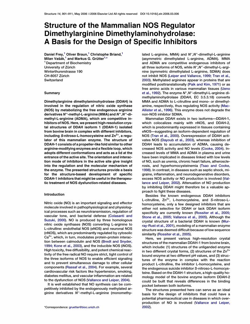
contacts, e.g., Phe75 to Leu74 in DDAH-2, as well as po-lar interaction, e.g., Glu77 to Gly76 or Arg144 to Trp143.The modeled active site of DDAH-2 superimposed on theexperimentally determined active site of DDAH-1 illus-trates these differences (Figure 8). The exchange ofAla28 for Glu27 in DDAH-2 might compensate for theAsp72-to-Leu71 mutation because both acidic sidechains are ideally positioned to form salt bridges to theCa-amino group of the substrate, whereas the distanceof the side chain of Trp143 is too far away to coordinatethe Ca-carboxyl of the substrate. Overall, three saltbridges between the enzyme and the ligand are alteredbetween the two isoforms. Although there are differ-
ences in the binding of the substrate to the enzyme, theoverall architecture of DDAH-2 is similar to the one ob-served in the DDAH-1 structures.
Discussion
Despite the low sequence identity between the mamma-lian DDAH-1 and the bacterial Pa DDAH (Murray-Rustet al., 2001), the overall fold is identical and the activesites are very similar. However, significant differencesexist that are important for a better understanding ofthe enzyme mechanism and critical for structure-baseddrug design. Our study of the mammalian enzyme
Figure 7. Alignment of DDAH Sequences
The sequence alignment of mammalian DDAH sequences and the bacterial Pa DDAH sequence (top, based on bacterial and mammalian enzyme
structures). The line above the sequences indicates identical, asterisk; positively charged, plus sign; negatively charged, minus sign; differently
charged, ‘‘0’’; tiny polar, colon; tiny nonpolar, open diamond; aliphatic, pound sign; and aromatic residues, closed diamond in mammalian DDAH
sequences. The green bars indicate residues involved in binding of L-citrulline, red residues indicate residues involved in conversion of the sub-
strate, blue indicates amino acids in the lid region, and yellow indicates residues with a large deviation in Ca position between DDAH-1 and Pa
DDAH. ClustalW (Thompson et al., 1997) was used for the mammalian sequence alignments, whereas Pa DDAH was structurally aligned to
DDAH-1 (crystal form I).
Structure908
Figure 8. Comparison ofDDAH-1andDDAH-2
Stereoview of the superimposed active sites
of the experimentally determined structure
of DDAH-1 (green) and the modeled DDAH-2
(gray). L-citrulline is shown in yellow. The
main chain, except for glycines and carbonyl
atoms involved in binding, is hidden. The
model was created with the program
Modeller 8v1 (Marti-Renom et al., 2000).
shows that the lid adopts a helical structure and an openconformation when no ligand or only Zn2+ is bound, andthat it closes the active site when L-citrulline, L-homo-cysteine, or S-nitroso-L-homocysteine are bound. Theisobutyl side chain of Leu29 in the lid blocks the en-trance to the active site completely, buries the ligands,and thereby stabilizes the enzyme ligand complex. Afterhydrolysis of MMA or ADMA, both products, L-citrullineand methylamine or dimethylamine, are released fromthe reaction center. L-citrulline leaves the enzymeupon the opening of the closing lid through the activesite, whereas, for both amines, two possibilities exist.First they could leave in a manner similar to L-citrul-line—through the active site entrance. Alternatively,they could leave through the pore opened by smallmovements of the side chains of Glu77 and Lys174.
As isolated, the enzyme contains one tightly boundzinc ion (Kd of w4 nM) that reversibly inhibits its enzy-matic activity. Knipp et al. (2001) reported that the coor-dination sphere of this Zn2+ ion contains two sulfuratoms and two lighter atoms (N or O). Coordination ofZn2+ by Sg of Cys221 and Cys273 was proposed sinceZn2+ protected both sulfurs from S-nitrosylation by gas-eous NO (Knipp et al., 2003). The presented crystalstructures of the Zn2+ ion containing DDAH-1 demon-strate that besides Cys273 no additional sulfur ligandis found in the proximity of the metal ion. All presentedstructures adopt the same overall fold, and, besidesthe loop region, no flexibility of the protein was found;therefore, it can be ruled out that Cys221, which is lo-cated 12 A away, participates in the binding of the metalion. The second sulfur ligand, observed by EXAFS, musttherefore be an exogenous ligand, introduced duringpurification of the enzyme. The other ligands of theZn2+ ion observed in the structures are water moleculesand His172. Since the second conformation of His172 isonly present in the structure at pH 9, we suggest that thisconformation rather reflects a high pKa value of this his-tidine that is similar to the pKa values of histidines in theactive sites of other cysteine proteases (Lakhdar-Ghazalet al., 2002). Water molecules positioned by the proteinas in the active site at pH 6.3 or pH 9.0 mediate tightbinding equal to that of a direct protein ligand; therefore,we assume that the metal ion is coordinated by one pro-tein ligand (Cys273) and three water molecules, whichare fixed to the protein by a hydrogen bonding network.
Reduction of NO production is desirable in diseasessuch as septic shock, pain, arthritis, or asthma (Vallanceand Leiper, 2002). In these cases, a specific inhibition ofNOS isoforms is sought. So far, the clinical use of NOSinhibitors has been limited due to the absence of spec-ificity or toxicity. Although new promising compoundsinhibiting iNOS have already been developed, a com-plete inhibition of NOS isoforms may be unwanted (Al-derton et al., 2005). Inhibition of DDAH isoforms offersan alternative approach to reduce NO levels in a tis-sue-specific manner. At present, three different classesof DDAH inhibitors, including pentalfluorophenyl sulfo-nates (Vallance et al., 2005), 2-chloroacetamidine (Stoneet al., 2005), and the class of improved DDAH inhibitors,derived from the weak reversible inhibitor S-2-amino-4(3-methylguanidino)butanoic acid (4124W) (Rossiteret al., 2005), are reported in the literature. Although,not selective or isoform specific, in combination withthe structures presented here, they can serve as a basisfor the development of selective/specific inhibitors.
Inhibitors having a distinct selectivity for DDAH should(1) form hydrogen bonds to the side chains of Asp72,Glu77, Asp78, Arg144, and Arg97 and to the main chaincarbonyls of Leu29 and Val267, (2) recognize the posi-tively charged side chain of Lys174, and (3) form hydro-gen bonds to His172 and the main chain carbonyl ofLeu270. These features should provide sufficient affinityto bind to DDAH and selectivity for DDAH compared tobinding to ADI or AT. When comparing the active sitesof the structure of DDAH-1 with the modeled activesite of DDAH-2, seven side chains forming the bindingsite are different. An inhibitor, which interacts with theindole ring of Trp143 (Arg144 in DDAH-1), should showpreferential binding to DDAH-2. A hydrogen bond toThr127 in DDAH-2 (Gly128 in DDAH-1) would further en-hance the specificity toward DDAH-2. In contrast, inhib-itors that form a hydrogen bond to the side chain ofGlu77 in DDAH-1 (Gly76 in DDAH-2) should bind prefer-entially to DDAH-1. In addition, the different polarity andsize of the side chains of Leu71 (DDAH-2) and Asp72(DDAH-1) could be exploited to increase the specificitytoward one of the isoforms. These differences could al-low for the design of selective and specific inhibitors foreach isoenzyme. Such inhibitors would be of high inter-est for DDAH research and treatment of diseases linkedto NO overproduction.

Crystal Structure of Mammalian DDAH909
Experimental Procedures
Protein Purification
Protein purification and activity measurements were performed as
described, with minor modifications (Knipp et al., 2001). All purifica-
tion steps, except the second ion exchange chromatography, were
performed at 4ºC. Tissue homogenization of bovine brains, ammo-
nium sulfate precipitation, and the subsequent hydrophobic interac-
tion chromatography were performed as previously described
(Knipp et al., 2001).
Anion Exchange Chromatography
The conductivity of the protein sample was adjusted with H2O to
%15 mU21cm21 and was then applied to a Fractogel EMD TMAE
(s) column (2.6 3 15 cm, Merck) equilibrated with 20 mM triethanol-
amine/HCl (pH 7.8). The protein was eluted with a linear salt gradient
from 0 to 200 mM KCl, and the active fractions were pooled.
Size Exclusion Chromatography
The protein solution was concentrated by ultrafiltration (Amicon) by
using a polyethersulfone membrane (Biomax, Milipore) with a molec-
ular weight cutoff of 5 kDa. The final protein sample (10 ml) was ap-
plied to a HiLoad 26/60 Superdex 75 column (Pharmacia) equili-
brated with 10 mM triethanolamine/HCl, 150 mM KCl (pH 7.4). The
active fractions were combined.
Anion Exchange Chromatography
The conductivity of the protein sample was adjusted with H2O to
%15 mU21cm21 and was then applied to a Mono Q HR 5/5 column
(Pharmacia) equilibrated with 10 mM triethanolamine/HCl, 10% (v/v)
glycerol (pH 7.4). The protein was eluted with a linear salt gradient
from 0 to 200 mM KCl, and the active fractions were pooled.
Protein homogeneity was routinely analyzed by SDS-PAGE and
mass spectrometry. Metal-to-protein ratios were determined by
measuring the Zn2+ concentration by flame atomic absorption spec-
troscopy (SpectrAA-110, Varian, Inc.), and that of the protein was
measured spectrophotometrically (3280 = 14,420 M21 cm21) (Bogu-
mil et al., 1998).
The fully Zn2+-loaded protein was prepared through protein dialy-
sis against Zn2+-containing buffer (Knipp et al., 2003).
Preparation of Zn2+-free DDAH-1
To remove Zn2+ from native DDAH-1, 200 ml protein sample (w50
mM) was dialyzed at 4ºC against 50 ml metal-free 100 mM imidaz-
ole/HCl and 10 mM DTT (pH 6.5). The outer solution was steadily
bubbled with argon. The dialysis buffer was changed four times
(every 2 hr), and dialysis was completed overnight. To remove imid-
azole and DTT, the protein was dialyzed against two changes of 50
ml metal-free 10 mM MES/NaOH, 150 mM KCl, 2 mM TCEP, 5 mM
EDTA (pH 6.5) (Knipp et al., 2001).
Preparation of L-Homocysteine and S-Nitroso-L-Homocysteine
L-homocysteine was prepared from L-homocysteine thiolacto-
ne$HCl as described by Duerre and Miller (1966) in a nitrogen atmo-
sphere. The yield (96%) was estimated through quantification of
thiols (Grasetti and Murray, 1967).
S-nitroso-L-homocysteine was always freshly prepared under an-
aerobic conditions by using the protocol of Feelisch and Stamler
(1996). Briefly, 150 ml 200 mM L-homocysteine was mixed with an
equal volume of 200 mM NaNO2. After completion of the reaction,
HEPES/EDTA was added to a final concentration of 50 mM
HEPES/5 mM EDTA, and the pH was adjusted to 7.3. The concentra-
tion of the S-nitroso-L-homocysteine formed was determined pho-
tometrically with the molar extinction coefficients reported by Feel-
isch and Stamler (1996).
Crystallization
The protein was concentrated to 2–3 mg/ml in protein storage buffer
(10 mM Tris/HCl, 150 mM KCl, 10% [v/v] glycerol [pH 7.4]). Crystal-
lization was done at 4ºC by using the sitting drop vapor diffusion
method combining 3 ml protein solution and 3 ml reservoir solution.
The final conditions were 100 mM MES/NaOH, 20%–40% PEG
8000 (pH 6.3) or 0.05 M glycine/HCl, 28% PEG 3350 (pH 9.0) for
the Zn2+-containing proteins and 100 mM citric acid/NaOH, 20%–
40% PEG 8000, 2 mM TCEP (pH 6.3) for the free form of the enzyme
and the cocrystallization experiments. For complex structures, 50
mM L-citrulline, 5 mM L-homocysteine, or 0.5 mM S-nitroso-L-ho-
mocysteine were added to the protein solution prior to crystalliza-
tion. The crystals were stabilized by adding 20%–25% ethylenegly-
col to the crystallization buffer and were flash frozen in liquid
propane.
Data Collection, Structure Determination, and Refinement
All data was collected on X06SA at the Swiss Light Source (SLS), Vil-
lingen, Switzerland, by using a MarCCD 165 mm detector. Images
were processed with XDS (Kabsch, 1993) or Denzo (Otwinoswki
and Minor, 1997). Structure determination was solved by molecular
replacement by using AMoRe (Navaza, 1994) and a poly-serine
model of the DDAH structure from P. aeruginosa (PDB code:
1H70). Model building and refinement were done with O (Jones
et al., 1991) and CNS (Brunger et al., 1998); the atomic resolution
structures (complexes with L-citrulline or S-nitroso-L-homocys-
teine) were further refined with SHELX (Sheldrick and Schneider,
1997). Cocrystallized small molecules were not included in the
search models, but clear positive density with contour levels higher
than 3s in difference Fourier maps appeared in the first round of
refinement. The position of the Zn2+ was confirmed by anomalous
difference Fourier maps contoured at 8s in Fourier maps. The re-
straints for L-citrulline, the N-thiosulfoximide derivative of cysteine,
L-homocysteine, and S-nitrosocysteine were calculated on the
ProDRG server (Schuttelkopf and van Aalten, 2004) and compared
with the small-molecule database (Allen, 2002). Data collection
and refinement statistics are shown in Table 1. Models were vali-
dated by using SFcheck and PROcheck (CCP4, 1994).
Acknowledgments
We gratefully acknowledge the Swiss Light Source, Paul Scherrer In-
stitut, Villigen, Switzerland, for providing synchrotron beamtime and
T. Tomizaki, A. Wagner, E. Pohl, and C. Schulze-Briese for their ex-
cellent support during data collection. This work was supported by
the Swiss National Science Foundation (Grant 6M3100A-1022181 to
M.G.G.; Grant 3100A0-100246/1 to M.V.) and the ‘‘Hartmann-Muller-
Stiftung’’ (to M.V.).
Received: October 12, 2005
Revised: March 10, 2006
Accepted: March 14, 2006
Published: May 16, 2006
References
Alberts, I.L., Nadassy, K., and Wodak, S.J. (1998). Analysis of
zinc binding sites in protein crystal structures. Protein Sci. 7,
1700–1716.
Alderton, W.K., Angell, A.D., Craig, C., Dawson, J., Garvey, E., Mon-
cada, S., Monkhouse, J., Rees, D., Russell, L.J., Russell, R.J., et al.
(2005). GW274150 and GW273629 are potent and highly selective in-
hibitors of inducible nitric oxide synthase in vitro and in vivo. Br. J.
Pharmacol. 145, 301–312.
Allen, F.H. (2002). The Cambridge Structural Database: a quarter of
a million crystal structures and rising. Acta Crystallogr. B 58, 380–
388.
Arita, K., Hashimoto, H., Shimizu, T., Nakashima, K., Yamada, M.,
and Sato, M. (2004). Structural basis for Ca(2+)-induced activation
of human PAD4. Nat. Struct. Mol. Biol. 11, 777–783.
Boger, R.H., Bode-Boger, S.M., Sydow, K., Heistad, D.D., and Lentz,
S.R. (2000). Plasma concentration of asymmetric dimethylarginine,
an endogenous inhibitor of nitric oxide synthase, is elevated in mon-
keys with hyperhomocyst(e)inemia or hypercholesterolemia. Arte-
rioscler. Thromb. Vasc. Biol. 20, 1557–1564.
Bogumil, R., Knipp, M., Fundel, S.M., and Vasak, M. (1998). Charac-
terization of dimethylargininase from bovine brain: evidence for
a zinc binding site. Biochemistry 37, 4791–4798.
Bredt, D.S., and Snyder, S.H. (1994). Nitric oxide: a physiological
messenger molecule. Annu. Rev. Biochem. 63, 175–195.
Brunger, A.T., Adams, P.D., Clore, G.M., DeLano, W.L., Gros, P.,
Grosse-Kunstleve, R.W., Jiang, J.S., Kuszewski, J., Nilges, M.,
Pannu, N.S., et al. (1998). Crystallography & NMR system: a new

Structure910
software suite for macromolecular structure determination. Acta
Crystallogr. D Biol. Crystallogr. 54, 905–921.
CCP4 (Collaborative Computational Project, Number 4) (1994). The
CCP4 suite: programs for protein crystallography. Acta Crystallogr.
D Biol. Crystallogr. 50, 760–763.
Colasanti, M., and Suzuki, H. (2000). The dual personality of NO.
Trends Pharmacol. Sci. 21, 249–252.
Cooke, J.P. (2004). Asymmetrical dimethylarginine: the Uber marker?
Circulation 109, 1813–1818.
Cruickshank, D.W. (1999). Remarks about protein structure preci-
sion. Acta Crystallogr. D Biol. Crystallogr. 55, 583–601.
Das, K., Butler, G.H., Kwiatkowski, V., Clark, A.D., Jr., Yadav, P., and
Arnold, E. (2004). Crystal structures of arginine deiminase with cova-
lent reaction intermediates; implications for catalytic mechanism.
Structure 12, 657–667.
Dayoub, H., Achan, V., Adimoolam, S., Jacobi, J., Stuhlinger, M.C.,
Wang, B.-y., Tsao, P.S., Kimoto, M., Vallance, P., Patterson, A.J.,
and Cooke, J.P. (2003). Dimethylarginine dimethylaminohydrolase
regulates nitric oxide synthesis: genetic and physiological evidence.
Circulation 108, 3042–3047.
DeLano, W.L. (2002). The PyMOL Molecular Graphics System (San
Carlos, CA: DeLano Scientific).
Duerre, J.A., and Miller, C.H. (1966). Preparation of L-homocysteine
from L-homocysteine thiolactone. Anal. Biochem. 17, 310–315.
Feelisch, M., and Stamler, J.S. (1996). Donors of nitrogen oxides. In
Methods in Nitric Oxide Research, M. Feelisch and J.S. Stamler, eds.
(Chichester, UK: John Wiley & Sons, Ltd.), pp. 71–115.
Grasetti, D.R., and Murray, J.F. (1967). Determination of sulfhydryl
groups with 2,20- or 4,40-dithiodipyridine. Archives Biochem. Bio-
phys. 119, 41–49.
Humm, A., Fritsche, E., Steinbacher, S., and Huber, R. (1997). Crystal
structure and mechanism of human L-arginine:glycine amidino-
transferase: a mitochondrial enzyme involved in cretine biosynthe-
sis. EMBO J. 16, 3373–3385.
Jones, T.A., Zou, J.Y., Cowan, S.W., and Kjeldgaard. (1991). Im-
proved methods for building protein models in electron density
maps and the location of errors in these models. Acta Crystallogr.
A 47, 110–119.
Kabsch, W. (1993). Automatic processing of rotation diffraction data
from crystals of initially unknown symmetry and cell constants. J.
Appl. Crystallogr. 26, 795–800.
Knipp, M., Charnock, J.M., Garner, C.D., and Vasak, M. (2001).
Structural and functional characterization of the Zn(II)-site in dimeth-
ylargininase-1 (DDAH-1) from bovine brain. Zn(II) release activates
DDAH-1. J. Biol. Chem. 276, 40449–40456.
Knipp, M., Braun, O., Gehrig, P.M., Sack, R., and Vasak, M. (2003).
Zn(II)-free dimethylargininase-1 (DDAH-1) is inhibited upon specific
Cys-S-nitrosylation. J. Biol. Chem. 278, 3410–3416.
Knipp, M., Braun, O., and Vasak, M. (2005). Searching for DDAH in-
hibitors: S-nitroso-L-homocysteine is a chemical lead. J. Am. Chem.
Soc. 127, 2372–2373.
Kone, B.C., Kuncewicz, T., Zhang, W., and Yu, Z.Y. (2003). Protein in-
teractions with nitric oxide synthases: controlling the right time, the
right place, and the right amount of nitric oxide. Am. J. Physiol. Renal
Physiol. 285, F178–F190.
Lakhdar-Ghazal, F., Blonski, C., Willson, M., Michels, P., and Perie,
J. (2002). Glycolysis and proteases as targets for the design of
new anti-trypanosome drugs. Curr. Top. Med. Chem. 2, 439–456.
Leiper, J., and Vallance, P. (1999). Biological significance of endog-
enous methylarginines that inhibit nitric oxide synthases. Cardio-
vasc. Res. 43, 542–548.
Lentz, S.R., Rodionov, R.N., and Dayal, S. (2003). Hyperhomocystei-
nemia, endothelial dysfunction, and cardiovascular risk: the poten-
tial role of ADMA. Atheroscler. Suppl. 4, 61–65.
MacAllister, R.J., Parry, H., Kimoto, M., Ogawa, T., Russell, R.J.,
Hodson, H., Whitley, G.S.J., and Vallance, P. (1996). Regulation of ni-
tric oxide synthesis by dimethylarginine dimethylaminohydrolase.
Br. J. Pharmacol. 119, 1533–1540.
Marti-Renom, M.A., Stuart, A.C., Fiser, A., Sanchez, R., Melo, F., and
Sali, A. (2000). Comparative protein structure modeling of genes and
genomes. Annu. Rev. Biophys. Biomol. Struct. 29, 291–325.
Murray-Rust, J., Leiper, J., McAllister, M., Phelan, J., Tilley, S., Santa
Maria, J., Vallance, P., and McDonald, N. (2001). Structural insights
into the hydrolysis of cellular nitric oxide synthase inhibitors by di-
methylarginine dimethylaminohydrolase. Nat. Struct. Biol. 8, 679–
683.
Navaza, J. (1994). AMoRe: an automated package for molecular re-
placement. Acta Crystallogr. A 50, 157–163.
Orengo, C.A., Michie, A.D., Jones, S., Jones, D.T., Swindells, M.B.,
and Thornton, J.M. (1997). CATH—a hierarchic classification of pro-
tein domain structures. Structure 5, 1093–1108.
Otwinoswki, Z., and Minor, W. (1997). Processing of X-ray diffraction
data collected in oscillation mode. Methods Enzymol. 276, 307–326.
Paik, W.K., and Kim, S. (1971). Protein methylation. Science 174,
114–119.
Rassaf, T., Feelisch, M., and Kelm, M. (2004). Circulating NO pool:
assessment of nitrite and nitroso species in blood and tissues.
Free Radic. Biol. Med. 36, 413–422.
Rossiter, S., Smith, C.L., Malaki, M., Nandi, M., Gill, H., Leiper, J.M.,
Vallance, P., and Selwood, D.L. (2005). Selective substrate-based in-
hibitors of mammalian dimethylarginine dimethylaminohydrolase.
J. Med. Chem. 48, 4670–4678.
Schuttelkopf, A.W., and van Aalten, D.M. (2004). PRODRG: a tool for
high-throughput crystallography of protein-ligand complexes. Acta
Crystallogr. D Biol. Crystallogr. 60, 1355–1363.
Selley, M.L. (2003). Increased concentrations of homocysteine and
asymmetric dimethylarginine and decreased concentrations of nitric
oxide in the plasma of patients with Alzheimer’s disease. Neurobiol.
Aging 24, 903–907.
Sheldrick, G.M., and Schneider, T.R. (1997). SHELXL: high resolution
refinement. Methods Enzymol. 277, 319–343.
Shirai, H., Blundell, T.L., and Mizuguchi, K. (2001). A novel superfam-
ily of enzymes that catalyze the modification of guanidino groups.
Trends Biochem. Sci. 26, 465–468.
Stone, E.M., Person, M.D., Costello, N.J., and Fast, W. (2005). Char-
acterization of a transient covalent adduct formed during dimethyla-
ginine dimethylaminohydrolase catalysis. Biochemistry 44, 7069–
7078.
Stuhlinger, M.C., Tsao, P.S., Her, J.-H., Kimoto, M., Balint, R.F., and
Cooke, J.P. (2001). Homocysteine impairs the nitric oxide synthase
pathway: role of asymmetric dimethylarginine. Circulation 104,
2569–2575.
Stuhlinger, M.C., Oka, R.K., Graf, E.E., Schmolzer, I., Upson, B.M.,
Kapoor, O., Szuba, A., Malinow, M.R., Wascher, T.C., Pachinger,
O., and Cooke, J.P. (2003). Endothelial dysfunction induced by hy-
perhomocyst(e)inemia: role of asymmetric dimethylarginine. Circu-
lation 108, 933–938.
Thompson, J.D., Gibson, T.J., Plewniak, F., Jeanmougin, F., and
Higgins, D.G. (1997). The CLUSTAL:X windows interface: flexible
strategies for multiple sequence alignment aided by quality analysis
tools. Nucleic Acids Res. 25, 4876–4882.
Tran, C.T.L., Fox, M.F., Vallance, P., and Leiper, J.M. (2000). Chro-
mosomal localization, gene structure, and expression pattern of
DDAH1: comparison with DDAH2 and implications for evolutionary
origins. Genomics 68, 101–105.
Tran, C.T.L., Leiper, J.M., and Vallance, P. (2003). The DDAH/ADMA/
NOS pathway. Atheroscler. Suppl. 4, 33–40.
Tsao, P.S., and Cooke, J.P. (1998). Endothelial alterations in hyper-
cholesterolemia: more than simply vasodilator dysfunction. J. Cardi-
ovasc. Pharmacol. 32 (Suppl. 3), S48–S53.
Ueno, S., Sano, A., Kotani, K., Kondoh, K., and Kakimoto, Y. (1992).
Distribution of free methylarginines in rat tissues and in the bovine
brain. J. Neurochem. 59, 2012–2016.
Vallance, P., and Leiper, J. (2002). Blocking NO synthesis: how,
where and why? Nat. Rev. Drug Discov. 1, 939–950.

Crystal Structure of Mammalian DDAH911
Vallance, P., and Leiper, J. (2004). Cardiovascular biology of the
asymmetric dimethylarginine:dimethylarginine dimethylaminohy-
drolase pathway. Arterioscler. Thromb. Vasc. Biol. 24, 1023–1030.
Vallance, P., Bush, H.D., Mok, B.J., Hurtado-Guerrero, R., Gill, H.,
Rossiter, S., Wilden, J.D., and Caddick, S. (2005). Inhibition of dime-
thylarginine dimethylaminohydrolase (DDAH) and arginine deimi-
nase (ADI) by pentafluorophenyl (PFP) sulfonates. Chem. Commun.
5563–5565.
Accession Numbers
Coordinates have been deposited in the Protein Data Bank with ac-
cession codes 2CI1, 2CI3, 2CI4, 2CI5, 2CI6, 2CI7, and 2C6Z.