student-directed investigations ofthe actin cytoskeleton hamster ovary v31reprint
TRANSCRIPT
This article reprinted from: Connerly, P. 2010. Student-Directed Investigations of the Actin Cytoskeleton in Chinese
Hamster Ovary Cells. Page(s) 462-468, in Tested Studies for Laboratory Teaching, Volume 31 (K.L. Clase, Editor). Proceedings of the 31st Workshop/Conference of the Association for Biology Laboratory Education (ABLE), 534 pages.
Compilation copyright © 2010 by the Association for Biology Laboratory Education (ABLE) ISBN 1-890444-13-8 All rights reserved. No part of this publication may be reproduced, stored in a retrieval system, or transmitted, in any form or by any means, electronic, mechanical, photocopying, recording, or otherwise, without the prior written permission of the copyright owner. Use solely at one’s own institution with no intent for profit is excluded from the preceding copyright restriction, unless otherwise noted on the copyright notice of the individual chapter in this volume. Proper credit to this publication must be included in your laboratory outline for each use; a sample citation is given above. Upon obtaining permission or with the “sole use at one’s own institution” exclusion, ABLE strongly encourages individuals to use the exercises in this proceedings volume in their teaching program. Although the laboratory exercises in this proceedings volume have been tested and due consideration has been given to safety, individuals performing these exercises must assume all responsibilities for risk. The Association for Biology Laboratory Education (ABLE) disclaims any liability with regards to safety in connection with the use of the exercises in this volume. The focus of ABLE is to improve the undergraduate biology laboratory experience by promoting the development and dissemination of interesting, innovative, and reliable laboratory exercises. Visit ABLE on the Web at: http://www.ableweb.org
INDIANA UNIVERSITYSOUTHEAST
Student-Directed Investigations of the Actin Cytoskeleton inChinese Hamster Ovary Cells
Pam Connerly, Indiana University SoutheastINDIANA UNIVERSITY
SOUTHEAST
AbstractMaterials and methods
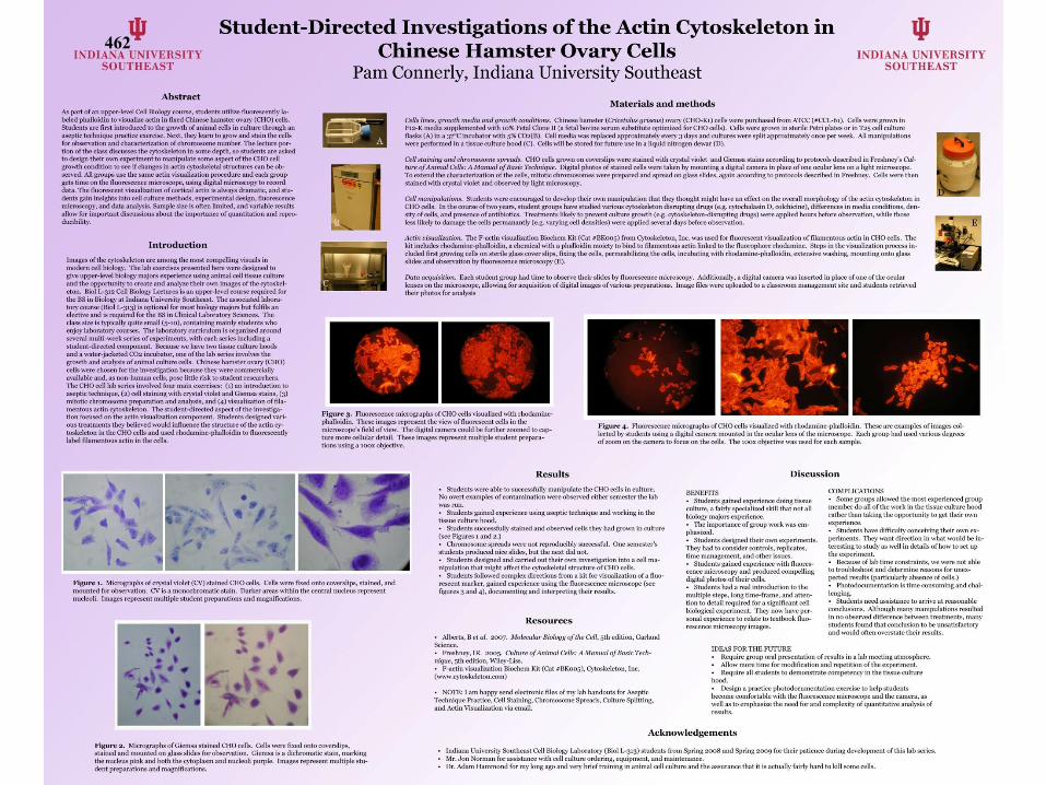
Filture4. t-1ooreseence micrographs ofCHO cells visualized with rhodamine-phalloidin. These are examples ofimag... oollectcd by students using a digital camera mounted in the ocular lensofthe microscope. Each group had uso..,) '"3riousd~'gteeS
ofO';(l()m on thecam..ra to focus onlhe cells. 111e lOOX objecth-e ""3S used forcach ~mpl..
Data ocquisih·o". Each b1udent group bad time toobse,,·e their slides by floorescenoe microscopy. Additionally. a digital camer.l ....as insert~"lI in p1aoeof One of the ocularl..nses on the microscope, alln"ing for acquisition of digital images ol ....rio'-"' preparations. Image fil"" .......... nploaded to a classroom manag..",..m site and stndents retri",'OOtbeir photos for anal)"sis
Acti" oisua/aanon. The F-aelinvisualization Biocbem Kit (Cat ,BKooS) from Cytoskeleton. Inc. was IISO.'d for fluorescent ,isualization of61amentousaetin in C.IOcells. Thekit includes rhodamine-phalloidin. a chemical "ith a phalloidin moiety to bind to filamentous actin linknl to the flooropbore rhodamine. Steps in the 'isuahuotion process included first gro...ing ""lis on sterile glass co,...r slips. fixing the eells. permeabilizing the oolls. incubating ...ith rhodamine-pballoidin. extensive ....""hing. mounting onto glassslides and obser>'3tion by floorescence micmscop)" (E).
Cell <Iaining alld chromosomespreads. CHO""lls gro....n on eo..-erslips ....ere stained ...ith cl)"5tal \"iolet and Giemsa stains according to protocols described in FreshllC)'< Cu/lut-e 0/Animal Cells: A Manual a/Bu8ic T....hnique. Digital photos ofstain~.,)cells ,,-ere taken by mounting a digital camera In place of one ocular 1"lI8on a light microscopeTo ",nend the CharaClenl'.3tion of the cells. mitotic chromosomes ....ere prel"lred and spread on glass slides. again acrording to protocols described in Freshney. Cells ....ere thenstained ..ith el)"5tal ,iolet and obser>'oo by light microscopy.
Cell manipulations. Students ....ere enoouraged to d"'·e!optbeiro....n manipulation thattbeythought might ba'-e an effect on the O'-erall morpbologyoftbeactin C)1oskelelonlnCHO cells. In the course oft....oy..ars. student groups ha'-estudioo ''3rioUSC)1oskeleton disrupting drugs (e.g. C)1ochalasin D. ookhicine). differences in mnlia conditions. den-sity ofcells. and presence ofantibioties. Treatments likel)' to pre...ent culture gro..1b ( g. C)106kelelon--disrupting drugs) ",,", applied hours before obse"lItion.....hile thoseless likely to <lamage the cells permanantl)" (e.g. ''lll)ing cell densities) were applinl s erJl days before obse"·ation.
Cells lines. growth media and growth co"ditioll5. Chincsc bamster (Cricemlus grise....) O\lIry (C.IQ-K,)cells were purebasOO from ATCC ("CCIA,,). Cells ....eregro....n inFI2-K media supplernentnl ..~tb 10910 Fetal Clone 11 (a fetal bmineserurn substitute optimized for CHO cells). Cells ......regro....n in sterile Pctri plates or inT2Scell cultureflasks (A) in a 37"C incubator "ith 5" C02{a). Cell media was replaced approximately we..y 3 da)"8 and cultures ....ere split approximat..lyonce per week. All manipulations...-ere perfo""ed in a tissue culture bood (C). Cells "ill be stored for future use in a liquid nitrogen de...." .. {D).
Figure 3. Fluorescence micrographs ofCHO cells 'isualized ..itb rhodaminephalloidin. These images represent the 'ie.... offlooresooent cells in themicroscope's field of ,-lew. The digital camera could be further zoomed to capture more cellular detail. These images represent multiple student prel"lrations using a IOOX objeel:i,-e.
Images of the C)106keleton are anlOng the most compelHng 'isuals inmodern cell biology. The lab exereises prescntnl here were designed togi"e upper-le--el biology majors experi"r>ee using animal cell tissue coltu",and the opportunity to create and analyze their own images of the C)106keleton. BioI 1.-312 Cell Biology Lectures is anupper-le-'el course required forthe BS in Biology at Indiana University Southeast. The assoeiatnllaboratory course (Bioi 1.-3'3) is optional for most biology majors but fulfils anelecti'-eand is required for the BS in Oini.... l Laboratory Sciences. TIleclass sire is typically quile small (S-IO), containing mainly stud..nts \\'110enjoy laboratory lXIUr8eli. 1be laboratorycurrieulum isorganiu.,) aroundseveralmulti-\\,,,,,kseries of experim..nts, ...ith each series including astudent--direct~'<I component. Ik-cause ...... han' two tissue culture hoodsand a ....ater-j;!cketnl CO2 incubator, one of the lab series in,'OI,·es thegro...1h and analysis of animal culture oolls. Chincsc hamsterovary(CHO)cells were chosen for the investigation because th..y were commerciallyavailable and. as non-human ""lls. pose little risk to student researehers.The CHOcelilabseries inw.h-ed four main e..ercises: (I) an Introduction toaseptic technique, (2) ""n staining ....ith el)"5tal ,·iol..t and Giemsa stains, (3)mitotic chromosome pn..-parntion and anal)"5is. and (4) ,~sualizatlonof fila"",ntous actin C)1oskeletoll. Th.. studcnt--directnl asllCCl of the ill\"CStigation focused on the actin 'isualization component. Students designnl various treatmcnts tbey heli",'oo would influenoe tbe structure of the actin cy-toskel..ton in theCHO oolls and IISO.'d rhodan,ine-phalloidin tn lluorescentlylahel filamentous actin in tbecells.
Introduction
As part of an upper-l",'el Cell Bio;>logy rourse, students utilize n""""""ntly la-beled phalloidin 10 'isualiu act;n in fixed Chinese hamster "'''lfy (CHO) cen•.Slu~nts are first introduced 10 the gro"1h of animal cells in culture through anaseptic I~hniquepractice cxcreise. Next. tlK-y learn logrow and stain the cellsforobservalion and charactcri:tation ofchl'llmosomc number. The 1eclure POl'·lion of the class discusses the C)1oskelcton in sorn<! depth, SO students are askedto design their own experiment tomanipulalc SOme aspect ofthcCHOcellgro",h eondilion \0 see if changes in actin C)1oskeletal stn..:Wrt'Scan beob!lCI"Td. All grouJlS usc the same actin \isualization procedure andcach groupgets time On the nuoresttnee micl'06COpe, using digital micto6COpy to reoorddata. The nUol'1':S<.'Cnt ,'isualization of cortical actin is always dramatic, and students sain insights into cell culture methods. e..po.'Timental design. fluorescencemicmscopy. and data analysis. Samplesize is often limited. and variable resultsallow for important discllSSions about the importance of quantitation and ",producibility.
Acknowledgements
Discussion
COMI'UCATIONS• Some groups allowed the most ~",pericncedgroupm..mberdnall of the ....ork in the tissue culture hoodratber tban taking tbe opportunitr to get their ownexperi..nce.• Students han diff....ultrcollCehing toorO\\ll experiments. They ....ant direction in what would be in_teresting to study as ...-ell in details ofho.... to set upthe experiment.• Ilceau:;eoflabtime constraints. ,,-e ...-ere not ableto troubleshoot and detennine reasons for unexpednl results (J"'lrticularly absence of cells.)• Photodocumentation is time consuming and challenging.• Students need assistanee to arri..e at ....asonableoonclusions. Although many manipulations resnlt~.,)
in no obse"-ed diffe....nce bet"......n t.....Jtments. manystudents found that conclusion to be unsatisfactol)·and would often m-en;tate their results.
IDEAS FORTHEFlITURE• !lequire group oral presentation of results in a lab mcetingattl1OSph..re• Aliow more time for modification and repetition of tbe experiment.• Relluire all students to demonstrate competenC)· in the tissue cuhurehood.• Design a praetice pootodocumentation exercise to hclp studentsbecome oomfortable "ith the fluorescen"" microscope and the camera. as.....11 as to emphasize the n~-ed for and complexity of quantitati'-e anal)"ls ofresults.
'''''eTI• Students gainnl ..xperiencedoing tissueculture. a fairly specialized skilltbatllOt allbiology majors experience.• The importance ofgroup ....ork was ..m-phaslzed.• Students designnltheirown experiments.llli.')' had to consider controls. replicates.time managem..nt. and oth..r issues• Students gainnl experience ...ith lluoreseence micmscopy and produced compellingdigital photos of their cells• Students had a real introduction to themultiple steps. long time-fr'Jme. and attention to detail required for a significant cellbiological experiment. Th~')" nOW ha,-e personal experience to ....late to t..xtbook fluorescence mieroocopy images.
Results
Resources
Students ,,""' able to successfully manipulate th.. CHO cells in C1Jltnre.No m-ert examples of contamination ......re obsen'oo ..ither semester the lab"<lSmn.• Students gain~.,) experi..nce using aseptic technique and working in thetissue culture hood.• Students successfully stained and observoo cells they had grow" in culture(see Figures, and 2.)• C'hromolSOme spreads w..re not reproducibly successful. Onesemestcr'sstud..nts produced ni.... slid.... but the n..xt did not.• Students design~.,)and earri~")out theiro"ll in,...-stigation intoa cell manipulation that might affect the C)t06keletal structure of CHO oolls.• Students follo"-ed oompk'll directions from a kit for ,~sualizationof a lIuotese('nl marker. gainnl experience using the lIuorescence microscope (seefigures 3 and 4), documenting and interpreting their results.
Alberts. B et al. 200'7. Molecular Bioi09Y oJthe Cell. 5th edition. GarlandScience.• Freshney.1R. 2OOS. Culture 0/Animal Cells: A Mmlual o/Basic Technique, 5th nlition. Wiley-Uss.• F-aetin ,isualization Bioch..m Kit (Cat #BKooS), C}1oskeleton. Inc.(www·CJ10iSkelcton.oom)
• NOTE: I am hapPl' send electronic files of my lab handouts for As<'pticTechnique Practice, Cell Staining, Chromoson'e Sp....ads. Culture Splitting.and Actin Visualization 'ia ernail.
Figure I, Micrograpbsof el)"5tal ,iolet (ev) stained CliO oolls. Cells ....ere fu:ed ontoOO\-erslips. stained. andmount~.,) forobse,,·ation. CV is a monochromatic stain. Darker areas "ithin the c.,ntral nucleus representnucleoli. Images represent mnltiple student preparations and magnifications.
Figure 2. Micrographs ofGiemsa stained CHO eells. Cells ...·ere fixnl ontoOO\·erslips.stained and mounted On glass slides forobse,.,..ation. Giemsa is a dichromatic stain. markingthe nucleus pink and both th.. eytnplasm and nncleoli purpl... Images ....present multiple stu·dent preparations and magnifications.
Indiana Unh-ersitr Soutbeast Celi Biology Laboratory (BioI 1.-313) students from Spring 2008 and Spring 2009 for their patiencedllringd~'\-elopmentof this lab S<.'Ties.Mr. Jon Norman for assista""" ...ith cell cultu,," ordering, equipment, and maintenar>ee.Dr. Adam Hammond for my long ago and '-ery brief training in animal cell culture and tbe assurance that it is actually fairly hard to kill SOme cells.
462

Student-Directed Investigations of the Actin Cytoskeleton in Chinese Hamster
Ovary Cells
Pamela L. Connerly
Biology Department
Indiana University Southeast
4201 Grant Line Rd.
New Albany, IN 47150
Abstract
As part of an upper-level Cell Biology course, students utilized fluorescently labeled phalloidin to visualize actin in fixed Chinese hamster ovary (CHO) cells. Students learned aseptic technique, then grew and stained CHO cells for observation and characterization of chromosome number. Students next designed their own experiment to manipulate CHO cell growth condition to induce changes in actin cytoskeletal structure. All groups used the same actin visualization procedure, using fluorescence microscopy and digital photography to record data. The fluorescent visualization of cortical actin is always dramatic, and students gained insights into cell culture methods, experimental design, fluorescence microscopy, and data analysis.
Introduction The advent of fluorescent markers that bind specifically to components of the cytoskeleton has
revolutionized cell biology and produced some of the most compelling visuals in modern biology. The actin cytoskeleton in particular is often easily visualized as various bundles and networks of fibers in the cell cortex, underlying the plasma membrane (Alberts et al., 2007.) The lab exercises presented here were designed to give upper-level biology majors experience using animal cell tissue culture and the opportunity to create and analyze their own images of the cytoskeleton. Biol L-312 Cell Biology Lectures is an upper-level course required for the BS in Biology at Indiana University Southeast. The associated laboratory courses (Biol L-313) is optional for most biology majors but fulfils an elective and is required for the BS in Clinical Laboratory Sciences. The class size is typically quite small (5-10), containing mainly students who enjoy laboratory courses. The laboratory curriculum is organized around several multi-week series of experiments, with each series including a student-directed component. Because we have two tissue culture hoods and a water-jacketed CO2 incubator, one of the lab series involves the growth and analysis of animal culture cells. Chinese hamster ovary (CHO) cells were chosen for the investigation because they were commercially available and, as non-human cells, pose little risk to student researchers. The CHO cell lab series involved four main exercises: (1) an introduction to aseptic technique, (2) cell staining with crystal violet and Giemsa stains, (3) mitotic chromosome preparation and analysis, and (4) visualization of filamentous actin cytoskeletal elements. The student-directed aspect of the investigation focused on the actin visualization component. Students designed various treatments they believed would influence the structure of the actin cytoskeleton in the CHO cells and used rhodamine-phalloidin to fluorescently label filamentous actin in the cells.
463
Instructor's Notes Materials and Methods
Cell lines, Growth Media and Growth Conditions Chinese hamster ovary (Cricetulus griseus) ovary cells (CHO-K1) were purchased from ATCC
(#CCL-61). Cells were grown in F12-K media supplemented with 10% Fetal Clone II (a fetal bovine serum substitute optimized for CHO cells.) Cells were grown in sterile Petri plates or in T25 cell culture flasks in a 37°C incubator with 5% CO2. Cell media was replaced approximately every three days and cultures were split approximately once per week. All manipulations were performed in a tissue culture hood. Cells were stored for future use in a liquid nitrogen dewar. Cell Staining and Chromosome Spreads
CHO cells grown on cover slips were stained with crystal violet and Giemsa stains according to protocols described in Freshney (2005). Digital photos of stained cells were taken by mounting a digital camera in place of one ocular lens on a light microscope. To extend the characterization of the cells, mitotic chromosomes were prepared and spread on glass slides, again according to protocols described in Freshney (2005). Cells were then stained with crystal violet and observed by light microscopy. Cell Manipulations
Students were encouraged to develop their own manipulation that they thought might have an effect on the overall morphology of the actin cytoskeleton in CHO cells. In the course of two semesters, student groups have studied various cytoskeleton disrupting drugs (e.g. cytochalasin D, colchicine), differences in media conditions, density of cells, and presence of antibiotics. Treatments likely to prevent culture growth (e.g. cytoskeleton-disrupting drugs) were applied hours before observation, while those less likely to damage the cells permanently (e.g. varying cell densities) were applied several days before observation. Actin Visualization
The F-actin visualization Biochem Kit (Cat #BK005) from Cytoskeleton, Inc. (www.cytoskeleton.com) was used for fluorescent visualization of filamentous actin in CHO cells. The kit includes rhodamine-phalloidin, a chemical with a phalloidin moiety, which binds specifically to filamentous actin, linked to the fluorophore rhodamine. Steps in the visualization process included first growing cells on sterile glass cover slips, fixing the cells, permeabilizing the cells, incubating with rhodamine-phalloidin, extensive washing, mounting onto glass slides, and observation by fluorescence microscopy. Data Acquisition
Each student group had time to observe their slides by fluorescence microscopy. Additionally, a digital camera was inserted in place of one of the ocular lenses on the microscope, allowing for acquisition of digital images of various preparations. Image files were uploaded to a classroom management site and students retrieved their photos for analysis.
Results
464

Each group of students was able to successfully manipulate the CHO cells in culture. No overt examples of contamination were observed either semester the lab was run. Students gained experience using aseptic technique and working in the tissue culture hood. Student successfully stained and observed cells they had grown in culture with both crystal violet (Figure 1) and Giemsa (Figure 2).
Figure 1. Micrographs of crystal violet (CV) stained CHO cells. Cells were fixed onto cover slips, stained, and mounted for observation. CV is a monochromatic stain. Darker areas within the central nucleus represent nucleoli. Images represent multiple student preparations and magnifications.
Figure 2. Micrographs of Giemsa stained CHO cells. Cells were fixed onto cover slips, stained, and mounted on glass slides for observation. Giemsa is a dichromatic stain, marking the nucleus pink and both the cytoplasm and nucleoli purple. Images represent multiple student preparations and magnifications.
465

Mitotic chromosome spreads were not reproducibly successful. One semester's students produced nice slides, but the next did not. Students designed and carried out their own investigation into a cell manipulation that might affect the cytoskeletal structure of CHO cells. Students chose to use variables including addition of cytoskeleton-disrupting drugs, changes in cell culture density, and presence of antibiotics in the growth medium. Students followed complex directions from a kit for visualization of fluorescent rhodamine-phalloidin, a fluorescence marker specific for filamentous actin. Students also gained experience using the fluorescence microscope to document their results (Figures 3 and 4).
Figure 3. Fluorescence micrographs of CHO cells visualized with rhodamine-phalloidin. These are examples of images collected by students using a digital camera mounted in the ocular lens of the microscope. These images represent the view of fluorescence cells in the microscope's field of view. The digital camera could be further zoomed to capture more cellular detail. These images represent multiple student preparations.
Figure 4. Fluorescence micrographs of CHO cells visualized with rhodamine-phalloidin. These are examples of images collected by students using a digital camera mounted in the ocular lens of the microscope. Each group used various degrees of zoom on the camera to focus on the cells.
466

Discussion Benefits of this series of experiments analyzing the actin cytoskeleton of CHO cells were
extensive. Students gained experience doing tissue culture, a fairly specialized skill that not all biology majors experience. The importance of group work was emphasized. Students designed their own experiments in which they had to consider controls, replicates, time management, and other issues. Students gained experience with fluorescence microscopy and produced compelling digital photos of their cells. Students had a real introduction to the multiple steps, long time-frame, and attention to detail required for a significant cell biological experiment. They now have personal experience to relate to textbook fluorescence microscopy images.
In addition to the many benefits described above, this activity does have several complications. Some groups allowed the most experienced group member to do all of the work in the tissue culture hood rather than encouraging all individuals to take the opportunity to get their own experience. Students had difficulty conceiving their own experiments, requesting direction in what manipulation to study as well as details of how to set up the experiment. Because of lab time constraints, we were not able to troubleshoot and determine reasons for some unexpected results (such as absence of cells in some treatments). Photo documentation was time consuming and challenging without dedicated equipment. Students required significant assistance to arrive at reasonable conclusions. Although many manipulations resulted in no observed difference between treatments, many students found that conclusion to be unsatisfactory and would often overstate their results. All of these complications can be addressed by the instructor. In fact, conversations regarding misconceptions about the results offer a powerful teaching tool, but sufficient time must be allowed for discussion and re-evaluation of results. As I continue to refine this series of lab activities, I plan to make several changes to address some of the mentioned complications while maintaining the positive aspects. To address student hesitancy to work in the tissue culture hood, I plan to require all students to demonstrate their competency at manipulating cells in the tissue culture hood before proceeding with their projects. To help all groups achieve interpretable photos of their cells, I am designing a practice photodocumentation exercise to help students become comfortable with both the fluorescence microscope and the digital camera, as well as to emphasize the need for and complexity of quantitative analysis of results. To facilitate communication about results and reasonable interpretations, I will require group oral presentation of results in a lab meeting atmosphere. I also plan to allow more time for modification and repetition of the experiment.
Acknowledgements I would like to than the Indiana University Southeast Cell Biology Laboratory (Biol L-313)
students from Spring 2008 and Spring 2009 for their patience during development of this lab series, Mr. Jon Normal for assistance with cell culture ordering, equipment, and maintenance, and Dr. Adam Hammond for my long ago and very brief training in animal cell culture and the assurance that it is actually fairly hard to kill some cells.
467
Literature Cited
Alberts, B., A. Johnson, J. Lewis, M. Raff, K. Roberts, and P. Walter. 2008. The Cytoskeleton. (Chapter 16). Pages 965-1052, in Molecular Biology of the Cell, Fifth edition. Garland Science, Taylor & Francis Group, New York, NY, 1268 pages.
Freshney, RI. 2005. Culture of Animal Cells: A Manual of Basic Technique, Fifth edition. Wiley-Liss,
New York, NY, 672 pages.
About the Author Pamela L. Connerly is an Assistant Professor of Biology at Indiana University Southeast, where she teaches Cell Biology and Introduction to Biological Sciences I. She actively involves undergraduates in two ongoing research projects investigating the relationship between the Golgi apparatus and the process of endocytosis in the methylotrophic budding yeast Pichia pastoris and the discovery and comparative genomics of novel bacteriophages infecting the gram negative bacterium Acinetobacter baylyi.
468