studies about phthalocyanine photosensitizers to be used in
TRANSCRIPT

Romanian Reports in Physics, Vol. 65, No. 3, P. 1032–1051, 2013
Dedicated to Professor Valentin I. Vlad’s 70th Anniversary
STUDIES ABOUT PHTHALOCYANINE PHOTOSENSITIZERS TO BE USED IN PHOTODYNAMIC THERAPY
A. STAICU1, A. PASCU1, A. NUTA2, A. SORESCU 2, V. RADITOIU2, M.L. PASCU1,3 1 National Institute for Lasers, Plasma and Radiation Physics, Laser Department, Atomistilor 409,
P.O. Box MG-36,077125 Magurele, Bucharest, ROMANIA, E-mail: [email protected] 2 INCDCP-ICECHIM, Spl. Independentei nr. 202, Bucuresti, O.P. 35, CP 174 c p 060021, Romania.
3 University of Bucharest, Physics Faculty, Romania
Received June 17, 2013
Abstract. A short description of the photodynamic therapy as an alternative cancer treatment procedure is made, highlighting the role of the photosensitiser and of the basic molecular processes resulting in active species. The synthesis path of metallated phthalocyanine photosensitizers used in this paper are presented and analyzed, considering the conventional and the microwave assisted procedures. Measurements are reported of some specific photophysical quantities characterizing the active species generation and properties (lifetime and quantum yield), when they are photogenerated by the synthesized compounds. The dedicated experimental set-ups are described. Comparative measured data of the mentioned photophysical quantities of the synthesized phthalocyanines are presented.
Key words: phthalocyanines, sinthesys, photophysics, singlet oxygen quantum yield, single oxygen lifetime.
1. INTRODUCTION
The photodynamic therapy (PDT) is an alternate or complementary method to the conventional anti-cancer treatments which is used more in recent years. The application methodology of PDT implies the administration of a photosensitizer which is a chemical compound with special photophysical properties, accumulated into the tumor. Through local irradiation with a suitable tailored light source an activation of the photosensitizer occurs; after a series of elementary intra and inter-molecular processes, molecules with high chemical reactivity are generated – the active species – that can destroy the tumor cells. Most researches in the field consider the singlet oxygen species as the major cytotoxic species involved in the therapy [1–3].

2 Studies about phthalocyanine photosensitizers
1033
The PDT involves three constitutive components: (i) the photosensitizer, (ii) the light radiation/source and (iii) the molecular oxygen. The way in which the active species are produced through photochemical reactions determines their classification as compounds of [4]:
– type I reaction, when the excited molecule of the photosensitizer reacts with the substrate to generate active radicals which interact at their turn with subcellular components.
– type II reaction, in which case the activated photosensitizer transfers energy to the oxygen molecule on an excited singlet state (singlet oxygen) that has a high chemical reactivity. Whichever of the two types reactions would occur, finally reactive oxygen species (ROS) are involved. De-oxygenation of the probe stops the photoactivity. One estimates that a sensitizer molecule can generate 103–105 singlet oxygen molecules in subsequent cycles before becoming inactive (through bleaching, oxydation or other phenomena of inactivation) [5.a]. Apart from the intersystem crossing from triplet to singlet states of the photosensitizer, the molecular processes involved in the photophysics and photochemistry of the active species generation take place in extremely short time intervals, which keep the excited photosensitizer molecules from interacting with any of the molecules present in the neighborhood. Consequently, the most likely photochemical reactions which involve photosensitizers can take place only while dye molecules are on their triplet state T1; the longer the lifetime of the T1 state, the greater the probability to encounter oxygen molecules and to produce energy transfer to them. The singlet oxygen generation can occur only if the triplet state energy of the photosensitizer exceeds, with at least 6-8 kJ/mol, the energy required to bring molecular oxygen to the singlet state. Taking into account that the forbidden back-emission singlet-triplet of oxygen (which yields phosphorescence radiation) occurs at λ = 1270 nm, one infer that virtually all molecules which absorb radiation at λ < 1260 nm and have triplet states can generate singlet oxygen. The value of the quantum yields of the singlet oxygen is the factor which defines a photosensitizer. From the information given above it results that the key element in the PDT is the series of elementary processes initiated in the photosensitizer molecules after absorbing activation photons. Data on photophysical properties specific for PDT of the photosensitizer are critical when a PDT application is targeted. Although the study of phtalocyanines Pc in different scientific fields is intense [5.b] this kind of data can be found with reasonable frequency in the literature for some of the basic phthalocyanines, e.g. for ZnPc, Chloroaluminum Pc or Hydroxyaluminium Pc, but rather scarce for other compounds as FePc, CoPc or NiPc [6, 7]. Moreover, the data reported in different works were obtained by different methods in different conditions so that they might be hardly compared. Therefore, the aim of this work was subsumed to a larger goal: to consistently synthesize and measure photophysical quantities of some basic metallated phthalocyanines.

A. Staicu et al. 3
1034
2. METTALATED PHTHALOCYANINES. SYNTHESIS AND PHOTOPHYSICS
2.1. THE PHOTOSENSITIZER: METALLATED PHTHALOCYANINES. SYNTHESIS
The main features that a photosensitizer must meet to have high therapeutic efficacy could be listed as: (1) low toxicity in the dark; (2) high phototoxicity when light activated; (3) high selectivity and specificity for target tumor; (4) rapid elimination from the body; (5) absorption in optical transmission window of biological tissues; (6) high quantum yield of singlet oxygen production; (7) solubility in water-based solutions; (8) stability under physiological conditions. Nowadays, some of the above requirements (low toxicity, specificity, water-solubility) could be fulfilled by associating the photosensitiser in different ways with specialized carriers (nanoparticles, liposomes, quantum dots, PEGylation) by encapsulation etc. This also refocused the interest for simple but effective photosensitizers having the drawback of the lack of some of the mentioned features (water-solubility e.g.) [8–10]. In recent years, the interest in the chemistry of macrocyclic compounds closely related to phthalocyanines paid considerable attention due to their properties and use in different fields [11]. Second and third-generation of photosensitizers are now under investigation, different derivatives of the basic phtalocyanines being synthetized and studied. The non-metallated phthalocyanine is represented by structural formula 1 while the structural formula 2 in Scheme 1 shows a metallated phthalocyanine, where M could be Cu, Ni, Fe, Al, Zn, Au, Ag, Co, Mn, Mg, etc. [12].
Scheme 1 – Structural formulae. 1: non-metallated. 2: metallated phthalocyanines.

4 Studies about phthalocyanine photosensitizers
1035
Formally, the phthalocyanine may be considered as a tetrabenzotetraazaporphyne, a product of condensation of four isoindol units. In terms of structure, phthalocyanines are similar to natural porphyrins as hemoglobin, chlorophyll and vitamin B12. Generally, phthalocyanine synthesis is performed in a single step reaction, starting from o-phthalic acid derivatives such as phthalic anhydride, phthalimide, or phthalonitrile, according to Scheme 2 [13–15]:
NH
O
OFtalimida
CN
O
NH2
o-Cianobenzamida
O
O
OAnhidr ida fta l ica
N
N
NM
N
N
N
N
N
PcM
N
N
N
N
N
N
N
N
LiLi
N
N
N
N
N
N
N
NH
H
P cH2 PcLi2
CN
CNFtaloni tril
NH
NH
NH
Isoindo lindiimina
Phthalimide
Phthalonitril
o-cyanobenzamide Phthalic anhydride
1,3-diiminoisoindoline Scheme 2 – General methods for the synthesis of phthalocyanines and their metallic derivatives.
The most important phthalocyanines derive from phthalonitrile, phthalic anhydride, phthalocyanine derivatives or phthalocyanine salts with alkali metals, as illustrated above. Recent data present the synthesis of metallated phthalocyanines without any solvent, through direct reaction of the phthalonitrile with a hydrated metal salt under microwave irradiation (υ = 2450 MHz) or the reaction of the unsubstituted phthalic anhydride with urea, as shown in Scheme 3 [16].

A. Staicu et al. 5
1036
CN
CN
4
MX2Fara solvent
microunde N
NN
NN
N
N
NM
M = Mg, Zn, Cd, Cu, Ni, Pd, Pt, Co, Fe, Ru,Rh,Ti , Cr, Mn, V, Mo, UO2, Eu
Randament: 7..92%
Pc(M)
without solvent
microwave
Yield =
X
X
X
X
O
O
Fara solvent
microundeN
NN
NN
N
X X
XX
X
X
X
X
X
XX
X
X
X
X
XN
NMO + NH2 C
O
NH2
MY2
X = H, ClM = FeCl, Co, Cu Randament = 86-93% X16Pc(M)
without solvent
microwave
Yield Scheme 3 – Synthesis of metallophthalocyanines under microwaves.
Table 1 includes the main reaction conditions used in laboratory for the synthesis of copper, nickel and chloroaluminium phthalocyanines through conventional procedure, using the phthalic anhydride as raw material. For copper, nickel, cobalt, zinc, iron and aluminum phthalocyanines synthesis under microwave, using phthalonitrile as raw material, Table 2 shows the main laboratory features in obtaining these metallated phthalocyanies.

6 Studies about phthalocyanine photosensitizers
1037
Table 1
Reaction conditions for synthesis of phthalocyanines through conventional method
No Compound Raw materials (g/M) Reaction time (h)
Yield (%)
1 Copper
phthalocyanine
-phthalic anhydride (10.5/0.071)-urea (17.9/0.3) -ammonium molybdate (0.25/0.0002)-CuCl2
(3/0.022) -trichlorobenzene (35 ml)
8
82
2 Nickel
phthalocyanine
- phthalic anhydride (10.5/0.071)-urea (17.9/0.3) - ammonium molybdate (0.25/0.0002)
-NiCl2 (2.96/0.012) -Na3PO4(5.4/0.033)
- trichlorobenzene (50 ml)
5
87
3 Chloro- aluminium
phthalocyanine
- phthalic anhydride (10.5/0.071)
11
85
Table 2
Reaction conditions for the synthesis of phthalocyanines under microwaves
Phthalocyanine Molar ratio Yield ( % )
Irradiation time (min)
Microwave power (W)
CoPc PN : CoCl2·6H2O = 4 : 1 86 2 500 NiPc PN : NiCl2·6H2O = 4 : 1 90 3.5 500 CuPc PN : CuSO4·5H2O = 4 : 1 91 3 500 ZnPc PN : ZnCl2·2H2O = 4 : 1 87 4 500 FePc PN : FeSO4·7H2O = 4 : 1 87 4 500 AlPc PN : AlCl3·6H2O = 4 : 1 88 4 500
where: CoPc: Cobalt phthalocyanine; NiPc: Nickel phthalocyanine; CuPc: Copper phthalocyanine; ZnPc: Zinc phthalocyanine; FePc: Iron phthalocyanine; AlPc: Aluminum phthalocyanine; PN: phthalonitrile. AlPc hydroxide was synthesized under mild hydrolysis of chloroaluminium phthalocyanine.
The obtaining of the Cu-tris(hydroxymethyl)-phthalocyanine and the Cu-tris(hydroxyethylaminomethyl) -phthalocyanine firstly supposes the synthesis of the Copper-tris-chloromethyl-phthalocyanine, followed by hydrolysis of this derivative, or condensation of the chloromethylated compound with monoethanolamine to obtain the final product.
2.2. THE PHOTOSENSITIZER PROPERTIES
2.2.1. Steady Absorption: IR and UV-VIS
The synthesized compounds were firstly analyzed by IR spectroscopy (using a FT-IR spectrometer JASCO 6300 and KBr disks) and by UV-VIS spectroscopy (Perkin Elmer Lambda 950 spectrometer) in solutions of organic solvents (DMSO

A. Staicu et al. 7
1038
and DMF) with concentrations around c = 10-5 M. As typical, Figs. 1 and 2 show the IR spectra of Copper-tris(hydroxymethyl)-Pc (Pc stands for phthalocyanine) and of Cobalt Phthalocyanine (CoPc), while Fig. 3 shows the UV-VIS electronic spectra of the CoPc for a sequence of concentrations in the range 1×10⁻⁶ M –5×10⁻⁵ M. Here one can notice the well structured Soret (~350 nm) and Q bands at around 670 nm.
Fig. 1 – IR spectrum of Cobalt phthalocyanine (KBr disk).
Fig. 2 – IR spectrum of of Copper-tris(hydroxymethyl)-phthalocyanine (KBr disk).

8 Studies about phthalocyanine photosensitizers
1039
The IR spectra in Figs. 1 and 2 exemplify the differences determined by using various central metals in the phthalocyaninic ring (structural formula 2 above). All the IR spectra reveal identifiable specific absorption bands of the compounds structure: C-N stretch and C-C stretching (e.g. for CoPc, AlPcCl and Cu-tris(hydroxymethyl)-trihidroximetil-Pc), C-C main ring deformation, vibrations outside the ring-plane etc. [17, 18].
Fig. 3 – UV-VIS spectra of Cobalt Phthalocyanine at increasing concentration
in the range 1×10⁻⁶ M – 5×10⁻⁵ M.
All the studied phthalocyanines showed similar electronic absorption spectra in the UV and visible domains, having well defined Q and Soret bands and un-split Q bands [19–21].
2.2.2. Specific properties
Referring to the photoreaction which produces active species in PDT, it is estimated that the photoreaction is running along the type I pathway for less than 1% of cases, while the type II reactions generate majority of active species – the molecular singlet oxygen. It is difficult to determine, particularly for the cases engaging type I reaction, the precise pathway involved or the contribution of each of the two types of mechanisms when both of them could be present. One of the major goals of the researches in PDT refers to the assessment of two main parameters featuring the phenomenon of photosensitization: (i) the quantum yield of the singlet oxygen generation and (ii) the singlet oxygen population lifetime.

A. Staicu et al. 9
1040
The main quantity involved in photogeneration of active species is the quantum yield of the process Φ∆, which defines the ability of a chemical compound to photogenerate reactive singlet oxygen species under optical radiation. This overall feature reflects the outcome of the successive series of the elementary intra and inter-molecular processes, such as the light absorption by the photosensitizer molecule, and the formation of the triplet state until the energy transfer to the oxygen molecule. Oxygen (solubility in water ~ 273 µM [22]) is one of very few substances whose molecule lies in a triplet state in its ground electronic state. In this state the oxygen molecule is not very reactive. When activated through transfer of energy (usually from another excited molecule lying in a triplet state) the oxygen molecule pass in a highly reactive singlet state. Because the singlet – triplet transition is a forbidden one, relatively long lifetime of the singlet excited state of the oxygen molecule develops (3–4 µs in aqueous media). This fact increases the possibility of the energetic singlet oxygen to interact with other molecules in the surrounding medium to produce damages to the organelles of the target cancerous cells. If the triplet electronic configuration of molecular oxygen in the ground state is a threshold to oxygen interaction with most biological molecules which are usually in a singlet state, then this barrier disappears when the oxygen molecule reaches the singlet excited state. Thus it is obvious the significance of the lifetime of the population of photosensitized active species that is the duration over which singlet oxygen could still destroy tumor cells. This feature determines e.g. the diffusion distance of the reactive species, and indicates in this way the optimum location of the intracellular photosensitizer accumulation ensuring effective destruction of the critical constituents of the cancer cells. Singlet oxygen detection methods are diverse, being based on different physical or chemical aspects of the production of the oxygen molecule in its singlet state, such as luminescence, electronic parametric resonance, calorimetry, chemical reactions etc. The oxygen molecule can return from the excited singlet state to the ground state non-radiatively or in a radiative manner giving off phosphorescence in the near IR. Based on this fact, one of the most sensitive and specific methods in measuring the singlet oxygen is the direct measurement of its phosphorescence at λ=1270 nm. The major advantage of this direct method is the capability to time-resolve the phenomena, revealing the kinetics of the generating process itself, and the possibility to be extended even for in-vivo measurements. This is why we have chosen this method for our objective, to study the kinetics of singlet oxygen photogenerated by different metallated phtalocyanines. The experimental device designed and set-up for this purpose is based on the basic right-angle geometry of the detection of the phosphorescence of singlet oxygen decay.
The experimental set-up contains a singlet oxygen generating section comprising, as shown in Fig. 4:

10 Studies about phthalocyanine photosensitizers
1041
– a pulsed YAG:Nd laser equipped with tripling (355 nm) optical nonlinear crystals as an optical excitation source (1 and 2 in Fig.4);
– an optical sub-assembly for tailoring the laser beam, including dichroic mirrors (4), a reverse Galilean telescope (6) with a divergent lens (avoiding air-breakdown by laser beam focalizing) and a set of optical filters (7) for the selection of the laser pulse energy according to the experimental requirements and beam splitters (3);
– a 10×10 mm cuvette (11) (fluorescence spectroscopy type, four-sides clear) for the dye solution sample.
The experimental set-up also contains a time-resolved phosphorescence measurement section which includes (Fig 4):
–a system for collecting and filtering the singlet oxygen phosphorescence (13) (narrow-band filters centered at λ = 1270 nm, long-pass and short-pass filters, a quartz cube-corner reflector (12) to increase the effectiveness of phosphorescence collection) housed in a light-sealed optical chamber (9) which is optically coupled to a photodetector;
– a photodetector (Hamamatsu H10330 Module) sensitive at 1270 nm; – a laser power-meter (Gentec, equipped with QE25LP measuring head) (8); – a digital oscilloscope (Tektronix type DPO-7254) (14); – a fast photodiode, triggering the measuring cycle (5).
Fig. 4 – Experimental setup for photosensitized generation and measuring of singlet oxygen species.

A. Staicu et al. 11
1042
The experiments were conducted so that the samples were in equilibrium with the atmosphere, without eliminating the naturally dissolved oxygen. The employed solvents for the experiments were Dymethyl sulfoxide (DMSO) and Dymethylformamide (DMF). Because the dimerization of the phthalocyanines in solutions is a serious drawback affecting the efficacy in generating active species, prior to measure lifetimes and quantum yields of singlet oxygen it was necessary to verify the occurrence of dimers in solutions in the range of utilized concentrations. The goal was accomplished by absorption spectroscopy, by studying the dependence of the optical absorption of phthalocyanine samples function of their concentration. As typical example, in the case of CoPc the absorbance of the Q-band peak (658 nm) versus concentration is depicted in Fig. 5, showing a good linear dependence which excludes the possibility of dimer formation over this concentration range.
Fig. 5 – Linear dependence of the absorbance vs concentration for Cobalt phthalocyanine.
The same kind of dependence was found for all the studied phthalocyanines over reasonable ranges of concentrations. This findings allow to document the fact that the specific-to-PDT photophysical quantities determined in this work (singlet oxygen quantum yield and singlet oxygen lifetime) are characteristic for the respective phthalocyanine monomers. Although the linearity of absorbance-concentration certifies the absence of dimers, to ensure that, at the laser pulse energies commonly used for experiments, no nonlinear effects occurred in the sample, a technique particularly sensitive to

12 Studies about phthalocyanine photosensitizers
1043
this type of influence was chosen, namely the optoacustics. The experimental system is shown in Fig. 6. It comprises a YAG:Nd laser (1) and a frequency tripling device (2) as optical source and a dedicated system for measuring the acoustic waves generated in the samples (PAS 1000, Quantum Northwest Inc, USA). The system contains a laser beam conveying and tailoring subassembly – with filters (3), lenses (4), shutters and beam splitters (6) – an energy meter (9) with two probes (8) to measure absorption in the sample. A fast photodiode (7) triggers the measurement cycle. The housing (10) accommodates the sample measurement cell (11) to which an ultrasonic transducer (12) is tightly coupled. The sound waves, converted in electric signal by this transducer are sent to an oscilloscope (15) and a computer (14). The system is computer controlled through an electronic controller (13). The computer runs a dedicated software (Quantum Northwest Inc, USA) to process the experimental data collected. As calorimetric reference, Potassium dichromate (K2Cr2O7) was employed [23].
Fig. 6 – Experimental setup for photoacoustic calorimetry.
The setup was used to measure the amplitude of the acoustic wave generated within the studied phthalocyanine samples as function of the laser pulse energy having the maximum value of 1.3 mJ. A typical example is the CoPc (Fig. 7). On the same layer of axes are depicted the dependence of the acoustic wave amplitude – laser pulse energy for the CoPc sample and for the potassium dychromate. The linear variation of the signal intensity (the amplitude of the first maximum minus amplitude of the first minimum) as a function of the laser pulse energy is a sound indicator of the lack of nonlinear effects, at least when the employed laser energy was up to 1.3 mJ and the absorbance of the sample at the laser wavelength purposely equals the absorbance of the calorimetric reference. Similar results were

A. Staicu et al. 13
1044
found for all the compounds of interest, supporting the statement that the data regarding the lifetime of singlet oxygen population and the quantum yields of the photosensitized singlet oxygen are not affected by saturation effects or other nonlinear effects.
Fig. 7 – Amplitude of acoustic wave function of laser energy for CoPc (c = 5×10⁻⁶ M)
and calorimetric reference (c = 2.21×10⁻³ M). Absorbance A = 0.13 at 355 nm.
2.3. ACTIVE SPECIES – PHOTOGENERATED SINGLET OXYGEN
2.3.1. Lifetime
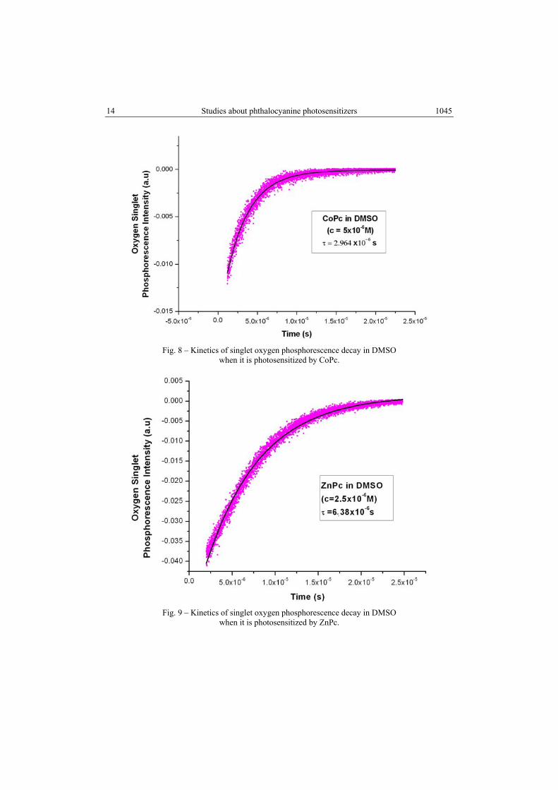
The above presented experimental setup (Figure 4) was used to measure the lifetime of singlet oxygen generated by the synhetized phthalocyanines by optical radiation excitation. Laser excitation of phthalocyanines was chosen in their Soret band at 355 nm. Maximum laser pulse energy was 1.2 mJ, in order to avoid saturation or photolysis. Fitting of the averaged experimental data – the intensity of phosphorescence given off by singlet oxygen population at 1270 nm versus time – with single exponential function produces lifetime of the population of oxygen molecules in singlet state. In Figs. 8 and 9 are presented the experimental data and the fitting exponential functions for CoPc and ZnPc. As it has been stated above, data on photophysical quantities are somehow conflicting; the lifetime found in the literature for the ZnPc in DMSO e.g. spreads over the range 19 µs (according to [24]), 1.8 µs (according to [25]) and 30 µs (according to [26]).
14 Studies about phthalocyanine photosensitizers
1045
Fig. 8 – Kinetics of singlet oxygen phosphorescence decay in DMSO
when it is photosensitized by CoPc.
Fig. 9 – Kinetics of singlet oxygen phosphorescence decay in DMSO
when it is photosensitized by ZnPc.
.

A. Staicu et al. 15
1046
For all the synthesized phthalocyanines the calculated lifetime of singlet oxygen are summarized in the Table 3 below.
Table 3
Synopsis of the measured singlet oxygen lifetime photosensitized by the studied phthalocyanines
No Compound name Solvent Lifetime of singlet oxygen (µs)
1. ZnPc DMSO 9.5 2. Cu-tris(hydroxyethylaminomethyl)-Pc DMF 20 3. Cu-tris(hydroxymethyl)-trihidroximetil-Pc DMSO 4.8 4. FePc DMSO 6.0 5. CoPc DMSO 3,0 6. CuPc DMSO 5.3 7. NiPc DMF 3.9 8. Chloroaluminum Pc (AlPcCl) DMSO 8.4 9. Hydroxyaluminium Pc (AlPcOH) DMSO 11.8
The analysis of the above table reveals that although one considers as a rule that the lifetime of the singlet oxygen depends only of the solvent of the sample, the measured lifetime of active species photogenerated by different phthalocyanines seems to point out a dependence of this lifetime on the properties of every photosensitizer and possible of its interactions with the solvent.
2.3.2. Quantum yield of photogenerated singlet oxygen
Usually, the measurement of the quantum yield of singlet oxygen generated by an unknown compound supposes reference to an accepted standard - a chemical compound for which this quantity is known. This was also our case, when a relative measuring method was used to measure quantum yield of singlet oxygen by comparing the intensity of the phosphorescence emission of the two compounds - the sample and the standard product, extrapolated at the zero moment of the laser pulse, as functions of the energy absorbed from the laser pulse. The two compounds are measured under strictly identical conditions (geometry of the set-up, optical absorption properties, excitation laser energy etc). The intensity of the phosphorescence is measured at the “zero” moment when the laser pulse excites the sample. The laser pulse duration (~ 5 ns) is very short as compared with the singlet oxygen phosphorescence duration (~ µs). Appropriate corrections were made when a different solvent was used (DMF instead of DMSO). As the standard compound was chosen Zinc phthalocyanine (Fluka), a compound for which quantum yields of singlet oxygen, largely accepted in the literature is Φ = 0.67 in DMSO solvent and Φ = 0.56 in the solvent DMF [27]. The absorbance of the standard and of each of the samples was kept at A = 0.28, so that the concentration of the sample was determined by this bond. By plotting the intensity of the phosphorescence of singlet oxygen at the moment of the laser

16 Studies about phthalocyanine photosensitizers
1047
pulse (zero moment) versus laser pulse energy, the experimental data fit well with straight lines, as shown in the Fig. 10, where this dependence is depicted on the same plot, as a typical example, for both the chosen standard ZnPc and CuPc.
Fig. 10 – Oxygen singlet phosphorescence intensity photosensitized by ZnPc
(standard) and CoPc.
If Φ∆ and Φ∆s are the quantum yields of the studied compound and of the standard, because the absorbances of the two are the same (thus the energies absorbed from the laser pulse are the same), the following relation holds:
Φ∆ / Φ∆s = m / ms , (1)
where m and ms are calculated slopes of the fitting straight lines The calculus of Φ∆ is thus straight forward. One may notice the different slopes of the fitting lines, corresponding in fact to different quantum yields for singlet oxygen generation of the two compounds. Similar computations led to the quantum yields values for all the synthesized phthalocyanines as shown in the Table 4.
Table 4
Synopsis of the calculated quantum yields for the phthalocyanines of concern
No Compound name Quantum yield of
photogenerated singlet oxygen
Solvent
1 ZnPc 0.67 DMSO 2 Cu-tris(hydroxyethylaminomethyl)-Pc 0.02 DMF 3 Cu-tris(hydroxymethyl)-trihidroximetil-Pc 0.05 DMSO 4 FePc 0.9 DMSO

A. Staicu et al. 17
1048
Table 4 (continued)
5 CoPc 0.3 DMSO 6 CuPc 0.10 DMSO 7 NiPc 0.02 DMF 8 Chloroaluminum Pc (AlPcCl) 0.23 DMSO 9 Hydroxyaluminium Pc (AlPcOH) 0.41 DMSO
From this table it results that the quantum yield of the singlet oxygen generated by different phthalocyanines highly depends on the diamagnetic metal used to metallate the phthalocyanine. Acceptable values of the quantum yield are for Zn and Al compounds, while the phthalocyanines with Cu, Fe, Co, Ni metals in the central ring have very small quantum yields in generating singlet oxygen, close to the quantum yields of singlet oxygen generated by endogenous constituents of the living cells (aromatic amino acids, proteins) [28].
3. CONCLUSIONS
Basic phthalocyanines with diamagnetic metals in the phthalocyanine ring (Cu, Al, Fe, Mn, Co and Ni) and a few derivatives with different moieties were selected and optimized from the point of view of the method for laboratory chemical synthesizing, using either the conventional (wet) method or the microwave assisted method. The synthesized compounds were studied using appropriate experimental setups from the point of view of their main photophysical characteristics as potential photosensitizers for PDT; samples were prepared in DMSO and DMF. Active species (singlet oxygen) are photogenerated by all the synthesized compounds by laser induced excitation. The photosensitized singlet oxygen was analyzed by studying quantum yields and lifetimes controlling processes. Experiments were made in similar conditions for all the products so that the measured quantities may be accurately compared to each other. It was confirmed that all considered metallated compounds generate singlet oxygen by photosensitization with different quantum yields, going down till a minimum value 0.03 for cobalt phthalocyanine. It was also found that although the lifetime of the photogenerated singlet oxygen is considered a property determined solely by the solvent, various lifetimes of the singlet oxygen populations were determined for the studied compounds (from about 3 µs for Cobalt phthalocyanine untill about 12 µs for the hydroxy-aluminium phthalocyanine in DMSO and 20µs for the compound named Cu-tris(hydroxyethylaminomethyl)-Pc in DMF).
These data may be correlated with other reports [29-38] regarding the use of medicines in solution, which are modified by exposure to laser radiation in order to produce new compounds that are either individually or synergistically effective

18 Studies about phthalocyanine photosensitizers
1049
against multiple drug resistant bacteria. In some cases [29, 33, 37] the role of the singlet oxygen as bactericide compound in solutions is mentioned and it results that further studies to more accurately identify the effects of it in the field of fighting multiple drug resistance (MDR) developed by bacteria are needed. This would be a complement of the singlet oxygen application in the photodynamic therapy of the malignant tumors.
Acknowledgements. The reported study was achieved as part of the research work financially supported by Romanian National Education Ministry, CNCS – UEFISCDI, project number PN-II-ID-PCE-2011-3-0922.
REFERENCES
1. M. C. DeRosa, R. J. Crutchley, Photosensitized singlet oxygen and its applications, Coordination Chemistry Reviews, 233–234, 351–371 (2002).
2. K. Ishii, Functional singlet oxygen generators based on phthalocyanines, Coordination Chemistry Reviews, 256, 1556–1568 (2012).
3. P. Di Mascio, S. P. Kaiser, T. P. A. Devasagayam, H. Sies, Biological Significance of Active Oxygen Species: In Vitro Studies on Singlet Oxygen-Induced DNA Damage and on the Singlet Oxygen Quenching Ability of Carotenoids, Tocopherols and Thiols, Advances in Experimental Medicine and Biology, 283, 71–77 (1991).
4. D. E. G. J. Dolmans, D. Fukumura, R. K. Jain, Photodynamic therapy for cancer, Nature Reviews Cancer, 3, 380–387 (2003).
5.a. E. Lempa, A. Canetea, G. Günthera, N. Pizarrob, A. L. Zanoccoa, Photosensitized generation of singlet molecular oxygen by aryloxazinones, Journal of Photochemistry and Photobiology A: Chemistry, 199, 2–3, 345–352 (2008).
5.b. P. Borker, A. V. Salker, Synthesis, characterization and photocatalytic studies of some metal phthalocyanines, Indian Journal of Chemical Technology, 13, 341–346 (2006).
6. M. Göksel, M. Durmuş, D. Atilla, A comparative study on photophysical and photochemical properties of zinc phthalocyanines with different molecular symmetries, J. Porphyrins Phthalocyanines, 16, 895–906 (2012).
7. P. M. Matlaba , Synthesis of zinc phthalocyanine derivatives for possible use in photodynamic therapy, Thesis, Rhodes University, Department of Chemistry, Grahamstown, South Africa, 2002.
8. M. Camerin, M. Magaraggia, M. Soncin, G. Jori, M. Moreno, I. Chambrier, M.J. Cook, D.A. Russell, The in vivo efficacy of phthalocyanine-nanoparticle conjugates for the photodynamic therapy of amelanotic melanoma, Eur. J. Cancer 46, 10, 1910–1918 (2010).
9. X. Jia, L. Jia, Nanoparticles improve biological functions of phthalocyanine photosensitizers used for photodynamic therapy, Curr. Drug. Metab., 13, 8, 119–122 (2012).
10. B. Kogan, A. Pankratov, A. Butenin, R. Feyzulova, T. Andreeva, O. Yuzhakova, R. Yakubovskaya, V. Negrimovsky, E. Lukyanets, G. Vorozhtsov, Targeted PDT with phthalocyanine nanoparticles, Photodiagnosis and Photodynamic Therapy, 8, 2, 209 (2011).
11. M. G. Waltera, A. B. Rudineb, C. C. Wamser, Porphyrins and phthalocyanines in solar photovoltaic cells, J. Porphyrins Phthalocyanines, 14, 759–792 (2010)
12. F.H. Moser, A.L. Thomas, The Phthalocyanines, Vols. 1–2, C.R.S. Press, Boca Raton, Florida, 1983.

A. Staicu et al. 19
1050
13. D. Villemin, M. Hammadi, M. Hachemi, N. Bar, Applications of Microwave in Organic Synthesis: An Improved One-step Synthesis of Metallophthalocyanines and a New Modified Microwave Oven for Dry Reactions, Molecules, 6, 831–844 (2001).
14. B. I. Kharisov, U. O. Mendez, J. R. Rosa, Low-temperature synthesis of phthalocyanine and its metal complexes, Russian Journal of Coordination Chemistry, 32, 9, 617–631 (2006).
15. H. Kantekin, Z. Biyiklioglu, E. Celenk, Synthesis and characterization of new metal-free and metallophthalocyanines peripherally fused to four 15-membered tetraoxamonoazamacrocycles by microwave irradiation, Inorg. Chem. Commun., 11, 633–635 (2008).
16. K. S. Jung, J. Y. Ro, J. Y. Lee, S. S. Park, Conventional versus microwave synthesis of phthalocyanine material, Journal of Materials Science Letters, 20, 24, 2203–2205 (2001).
17. A. V. Ziminov, S. M. Ramsh, E. I. Terukov, I. N. Trapeznikova, V. V. Shamanin, T. A. Yurre, Correlation Dependences in Infrared Spectra of Metal Phthalocyanines, Semiconductors, 40, 10, 1131–1136 (2006).
18. K. Sakamoto, E. Ohno-Okumura, Syntheses and Functional Properties of Phthalocyanines, Materials, 2, 1127–1179 (2009).
19. T. Fukuda, N. Kobayashi, Electronic absorption spectra – phthalocyanines, Vol 9 of the Series: Handbook of porphirin science, K. M. Kadish, K. M. Smith, R. Guilatd (editors), World Scientific Publishing Co., Singapore, 2010.
20. S. E. Maree, Effects of Substituents on the Photosensitizing and Electrocatalytic properties of Phthalocyanines, Thesis, Rhodes University, Grahamstown, South Africa, 2001.
21. T. Nyokong, Effects of substituents on the photochemical and photophysical properties of main group metal phthalocyanines, Coordination Chemistry Reviews, 251, 13–14, 1707–1722 (2007).
22. R. Battino, T. R. Rettich, T. Tominaga, The solubility of Oxygen and Ozone in Liquids, J. Phys. Chem. Ref. Data, 12, 163–178 (1983)
23. H. L. Chen, J. C. Hsu, M. H. Viet, M. S. Li, C. K. Hu, C. H. Liu, F.Y. Luh, S. S. Chen, E. S. Chang, A.H. Wang, M. F. Hsu, W. Fann, R. P. Chen, Studying submicrosecond protein folding kinetics using a photolabile caging strategy and time-resolved photoacoustic calorimetry, Proteins, 78, 14, 2973–83 (2010).
24. M. Hajimohammadi, N. Safari, H. Mofakham, A. Shaabani, A new and efficient aerobic oxidation of aldehydes to carboxylic acids with singlet oxygen in the presence of porphyrin sensitizers and visible light, Tetrahedron Lett., 51, 4061–4065 (2010).
25. M. Korinek., R. Dedic, A. Molnar, A. Svoboda, J. Hala, A comparison of photosensitizing properties of meso-tetraphenylporphin in acetone and in dimethyl sulfoxide, J. Molec. Struct., 744–47, 727–731 (2005).
26. R. Nilsson, D. R. Kearns, Role of singlet oxygen in some chemiluminescence and enzyme oxidation reactions, J. Phys. Chem., 78, 1681–1683 (1974).
27. N. Kuznetsova, N. Gretsova, E. Kalmkova, E. Makarova, S. Dashkevich, V. Negrimovskii, O. Kaliya, E. Lukyanets, Relationship between the Photochemical Properties and Structure of Pophyrins and Related Compounds, Russ. J. Gen. Chem., 70, 133–140 (2000).
28. K. K. Chin, C. C. Trevithick-Sutton, J. McCallum, S. Jockusch, N. J. Turro, J. C. Scaiano, C. S. Foote, M. A. Garcia-Garibay, Quantitative Determination of Singlet Oxygen Generated by Excited State Aromatic Amino Acids, Proteins, and Immunoglobulins, J. Am. Chem. Soc., 130, 6912–6913 (2008).
29. R. A. Pascu, In vivo study of the effects of alkylphenylpiridinium compounds exposed to optical radiation, The South- East European Journal of Ophthalmology, 1, 3–4, 37–40 (2006).
30. V. Nastasa, K.Samaras, I.R.Andrei, M.L.Pascu, T.Karapantsios, Study of the formation of micro and nano-droplets containing immiscible solutions, Colloids and Surfaces A: Physicochem. Eng. Aspects, 382, 246–250 (2011).
31. V. Nastasa, V. Pradines, I. R. Andrei, M. Boni, M. L. Pascu, R. Miller, Studies about the generation and characterization of microdroplets with a controlled content, Optoelectronics and advanced materials-Rapid Communications, 4, 11, 1916–1919 (2010).

20 Studies about phthalocyanine photosensitizers
1051
32. M.L. Pascu, I.R. Andrei, M. Ferrari, Angela Staicu, Adriana Smarandache, A. Mahamoud, V. Nastasa, L. Liggieri, Laser beams resonant interaction with micro-droplets which have a controlled content, Colloids and surfaces A: Physicochem. Eng. Aspects, 365, 83–88 (2010).
33. M.L. Pascu, A. Pascu, A. Staicu, I.R. Andrei, V. Nastasa, Tunable lasers at the laser spectroscopy group: short form history from the beginnings to date, Rom.Rep.Phys., 62, 3, 1455–480 (2010).
34. M.L. Pascu Adriana Smarandache, M. Boni, Jette kristiansen, V. Nastasa, I. R. Andrei, Spectral properties of molecular solutions, Rom. Rep.Phys., 63 (Suppl.), 1267–1284 (2011).
35. A. Hunyadi, B. Danko, M. Boni, A. Militaru, T. Alexandru, V. Nastasa, I. R. Andrei, M. L. Pascu, L. Amaral, Rapid, Laser-Induced Conversion of 20-Hydroxyecdysone and its Diacetonide – Experimental Set-up of a System for Photochemical Transformation of Bioactive Substances, Antic. Res., 32, 1291–1298 (2012).
36. M.L. Pascu, M. Boni, Adriana Smarandache, V. Nastasa, Andra Militaru, Angela Staicu, I.R. Andrei, Laser beams interaction with liquids in optofluidic experiments, Rom. Rep. Phys., 64 (Suppl.), 1179–1194 (2012).
37. M. L. Pascu, B. Danko, A. Martins, N. Jedlinszki, T. Alexandru, V. Nastasa, M.Boni, A. Militaru, I. R. Andrei, A. Staicu, A. Hunyadi, S. Fanning, L. Amaral, Exposure of chlorpromazine to 266 nm laser beam generates new species with antibacterial properties: contributions to development of a new process for drug discovery, PLOS ONE, 8, 2, e55767; doi:10.1371/journal.pone.0055767 (2013).
38. M.L. Pascu, V. Nastasa, A. Smarandache, A. Militaru, A. Martins, M. Viveiros, M.Boni, I. R. Andrei, A. Pascu, A. Staicu, J. Molnar , S. Fanning L. Amaral, Direct Modification of Bioactive Phenothiazines by Exposure to Laser Radiation, Rec. Pat. Anti-Infect. Drug Discov., 6, 147–157 (2011).